Copyright © 2000, American Society for Microbiology. All Rights Reserved.
Selective Cleavage of AAVS1 Substrates by the Adeno-Associated
Virus Type 2 Rep68 Protein Is Dependent on Topological and
Sequence Constraints
STEFANIA LAMARTINA, GENNARO CILIBERTO,
ANDCARLO TONIATTI*
Department of Gene Therapy, Istituto di Ricerche di Biologia Molecolare, 00040 Pomezia (Rome), Italy
Received 7 February 2000/Accepted 26 June 2000
The adeno-associated virus type 2 (AAV-2) Rep78 and Rep68 proteins are required for replication of the
virus as well as its site-specific integration into a unique site, called AAVS1, of human chromosome 19. Rep78
and Rep68 initiate replication by binding to a Rep binding site (RBS) contained in the AAV-2 inverted terminal
repeats (ITRs) and then specifically nicking at a nearby site called the terminal resolution site (
trs
). Similarly,
Rep78 and Rep68 are postulated to trigger the integration process by binding and nicking RBS and
trs
homologues present in AAVS1. However, Rep78 and Rep68 cleave in vitro AAVS1 duplex-linear substrates
much less efficiently than hairpinned ITRs. In this study, we show that the AAV-2 Rep68 endonuclease activity
is affected by the topology of the substrates in that it efficiently cleaves in vitro in a site- and strand-specific
manner the AAVS1
trs
only if this sequence is in a supercoiled (SC) conformation. DNA sequence mutagenesis
in the context of SC templates allowed us to elucidate for the first time the AAVS1
trs
sequence and position
requirements for Rep68-mediated cleavage. Interestingly, Rep68 did not cleave SC templates containing RBS
from other sites of the human genome. These findings have intriguing implications for AAV-2 site-specific
integration in vivo.
Human adeno-associated virus type 2 (AAV-2) is a
non-pathogenic parvovirus which establishes latency in cultured
human cell lines (4, 46). It integrates into the genome of
infected cells, with a high preference for a specific site,
AAVS1, on human chromosome 19 (22, 23, 46, 47). AAV-2
replication is stimulated either by coinfection with an
adeno-virus or herpesadeno-virus as a helper or by genotoxic stimuli such as
X-ray and UV treatment (3, 4). Infection of a latently infected
cell line with a helper virus leads to rescue and replication of
the integrated AAV genome, with the generation of infective
progeny (3, 4).
AAV-2 has a single-stranded genome approximately 4.7 kb
in length, which contains two open reading frames,
rep
and
cap
(55). The whole genome is flanked by 145-bp terminal repeats
(ITRs) which fold back into a hairpin-like structure and are
required for AAV-2 DNA replication, packaging, and
site-specific integration (3, 46). Crucial for the AAV-2 life cycle is
the activity of the viral Rep78 and Rep68 proteins: these are
translated from unspliced and spliced transcripts initiated from
the p5 promoter and differ only at the C terminus (55). The
two proteins, which probably function as multimers, have
sev-eral biochemical properties in common and are essential for
AAV-2 replication and site-specific integration (4, 14, 51, 55).
AAV-2 replication occurs via a unidirectional,
leading-strand DNA synthesis which closely resembles rolling-circle
replication (RCR) (3). During AAV-2 replication, Rep78 and
Rep68 bind the ITRs at a specific DNA sequence, the Rep
binding site (RBS), whose core region consists of four tandem
repeats of the GAGC tetramer (6, 18, 19, 45). Upon binding
the ITRs, Rep78 and Rep68 cleave in a site- and
strand-specific manner between the two thymidine residues of the
AGTTGG sequence, at the terminal resolution site (
trs
), which
is located near the RBS in the ITRs (5, 18, 49, 53). This nicking
provides the 3
⬘
-OH terminus, which serves as a primer for
replication and is followed by unwinding of the terminal
hair-pins, probably mediated by the helicase activity of Rep68 or
Rep78; the ITRs are thus converted to a blunt-ended and
double-stranded form in a process, called terminal resolution,
which allows the replication of the AAV-2 termini (5, 54).
Rep78 and Rep68 have also the capacity to hydrolyze ATP,
and the helicase activity is ATP dependent (18, 67, 69).
Several lines of evidence have identified Rep78 and Rep68
and the ITRs as the only viral elements required for
integra-tion into human chromosome 19 (27, 29, 68). Recombinant
AAV vectors lacking the
rep
gene do not integrate site
specif-ically (10, 21). In contrast, transgenes flanked by the AAV
ITRs integrate preferentially into AAVS1 when introduced
into cell lines together with Rep68 or Rep78 expression vectors
or recombinant proteins (2, 25, 39, 42, 43, 49, 56). An RBS
flanked by a
trs
-like GGTTGG sequence is also present in
AAVS1, and genetic analysis has demonstrated that these two
cis
-acting elements on chromosome 19 are necessary and
suf-ficient to dictate AAV-2 site-specific integration (12, 28, 29).
Rep78 and Rep68 mediate the formation in vitro of a complex
between an AAV-2 ITR and an AAVS1 oligonucleotide by
simultaneously binding the RBS contained in the two DNA
substrates (7, 63). This has led to the proposal that AAV
integration initiates when multimeric Rep78-Rep68 complexes
direct an AAV circular genome toward AAVS1 by
juxtaposi-tioning the two DNA substrates via Rep binding (10, 63).
Subsequently, Rep78 and Rep68 nick the
trs
at AAVS1, thus
leaving a free 3
⬘
-OH terminus, which serves as a primer for
replication mediated by the cellular replication machinery.
Two Rep-mediated strand switchings produce a
nonhomolo-gous recombination ITR/AAVS1 junction which allows the
replication complex to proceed through the AAV-2 genome,
which is thus inserted 3
⬘
to the RBS in AAVS1. A third
strand-* Corresponding author. Mailing address: Istituto di Ricerche di
Biologia Molecolare, IRBM-P. Angeletti, Via Pontina Km 30,600,
00040 Pomezia (Rome), Italy. Phone: 91093668. Fax:
39-06-91093654. E-mail: [email protected].
8831
on November 9, 2019 by guest
http://jvi.asm.org/
switching event translocates the replication complex to the
chromosomal DNA and terminates integration (10, 27, 28).
This model accounts for a number of features of AAV-2
integration, but there are still some issues which need to be
clarified. In particular, there is no evidence so far that Rep78
and Rep68 can indeed efficiently nick the AAVS1 at the
trs
. In
fact, the two proteins cleave in vitro a duplex linear AAVS1
template with very low efficiency (59, 60). Furthermore, the
trs
sequence and distance from the RBS are different in AAVS1
and the ITRs, and it is not yet clear whether this affects the
endonuclease activity of Rep78 and Rep68 at AAVS1 (59). To
fill these gaps in our information, the development of a
sensi-tive in vitro assay for studying Rep activity at AAVS1 is highly
desirable.
Rep78 and Rep68 share several functional properties with
RCR initiator proteins involved in the replication of small
prokaryotic genomes: they bind DNA at a specific site of the
replication origin, nick a nearby sequence in a site- and
strand-specific manner, and remain covalently bound through a
phos-photyrosyl linkage with the 5
⬘
-end phosphate at the nick (38).
In common with RCR initiators, Rep78 and Rep68 also have
the two-His structural motif (HuHuuu, where u is any
hydro-phobic residue) which is believed to be important in metal ion
coordination required for the activities of replication proteins
(17). Starting from the observation that RCR initiators nick
their DNA substrates only if they are supercoiled (38), we
tested whether also Rep-mediated cleavage at AAVS1
trs
might be affected by the DNA topology.
MATERIALS AND METHODS
Expression and purification of Rep68.Recombinant Rep68 was produced and purified as previously described (7, 25), with minor modifications. Briefly, the Rep68 coding region was amplified by PCR using plasmid pCMV/Rep68 as a template (25, 43). The fragment obtained was cloned in frame with the C terminus of maltose binding protein (MBP) into the unique BamHI site of pMAL-cRI vector (New England Biolabs). The MBP-Rep68 fusion was pro-duced as a soluble protein and partially purified by amylose affinity chromatog-raphy as described previously (7, 25). The fusion protein was then dialyzed against TN buffer (20 mM Tris-HCl [pH 8.0], 100 mM NaCl). To remove the maltose-binding moiety, CaCl2(2 mM, final concentration) was added to TN
buffer, and the MBP-Rep68 fusion was incubated with Factor Xa protease at an MBP-Rep68 Factor Xa weight ratio of 100 to 0.5 for 3 h at 4°C. The reaction was stopped by adding EGTA (final concentration, 10 mM [pH 8.0]), and the sample was loaded on a prepacked Mono Q HR 5/5 (anion exchange; Amersham, Pharmacia Biotech) equilibrated in TN buffer. The column was developed with 10 ml of linear gradient (20 mM Tris-HCl [pH 8.0], 100 mM NaCl, 2 mM CaCl2,
10 mM EGTA to 20 mM Tris-HCl [pH 8.0], 500 mM NaCl, 2 mM CaCl2, 10 mM
EGTA) at a flow rate of 1 ml/min. The peak corresponding to Rep68 was collected and further purified by gel filtration onto a prepacked Superdex 75 HR 10/30 column (Amersham, Pharmacia Biotech) equilibrated in 20 mM Tris-HCl (pH 8.5) and 150 mM NaCl. As previously reported, the purity of the protein was ⬎99%, as judged by silver staining of sodium dodecyl sulfate (SDS)-polyacryl-amide gels (25).
Preparation of SC plasmids.All supercoiled (SC) plasmids were prepared by the Triton lysis method and purified by double CsCl gradient centrifugation as described elsewhere (1).
Plasmid construction.To obtain plasmid pBS/trs, two complementary oligo-nucleotides were designed and annealed, to generate a double-stranded frag-ment spanning nucleotides (nt) 379 to 434 of the AAVS1 region and flanked at its 5⬘and 3⬘ends byBamHI andXbaI sites, respectively. This region was inserted into theBamHI andXbaI sites of plasmid pBluescript II KS(⫹) (Stratagene), thus obtaining plasmid pBS/trs. Plasmids containingtrseither mutated in se-quence or located at various distances from the RBS were obtained according to the same strategy but using oligonucleotides containing the desired mutations. Plasmids pBSmut1 and pBSmut2, also obtained by using this strategy, contain the AAVS1 region spanning nt 379 to 434 in which the wild-type RBS was mutated to GCTCGCGATAGATCTG (pBSmut1) and TAGAGCGATAGAT CTG (pBSmut2) (35), as indicated by underlining. Plasmids pIGFBP-2, pInh, pILF, pBRCA-1, and pERCC-1 contain RBSs identified in different regions of the human genome (65); insulin-like growth factor binding protein 2 (IGFBP-2) gene, inhibin gene, interleukin-2 enhancer binding factor (ILF) gene,BRCA1, andERCC1, respectively. As done for the AAVS1 region, these sequences were obtained by annealing of complementary oligonucleotides and cloned into pBluescript II KS(⫹) vector.
Electrophoretic mobility shift assays (EMSAs).The various radiolabeled sub-strates (15,000 cpm) were incubated with increasing concentrations of Rep68 in reaction mixtures (20l) that contained 10 mM HEPES-NaOH (pH 7.9), 8 mM MgCl2, 1g of poly(dI-dC), 40 mM KCl, and 0.2 mM dithiothreitol (DTT).
Following a 30-min incubation at room temperature, 4l of 20% Ficoll was added; samples were then loaded on a 4% polyacrylamide gel (acrylamide/ bisacrylamide ratio, 29:1; 0.5⫻Tris-borate-EDTA) and electrophoresed in 0.5⫻ Tris-borate-EDTA at room temperature and 10 V/cm. Gels were then dried and subjected to autoradiography at⫺80°C.
Nicking assay on SC templates.The standard SC nicking assays were per-formed in 30l of a solution containing 30 mM HEPES (pH 7.5), 7 mM MgCl2,
0.5 mM DTT, 4 mM ATP, 40 mM creatine phosphate, and 1g of creatine phosphokinase. The reaction mixtures also contained SC plasmid DNA and purified Rep68 at the concentrations indicated in the figure legends. The reac-tions were carried out at 37°C for 1 h and then terminated by adding 40l of stop solution (proteinase K [1.2g/l], 0.5% SDS, 30 mM EDTA [pH 7.5]). After incubation at 37°C for 1 h, the DNA samples were subjected to phenol-chloro-form extraction and ethanol precipitation. Precipitated DNA samples were re-suspended in water and resolved on a 1% agarose gel (1% agarose, 1⫻ Tris-acetate-EDTA [TAE]) which was subsequently stained by incubation at room temperature for 30 min in 1⫻TAE containing ethidium bromide (0.3g/ml).
Preparation of RC topoisomers and separation of SC, NC, and RC molecules.
Three hundred-nanogram aliquots of SC plasmids were relaxed by treatment with 6 U of calf thymus topoisomerase I (GibcoBRL) for 2 h at 37°C in 25l of a reaction mixture containing 50 mM Tris-HCl (pH 7.5), 50 mM KCl, 10 mM MgCl2, 0.5 mM DTT, 0.1 mM EDTA and 30g of bovine serum albumin per ml.
Subsequently, reaction mixtures were first adjusted to 35l containing 4 mM ATP, 40 mM creatine phosphate, and 1.2g of creatine phosphokinase and then incubated for an additional hour at 37°C in the presence or absence of 300 ng of recombinant Rep68. The reactions were terminated by treatment with proteinase K, extracted with phenol, precipitated with ethanol, and resuspended in 2⫻ TAE. The negative SC, relaxed circular (RC), and nicked circular (NC) forms of the template plasmid were resolved on agarose gels as described elsewhere (15). Briefly, samples were electrophoresed on a 1% agarose gel in 2⫻TAE buffer in the absence of ethidium bromide for 5 h at 3 V/cm. Gel was then stained with ethidium bromide (0.5g/ml), and electrophoresis was continued for an addi-tional hour under the same conditions but in a running buffer containing ethidium bromide (0.5g/ml). Under these conditions, the first electrophoresis step separates the SC molecules from the RC and NC forms; in the second step, the RC topoisomeres migrate faster than the NC form (15).
Mapping of the nicking site on SC templates.A standard SC nicking reaction was performed by incubating 1g of SC plasmid substrates (plasmids pRVK, pBS/trsand its derivatives containingtrsmutants, and psub201) with 300 ng of Rep68 protein. After proteinase K treatment, phenol-chloroform extraction, and ethanol precipitation, the reaction products (NC forms of the plasmids) were dissolved in water and used as templates for sequencing reactions, which were performed by the dideoxy method using the Sequenase version 2.0 polymerase (U.S. Biochemical Corporation). A32P-labeled oligonucleotide annealing with
thetrs-containing (trs⫹) strand was used as the primer for the sequencing reac-tions. The primer was centered on the T7 promoter region in the case of plasmid pBS/trsand its derivatives. In the case of plasmid pRVK, the primer spanned positions 495 to 479 of AAVS1. In the case of plasmid psub201, the primer spanned positions 4686 to 4672 of the AAV-2 genome contained in this plasmid (48). Reaction products were analyzed on 8% denaturing polyacrylamide gels.
Covalent attachment of Rep68 to the 5ⴕend of the cleavage site.Covalent attachment of Rep68 to the 5⬘end was assessed as described elsewhere (40), with some modifications. Three hundred nanograms of Rep68 was incubated with 1 g of SC plasmid pRVK in a standard SC nicking reaction for 1 h at 37°C. The reaction product was digested with restriction enzymeSmaI; the 3⬘ends of the digested fragments were labeled with [␣32-P]ddATP by using terminal
de-oxynucleotidyltransferase (TdT). After 1 h at 37°C, the labeled products were immunoprecipitated in 0.05% Tween 20 in phosphate-buffered saline with a polyclonal rabbit antiserum against Rep68 (25). After a 6-h incubation at 4°C, samples were washed extensively with 0.05% Tween 20 in phosphate-buffered saline. The immunoprecipitates were then divided into two aliquots, one of which was digested with proteinase K. Both aliquots were then subjected to phenol-chloroform extraction and ethanol precipitation. Finally, samples were resus-pended in 0.1% SDS–30% formamide–6.5 mM EDTA (pH 8.0) and resolved on an 8% sequencing gel.
Determination of strand- and site-specific nicking on AAVS1 SC templates.
One microgram of SC plasmid pRVK was incubated in the standard SC nicking reaction with or without 300 ng of Rep68 for 1 h at 37°C. After proteinase K treatment, phenol-chloroform extraction, and ethanol precipitation, each reac-tion product (NC plasmid) was divided into two aliquots, and thetrs⫹andtrs⫺ strands were selectively labeled. To label the strand not containing thetrs, NC pRVK was digested withPvuII; this digestion released three fragments, one of which contains nt 1 to 513 of AAVS1 flanked at its 5⬘end by an additional 175 bases derived from the vector (plasmid pBluescript) backbone. All of the frag-ments were dephosphorylated by treatment with calf intestinal alkaline phospha-tase and 5⬘-end labeled with T4 polynucleotide kinase and [␥-32P]ATP. After
phenol-chloroform extraction and ethanol precipitation, the reaction mixture was digested with restriction enzyme EcoRI, whose unique recognition site
8832
LAMARTINA ET AL.
J. V
IROL.
on November 9, 2019 by guest
http://jvi.asm.org/
FIG. 1. AAV-2 Rep68 poorly cleaves duplex-linear AAVS1 DNA substrates. (A) Schematic representation of the three AAVS1 linear substrates (AAVS1/79, AAVS1/109, and AAVS1/304) used in binding and nicking experiments. Positions of the RBS and thetrsare indicated. (B) EMSAs. AAVS1 duplex-linear templates (15,000 cpm; corresponding to 3.7, 5.7, and 8.3 fmol) were incubated with increasing amounts (5, 10, 100, and 1,000 ng; corresponding to 0.09, 0.18, 1.8, and 18 pmol) of recombinant Rep68 in a standard binding reaction buffer (see Materials and Methods). Reaction products were resolved on a nondenaturing 5% polyacrylamide gel. In the absence of protein, no shifted complexes were detected (lanes 1, 6, 11, and 16). (C) Rep68 nicking on linear AAVS1 substrates. Rep68 (1g; 18 pmol) was incubated with 4 fmol of radiolabeled AAVS1/79, AAVS1/109, and AAVS1/304 linear substrates (20,000, 12,000, and 5,000 cpm, respectively), in the presence (lanes 3, 4, 7, 8, 11, and 12) or absence (lanes 1, 2, 5, 6, 9, and 10) of 1g of unspecific competitor poly(dI-dC). Standard endonuclease reactions were performed for 60 min at 37°C, followed by proteinase K digestion and phenol-chloroform extraction. Reaction products were resolved on an 8% denaturing polyacrylamide gel. The triangle indicates the released products of the expected size observed with template AAVS1/79. Fragments released from AAVS1/109 and AAVS1/304 substrates were observed only after longer exposures (not shown).
on November 9, 2019 by guest
http://jvi.asm.org/
constitutes the 5⬘end of the AAVS1 site as cloned into plasmid pRVK (nt 1 to 6) (23). This digestion thus selectively removed the radioactively labeled 5⬘end of the trs⫹strand; the resultingEcoRI-PvuII fragment was thus selectively labeled only at the 5⬘end of thetrs⫺strand. This end-labeled fragment was purified from an agarose gel and loaded on a 6% sequencing gel. To selectively label thetrs⫹strand, NC (Rep68-treated) pRVK was digested withPvuII as described above. The released fragments were then labeled at their 3⬘ends by treatment with the TdT and [␣32-P]ddATP. The labeled fragments were then
digested withEcoRI; in this case, the digestion selectively removed the radioac-tively labeled 3⬘end of thetrs⫺strand. Therefore, the resultingEcoRI-PvuII fragment was selectively labeled only at the 3⬘end oftrs⫹strand. Again, the labeled fragment was purified and loaded on a 6% sequencing gel.
DNase I footprinting analysis.The DNase footprinting analysis on SC or linear AAVS1 templates was performed as described elsewhere (58), with some modifications. Plasmid pRVK was used as the SC template, while anMscI-PvuII duplex-linear fragment derived from plasmid pRVK and spanning nt 210 to 513 of AAVS1 was used as the linear template. One hundred-nanogram aliquots of SC or duplex-linear templates were incubated with 1g of Rep68 for 30 min at room temperature in 30l of a solution containing 10 mM HEPES (pH 7.9), 8 mM MgCl2, 40 mM KCl, 0.2 mM DTT, and 1.5g of poly(dI-dC). CaCl2was
then added to a final concentration of 2.5 mM, and the samples were digested with 5 ng of DNase I (Boehringer Mannheim catalog no. 104 159; conversion factor, 1 ng⫽2 mU) for 2 min at room temperature. Digestion was stopped by adding 1 volume of DNase I stop buffer (10 mM HEPES [pH 7.9], 1% SDS, 30 mM EDTA [pH 8.0]). After phenol-chloroform extraction and ethanol precipi-tation, specific cleavages were detected by PCR-mediated primer extension on the DNase I-treated DNA using a32P-labeled primer. Analysis of thetrs⫹strand was performed using as a primer an oligonucleotide (5⬘-CCCCACTGCCGCA GCTGC-3⬘) annealing to this strand at the level of the AAVS1 sequence from nt 527 to 510. For analysis of thetrs⫺strand, we used a primer (5⬘-CCGGGAGA TCCTTGGGGCGGTGGGG-3⬘) annealing to this strand at the level of the AAVS1 region spanning nt 310 to 334. Since the selected AAVS1 region is enriched in G⫹C sequences (23), primer extension was performed by using the thermostable DNA polymerases and the additional reagents contained in the Advantage-GC2 PCR kit (Clontech) that we have successfully used to efficiently
amplify GC-rich sequences (S. Lamartina and C. Toniatti, unpublished results). Specifically, samples were resuspended in a buffer containing 1% glycerol, 0.8 mM Tris-HCl (pH 7.5), 1.0 mM KCl, 0.5 mM (NH4)2SO4, 2M EDTA, 0.1 mM
-mercaptoethanol, 0.005% Thesit, 40 mM Tricine-KOH, 15 mM potassium acetate, 3.5 mM magnesium acetate, 5% dimethyl sulfoxide, 3.75g of bovine serum albumin per ml, 1 M GC-Melt reagent, 0.2 mM each dATP, dCTP, dGTP, and dTTP, and the mixture of KlenTaq-1 DNA polymerase, Deep VentR, and
TaqStart antibodies as supplied by the manufacturer (Clontech). Reaction mix-tures also contained 1.5⫻106cpm (2 pmol) of the primers labeled at the 5⬘end
with [␥-32P]ATP by T4 polynucleotide kinase. After a preheating step at 94°C for
1 min, the reaction was allowed to proceed for 30 cycles of amplification and extension (1 min at 94°C, 30 s at 94°C, and 3 min at 72°C) and then stopped with 50l of DNase I stop buffer. Reaction products were extracted with phenol-chloroform and ethanol precipitated. Samples were then resuspended in 4l of denaturing loading buffer (95% formamide, 20 mM EDTA, 0.05% bromophenol blue, 0.05% xylene cyanol), denatured for 5 min at 100°C, and electrophoresed on a 6% sequencing gel.
RESULTS
[image:4.612.97.499.73.314.2]Rep68 poorly cleaves a linearized AAVS1 template.
In
vitro-translated AAV-2 Rep78 poorly nicks the potential target site
present in a 57-bp-long linear duplex DNA fragment spanning
the AAVS1 RBS-
trs
region (23). Experiments performed using
AAV ITRs as DNA substrates have demonstrated that Rep78
and Rep68 nick the
trs
in a linear template containing only the
stem of the ITR with 50- to 100-fold lower efficiency than the
hairpinned ITR, which also includes the ITR loop (7, 35, 53,
54). This has been attributed to additional contacts that Rep78
and Rep68 make with sequences contained in the ITR loop but
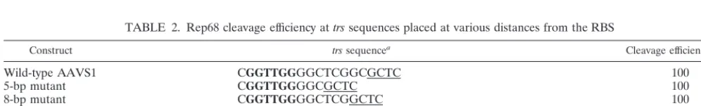
FIG. 2. Rep68 efficiently nicks an SC plasmid containing the AAVS1 RBS-trsregion. (A) Schematic representation of the AAVS1 region contained in plasmids pRVK and pBS/trs. TheBamHI andXbaI sites of pBS/trs originate from the cloning procedure and do not refer to the original AAVS1 sequence (23). (B) Rep68-mediated cleavage of pRVK. One hundred nanograms of plasmid pRVK (6,485 bp long) was incubated in a standard endonuclease reaction with 10 (lane 2), 30 (lane 3), 50 (lane 4), and 100 (lane 5) ng of recombinant Rep68. After 60 min at 37°C, reaction products were digested with proteinase K, purified by phenol-chloroform extraction, and concentrated by precipitation with ethanol. Samples were then resolved on a 1% agarose gel, which was stained by 30 min of incubation in TAE buffer containing ethidium bromide (0.3g/ml). Lane 1, untreated pRVK. The SC and NC forms of the plasmid are indicated by arrows. M, size markers. (C) Rep68 does not cleave RC templates. Three hundred nanograms of SC pRVK was converted to RC form by topoisomerase I treatment and then incubated with or without 300 ng of Rep68 in a standard endonuclease reaction (see Materials and Methods). In control experiments, 300 ng of SC pRVK was incubated with or without 300 ng of Rep68. Reaction products were resolved by electrophoresis on agarose gels as described elsewhere (15). Lanes 1 and 2, SC pRVK incubated without and with Rep68, respectively; lanes 3 and 4, RC pRVK topoisomers incubated without and with Rep68, respectively. (D) Rep68-mediated cleavage of pBS/trs. One hundred nanograms of plasmid pBS/trs(3,011 bp long) was incubated with 5 (lane 3) and 20 (lane 4) ng of recombinant Rep68. Endonuclease reactions were performed as described for panel B. Lane 1, linearized pBS/trs; lane 2, untreated pBS/trs; lane 5; control plasmid pBS treated with 100 ng of Rep68. Sizes are indicated in kilobases.8834
LAMARTINA ET AL.
J. V
IROL.
on November 9, 2019 by guest
http://jvi.asm.org/
outside the consensus RBS (45, 67). We thus checked whether
Rep endonuclease might be more active on longer templates.
AAV-2 Rep68 was produced in
Escherichia coli
, purified to
near homogeneity as described previously (25), and tested for
its ability to bind and nick three AAVS1 fragments of different
lengths (79, 109, and 304 bp). Figure 1A shows the three
fragments and the location of the RBS-
trs
region within them.
Rep68 binding was monitored by incubating equivalent
amounts of double-stranded probes with increasing
concentra-tions of the protein. By EMSA, Rep68 bound all three linear
fragments with similar affinities (Fig. 1B). However, even at the
highest Rep68/DNA molar ratio (higher than 2,000:1) at which
more than 95% of DNA is bound (Fig. 1B, lanes 5, 10, and 15),
only minimal cleavage was observed with the three linear
AAVS1 substrates (Fig. 1C). These findings, which confirm
and extend previous results (60), demonstrate that Rep68
cleavage in vitro of an AAVS1 linear fragment is a largely
inefficient process, regardless of the length of the DNA
sub-strate.
Rep68 efficiently nicks an SC AAVS1 substrate.
Rep68
shares protein motifs and functional properties with initiator
proteins involved in RCR (17). These proteins are known to
start replication upon cleavage of a specific site but only if the
substrate is supercoiled (38). We therefore asked whether also
Rep68 might preferentially cleave an SC AAVS1 target
se-quence. To test this possibility, 100 ng of SC plasmid pRVK (a
gift from K. I. Berns, Cornell University Medical College,
Ithaca, N.Y.), which is a pBluescript vector containing the
AAVS1 sequence from nt 1 to 3525 (schematically represented
in Fig. 2A), was incubated in a classical endonuclease reaction
with increasing concentrations of Rep68. After 1 h at 37°C, the
plasmid was digested with proteinase K, purified by
phenol-chloroform extractions, precipitated with ethanol, and then
loaded onto an agarose gel. It was expected that if Rep68 had
cleaved the
trs
in AAVS1 in a strand- and site-specific manner,
the plasmid conformation would have changed from SC to NC.
As shown in Fig. 2B, this is in fact what was observed: in the
presence of Rep68, the monomeric SC pRVK (Fig. 2B, lane 1)
was converted to NC. The modification was already evident at
a Rep68/pRVK molar ratio of 8:1 (10 ng of Rep68:100 ng of
pRVK [Fig. 2B, lane 2) and was complete with as low as a
23-fold molar excess (30 ng) of Rep68 (Fig. 2B, lane 3).
No-tably, RC pRVK was not cleaved (Fig. 2C, lanes 3 and 4), a
further indication that supercoiling of the template is required
for efficient nicking.
To rule out the possibility that formation of NC forms of
pRVK was due to Rep68 nicking at sites other than the
ex-pected target region, a 56-bp fragment containing the RBS and
trs
of AAVS1 was cloned into the pBluescript vector (plasmid
pBS/
trs
[Fig. 2A]). As shown in Fig. 2D, this substrate was
converted from the SC to the NC form by Rep68 as efficiently
as pRVK (Fig. 2D, lanes 3 and 4). No cleavage was observed
with the empty vector (Fig. 2D, lane 5), thus demonstrating
that Rep68 nicking was restricted to the RBS-
trs
region. RC
pBS/
trs
was also not cleaved by Rep68 (not shown).
Rep68 nicks an SC AAVS1
trs
between the two T residues
(GGT/TGG).
To verify that conversion from the SC to the NC
form was due to site-specific nicking at the GGTTGG
trs
se-quence, the Rep68-generated NC form of pRVK was purified
and used as a template for sequencing by the dideoxy-chain
termination method (1). An oligonucleotide annealing with the
trs
⫹strand and 3
⬘
to the RBS was used as a primer. A nick in
the
trs
⫹strand at the target GGTTGG sequence would halt
synthesis of the complementary DNA strand and lead to
ac-cumulation of DNA strands terminated at the nick. As shown
in Fig. 3A, in the case of the NC form, polymerization of the
new strand was indeed blocked at the level of the
trs
(lanes 1 to
4). By comparison with the DNA sequence ladder obtained in
a similar sequencing reaction but using an SC, not
Rep68-treated pRVK as a template (Fig. 3A, lanes A, G, C, and T),
the cutting site apparently mapped between the guanosine and
the first thymidine residue (GG/TTGG). However, the
Seque-nase DNA polymerase used in the sequencing reaction displays
a TdT activity which adds an extra nucleotide once it reaches
the end of the template DNA (33). Notably, we confirmed this
FIG. 3. Mapping of the Rep68 nicking site. (A) Mapping of the cleavage site on SC AAVS1 templates. SC plasmid pRVK (1g) was converted to NC by treatment with 300 ng of Rep68 protein. The purified NC form was used as a template for a standard sequencing reaction performed by using Sequenase and a primer, schematically represented by an arrow, which spanned nt 495 to 479 of AAVS1 and annealed with thetrs⫹strand. Reaction products were loaded on a 8% denaturing gel. Lanes 1, 2, 3, and 4 correspond to A, G, C, and T sequencing reactions, respectively, performed using the NC (Rep68-treated) form of pRVK; the DNA sequencing ladder was too faint to be seen in the gel. Lanes A, G, C, and T represent the sequencing ladder obtained in a control sequencing reaction performed by using the same primer on an SC (not Rep68-treated) form of plasmid pRVK. The AAVS1 RBS-trsregion is schematically represented at the bottom; the triangle indicates the apparent nicking site, and the arrow indicates the cutting site deduced from the TdT activity of the polymerase (33). (B) Mapping of the nicking site on the AAV-2 ITR contained in SC plasmid psub201. SC plasmid psub201 (1g) was converted to the NC form by treatment with 300 ng of Rep68 protein. The nick site in the AAV ITR was mapped as described in the legend to Fig. 3A by using a primer annealing with the 4686–4672 region of the AAV-2 genome contained in plasmid psub201 (48). Reaction products were resolved on an 8% denaturing gel. Lanes 1, 2, 3, and 4 correspond to A, G, C, and T sequencing reactions using Rep68-treated psub201; the DNA sequencing lad-der was too faint to be seen in the gel. Lanes A, G, C, and T represent the sequencing ladder obtained by sequencing SC plasmid psub201. The AAV-2 ITR RBS-trsregion is also represented; the triangle and arrow indicate the apparent and deduced nicking sites, respectively.on November 9, 2019 by guest
http://jvi.asm.org/
TdT activity by using the same technique to map the nicks
introduced by restriction enzymes
Pst
I,
Bam
HI,
Sma
I, and
Eco
RI in the context of pBluescript: in all cases, Sequenase
was found to promote a nontemplated addition of one
nucle-otide once it reached the end of the template DNA (not
shown). Based on this evidence, therefore, the cutting site in
AAVS1
trs
should probably be moved one nucleotide to the 3
⬘
side, with the nick occurring between the two T nucleotides
(GGT/TGG [Fig. 3A]). To confirm this supposition, we used
the same technique to map the Rep68 nicking site in the
AAV-2 ITR
trs
. SC plasmid psub201, which contains the
AAV-2 genome, was nicked with Rep68, and the site of strand
interruption in the context of the AAV-2 ITR
trs
was mapped
(Fig. 3B). Also in this case, the apparent cutting site (AG/
TTGG [Fig. 3B]) was shifted by one nucleotide with respect to
the previously mapped AGT/TGG cleavage at the AAV-2 ITR
trs
(18, 53).
Rep68 cleaves the AAVS1
trs
contained in an SC plasmid in
a site- and strand-specific manner.
Strand polymerization was
not stopped in sequencing reactions performed using as a
primer an oligonucleotide annealing with the
trs
⫺strand (not
[image:6.612.55.544.69.496.2]shown), suggesting that Rep68 cleavage at the
trs
was strand
FIG. 4. Strand-specific nicking of SC AAVS1 templates and covalent linkage of Rep68 to the 5⬘end of the nicking site. (A) Strand-specific nicking. SC plasmid pRVK (1g) was incubated with or without 300 ng of Rep68 for 60 min at 37°C in a standard endonuclease reaction. Following proteinase K digestion, plasmid was purified and digested with restriction enzymePvuII, which released a fragment containing the AAVS1 RBS-trsregion. The two strands of this fragment were selectively labeled in two distinct reactions. The 3⬘ends of the fragments derived from thetrs⫹strand (Strand⫹) were selectively labeled by using TdT, while thetrs⫺strand (Strand⫺) was labeled at its 5⬘end by treatment with T4 polynucleotide kinase (see Materials and Methods for further details). Labeled products were resolved on a 6% denaturing gel. (B) Covalent linkage of Rep68 to the 5⬘end of the nick site. SC plasmid pRVK was incubated with 300 ng of Rep68 in a standard endonuclease reaction. Plasmid was then digested with the restriction enzymeSmaI, and the 3⬘ends of the digestion products were32P labeled with TdT. The double-strandedfragment containing the cleavedtrs(shown at the left) was coimmunoprecipitated with the covalently linked Rep68 protein by using an anti-Rep68 polyclonal serum. The immunoprecipitated material was digested (lane 4) or not (lane 2) with proteinase K (PK) and then resolved on a 6% polyacrylamide denaturing gel. In control experiments, SC pRVK was digested with restriction enzymeSmaI, and the digestion products, previously labeled with TdT, were incubated with anti-Rep68 serum. In this case, no labeled material was present in the immunoprecipitate (lane 1 and 3).
8836
LAMARTINA ET AL.
J. V
IROL.
on November 9, 2019 by guest
http://jvi.asm.org/
specific. This was confirmed in additional experiments. SC
pRVK was first converted to NC by treatment with Rep68 and
then treated with proteinase K and purified. A 512-bp-long
DNA segment containing the RBS and the cleaved
trs
(sche-matically represented in Fig. 4A) was then excised and divided
into two aliquots; which were selectively labeled at either the
trs
⫹or the
trs
⫺strand and resolved on a denaturing
polyacryl-amide gel. Figure 4A shows that no cleavage at the
trs
⫺strand
was observed, while two major fragments were released from
the
trs
⫹strand, and their sizes were compatible with cleavage
occurred at the
trs
. However, besides a major released product,
additional (from one to two, in different experiments) faint and
apparently longer fragments were also detectable (Fig. 4A). It
is possible that this observation reflects a low specificity of
cleavage at the
trs
or the presence of contaminating bacterial
nucleases in the protein preparation. However, we rather
be-lieve that these additional fragments represent the expected
120-bp cleavage product covalently linked at its 5
⬘
end to
Rep68 polypeptides of various lengths that remain after
pro-teinase K digestion and reduce the electrophoretic mobility of
the DNA segment (18, 52, 60).
To further verify this hypothesis and, more generally, to rule
out the possibility that cleavage at the
trs
was due to a
con-taminant present in the protein preparation, we checked
whether Rep68 established a covalent linkage with the 5
⬘
end
of the nick site in an SC AAVS1 template (60). SC pRVK was
converted to NC by Rep68, and a fragment spanning the RBS
and the cleaved
trs
was selectively labeled at its 3
⬘
end by using
TdT. The labeled fragment (Fig. 4B) was then
immunoprecipi-tated along with the potentially covalently linked Rep68
pro-tein by using a polyclonal anti-Rep68 serum. The
immunopre-cipitate was treated or not with proteinase K, purified, and
resolved on a denaturing polyacrylamide gel. As shown in Fig.
4B, in the absence of proteinase K digestion, the cleavage
product expected to be Rep68 linked at its 5
⬘
end did not enter
the denaturing gel (Fig. 4B, lane 2), indicating that it was
tightly associated with a high-molecular-weight material. Upon
proteinase K digestion, a major cleavage product (Fig. 4B, lane
4, band b) was detectable. Interestingly, also in this case, one to
two additional and fainter fragments were detectable in
differ-ent experimdiffer-ents, in full agreemdiffer-ent with previous results
(com-pare Fig. 4A and B). Taken together, these results
demon-strated that Rep68 cleaved the plasmids containing the
AAVS1 RBS-
trs
region at the expected site, in a strand-specific
manner and according to molecular mechanisms similar to
those already characterized with linear and hairpinned DNA
substrates (18, 52, 60).
Rep68 nicking of SC templates is ATP and DNA binding
dependent.
Rep68 nicking activity on an SC template was
clearly ATP dependent (Fig. 5A, lanes 2 and 3), although some
nicking could be observed in the absence of ATP with high
Rep68 concentrations (Fig. 5A, lanes 4 and 5; see Discussion).
Notably, the nicking reaction was fully and specifically
com-peted by adding in solution double-stranded oligonucleotides
containing the RBS (Fig. 5A, lanes 6 and 7). Furthermore,
derivatives of plasmid pBS/
trs
, called pBSmut1 and pBSmut2,
which contain the wild-type
trs
flanked by binding-deficient
mutant of the RBS (35), were not cleaved (Fig. 5B, lanes 5 to
12). These results strongly suggest that binding to the RBS was
necessary for nicking.
Rep68 footprinting on SC and linear AAVS1 templates.
To
test whether the more efficient cleavage on an SC rather than
a linear template reflected qualitative differences in the
bind-ing mode to the two substrates, DNase I footprintbind-ing analyses
were performed using either SC or linear forms of a DNA
substrate centered on the AAVS1 RBS-
trs
element. No
differ-ence was observed between the two templates: the same
re-gions were protected in the SC and linear template on the
trs
⫹and
trs
⫺strands. In both cases, the four repeats of the
non-perfect GAGC tetramer constituting the core of the RBS were
fully protected (Fig. 6). Footprinting was broader on the
trs
⫺strand, where protection spanned the entire
trs
-complementary
sequence and extended up to about 18 bp from the 5
⬘
end of
the core of the RBS (Fig. 6). Only partial protection of the
trs
hexamer was observed on the
trs
⫹strand (Fig. 6). Therefore,
the binding features of Rep68 to SC and linear AAVS1
tem-plates are similar and probably do not account for the observed
difference in nicking efficiency.
Mutagenesis of the AAVS1-
trs
sequence.
Having established
a fast and sensitive nicking assay using the SC template, we
decided to study the sequence specificity of the Rep68
endo-nuclease activity in this experimental system. To this end, the
wild-type
trs
sequence (GGTTGG) in the context of plasmid
pBS/
trs
(Fig. 2A) was extensively mutagenized, and the
corre-sponding SC plasmids were used in the nicking assay. Table 1
summarizes the results obtained; the cleavage sites within each
trs
mutant are also indicated.
[image:7.612.319.539.74.293.2]We first analyzed the effects of mutations in the TT dimer.
Substitution of the two thymidine residues with a CC or AA
dimer resulted in a complete loss of cleavage (Table 1). In
contrast, mutation of only one of the two T residues with an A
or a C was quite well tolerated, and the resulting sequences
could still be cleaved, although less efficiently than the
wild-type sequence (Table 1). However, substitution of the first T
residue, which is the 5
⬘
end of the nick site (Fig. 3), was slightly
more detrimental than mutation of the second T nucleotide;
interestingly, cleavage always occurred 3
⬘
to the remaining
FIG. 5. ATP and DNA binding-dependent cleavage of SC pBS/trs. (A) SC pRVK (100 ng) was incubated with 10 ng (lanes 2, 4, 6, and 8) and 100 ng (lanes 3, 5, 7, and 9) of Rep68 in an endonuclease reaction. Lane 1, SC pRVK; lanes 2 and 3, standard reaction; lanes 4 and 5, no ATP in the reaction buffer; lanes 6 and 7, 200 ng of double-stranded oligonucleotide spanning the RBS added to the reaction buffer; lanes 8 and 9, reaction mixture containing 200 ng of an unspecific double-stranded oligonucleotide. (B) Rep68 (5, 20, and 200 ng) was incubated in a standard endonuclease reaction with 100 ng of plasmids pBS/trs(lanes 1 to 4), pBSmut1 (lanes 5 to 8), and pBSmut2 (lanes 9 to 12). Plasmids pBSmut1 and pBSmut2 contain mutant RBS sequences which have been reported to strongly impair Rep binding (36). See Materials and Methods for further details. wt, wild type.on November 9, 2019 by guest
http://jvi.asm.org/
thymidine (Table 1). The role of the G residues flanking the
TT dimer was also studied. Modifications of the flanking
nu-cleotides did not significantly hamper Rep68 nicking at the
trs
;
in fact, templates in which the guanosines were replaced by
either AC or CC dimers (ACTTAC and CCTTCC sequences,
respectively) were still nicked by Rep68, although with a
slightly reduced efficiency (Table 1). The same applied also to
mutants CGTTCC and CCTTGC, in which all but one of the
flanking guanosine residues were mutated (Table 1). Taken
together, these results demonstrated that the GGTTGG
se-quence is the best target for Rep68 nicking; nevertheless,
several substitutions are tolerated, provided that at least one
thymidine is maintained.
Activity of Rep68 on
trs
positioned at various distances from
the AAVS1 RBS.
In AAVS1, the TT dimer within the
trs
is
located at 10 bp from the core of the RBS, as opposed to the
15 bp in the AAV-2 ITRs (18, 60). To test whether the distance
between the RBS and the
trs
might affect the efficiency of
cleavage, mutants were generated in the context of plasmid
pBS/
trs
in which the TT dimer, flanked by the wild-type GG
dimers, was positioned at distances of 5, 8, 13, 15, and 20 nt
from the RBS (5-, 8-, 13-, 15-, and 20-bp mutants, respectively).
All but the 20-bp mutant represented excellent substrates for
Rep68 nicking and were cleaved as efficiently as the wild type
sequence (Table 2). In contrast, the longer-distance 20-bp
de-rivative was still nicked, but with a significantly reduced
effi-ciency (about 10% of the wild-type level). According to our
footprinting analysis, in this mutant the
trs
-complementary
re-gion is so far from the RBS core that it should not interact with
Rep68 (Fig. 6). This suggests that direct contacts between
Rep68 and the
trs
-complementary sequence might be crucial
for Rep68 cleavage at AAVS1 (see Discussion).
Rep68 does not cleave SC plasmids containing RBSs
de-rived from other regions of the human genome.
Several
poten-tial RBSs are present within the human genome, but it is not
clear whether these may function as alternative and
lower-efficiency AAV-2 integration sites (8, 64, 65). Interestingly, all
of these sites are not flanked by a canonical
trs
(64, 65).
How-ever, our finding that some variations of the canonical
trs
sequence as well as its distance from the RBS do not
dramat-ically affect Rep68 nicking prompted us to verify in the SC
nicking assay whether Rep68 could cut also some of these sites.
We focused on the RBSs identified in the
ERCC1
locus
(chro-mosome 19) and in the genes coding for IGFBP-2
(chromo-some 2), inhibin (chromo(chromo-some 2), ILF (chromo(chromo-some 17), and
BRCA1 (chromosome 17). These sites, to which Rep68 binds
as efficiently as or even better than the AAVS1 RBS (reference
68 and data not shown), were selected among several others
FIG. 6. Rep68 footprinting on SC and linear AAVS1 templates. SC, supercoiled template, plasmid pRVK; L, linear template, a duplex-linear fragment derived from plasmid pRVK and spanning nt 210 to 513 of AAVS1 (see Materials and Methods). SC and linear templates were incubated with (lanes⫹) or without (lanes⫺) purified Rep68 protein and then subjected to DNase I treatment. Primer extensions of digested products were then performed by using thermostable polymerases and a32P-labeled primer which annealed to thetrs⫹strand at positions 527 to 510 of AAVS1 (see Materials and Methods for further details). The same primer was also used to perform sequencing reactions to be used as size markers (A, G, and C for thetrs⫹strand; C and T for thetrs⫺strand). Reaction products were then resolved on a 6% polyacrylamide denaturing gel. Continuous lines indicates the AAVS1 segments fully protected by Rep68; dotted lines indicates partially protected regions.
8838
LAMARTINA ET AL.
J. V
IROL.
on November 9, 2019 by guest
http://jvi.asm.org/
because AAV-2 integration at chromosomes 2 and 17 has been
reported (65). The sequences flanking the selected RBSs (Fig.
7A) include single thymidine residues or TT dimers (BRCA1)
which, based on our results, might represent low-efficiency
Rep68 cleavage sites. However, none of them were nicked by
Rep68 when introduced into SC vectors (Fig. 7B), thus
pro-viding additional evidence that AAV-2 site-specific integration
is dictated by the capacity of Rep68 to efficiently nick only at
the AAVS1 region.
Interestingly, also the TT dimer within the CCTTGC
se-quence and located 15 bp from the RBS in BRCA1 was not
cleaved at all (Fig. 7B), while the same sequence was nicked
when placed at the wild-type distance of 10 bp from the RBS
in the AAVS1 template (CCTTGC mutant [Table 1]). This
suggested that at least in our experimental system, some
vari-ations from the wild-type
trs
sequence are tolerated only when
the
trs
is properly positioned with respect to the RBS. In line
with this interpretation is the finding that the wild-type GGT
TGG sequence but not the CCTTGC hexamer was cleaved by
Rep68 when located at 15 bp from the AAVS1 RBS (data not
shown).
DISCUSSION
In this study, we report that AAV-2 Rep68 cleavage at the
AAVS1
trs
is strongly affected by the template topology. A
linear double-stranded DNA sequence containing the AAVS1
RBS-
trs
region is poorly cleaved by Rep68; the same element
inserted into an SC plasmid is an excellent template for Rep68
endonuclease. This finding reveals a novel biochemical
prop-erty of Rep68, suggests the close evolutionary relationship
between AAV-2 Rep68 and prokaryotic RCR initiators, and
has interesting implications for AAV-2 site-specific integration
in vivo. The features of Rep68 nicking at SC AAVS1
trs
closely
resembles those at the hairpinned ITR
trs
in terms of specificity
and efficiency of cleavage. This validates results of the in vitro
SC nicking assay, and we believe that its use will facilitate the
elucidation of the molecular mechanisms underlying Rep68
nicking at AAVS1 and, ultimately, of Rep-mediated
integra-tion at this site.
The main cleavage site in the SC AAVS1
trs
was located
between the two T residues (GGT/TGG), in agreement with
the cleavage site in the AAV-2 ITR
trs
(AGT/TGG). Urabe
and coworkers (59) have recently reported that in
vitro-trans-lated AAV-2 Rep78 cleaves AAVS1 linear substrates at low
efficiency not only between the two T residues (GGT/TGG)
but also upstream of the first T (GG/TTGG), but we did not
observe this fluctuation in the nicking site in our experimental
system. The reasons for this partial discrepancy are not clear
but might be due to the use of different Rep proteins produced
by alternative systems (in vitro-translated Rep78 versus
bacte-rially expressed Rep68).
Mutagenesis of the AAVS1
trs
sequence demonstrated that
sequence mutations are quite well tolerated and that
appar-ently the only prerequisite for cleavage is the presence of at
least one thymidine residue. When only one thymidine is
present, cleavage occurs at the 3
⬘
end, with the 5
⬘
end of the
nick being an A, C, or G nucleotide (Table 1). It will be of
interest to check whether Rep68 also remains covalently linked
to nucleotides other than the canonical thymidine.
Interest-ingly, independent substitutions of each of the two thymidine
residues of the
trs
are apparently less detrimental in AAVS1
(Table 1) than in AAV-2 ITRs (5). This might be related to the
differences in template topology or might reflect an influence
of the flanking regions, as the DNA sequences flanking the
AAVS1
trs
are different from those flanking the AAV-2 ITR
trs
(18, 60). It is possible that the sequences surrounding the
trs
are not functionally inert but affect the efficiency and specificity
of nicking, thus compensating for mutations at the target
trs
in
AAVS1.
TABLE 1. Efficiencies and sites of cleavage by Rep68
on mutant AAVS1
trs
sequences
trssequencea Cleavage efficiencyb Cleavage sitec
GGTTGG (wild type)
⫹⫹⫹⫹
GGT TGG
GGCCGG
⫺
None
GGAAGG
⫺
None
GGCTGG
⫹⫹
GGCT GG
GGTCGG
⫹⫹⫹
GGT CGG
GGATGG
⫹⫹
GGAT GG
GGTAGG
⫹⫹⫹
GGT AGG
ACTTAC
⫹⫹⫹
ACT TAC
CCTTCC
⫹⫹⫹
CCT TCC
CCTTGC
⫹⫹⫹
CCT TGC
CGTTCC
⫹⫹⫹
CGT TCC
aDerivatives of plasmid pBS/trscontaining the indicatedtrssequence mutants were challenged with Rep68 protein as indicated in the legend to Fig. 7. For each mutant, the data summarize the results of at least five distinct experiments performed with two different plasmid preparations. In the various experiments, different concentrations of Rep68 were used to allow carefully comparison of the nicking proficiencies of the various mutants.
bCalculated with respect to the cleavage efficiency of the wild-typetrs se-quence:⫹⫹⫹⫹, 100% efficiency;⫹⫹⫹, 75% efficiency;⫹⫹, 50% efficiency;⫺, no cleavage.
[image:9.612.52.293.92.213.2]cDetermined by the dideoxy sequencing method and by taking into account the TdT activity of Sequenase DNA polymerase (see the legend to Fig. 3).
TABLE 2. Rep68 cleavage efficiency at
trs
sequences placed at various distances from the RBS
Construct trssequencea Cleavage efficiencyb(%)
Wild-type AAVS1
CGGTTGGGGCTCGGCGCTC
100
5-bp mutant
CGGTTGGGGCGCTC
100
8-bp mutant
CGGTTGGGGCTCGGCTC
100
13-bp mutant
CGGTTGGGGCTCGGCTCGGCTC
90
⫾
5
15-bp mutant
CGGTTGGGGCTCGGCTCGGCGCTC
90
⫾
5
20-bp mutant
CGGTTGGGGCTCGGCTCGGCTCGGCGCTC
8
⫾
2
apBS/trsplasmid derivatives (100 ng) containing the indicated sequences were incubated in a standard endonuclease reaction with 30 ng of Rep68 in a standard SC nicking assay. Thetrssequence is indicated in bold, and the initial nucleotides of the RBS core are underlined. In all cases, cleavage site was mapped between the T nucleotides (GGT/TGG) by taking into account the TdT activity of Sequenase DNA polymerase (see also the legend to Fig. 3).
bCalculated as the percentage of SC plasmid converted to the NC form. This percentage was measured by densitometric analysis of ethidium bromide-stained agarose gels using the Electrophoresis Documentation and Analysis System 120 (Kodak Digital Science). Data represent the mean⫾standard deviation of at least three experiments performed with two different plasmid preparations.
on November 9, 2019 by guest
http://jvi.asm.org/
[image:9.612.53.551.602.677.2]An interesting question raised by our results is why Rep68
preferentially nicks SC rather than linear AAVS1 templates.
This is probably not due to a major difference in binding
features. In fact, cleavage of linear substrates was barely
de-tectable even at saturating concentrations of Rep68, which
were sufficient to bind all of the DNA template molecules used
in the reactions. In addition, footprinting analysis did not
re-veal any difference in Rep68 protection on both linear and SC
templates; therefore, it is probably not major qualitative
dif-ferences in binding mode that cause preferential cleavage of an
SC rather than a linear AAVS1 substrate. Additional
experi-ments and different assays will be required to more carefully
address this point and possibly identify more subtle differences
in the binding features of the two substrates.
One possible explanation for the high preference exhibited
by Rep68 endonuclease for an SC template is provided by
analysis of the behavior of other RCR initiators. In the context
of SC DNA, RCR initiators cleave preferentially
single-stranded substrates that they actively generate, in the majority
of cases, by melting the nick region (38). These target regions
are either extruded as cruciform elements upon binding of the
RCR initiators (37, 44) or centered in an AT-rich region and
therefore prone to spontaneous superhelix-driven melting
(16).
In the case of AAVS1, no sequences capable of forming
stable cruciform structures are present near the RBS and the
trs
, which is centered in a GC-rich region and thus unlikely to
be spontaneously melted by superhelix-driven denaturation
(38, 61). However, since Rep68 has both endonuclease and
helicase activities, it is possible that sequence requirements for
Rep68 cleavage at an SC template are less stringent than for
other RCR initiators. It can be hypothesized that upon binding
to the RBS, Rep68 might destabilize the
trs
region and possibly
promote its partial extrusion as a single-stranded sequence. In
line with this hypothesis is the result of footprinting analysis,
which revealed that Rep68 makes contacts with the GGTTGG
target sequence on both strands (Fig. 6). The extrusion of a
single-stranded
trs
region is energetically improbable and
therefore requires the free energy provided by superhelix
twist-ing (61). Nevertheless, the unstable and possibly short stwist-ingle-
single-stranded
trs
sequence might well be an effective and properly
positioned substrate for the Rep68 ATP-dependent helicase
activity which would complete the
trs
melting process, thus
resembling the behavior of the simian virus 40 large-T-antigen
helicase (13, 34, 57). Finally, Rep68 nicks the properly
posi-tioned single-stranded
trs
which, as for other RCR initiators,
might be the true substrate of the Rep68 endonuclease (38).
Contrary to SC substrates, binding of Rep68 to duplex-linear
AAVS1 substrates does not cause the initial extrusion of a
single-stranded
trs
in the absence of the free energy provided
by supercoiling; this would explain why Rep68 poorly cleaves
double-stranded linear templates (59, 60). In support of this
model is the observation that limited nicking at the
trs
was also
observed in the absence of ATP (Fig. 5A), possibly suggesting
that in the context of an SC template, the
trs
sequence has
some propensity to be exposed as a single-stranded region and
therefore cleaved by Rep68 in an ATP-independent manner
(53). More experiments will be required to clarify this issue.
Our in vitro results also have interesting implications for
AAV-2 site-specific integrations in vivo. In contrast with the
genome of
E. coli
, which has a net superhelical density (
) of
⬇⫺
0.05 (supercoils per turn), no net superhelical tension
ap-pears in the genomes of eukaryotes (50). This is because the
negative superhelical stress present in topologically isolated
chromatin domains is, on average, restrained by bound
nucleo-somes (50). Furthermore, the global superhelical state of
in-tracellular DNA is controlled by eukaryotic topoisomerases
which relax supercoiling (62). In spite of this, however, it is now
very well established that localized regions of unrestrained
supercoiling are present in the human chromatin (11, 24, 31,
36). In particular, transcriptionally active DNA contains high
levels of localized torsional tension, consistent with the
obser-vation that transcription in vivo results in the generation of a
twin supercoiled domain with a positively and negatively
su-percoiled domain, respectively, in front and behind the
tran-scription complex (20, 30, 32, 41, 66). Negative supercoiling,
possibly due to the absence of canonical nucleosomes, has also
been associated with DNase I-hypersensitive,
transcription-regulatory regions (20). Interestingly, a transcribed open
read-ing frame has been detected in the context of AAVS1 (23), and
we have recently demonstrated that a DNase I-hypersensitive
site with transcriptional enhancer-like properties localizes
im-mediately upstream of the RBS in AAVS1 (26). Therefore,
although the specificity of Rep-mediated integration at
AAVS1 is primarily dictated by the DNA sequence, it might be
facilitated by structural features; possibly (i) the RBS is present
in an exposed (DNase I-hypersensitive) region of the
chroma-tin and is therefore potentially easily accessible to Rep78 and
Rep68 and (ii) the same region has an SC conformation which
would be an optimal substrate for Rep cleavage at the AAVS1
trs
. In vitro chromatin reconstitution experiments as well as in
vivo determination of DNA topology at AAVS1 will be
re-quired to clarify all these issues. Remarkably, a recently
devel-oped in vitro assay for Rep68-mediated formation of AAV-2/
AAVS1 junctions uses an SC plasmid containing the AAVS1
preintegration locus as the acceptor substrate (9). In light of
our results, it would be of interest to check whether the
utili-zation of a linear substrate reduces the efficiency of the
pro-cess.
[image:10.612.54.294.73.266.2]Finally, we believe that the SC nicking assay may be useful to
identify in vitro alternative, low-efficiency AAV-2 integration
sites. Analyzing a few selected RBSs present in the human
genome showed that they are not good substrates for Rep68
endonuclease. At this stage, we cannot rule out that cleavage at
FIG. 7. Rep68 does not cleave in vitro at selected genomic sites other thanAAVS1. (A) Sequences of the RBSs plus flanking regions derived from the human genome and inserted into plasmids (65). The sequences are written in 3⬘-5⬘ polarity. See text for further details. (B) Endonuclease reactions were carried out with 5, 20, and 200 ng of Rep68 and 100 ng of SC plasmids carrying the indicated sequences.
8840
LAMARTINA ET AL.
J. V
IROL.
on November 9, 2019 by guest
http://jvi.asm.org/
these sites fails to occur simply because they are efficiently
bound by Rep68 when contained in duplex-linear templates
(65) but not bound when inserted into an SC plasmid;
foot-printing analysis would help to resolve this issue. However, we
favor the hypothesis that the lack of cleavage does not reflect
lack of binding but is due to suboptimal sequence and
posi-tioning of the putative
trs
s flanking these alternative genomic
RBSs. This is suggested by the observation that the putative
CCTTGC
trs
contained in the BRCA1 substrate is cleaved
when located at 10 bp from the AAVS1 RBS (which we have
demonstrated to be bound by Rep68 in the context of an SC
plasmid) but not when it is placed at 15 bp. This indicates that
the specificity and efficiency of Rep68 cleavage is not simply
dictated by the
trs
nucleotide sequence. This decreases the
chance that Rep proteins might cleave at sites other than
AAVS1. The in vitro nicking assay described in this report will
contribute to elucidating the sequence and position
require-ments for efficient
trs
nicking in future studies.
ACKNOWLEDGMENTS
We thank J. Clench for editing the manuscript and M. Emili for
contributing graphical work.
REFERENCES
1.Ausubel, F. M., R. Brent, R. E. Kingston, D. D. Moore, J. G. Seidman, J. A. Smith, and K. Struhl (ed.).1995. Current protocols in molecular biology. John Wiley & Sons, New York, N.Y.
2.Balague´, C., M. Kalla, and W.-W. Zhang.1997. Adeno-associated virus Rep78 protein and terminal repeats enhance integration of DNA sequences into the cellular genome. J. Virol.71:3299–3306.
3.Berns, K. I.1990. Parvovirus replication. Microbiol. Rev.54:316–329. 4.Berns, K. I., and R. M. Linden.1995. The cryptic life stile of
adeno-associ-ated virus. Bioessays17:237–245.
5.Brister, R. J., and N. Muzyczka.1999. Rep-mediated nicking of the adeno-associated virus origin requires two biochemical activities, DNA helicase activity and transesterification. J. Virol.73:9325–9336.
6.Chiorini, J. A., S. M. Wiener, R. A. Owens, S. R. M. Kyo¨stio¨, R. M. Kotin, and B. Safer.1994. Sequence requirements for stable binding and function of Rep68 on the adeno-associated virus type 2 inverted terminal repeats. J. Virol.68:7448–7457.
7.Chiorini, J. A., M. D. Weitzman, R. A. Owens, E. Urcelay, B. Safer, and R. M. Kotin.1994. Biologically active Rep proteins of adeno-associated virus type 2 produced as fusion proteins inEscherichia coli. J. Virol.68:797–804. 8.Chiorini, J. A., L. Yang, R. M. Kotin, and B. Safer.1995. Determination of
adeno-associated virus Rep68 and Rep78 binding sites by random sequence oligonucleotide selection. J. Virol.69:7334–7338.
9.Dyall, J., P. Szabo, and K. I. Berns.Adeno-associated virus (AAV) site-specific integration: formation of AAV-AAVS1 junctions in an in vitro system. Proc. Natl. Acad. Sci. USA96:12849–12854.
10. Flotte, T. R., and B. J. Carter.1995. Adeno-associated virus vectors for gene therapy. Gene Ther.2:357–362.
11. Giaever, G. N., and J. C. Wang.1988. Supercoiling of intracellular DNA can occurr in eukaryotic cells. Cell55:849–856.
12. Giraud, C., E. Winocour, and K. I. Berns.1994. Site-specific integration by adeno-associated virus is directed by a cellular DNA sequence. Proc. Natl. Acad. Sci. USA91:10039–10043.
13. Goetz, G., F. Dean, J. Hurwitz, and S. Matson.1988. The unwinding of duplex regions in DNA by the simian virus 40 large tumor antigen-associated DNA helicase activity. J. Biol. Chem.263:383–392.
14. Hermonat, P. L., and R. B. Batchu.1997. The adeno-associated virus Rep78 major regulatory protein forms multimeric complexes and the domain for this activity is contained within the carboxy-half of the molecule. FEBS Lett.
401:180–184.
15. Higashitani, A., D. Greenstein, and K. Horiuchi.1992. A single aminoacid substitution reduces the superhelicity requirement of a replication initiator protein. Nucleic Acids Res.20:2685–2691.
16. Higashitani, A., D. Greenstein, H. Hirokawa, S. Asano, and K. Horiuchi.
1994. Multiple DNA conformational changes induced by an initiator protein precede the nicking reaction in rolling circle replication origin. J. Mol. Biol.
237:388–400.
17. Ilyina, T. V., and E. V. Koonin.1992. Conserved sequence motifs in the initiator proteins for rolling circle DNA replication encoded by diverse replicons from eubacteria, eucaryotes and archaebacteria. Nucleic Acids Res.20:3279–3285.
18. Im, D.-S., and N. Muzyczka.1990. The AAV origin-binding protein Rep68 is an ATP dependent site-specific endonuclease with DNA helicase activity. Cell61:447–457.
19. Im, D.-S., and N. Muzyczka.1992. Partial purification of adeno-associated virus Rep78, Rep68, Rep52, and Rep40 and their biochemical characteriza-tion. J. Virol.66:1119–1128.
20. Jupe, E. R., R. R. Sinden, and I. L. Cartwright.1995. Specialized chromatin structure domain boundary elements flanking aDrosophilaheat shock gene locus are under torsional strain in vivo. Biochemistry34:2628–2633. 21. Kearns, W. G., S. A. Afione, S. B. Fulmer, M. G. Pang, D. Erikson, M. Egan,
M. J. Landrum, T. R. Flotte, and G. R. Cutting.1996. Recombinant adeno-associated (AAV-CTFR) vectors do not integrate in a site-specific fashion in an immortalized epithelial cell line. Gene Ther.3:748–755.
22. Kotin, R. M., M. Siniscalco, R. J. Samulski, X. D. Zhu, L. Hunter, C. A. Laughlin, S. M. Laughlin, N. Muzyczka, M. Rocchi, and K. I. Berns.1990. Site-specific integration by adeno-associated virus. Proc. Natl. Acad. Sci. USA87:2211–2215.
23. Kotin, R. M., R. M. Linden, and K. I. Berns.1992. Characterization of a preferred site on human chromosome 19q for integration of adeno-associ-ated virus DNA by non-homologous recombination. EMBO J.11:5071–5078. 24. Kramer, P. R., and R. R. Sinden.1997. Measurement of unrestrained neg-ative supercoiling and topological domain size in living human cells. Bio-chemistry36:3151–3158.
25. Lamartina, S., G. Roscilli, D. Rinaudo, P. Delmastro, and C. Toniatti.1998. Lipofection of purified adeno-associated virus Rep68 protein: toward a chro-mosome targeting nonviral particle. J. Virol.72:7653–7658.
26. Lamartina, S., E. Sporeno, E. Fattori, and C. Toniatti.2000. Characteristics of the adeno-associated virus preintegration site in human chromosome 19: open chromatin conformation and transcription-competent environment. J. Virol.74:7671–7677.
27. Linden, R. M., and K. I. Berns.1997. Site-specific integration by adeno-associated virus: a basis for a potential gene-therapy vector. Gene Ther.
4:4–5.
28. Linden, R. M., E. Winocour, and K. I. Berns.1996. The recombination signals for adeno-associated virus site-specific integration. Proc. Natl. Acad. Sci. USA93:7966–7972.
29. Linden, R. M., P. Ward, C. Giraud, E. Winocour, and K. I. Berns.1996. Site-specific integration by adeno-associated virus. Proc. Natl. Acad. Sci. USA93:11288–11294.
30. Liu, L. F., and J. C. Wang.1987. Supercoiling of the DNA template during transcription. Proc. Natl. Acad. Sci. USA84:7024–7027.
31. Ljungman, M., and P. C. Hanawalt.1992. Localized torsional tension in the DNA of human cells. Proc. Natl. Acad. Sci. USA89:6055–6059. 32. Ljungman, M., and P. C. Hanawalt.1995. Presence of negative torsional
tension in the promoter region of the transcriptionally poised dihydrofolate reductase gene in vivo. Nucleic Acids Res.23:1782–1789.
33. Llosa, M., G. Grandoso, and F. de la Cruz.1995. Nicking activity of TrwC directed against the origin of transfer of the IncW plasmid R388. J. Mol. Biol.246:54–62.
34. Lohman, T. M., and K. P. Bjornson.1996. Mechanisms of helicase-catalyzed DNA unwinding. Annu. Rev. Biochem.65:169–214.
35. McCarty, D. M., D. J. Pereira, I. Zolotukhin, X. Zhou, J. H. Ryan, and N. Muzyczka.1994. Identification of linear DNA sequences that specifically bind the adeno-associated virus Rep protein. J. Virol.8:4988–4997. 36. Michelotti, G. A., E. F. Michelotti, A. Pullner, R. C. Duncan, D. Eick, and D.
Levens.1996. Multiple single-strandedciselements are associated with ac-tivated chromatin of the human c-mycgene in vivo. Mol. Cell. Biol.16:2656– 2669.
37. Noirot, P., J. Bargonetti, and R. Novick.1990. Initiation of rolling-circle replication in pT181 plasmid: initiator protein enhances cruciform extrusion at the origin. Proc. Natl. Acad. Sci. USA87:8560–8564.
38. Novick, R. P.Contrasting lifestyles of rolling-circle phages and plasmids. Trends Biol. Sci.23:434–438.
39. Palombo, F., A. Monciotti, A. Recchia, R. Cortese, G. Ciliberto, and N. La Monica.1998. Site-specific integration in mammalian cells mediated by a new hybrid baculovirus–adeno-associated virus vector. J. Virol.72:5025– 5034.
40. Pansengrau, W., F. Schoumacher, B. Hohn, and E. Lanka.1993. Site-specific cleavage and joining of single-stranded DNA by VirD2 protein of Agrobac-terium tumefaciens Ti plasmids: analogy to bacterial conjugation. Proc. Natl. Acad. Sci. USA90:11538–11542.
41. Rahmouni, A. R., and R. D. Wells.1992. Direct evidence for the effect of transcription on local DNA supercoiling in vivo. J. Mol. Biol.223:131–144. 42. Recchia, A., R. J. Parks, S. Lamartina, C. Toniatti, L. Pieroni, F. Palombo, G. Ciliberto, F. L. Graham, R. Cortese, N. La Monica, and S. Colloca.1999. Site-specific integration mediated by a hybrid adenovirus/adeno-associated virus vector. Proc. Natl. Acad. Sci. USA96:2615–2620.
43. Rinaudo, D., S. Lamartina, G. Roscilli, G. Ciliberto, and C. Toniatti.2000. Conditional site-specific integration into human chromosome 19 by using a ligand-dependent chimeric adeno-associated virus/Rep protein. J. Virol.74:
281–294.
44. Ruzhong, J., M.-E. Fernandez-Beros, and R. P. Novick.1997. Why is the initiation nick site of an AT-rich rolling circle plasmid at the tip of a GC-rich cruciform? EMBO J.16:4456–4466.
45. Ryan, J. H., S. Zolotukhin, and N. Muzyczka.1996. Sequence requirements
on November 9, 2019 by guest
http://jvi.asm.org/
for binding of Rep68 to the adeno-associated virus terminal repeats. J. Virol.
70:1542–1553.
46.Samulski, R. J.1993. Adeno-associated virus: integration at a specific chro-mosomal locus. Curr. Opin. Genet. Dev.3:74–80.
47. Samulski, R. J., X. Zhu, X. Xiao, J. D. Brook, D. E. Housman, N. Epstein, and L. A. Hunter.1991. Targeted integration of adeno-associated virus (AAV) into human chromosome 19. EMBO J.10:3941–3950. (Erratum,
11:1228, 1992.)
48. Samulski, R. J., L. S. Chang, and T. Shenk.1987. A recombinant plasmid from which an infectious adeno-associated virus genome can be excised in vitro and its use to study in vitro replication. J. Vir