DISSERTATION ON
“SPIROMETRY IN ASYMPTOMATIC SMOKERS” SUBMITTED FOR M.D. DEGREE EXAMINATION
BRANCH I
(GENERAL MEDICINE) EXAMINATION
IN APRIL – 2013
ASYMPTOMATIC SMOKERS” is the bonafide record work done by Dr. I.LAKSHMI, submitted as partial fulfillment for the requirements of M.D. Degree Examinations, General Medicine (Branch I) to be held in APRIL 2013.
Prof. Dr. S.ALAGESAN, M.D.,D.M (NEURO) Prof. Dr. R. GEETHARANI, M.D.,
Additional Professor of Medicine, Professor and H.O.D,
Unit Chief M VI, Department of Medicine,
Department of Medicine, Tirunelveli Medical College hospital,
Tirunelveli Medical College Hospital, Tirunelveli.
Tirunelveli.
THE DEAN,
Tirunelveli Medical college Hospital, Tirunelveli.
I express my sincere gratitude to the professor and the Head of the Department of medicine Prof. Dr.R.GEETHARANI, M.D., for her valuable support and guidance in preparing this dissertation.
I am greatly indebted to my unit chief and beloved teacher Prof.Dr.S.ALAGESAN.M.D,D.M.(Neuro), who inspired, encouraged and guided me in every step of this study.
I am thankful to beloved Assistant Professors of my unit, Dr.S.MANIKANDAN M.D. and Dr. T.Grashia, M.D., for their
guidance and help throughout this work.
I thank the Thoracic Medicine Department for their help in investigation aspects.
S.NO. CONTENTS PAGE NO.
1. INTRODUCTION 1
2. AIM OF THE STUDY 2
3. REVIEW OF LITERATURE 3
4. MATERIALS AND METHODS 54
5. RESULTS AND OBSERVATIONS 56
6. DISCUSSION 73
7. CONCLUSION 76
BIBLIOGRAPHY
PROFORMA
MASTER CHART
FVC - Forced vital capacity VC - Vital capacity
FEV1 - Forced expiratory volume in 1s FEV1/FVC - Ratio of FEV1 to FVC
FEF25-75% - Forced expiratory flow rate between 25 and 75% of the VC
TLC - Total lung capacity RV - Residual volume
1
INTRODUCTION
Chronic obstructive pulmonary disease (COPD) is at present, the
sixth leading cause of mortality and twelfth leading cause of morbidity
worldwide. Smoking is one of the most important major risk factor
leading to the development of COPD. The risk of morbidity and
mortality increases with both the quantum of smoking and duration in
years.
Therefore the incidence of COPD can be prevented of if we do
screening of the smokers without any respiratoy symptoms in their early
age itself. This can be achieved by doing spirometry in smokers.
Spirometry measures the pulmonary function by measuring the air flow
rates, lung volumes in the form of measuring FEV1, FVC, PEF 25 – 75%
FEV1/ FVC. It should be compared to their normal predicted values.
If there is a decline in any of the above values, then the risk of
occurrence of COPD in the near future can be assessed. Patient can be
advised to quit smoking, as cessation of smoking can halt the incidence
2
AIM OF STUDY
1.
To evaluate the pulmonary function test parameters inasymptomatic smokers.
2.
To compare the spirometric findings in asymptomatic smokers totheir expected values.
3.
To identify the asymptomatic COPD among smokers, so thatcessation of smoking would halt their progression.
4.
To identify the degrees of deterioration in PFT.3
REVIEW OF LITERATURE
Anatomy of the lungs:35
The respiratory system is made up of units for gas exchange (ie)
the lungs and a pump which is necessary to ventilate the lungs. The
pump is made up of the wall of the chest, the muscles of respiration
which can either increase or decrease the size of thoracic carity, the brain
which has the central control over respiration.
At the rest, man breathes normally 12-15 times per minute. Every
time about 500ml of air enters the lung lion 6 lt/min of air is inspired or
expired. So 250ml of O2 enters the body and 200ml of Co2 is excreted
from the body per minute.
Air Passages :
The air passes through the nose, pharynx where it is warmed, then
it pass down through trachea and bronchioles, respiratory bronchiolos,
alveolar ducts and to the alveoli. It is the alveoli, where gas exchange
ocurs. The first 16 generations are mainly concerned with the transport
of gas from and to the exterior. They are constituted by bronchi,
bronchioles and terminal bronchioles.
The remaining seven generations which forms the transititional and
respiratory passages are concerned with gas exchange. They are
4
cross sectional area is increased form 2.5cm2 in trachea to 11,800 cm2 in
alveoli due to these multiple divisions. So, the velocity of the flow of the
air is very much declined.
Blood supply to the lungs :
All most, all the blood pass through the pulmonary artery which
reaches the capillary bed. Here blood is oxygenated. Then it returns to
the left atrium of the heart through the pulmonary veins. Bronchial
arteries from systemic circulation also supplies the lungs.
Mechanics of respiration :34
The lungs and chest wall are elastic structures. Inspiration is an
active process expiration is a passive process.
Respiratory volumes and Capacities :
1) Tidal Volume :
It is the amount of air which is inhaled or exhaled in one breath,
while the person being relaxed and breathing quietly, normally, it is about
500ml.
2) Inspiratory Reserve Volume (IRV) :
Amount of air which can be inspired in excess of that tidal volume
5 3) Expiratory Reserve Volume (ERV) :
It is the amount of air which is expired in excess to that of tidal
expiration which can be exhaled with a maximal expiratory effort. It is
about 1200ml.
4) Residual Volume (RV):
It is the amount of air which remains in the lungs after a maximum
exhalation. It is about 1200ml.
5) Vital Capacity (VC) :
It is the amount of air which can be expired with a maximum effort
after a maximum inspiration. (ie) ERV + TV + IRV. It is about 4700ml.
6) Inspiratory Capacity (IC) :
It is the maximum amount of air that can be inspired after a normal
expiration. It is about 3500ml.
7) Functional Residual Capacity :
It is the amount of air that remain in the lungs after a normal
expiration. It is about 2400ml.
8) Total Lung Capacity :
It is the maximum amount of air which the lung can accomodate. It
6
SPIROMETRY
Procedure
The test is performed in the sitting position and airflow is recorded
as forced and sustained expiration followed by inspiration.3 efforts which
have less than 5% variability between each other are selected and the
7 Spirometry Measurements:4
FVC-Total volume of air exhaled with a maximal effort after deep
inspiration.
FEV1-Volume of air expired in the first second after deep
inspiration.
Other parameters are FEF25-75%,which is the midportion of
expiration and vital capacity.
Spirometry
Spirometry is the measurement of airflow during inspiration and
expiration. It is used to evaluate the pulmonary function in people with
obstructive or restrictive lung diseases such as COPD, bronchial asthma,
8 Indications:
To confirm the presence or absence of respiratory disease in a
person with signs or symptoms or presence of abnormal chest x –
ray or ABG.
To quantify the degree of known lung diseases.
To follow up the change in severity of the lung functions.
To assess the response to environmental and occupational
response.
To assess the risk of surgical procedures which can affect the
lungs.
To differentiate between obstructive and restrictive pattern of lung
diseases.
Contra indications:
Haemoptysis of unknown origin.
Pnemothorax.
Unstable cardiac status/ recent myocardial infarction.
Pulmonary embolism.
Thoracic,cerebral and abdominal aneurysm.
Recent cataract surgery.
Presence of any acute disease conditions.
Complications:
Pnemothorax.
Raised intracranial t
Syncope,light heade
Chest pain.
Paroxysmal cough.
Nosocomial infectio
Bronchospasm.
Normal Spirometric Valu
Parameters
FVC (lt)
FEV1 (lt)
FEV1/FVC(lt/Sec)
FEF 25-75%
PEFR (lt / Sec)
PATTERNS INTERPRE TYPES Obstructive Restrictive 9 anial tension. headedness,dizziness. ough. nfections. ic Values Predicted Normal Values % Predicted Baseline
5 70%
4 50%
80 --
4 50
8 75
RPRETED BY SPIROMETRY
PES FEV1 FVC FEV1/FVC
/N
/N
cted
10 Smoking16
Smoking is the practice of burning tobacco and inhaling the
resulting smoke (consisting of particle and gaseous phases). It
causesdamage to cells on the human body, no matter in which form it is
packaged. However, it is said some forms of smoking tobacco are less
harmful than others.
Smoking in India :
30% of the population are 15 years or older in age group.
47% men and 14% of women—either smoke or chew tobacco.
Tobacco consumption was significantly higher in lower
socioeconomic population.
Types of Smoking Tobacco:
I. Cigarettes:
Manufactured cigarettes are the most popular forms of smoking
tobacco worldwide.
They are made by machine and put together using shredded or
re-used tobacco along with lots of different chemicals.
11 II. Cigars:
Cigars are made up of tobacco that has been air-cured and
fermented. The filling is held together in a wrapper that also made
from tobacco.
Cigars can come in a variety of sizes, though generally they are
bigger than cigarettes. They were previously thought of as an item
smoked by wealthier, older men.
Trends have changed in recent years with much younger people,
including women.
They are said to be a safer alternative to cigarettes because the user
should not inhale when smoking a cigar, meaning no smoke
reaches the lungs.
However, they are still a danger to a person's health and can still
cause cancer in and around the mouth
III. Pipe :5
Pipe tobacco is normally a blend of tobaccos that may also contain
different kinds of additives.
Pipes are made from a variety of different materials. The user
places a flame directly onto the tobacco and smokes the fumes
12
They are also thought to be a safer alternative to cigarettes because
the smoke does not have to be inhaled by the user.
However, cancer of the lip is more prevalent in pipe smokers than
any other form of tobacco smoking.
Bidis :
Bidis are the most popular form of smoking tobacco in India.
They have a only a small amount of tobacco, which is wrapped in
dried temburni leaves.
They can come in either flavoured or unflavoured form and are
imported into the United States from South Asia.
Bidis are said to have a higher concentration of tar and nicotine
than manufactured cigarettes
The user usually has to smoke it harder to keep it alight.
Chemicals associated with smoking: 30
The chemicals in cigarettes and tobacco smoke make smoking
harmful.
Tobacco smoke contains over 4,000 different chemicals, at least 50
are known to be carcinogens (cause cancer in humans) and many
13
CHEMICALS IN CIGARETTE
Benzene (petrol additive)
A colourless agent which is a cyclic hydrocarbon and it is
obtained from coal and petroleum.It is used as a solvent for fuels
and chemicals which is present in cigarette smoker also.
It is a well known carcinogen which is associated with the
development of leukaemia.
Formaldehyde (embalming fluid)
A colourless liquid, which is highly poisonous,and it is used to
preserve dead bodies by embalmation. – which is present in
14
Known to cause carcinoma, respiratory, dermatological and gastrointestinal problems.
Ammonia (Toilet Cleaner)
Used as a flavouring agent,can liberate nicotine from tobacco and
turn it into a gas.
Most frequently seen in dry cleaning fluids.
Acetone (Nail Polish Remover)
It is a fragrant volatile liquid which is a ketone, and used as a
solvent, for example, to remove nail polish.
Present in tobacco smoke.
Tar
Particulate matter which can be drawn into lungs during inhalation
from a lighted cigarette. After inhalalation,in the lung of smokers,
the tar condenses more than 75 per cent and gets deposited in the
respiratory tract .
Nicotine (Insecticide/ Addictive Drug)
One of the most common addictive substances commonly amneble
to man,which is a powerful and fast-acting poison.
15 Carbon Monoxide (car exhaust fumes)
An odourless, tasteless gas which is a poisonous gas,and is rapidly
fatal when inhaled in huge amounts.
The same gas is also produced in car exhausts
The main gas in tobacco smoke, formed when the cigarette is
lighted.
Diseases associated with smoking: 16
Smokers frequently suffer from respiratory infections
Smoking affects almost all organs of the body. Smoking causes
several diseases and detoriates the health of smokers .
Smoking and Increased Health Risks32
Compared with nonsmokers, smoking is estimated to increase the risk of
Increase in incidence of coronary heart disease by two to four
times,
Increase in incidence of cerebro vascular accidents by two to four
times,
Males developing bronchogenic carcinoma by twenty three times,
Females developing bronchogenic carcinoma by thirteen times,
and Death due to chronic obstructive lung diseases (such as chronic
16 Smoking and Diseases related to heart:
Cigarette smoking leads to reduction of circulation by occlusion of
the arteries and leads to increased risk of development of
peripheral arterial disease like gangrene of the extremities that can
lead to symptoms like pain .
Smoking can lead to the development of aneurysm of the
abdominal aorta (i.e., thinning of the arterial walls of the important
vessel of the body - the aorta in its course through the abdomen).
Smoking and Respiratory Disease
Smoking causes bronchogenic carcinoma.
Smoking causes respiratory diseases (e.g., emphysema, bronchitis,
chronic airway obstruction) by destruction of the airways and
alveoli (i.e., small air sacs) of the lungs.
Smoking and Cancer :31
Smoking causes the following cancers:
Leukemias like AML.
Carcinoma of urinary bladder.
Cervical carcinoma.
Esophageal carcinoma.
Renal cell carcinoma.
17
Bronchogenic carcinoma.
Cancer of the cheek,tongue,lips.
Pancreatic carcinoma.
Pharyngeal carcinoma.
Stomach carcinoma.
Hepatocellular carcinoma
Smoking and Other Health Hazards:
Smoking has several deleterious effects during pregnancy and early
childhood period, including increased risk for
Infertility,
Premature labour
Intrauterine growth retardation,
Stillbirth,
Birth weight lower than normal, and
Sudden infant death syndrome (SIDS).
Smoking is associated with the following deleterious health effects:
Postmenopausal women with h/o smoking develop osteoporosis
more frequently than women who had never smoked.
Women with h/o smoking will have an increased risk for fracture
18 Other Disease Associated with Smoking
Acute coronary syndrome
Coronary heart disease
Cardiovascular disease
Congestive cardiac failure
Cerebrovascular disease
Atherosclerosis
Aneurysm of the abdominal aorta.
Bronchial asthma
Diabetes mellitus
Peptic ulcers
Cataracts
Gum disease
Systemic hypertension
Crohn's disease
Peripheral artery disease
Ischaemic heart disease
Angina pectoris
Haematopoietic cancers
Emphysema
Chronic bronchitis
Pneumonia
Premature aging of the skin
Loss of smell and taste
Decrease in bone density (women)
Gangrene
Impotence
infertility
19 COPD
In clinical practice, COPD is defined by its characteristically low
airflow on lung function test. In contrast to asthma, this limitation is
poorly reversible and usually gets progressively worse over time.
It includes all of the following
1. Chronic bronchitis
2. Emphysema
3. Chronic airway obstruction
4. Chronic non-specific lung disease
5. Non-reversible obstructive airways disease
6. Small airway disease( obliterative bronchiolitis)
7. Cases of chronic asthma with fixed airway obstruction
Risk factors 2
Exposures
1. Tobacco smoking
2. Biomass solid fuel fires
3. Coal miners and who work with cadmium
4. Outdoor and indoor pollution
5. Childhood infections and maternal smoking—affect lung growth
20
6. Recurrent infection – accelerate decline in FEV1. Persistence of
adenovirus in lung tissue alter local inflammatory response
7. Low birth weight – reduce maximally attained lung function
Host factors
1. Genetic factor like alpha 1 – antiproteinase deficiency
2. Airway hyper-reactivity
Cigarette smoking :
The major risk factor for mortality from chronic bronchitis and
emphysema is cigarette smoking. There are numerous studies which show
a significant reduction of FEV1 (ie) the volume of air exhaled within the
first second of the forced expiration in a dose response relationship to the
intensity of cigarette smoking which is expressed as pack years. (average
number of packs of cigarettes smoked per day multiplied by the total
number of years of smoking.) Thus, cigarette smoking is a significant
predictor of FEV1 which is very much declined in COPD.
Pathology : 2
Cigarette smoke exposure can affect both large & small air way
(< 2mm diameter) & alveoli.
Changes in large airways cause. Cough & Sputum.
Changes in small airways & alveoli are responisble for
21
Emphysema & small airway pathology – both are present in
most persons with COPD.
Large airway :
Smoking leads to mucous gland enlargement and goblet cell
hyperplasia leading to cough and mucus production.
Bronchi can also undergo squamous metaplasia, predisposing to
carcinogenesis and disrupting mucociliary clearance.
Neutrophil influx leads to purulent sputum production of upper
respiratory tract infections.
Small airways :
Major site of increased resitance in COPD is in airways < 2mm
diameter.
Goblet cell metaplasia which replaces the surfactant secreting clara
cells and infiltration of mononuclear phagocytes occur.
All these changes lead to luminal narrowing by fibrosis or collapse.
Lung Parenchyma :
Emphysema is characterized by destruction of gas – exchanging
air spaces ie – the respiratory bronchioli alveolar ducts and
alveoli.
The walls of these airways perforate and later obliterate with
Macrophages accum
Emphysema is class
1. Centriacinar emphyse
Most frequent type
Characterized by
respiratory bronchi
Most prominent in
lobes.
Often quiet focal.
2. Panacinar Emphysema
Abnormally large
across acinar unit
Seen in patients w
It has predilection
Pathogenesis :19
I.Inhaled noxious pa
Induce tissue destr
22
ccumulate.
s classified into district pathologic types. They
physema
t type associated with cigarette smoking.
d by enlarged air spaces in association
ronchioles.
ent in the upperlobes and superior segments of
ocal.
ysema
y large air spaces are evenly distributed in th
ar units.
ients with ∝1 anti trypsin deficiency.
lection for the lower lobes.
ous particles and gases result in respiratory trac
inflammation.
e destruction, and impair defense mechanisms They are
ciation with
ents of lower
d in this and
tract
1. Leads to the mucous
inflammatory cell infil
chronic bronchitis)
2. narrowing of the airwa
3. Damage to parenchym
4. Changes in the pulmon
impaired cardiac functi
These pathological c
Other changes incl
neutrophils and T-lymphoc
Release a variety of
leukotreine B4, interleukin
Destruction of the
inflammation.
23
ucous hyper secretion, increase in goblet cel
ll infiltrate --- increased sputum production ( le
airways and leads to fibrosis,
nchyma of the lungs and
ulmonary vascular tree and remodelling ( lea
function.)
gical changes lead to airflow limitation.
s include ----increase in influx of macrop
mphocytes in most of the parts of the lung .
iety of chemical mediators, many of which (e.g
rleukin- 8, and tumour necrosis factor)
of the lung structures with sustained neutr
let cells and
ion ( leads to
g ( leading to
acrophages,
h (e.g.
PICTURE SHOW
Another important p
is an imbalance in the pr
lung, and oxidative stress.
Unopposed action of p
Ap
24
SHOWING THE PATHOLOGY OF COPD :
rtant process involved in the pathogenesis of
the production of elastases and antielastases
stress.
on of proteases and oxidants, reactive free radic
Destruction of alveoli
Appearance of emphysema
sis of COPD
stases in the
25 Clinical features: 31
Pulmonary features
Chronic cough --It is defined as cough for at least 3 months in a
year for 2 or more consecutive years which may be intermittent or
continuous during the daytime.
If COPD is associated with cardiac failure,then dyspnoea occur
more during nights
Sputum production –which is of either mucoid or mucopurulent in
nature.
It may be of copious amount in the early morning, especially on
getting up from bed.
Dyspnoea develops during later course of the disease and
progresses over time.
Breathlessness gets worsened on doing exercise and during acute
exacerbations of the disease
Acute exacerbations of COPD
Repeated episodes of acute bronchitis leads to worsening of
26
Most patients would seek medical help only during these episodes of worsening .The systemic features are
1. Muscular weakness due to deconditioning and cellular changes in
skeletal muscles
2. Increased circulatory inflammatory markers
3. Impaired sodium and water retention – peripheral oedema
4. Altered lipid metabolism leading to weight loss
5. Increased incidence of osteoporosis
Physical examination :2
Physical signs of pulmonary obstruction are rarely present until
significant detoriation of lung function has occurred
1. Hyperinflated barrel shaped chest
2. Pursed lips
3. Prolonged expiration
4. Central cyanosis
5. Inward movements of lower ribs and intercostal indrawing
during inspiration
6. Palpation --- cardiac apex not palpable
7. Percussion --- loss of cardiac dullness
8. Hyper resonant chest
9. Auscultation --- reduced breath sounds—wheeze
27 Severity of COPD GOLD CRITERIA
GOLD
Stage Severity Symptoms Spirometry
0 At Risk Chronic cough, sputum
production
Normal
I Mild With or without chronic
cough
or sputum production
FEV1/FVC <0.7 and FEV1 80%
predicted
II Moderate With or without chronic
cough or sputum production
FEV1/FVC <0.7 and 50%
FEV1<80% predicted
III Severe With or without chronic
cough or sputum production
FEV1/FVC <0.7 and 30%
FEV1<50% predicted
IV Very
Severe
With or without chronic
cough or sputum production FEV1/FVC <0.7 and FEV1<30%
predicted
or
FEV1<50% predicted with
respiratory failure or signs of
right heart failure
Abbreviation: GOLD, Global Initiative for Lung Disease.
28 GOLD Executive Summary – updates
The newly revised document emphasizes the following facts :
1. COPD is characterized by chronic airflow limitation and a range of
pathologic changes in the lung.
2. The spirometric classification of COPD includes 4 stages.
Stage I – mild
Stage II – Moderate
Stage III – Severe
Stage IV – Very severe
3. It recommends the use of fixed post – bronchodilator FEV, / FVC
ratio of < 0.7 to define airflow limitation. But this can not be applied
to elderly patients because the normal process of aging affects lung
volumes.
4. The Burden of COPD is about 15- 25% in adults of age more than 40
years. They may show airflow limitation of stage I COPD or more
than it.
5. Cigarette smoking is a definite risk factor and elimination of this risk
29
6.
COPD should be managed in four ways :31
Assess and monitor disease.
Reduce the risk factors.
Manage stable COPD.
To manage acute exacerbations.
7. Definition of Acute exacerbation “ An event in the natural course of
the disease which is characterized by a change in the patient’s
dyspnoea, cough, and / or sputum which is beyond normal day to day
variations, which is acute in onset, and may indicate a change in
regular medication in a patient with underlying COPD.
Classical phenotypes
Pink puffers - Thin and breathless
May over-ventilate
Maintain normal PaCO2 until the late stages
Blue bloater - Tolerate / develop hypercapnia earlier
Abundant purulent sputum
30
Comparison of Two Types of Chronic Obstructive
Pulmonary Diseases.
Predominantly
Chronic Bronchitis
(Blue – Bloater)
Predominantly
Emphysema
(Pink – Puffer)
Predominant
Symptom Cough Dyspnoea
Sputum Copious and purulent Scant and mucoid
Episodes of bronchial
infection More frequent Less frequent
episodes of
respiratory
insufficiency
Frequent Often terminally
CXR
Increased BV markings
at lung bases, large
heart
Hyperinflation, bullous
changes, small and
tubular heart.
Lung compliance Normal Increased.
Airway resistance High Normal to slight
increase.
Arterial blood gases Abnormality early in
course of disease. Normal until late
Pulmonary
Hypertension Moderate to severe None to mild
Chronic Cor
Pulmonale Common Rare
31 Complications of COPD :31
1. Pneumothorax :
The commonest cause for secondary spontaneous pneumothorax is
COPD. It may be due to rupture of a bleb or can occur during an acute
exacerbation.
It leads to respiratory failure which is life threatening. It should be
treated as an emergency, since the condition is curable. Treatment is by
doing a Thoracostomy. In patients with advanced COPD, recurrence is
common. So they should be treated b;y doing pleurodesis.
Cor Pulmonale:
Chronic alveolar hypoxia leads to the development of Pulmonary
hypertension, Right ventricular failure and consequently corpulmonale.
Right heart failure signs appear later in the course. After development of
cor pulmonale, the median surrival period is diminished. When the
pulmonary artery pressure is more than 45mm of Hg, the average survival
rate is diminished to less than 2 years.
The condition is treated with continous oxygen therapy for
32 Supra ventricular Arrhythmias :
It is common in COPD.
Causes are due to
1. Right atrial enlargement.
2. Increased sympathetic tone.
3. Hypoxemia.
4. Iatrogenic due to drugs like theophyllire and ipratropium.
Hypercapnia :
It is an adaptive response. It is due to decreased work of breathing
to reduce the fatiguability of respiratory muscles. It leads to decreased
sensation of breathlessness.
The adverse effects are alveolar hypoxia and pulmonary
hypertension.
Treatment is to give oxygen in controlled concentrations with
33 Death in COPD :
Due to
1. Respiratory acidosis and coma
2. Massive collapse of the lungs secondary to pneumothorax
3. 3.Right heart failure.
Investigations :31
Two main investigations are X Ray chest and spirometry.
A) X Ray chest :
There are 4 criterias to diagnose emphysema in chest X Ray. They
are
a. The retrosternal space should be more than 2.54 cm in lateral
radiograph.
b. Hypertranslucency of pulmonary vessels.
c. Low and flat diaphragm below 10th posterior intercostal space.
d. One or more bullae.
Atleast 2 Criteria should be present to diagnose COPD.
B). Spirometric measurement of FEV1, and FVC is diagnostic of COPD.
It also can assess the severity and progression of the disease.
The hallmark of COPD is airflow obstruction.
Pulmonary function test shows airflow obstruction with a reduction
34
With worsening of the disease, lung volumes may increase,
resulting in an increase in total lung capacity functional residual capacity
and residual volume.
In patients with emphysema, the diffusing capacity will reduce,
which reflects the destruction of the lung parenchyma.
The degree of airflow obstruction is an important prognostic factor
in COPD.
More recently, a multifactorial index incorporating airflow
obstruction, exercise performance dyspnoea and body mass index is a
better predictor of mortality rate than spirometry abone.
Arterial Blood gases and oximetry can demonstrate resting or
exertional hypoxemia.
The charge in PH with Pco2 is 0.08 units / 10 mm Hg acutely and
0.03 units / 10 mm Hg in the chronic state.
Determination of arterial PH allows the classification of ventilatory
failure, defined as Pco2 > 45mm Hg, into acute or chronic states.
Elevated haematocrit indicates the presence of chronic hypoxemia
as does the presence of sign of right ventricular hypertrophy.
Computed Tomography is the current definitive test for
35
Recent guidlines suggest testing for ∝1 Antitrypsin deficiency in all
persons with COPD with chronic airflow obstruction.
Differential Diagnosis :
1. Bronchial Asthma –
Usually the patients are young, not a smoker with h/o atopy.
Family members can have similar illness – wheezing is the main
complaint. Broncho dilators andcortico steroids give excellent relief.
Corpulmonale is rare.
2. Congestive Cardiac Failure:
B/L crepitations present on auscultation. Chest X Ray may show
cardiomegaly with pulmonary edema. Spirometry shows volume
restriction, but there will not be any airflow limitation.
3. Bronchiectasis :
Copious amount of purulent sputum which is most frequently
associated with bacterial infections. Coarse leathery crackles will be
present on auscultation.
4. Tuberculosis :
Can occur in all ages. CXR will show lung infiltrates. Sputum
36 5. Obliterative bronchiolitis:
Young age of onset in non smokers. May give h/o rheumatoid
arthritis or exposure to fumes. CT may show hypodense areas.
6. Diffue panbronchiolitis :
Most patients are non smokers with h/o chronic sinusitis. CXR and
HRCT shows diffuse small centrilobular nodular opacities and
hyperinflation.
Difference between Bronchial Asthma and COPD
Bronchial Asthma COPD
Patient is relatively young. Middle age or older.
Non smoker Invariably smokers.
H/o Atopy or family history Atopy is not essential.
Inbetween symptom free. Symptoms are invariably persistent
and more in winter.
Wheezing is the main symptom Cough, sputum and dyspnoea are
predominant symptom.
Response to bronchodilators and
sterodis – excellent. May not be excellent.
Eosinophilia, sputum eosinophils
and positive skin tsts. Unusual.
Corpulmonale unusual Very common.
Hypercarbia and Hypoxia –
uncommon Common
Reversibility to bronchodiators is
charecteristic.
Some reversibility present, but not
more than 20%
Diffusing capacity normal. Low.
37 Excluding Alternate Diagnosis:
Tuberculosis should be excluded in all patients with complaints of
chronic cough. So sputum should be examined for AFB for a minimum
of three times.
Other conditions like fibrocavitatory lesions of TB, bronchiectasis
and mass lesions of lungs can be identified by chest X-Ray.
It can also diagnose the other complications like corpulmonale,
pneumothorax and bronchopneumonia.
If Bronchial Asthma is suspected, it should be ruled out by doing
spirometry. Bronchodilator reversibility test is the best method to
exclude bronchial asthma. If spirometry is not available, PEFR with
reversibility can be done.
Glucocorticoid reversibility test should be performed in patients
with inadequate response to therapy or in patients with suspicion of
asthma. Oral prednisolone should be given for 2 weeks and spirometry
should be repeated. The criteria for reversibility are
1. Increase in FEV1 by 200ml.
38 Confiming the Diagnosis :
The gold standard test for confirmation of COPD is spirometry. It
measures FVC, FEV1 and ration of FEV1/FVC. The presence of post
bronchodilator FEV1 < 80% of the predited value in combination with
FEV1 / FVC ratio < 70% confirms the presence of air flow limiation
which is not fully reversible.
Treatment:31
a. A.Non pharmacological method
Cessation of smoking
Avoiding precipitating factors
Pulmonary rehabilitation
b. Pharmacological method
Bronchodilators
Corticosteroids
Theophylline and aminophylline
c. Surgical intervention
Bullectomy
Lung volume reduction surgery
39
d. Others
Offering vaccination
Palliative care
Smoking cessation :
It can be attained by
1. Non pharmacological
2. Pharmacological
Non pharmacological
Educating the patient about the hazards of smoking
Group counselling
Hypnosis
Drug therapy:
Nicotine replacement therapies -- in the form of transdermal
patch , lozenges etc.
Bupropion;
Nicotine replacement therapyavailable as gum, transdermal
patch, inhaler, and nasal spray; and
40 Avoidance of precipitating factors
Masks can be weared at work place where dust is generated.
Use of water to suppress dust
Avoiding open burning of crop residue
Drug treatment :
a. Bronchodilators :31
It is central and most important drug used in the symptomatic
management of COPD.
Inhaled drugs are usually preferred to oral preparations.
Short acting bronchodilators can be used prophylactically to relieve
intermittent or worsening symptoms and when used on regular basis it
can prevent or reduce the severity of persistent symptoms.
Regular treatment with short-acting bronchodilators is even though
cheaper but it is less convenient for usage than long-acting
bronchodilators
The long acting inhaled beta agonist salmeterol can improve the
symptoms significantly in doses of 50µg twice daily
In addition if theophylline is given along with bronchodilators or
anticholinergics (ipratropium bromide and tiotaropium) it may give an
41 Step by Step pharmacological therapy :
I. Mild, variable symptoms :
Selective Beta – 2 agonist metered dose inhaler (M D I) aerosol, 1 -
2 puffs every 2 – 6 hours as needed. Maximum dose 8 – 12 puffs over
24 hours.
II. Mild to moderate continuing symptoms :
Ipratropium MDI aerosol
2 to 6 puffs / 6 – 8th hourly.
Plus Selective Beta 2 agonist
MDI aerosol 1- 4 puffs / 6th hourly.
III. If no response :
Add sustained release theophylline, 200 – 400mg b.d or 400 –
800mg at bed time for nocturnal broncho spasm And / or Sustained
release salbutamol 4 – 8mg bd or bed time only. Mucolytics can be
added.
IV. If control of symptoms are not optimal :
Course of prednisolone 40mg / day for 10 14 days.
If improvement occurs, taper the drug.
If no improvement, it can be stopped abruptly and inhaler
42 V. Severe acute exacerbation :
Increase β2 agonist dose (MDI with spacer 6-8 puffs every ½ - 2
hours or subcutaneous injection of adrenaline or salbutamol or terbutaline
in the dose of 0.1 – 0.5 ml. And / or
Increase ipratropium dosage (MDI) 6-8 puffs / 3-4th hourly or
inhalation every 4-8 hours and Intravenous theophylline to make serum
level of 10-12 mg /ml and Intravenous methyl prednisolone 50-100mg,
repeated 6-8 15 hourly should be tapered as early as possible and
hydrocortisone should be substituted. Add Antibiotics and mucolytics if
indicated.
b. Corticosteroids
Inhaled corticosteroids cannot change the rate of decline in
pulmonary function, but can
increase postbronchodilator FEV1
reduce the number of occurrence of acute exacerbations
slow the rate of decline in morbidity.
It is indicated for
patients with a documented evidence of spirometric response of
reversibility to glucocorticosteroids
FEV1<50% of the predicted value.
Patients with frequent acute severe exacerbations
43
increased rates of oropharyngeal candidiasis and an increased rate of
osteoporosis.
Common side effects :
Loss of bone mineral density,
weight gain,
cataracts of the lens,
glucose intolerance,
risk of serious and recurrent infection,
If the dose of steroids are reduced and tapered to less thas
10mg/day,then the side effects can be minimized.
c. Oxygen
Supplemental O2 is the only pharmacologic therapy demonstrated
to unequivocally decrease mortality rates in patients with COPD.
For patients with resting hypoxemia (resting O2 saturation 88% or
<90% with signs of pulmonary hypertension or right heart failure),
the use of O2 has been demonstrated to have a significant impact on
mortality rate
Patients meeting these criteria should be on continual oxygen
supplementation, as the mortality benefit is proportional to the
44 Exacerbations of COPD:31
Exacerbations are a prominent and quiet often feature seen
frequently in the natural history of COPD.
Exacerbations are frequent episodes associated with increased
breathlessness and cough with change in the amount and character
of sputum.
Sometimes they are associated with fever, sore throat and malaise.
The frequency of exacerbations increases as airflow obstruction
increases;
Patients with moderate to severe airflow obstruction [GOLD
stages III, IV (Table 260-1)] on average have one to three episodes
per year.
However, some individuals with very severe airflow obstruction
do not have frequent exacerbations;
The history of prior exacerbations is a strong predictor of future
exacerbations.
Indication for hospitalization of COPD :
Hospitalization :
1. Acute exacerbation with one or more of the following :
Inadequate response to outpatient management.
Inability to eat or sleep due to dyspnoea.
45
When patient cannot be managed at home.
High risk comorbid conditions.
Altered sensorium.
Worsening hypoxemia.
New or worsening hypercarbia.
2. New or worsening of corpulmonale which is unresponsive to
outpatient management.
3. During surgery which require sedatives that may worsen the lung
function.
4. Comorbid conditions like severe steroid induced myopathy,
compression fracture of the vertebra which can worsen the lung
functions.
ICU Admission :
1. Severe dyspnoea that do not respond adequately to initial
emergency therapy.
2. Confusion, lethargy or respiratory muscle fatigue.
3. Persistent or worsening hypoxemia inspite of O2 therapy.
4. When mechanical ventilation is required.
5. General approach to management of COPD :
6. Establish the diagnosis and assessment of symptoms.
46
8. Encourage healthy life style pattern, exercise and immunization.
9. Treatment of obstruction by drugs.
10.Assessment of hypoxemia, O2 therapy given if needed.
11.Assess the response to therapy.
12.Referral to higher centre if necessary.
13.Patient education.
Management of acute exacerbation:31
Bronchodilators :
Patients are treated with an inhaled agonist, often with the addition
of an anticholinergic agent.
The frequency of administration depends on the severity of the
exacerbation.
Patients are treated initially with nebulized therapy, as such
treatment is often easier to administer in older patients or to those in
respiratory distress.
The addition of methylxanthines (such as theophylline) to this
regimen can be considered.
Antibiotics :
Patients with COPD are frequently colonized with potential
47
Bacteria frequently implicated in COPD exacerbations include
Streptococcus pneumonia
Haemophilus influenza
Moraxella catarrhalis.
In addition, Mycoplasma pneumoniae or Chlamydia pneumoniae
are found in 5-10% of exacerbations.
The choice of antibiotic should be based on local patterns of
antibiotic susceptibility of the above pathogens as well as the patient's
clinical condition.
Glucocorticords
The use of glucocorticoids has been demonstrated to reduce the
length of stay, hasten recovery, and reduce the chance of subsequent
exacerbation or relapse for a period of up to 6 months.
A study demonstrated that 2 weeks of glucocorticoid therapy
produced benefit indistinguishable from 8 weeks of therapy.
The GOLD guidelines recommend 30–40 mg of oral prednisolone
or its equivalent for a period of 10–14 days.
Hyperglycemia, particularly in patients with preexisting diagnosis
of diabetes, is the most frequently reported acute complication of
48 Oxygen
Supplemental O2 should be supplied to keep arterial saturations
90%
Hypoxemic respiratory drive plays a small role in patients with
COPD.
Studies have demonstrated that in patients with both acute and
chronic hypercarbia, the administration of supplemental O2 does not
reduce minute ventilation.
It does, in some patients, result in modest increases in arterial PCO2,
chiefly by altering ventilation-perfusion relationships within the lung.
This should not deter practitioners from providing the oxygen
needed to correct hypoxemia.
Indications for long term oxygen therapy :
I. Continous Oxygen :
Resting Pa O2 of 55mm of Hg or less or oxygen saturation of 88%
or less.
Resting Pa O2 of 56-59mm Hg or oxygen saturation of 89% in the
presence of any of the following :
Dependent edema suggesting cardiac failure.
ECG showing P pulmonale.
49 II. Non continous oxygen :
During exercise – Resting Pa O2 of 55mm Hg or less or oxygen.
Saturation of 88% or less with a low level of exertion.
During Sleep :
Resting Pa O2 < 88% with associated complications like pulmonary
hypertension, day time somnolence and cardiac arrythmias.
Mechanical ventilatory support
When PaCO2>45 mmHg, patient is said to be in respiratory failure
and immediate initiation of non invasive positive pressure ventilation is
recommended. It significantly leads to a reduction in mortality rate.
Indications for NIPPV :
Selection Criteria :
Moderate to severe dyspnoea with use of acessory muscles and
paradoxical abdominal movements.
Moderate to severe acidosis of PH < 7.35 and / or hypercapnia
of Pa Co2 > 6 K pa, 45mm of Hg.
Respiratory rate > 25 / minute.
Indications for invasive mechanical ventilation :
Unable to tolerate NIPPV or NIPPV failure.
Severe dyspnoea with use of accessory muscles and paradoxical
50
Respiratory Rate > 35 / minute.
Life threateing hypoxaemia.
Severe Acidosis PH < 7.25 and / or hypercapnia Pa Co2 > 8 k
Pa,60mm Hg.
Respiratory arrest.
Worsening in mental status despite optimal therapy.
Complications like shock, hypotension.
Other compliations like electrolyte, imbalance, sepsis,
pneumonia, pulmonary embolism, barotrauma, massive pleural
effusion.
Discharge criteria for patients with COPD exacerbations :
Inhaled β2 agonist therapy is required no more frequently than
every 4 hourly.
Patient is able to walk across the room.
Patient is able to eat and sleep without frequent awakening by
breathlessness.
Patient has been clinically stable for 12 – 24 hours.
Patient and attenders understand the correct use of medicatins and
51 Follow – up visits :
Done 4-6 weeks after discharge.
Measurement of FEV, done.
Reassessment of inhaler technique.
Understanding of treatment regimen.
Need for longterm O2 therapy and / or home nebulizer
espescially for stage IV Patients.
Prognosis of COPD :
If COPD is clinically evident, then the median survival period is
only about 10 years.
The factors which are associated with poor prognosis are
1. Low FEV1
2. Presence of Cor Pulmonale
3. Severe Breathlessness
4. Resting Tachycardia.
5. Hypoxemia
6. Continuos active smoking
7. Poor nutrition
8. Anaemia
9. Poor quality of life
52
11. Frequent Acute Severe exacerbations
12. Co-morbid illness
13.Low DLCO
There is also a multidimensional prognostic index – BODE Index
which includes:
1. Body mass index
2. Obstructive ventilatory defect severity
3. Dyspnoea severity
4. Exercise capacity
Score :
1. 7 – 2 year mortality is 30%
2. 5 – 6 – 2 year mortality is 15%
3. < 5 – 2 year mortality i less than 10%
No drug treatment has shown to alter the natural history of COPD.
Smoking cessation is the only intervention that can arrest the rapid
decline in lung function. Home Oxygen therapy can also improve the
prognosis in patients suffering from hypoxemia.
The best guide to the progression of COPD is the change in FEV1,
over time. FEV1, declines with normal aging at about 30ml / year, but
this decline is increased to 45ml/year in smokers. Cessation of smoking
53
in FEV1, progressively slows to the rate of 30ml/year, as that of in non
smokers.
Depending upon the diease severity, the 5 year mortality rate of
patients with COPD is 40-70%. The three major causes of death are
COPD itself, Bronchogenic Carcinoma and Corpulmonale. The age and
54
MATERIALS AND METHODS
PLACE OF STUDY
This study was conducted on 50 patients in the general medical ward.
PERIOD OF STUDY
From NOV 2011 to NOV2012.
DESIGN
Observational prospective cohort study of patients who are
smokers admitted in the hospital for complaints other than respiratory
symptoms. A total of 50 patients were included in the study.
METHODOLOGY
A. Subject selection
1. Inclusion criteria
a. Subjects randomly selected from medical ward in age group
20 -60 years.
b. with h/o smoking of atleast on ten pack years(ie.. > 10 cigarettes
or 20 beedies per day for atleast ten years).
2. Exclusion criteria
a. Patients who had symptoms as per American
b. Thoracic Society scale for dyspnoea
c. Known case of cardiac disease.
55
e. Patients with spinal deformity.
f. Age > 60 years
g. Women
All patients included in the study were subject to thorough clinical
examination. All were subject to laboratory investigation as per the
profoma.
UDWADIA’S FORMULA
VARIABLES MALE FEMALE
FEV 1(L) Adults <30years Adults <30years
0.039×H-0.010 × A-3.266 0.025× H-0.011 × A-1.424
Adults >30years Adults >30years
0.037×H-0.022× A-2.650 0.032×H-0.012×A-2.580
FVC(L) Adults <30years Adults <30years
0.055×H+0.019×A-6.058 0.030×H+0.006×A-2.284
Adults >30years Adults >30years
0.054×H-0.018×A-4.832 0.043×H-0.010×A-3.755
PEF 0.085×H-0.0187×A-6.2083 0.0497×H-0.0336×A-0.1399
FEF25-75% 0.0173×H-0.0407×A+1.6108 0.0245×H-0.0336×A0.1399
FEF50 0.0195×H-0.0365× A+1.7383 0.0272×H-0.279×A-0.2704
FEF75 0.0088×H-0.0301×A+1.0402 0.0113×H-0.0288×A+0.5012
OBSERV
Agewi
Age in
31
41
51
Tota
Maximum percentag
group of 51 to 60 years an
of age.
44%
56
[image:63.612.116.494.110.626.2]SERVATIONS AND RESULTS
Table - 1
Agewise distribution of smokers
ge in years Frequency Percent
31-40 12 24.0
41-50 16 32.0
51-60 22 44.0
Total 50 100.0
rcentage of smokers in the study belongs to the
ars and minimal percentage belongs to 31 to 40
24%
32%
to the age
1 to 40 years
31-40
41-50
Sm
Occupa
Farmer
Manual labo
Skilled/ Prof
Total
Manual labourers ar
percentage of smoking in t
28%
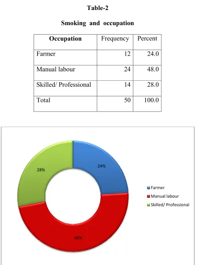
[image:64.612.111.504.80.608.2]57 Table-2
Smoking and occupation
ccupation Frequency Percent
12 24.0
al labour 24 48.0
d/ Professional 14 28.0
50 100.0
rers are the group who are associated with inc
ng in this study.
24%
48%
Farmer
Manual labou
Skilled/ Profe
ith increased
abour
FEV1
Normal Mild
Modera
Total
FEV1 is normal in
decreased in 4% of the
obstruction.
54%
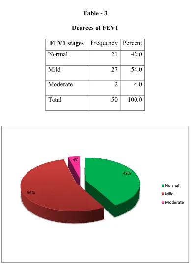
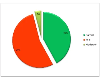
[image:65.612.112.501.69.607.2]58 Table - 3
Degrees of FEV1
EV1 stages Frequency Percent
ormal 21 42.0
27 54.0
oderate 2 4.0
50 100.0
al in 42%,mildly decreased in 54% and mod
the population under study. no one had
42% 4%
Nor
Mil
Mo
d moderately
had severe
Normal
Mild
FEV1/FEC
FEV1/
Normal Mild
Moderate
Total
FEV1/FVC ratio is m
in 4% of the population in
and none showed severe o
54%
59 Table - 4
1/FEC ratio in various individuals
EV1/FVC Frequency Percent
rmal 21 42.0
27 54.0
derate 2 4.0
50 100.0
tio is mildly decreased in 54%,moderately decr
in this study.It is normal in the remaining p
vere obstruction.
42% 4%
Norm
Mild
Mode
ly decreased
ning persons
ormal
ild
FE
Normal Mild
Moderat
Total
FEF25-75% also sho
FEV1/FVC ratio.
54%
[image:67.612.130.482.352.631.2]60 Table - 5
FEF values
FEF Frequency Percent
ormal 21 42.0
27 54.0
oderate 2 4.0
50 100.0
lso showed similar results as that of FEV1 and
42% 4%
Normal
Mild
Modera
1 and
mal
Ty
Beedi Cigarette
Total
In the study group, a
bidis.
62%
[image:68.612.124.490.318.580.2]61 Table -6
Types of smoking
Type Frequency Percent
19 38.0
igarettes 31 62.0
50 100.0
roup, about 62% smoked cigarettes and 38% sm
38%
Beedi
Cigare
8% smoked
edi
showing the
Duration of smo
10 15 20 25 30 35 > Tota
In this study,maxim
15-19 years and only 3% o
[image:69.612.141.474.146.619.2]years. 14% 6% 12% 10 62
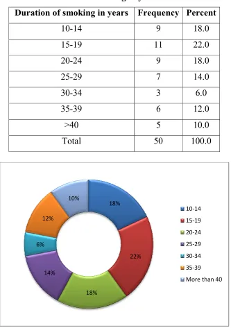
Table - 7
g the percentage of smokers with duration o
smoking in years
of smoking in years Frequency Percent
10-14 9 18.0
15-19 11 22.0
20-24 9 18.0
25-29 7 14.0
30-34 3 6.0
35-39 6 12.0
>40 5 10.0
Total 50 100.0
aximum number of smokers, (i.e) 22% smoke
y 3% of smokers smoked for a long duration of
18% 22% 18% 10% 10-14 15-19 20-24 25-29 30-34 35-39
More than 40 tion of
smoked for
ion of 30-34
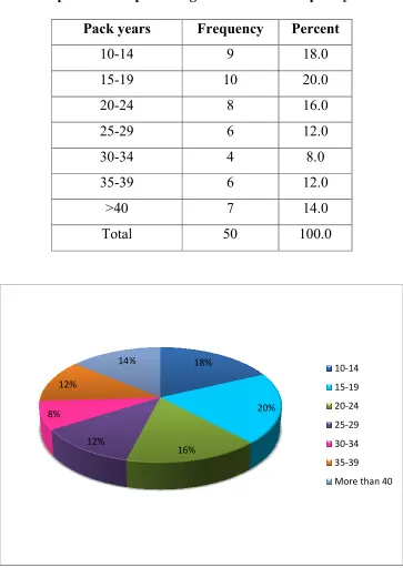
Comparison of p Pack yea 10-14 15-19 20-24 25-29 30-34 35-39 >40 Total
20% of smokers had
34 pack years.
12% 8%
12%
14%
[image:70.612.124.487.131.643.2]63
Table – 8
of percentage of smokers with pack years
k years Frequency Percent
14 9 18.0
19 10 20.0
24 8 16.0
29 6 12.0
34 4 8.0
39 6 12.0
7 14.0
Total 50 100.0
ers had 15-19 pack years of smoking and 8% ha
18% 20% 16% 14% 10-14 15-19 20-24 25-29 30-34 35-39 More tha years
8% had
Comparison of FEV Occupation Farmer Manual labour Skilled/ Professional Total
χ2 =0.519 df=2
There is no sign
reduction in FEV1.
0 10 20 30 40 50 60 70 Farmer 33.3 66.7 P e rc e n ta g e De 64 Table – 9
f FEV1 reduction in relation to occupatio
FEV1
Tota Normal Mild/ Moderate
N % N % N
4 33.3 8 66.7 12
11 45.8 13 54.2 24
6 42.9 8 57.1 14
21 42 29 58 50
p=0.771
significant association between occupatio
Manual labour Skilled/ Professional 45.8
42.9
54.2 57.1
Degrees of obstruction of FEV1 No M upation Total % 100 100 100 100
upation and
Normal
Comparison of
Age Normal
N %
31-40 12 100.
41-50 7 43.
51-60 2 9.
Total 21 42.
There is a significa
FEV1.Smokers of the age
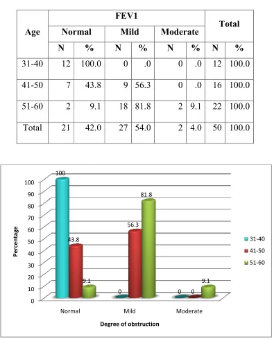
[image:72.612.111.506.134.623.2]flow rates. 0 10 20 30 40 50 60 70 80 90 100 Normal 100 43.8 9.1 P e rc e n ta g e 65 Table - 10
on of FEV1 reduction in relation to age
FEV1
Total rmal Mild Moderate
% N % N % N %
100.0 0 .0 0 .0 12 100.
43.8 9 56.3 0 .0 16 100.
9.1 18 81.8 2 9.1 22 100.
42.0 27 54.0 2 4.0 50 100.
gnificant association between age and reduc
e age group 51-60 years had marked limitatio
Mild Moderate 0 0 56.3 0 9.1 81.8 9.1
Degree of obstruction
otal % 100.0 100.0 100.0 100.0
reduction in
itation in air
31-40
41-50
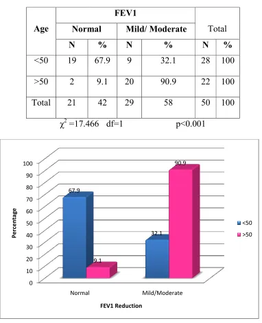
Comparison of mild an
moderate red
Age Norm
N
<50 19
>50 2
Total 21
χ2 =17.46
Smokers of more th
FEV1 when compared
[image:73.612.112.494.168.639.2]very significant. 0 10 20 30 40 50 60 70 80 90 100 Normal 67.9 P e rc e n ta g e 66 Table - 11
ild and moderate reduction of FEV1 in mil
e reduction of FEV1 in relation to age.
FEV1
Total
Normal Mild/ Moderate
% N % N %
67.9 9 32.1 28 100
9.1 20 90.9 22 100
42 29 58 50 100
17.466 df=1 p<0.001
ore than 50 years of age had significant reduc
to smokers of less than 50 years of age.
al Mild/Moderate 32.1
9.1
90.9
FEV1 Reduction
in mild and
reduction in
age. This is
<50
Comparison of
Age Normal
N %
31-40 12 100.
41-50 7 43.
51-60 2 9.
Total 21 42.
FEV1/FVC ratio is
degrees in 51-60 years ag
the age group of 41-50 yea
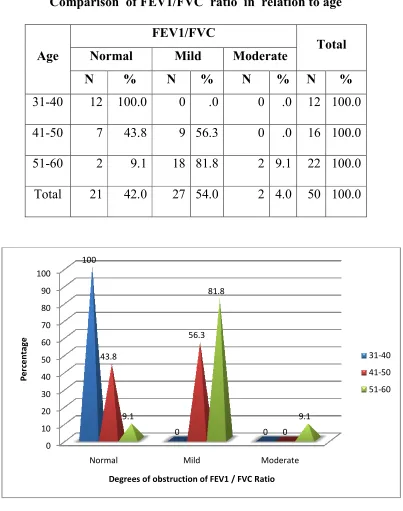
[image:74.612.110.513.124.635.2]0 10 20 30 40 50 60 70 80 90 100 Normal 100 43.8 9.1 P e rc e n ta g e Degree 67 Table - 12
on of FEV1/FVC ratio in relation to age
FEV1/FVC
Total rmal Mild Moderate
% N % N % N %
100.0 0 .0 0 .0 12 100.
43.8 9 56.3 0 .0 16 100.
9.1 18 81.8 2 9.1 22 100.
42.0 27 54.0 2 4.0 50 100.
atio is very much declined to mild and mo
age of smokers.It is declined to a lesser ex
50 years. Mild Moderate 0 0 56.3 0 9.1 81.8 9.1
grees of obstruction of FEV1 / FVC Ratio
otal % 100.0 100.0 100.0 100.0
nd moderate
sser extent in
31-40
41-50
Comparison of redu
Age Normal
N %
31-40 12 100.
41-50 7 43.
51-60 2 9.
Total 21 42.
FEF25-75% also s
parameters with the age gr
[image:75.612.109.522.129.657.2]100 43.8 9.1 0 20 40 60 80 100 120 Normal P e rc e n ta g e De 68 Table - 13
f reduction of FEF 25%-75% in relation to a
FEF 25-75%
Total rmal Mild Moderate
% N % N % N %
100.0 0 .0 0 .0 12 100.
43.8 9 56.3 0 .0 16 100.
9.1 18 81.8 2 9.1 22 100.
42.0 27 54.0 2 4.0 50 100.
also shows similar distribution as that of
age group under study.
0 0 56.3 0 9.1 81.8 9.1 Mild Moderate
Degress of obstruction of FEF 25%-75%
n to age
otal % 100.0 100.0 100.0 100.0
at of other
31-40
41-50
Comparison of mild an
Age No
N <50 19 >50
Total 21
χ2 =17.46
FEF25-75% shows
years when compared with
[image:76.612.123.493.167.632.2]0 10 20 30 40 50 60 70 80 90 100 Less than5 67.9 P e rc e n ta g e Reduction of 69 Table - 15
ild and moderate reduction of FEF
25%-relation to age
FEF25-75%
Total Normal
Mild/ Moderate
% N % N %
19 67.9 9 32.1 28 100
2 9.1 20 90.9 22 100
21 42 29 58 50 100
17.466 df=1 p<0.001
shows a significant reduction in age more th
with age less than 50 years.
an50 More than 50 9.1 32.1
90.9
n of FEF 25%-75% relation to age
Normal
Mild/Mode
-75% in
ore than 50
70 Table – 16
Comparison of FEV1 reduction in relation to duration of
smoking in years
Duration of
smoking in years
FEV1
Total Normal Mild/ Moderate
N % N % N %
10-19 17 85.0 3 15.0 20 100.0
20-29 4 25.0 12 75.0 16 100.0
>30 0 .0 14 100.0 14 100.0
Total 21 42.0 29 58.0 50 100.0
χ2 =27.217 df=2 p<0.001
FEV1 shows a significant reduction with prolonged duration of
smoking. More than 30 years of smoking in this study showed significant
airfiow Limitation when compared with 10-19 years of smoking
Comparison of FEV
FEV1 shows a sig
smoking. More than 30 ye
airfiow Limitation whe
duration. 0 10 20 30 40 50 60 70 80 90 100 10-19 85 15 P e rc e n ta g e 71
f FEV1 reduction in relation to duration
smoking in years
a significant reduction with prolonged durat
30 years of smoking in this study showed sign
when compared with 10-19 years of sm
20-29 More than 30 25
0 75
100
FEV1 Reduction related to age
Normal
Mild/Mod tion of
duration of
d significant
of smoking