Copyright © 1997, American Society for Microbiology
Regression of Papillomas Induced by Cottontail Rabbit
Papillomavirus Is Associated with Infiltration of CD8
1
Cells
and Persistence of Viral DNA after Regression
R. SELVAKUMAR,1A. SCHMITT,2T. IFTNER,2R. AHMED,1,3†ANDF. O. WETTSTEIN1,3,4*Department of Microbiology and Immunology, School of Medicine,1Jonsson Comprehensive Cancer Center,3and
Molecular Biology Institute,4University of California, Los Angeles, California 90095-1747, and Institut fu¨r Klinische
und Molekulare Virologie der Universita¨t Erlangen-Nu¨rnberg, 91054 Erlangen, Germany2
Received 16 December 1996/Accepted 24 March 1997
Cottontail rabbit papillomavirus (CRPV) is a highly oncogenic papillomavirus and has been successfully used as a model to develop protective vaccines against papillomaviruses. Papillomas induced by the virus may spontaneously regress, suggesting that CRPV can also serve as a model to develop therapeutic vaccines. As a first step toward this goal, we have analyzed immunologic and viral aspects associated with papilloma regression and have identified several features unique to regression. Immunohistochemical staining of biopsies from growing and regressing papillomas and from sites after complete regression showed infiltration of CD81
cells into the basal and suprabasal layers of the epidermis only during active regression. In situ hybridizations with mRNA-specific probes were strongly positive for E6 and E7 mRNAs during regression, but no late mRNA was present. Viral DNA was detected by in situ hybridization during regression but not after regression. However, analysis by PCR revealed persistence of viral DNA for several months at the majority of regression sites. The results suggest that stimulation of a strong CD81response to virus-infected cells is important for
an effective therapeutic vaccine and that special attention should be given to the suppression of latent infection.
Papillomaviruses (PVs) are small DNA tumor viruses with a circular genome of about 8 kb. Replication of the viruses is highly species and tissue specific, and the viral life cycle is tightly linked to the differentiation of the stratified epithelium (38). Cottontail rabbit PV (CRPV) was the first PV identified. Its natural host is the cottontail rabbit; however, papillomas can be induced with equal efficiency in domestic rabbits. Pap-illoma formation in both host species is strictly limited to the hairy skin epithelium. The basis for this specificity has recently been elucidated, as it was shown that cells permissive for a high level of early viral gene expression are located in the hair follicles from which papillomas grow out (33). In papillomas induced in domestic rabbits, only low levels of mRNA encod-ing viral structural proteins are present (50), and little or no infectious virus can be recovered. Papillomas induced by CRPV can progress to invasive cancers (41), a property shared by a subset of genital (51) and cutaneous (31) human PVs (HPVs), the so-called high-risk HPVs. Furthermore, some papillomas induced by CRPV regress spontaneously, a fate also observed with HPV-induced lesions. Thus, in several as-pects CRPV can serve as a model to study various asas-pects of HPV infection.
CRPV was extensively used to develop prophylactic PV vac-cines. First, it was shown that both structural proteins L1 and L2 provided protection (5, 24), although immunization with L1 was superior. However, protection by L1 was provided only by the full-length, nondenatured protein (24). Particularly effec-tive were virus-like particles produced with baculovirus vectors in insect cells (3, 6) or in yeast cells (18). Protection was also
achieved by immunization with L1-expressing recombinant vaccinia virus (25) and by DNA vaccination with an L1-ex-pressing vector (10). Sera from L1 protein-immunized animals contained neutralizing antibodies and were effective in passive immunization (3). This suggests that protection by L1 immu-nization is based on neutralization of the infecting virus, and it may not involve a response to virus-infected cells. This sugges-tion is supported by the finding that L1-immunized rabbits are not protected against DNA-induced papilloma development. Furthermore, when infection sites of L1-immunized rabbits were probed for viral DNA by PCR 3 months after infection, no viral DNA was detected (24). Since receptors for PVs are rather ubiquitous (27), virus will be taken up by permissive keratinocytes as well as by nonpermissive cells. While permis-sive cells containing viral DNA may potentially be eliminated by an immune response to virus-infected cells, this is less likely the case with nonpermissive cells. Thus, the complete elimina-tion of viral DNA in L1-immunized rabbits strengthens the notion that L1 vaccines completely prevented uptake of the virus. Effective vaccination with L1 of the natural host was also demonstrated with bovine PV types 1 (32) and 4 (4) and with canine oral PV (40).
PV infections in humans and animals usually result in a persistent infection lasting for a prolonged period of time, sometimes for the lifetime of the host. Although antibodies against structural and nonstructural viral proteins are often detected in hosts with persisting papillomas, the immune re-sponse does not eliminate the virus-infected cells. However, in humans (1, 42, 43) as well as in animals (4, 11, 19, 22, 37, 41), papillomas may regress spontaneously. In regressing human warts there was evidence of CD41-T-cell infiltration (7), and
the spontaneous regression of an HPV type 31 condyloma was transiently associated with an in vitro proliferation response of peripheral blood mononuclear cells (PBMCs) to an E7 peptide (20). In domestic rabbits, genetic analyses suggested that spon-taneous regression was linked to certain major
histocompati-* Corresponding author. Mailing address: Department of Microbi-ology and ImmunMicrobi-ology, UCLA School of Medicine, 10833 Le Conte Ave., Los Angeles, CA 90095-1747. Phone: (310) 825-7239. Fax: (310) 206-3865.
† Present address: Department of Microbiology and Immunology, Emory University School of Medicine, Atlanta, GA 30322.
5540
on November 9, 2019 by guest
http://jvi.asm.org/
bility complex class II (MHCII) allotypes, but the molecular analysis showed that it was not the MHCII gene itself but rather a gene linked to certain MHCII alleles (15, 16). Immu-nohistochemical analysis of spontaneously regressing rabbit papillomas indicated that regression was associated with some infiltration of leukocytes (30) and an accumulation of T cells and of cells positive for tumor necrosis factor alpha (TNF-a) mRNA (14) in the dermis below the regressing papilloma (29). Furthermore, there was a suggestion from skin tests of an increased cellular reactivity to viral structural protein in gressors (17). In vitro proliferation assays with PBMCs re-vealed that the regression was associated with an immune response to E2 (34). Furthermore, papilloma regression could be increased by immunization. First it was shown that vaccines prepared from papillomas increased the frequency of regres-sion (11), and subsequently it was shown that this could be accomplished by immunization with E1 and E2 (36), the two viral proteins required for DNA replication (44), as well as by immunization with E6, the product of one of the viral onco-genes (23). These observations, together with the finding that regressor animals were resistant to infection with DNA (12), clearly indicate that regression is associated with an immune response to virus-infected cells.
Here, we have further defined steps associated with papil-loma regression. It is shown that the infiltrating T cells are CD8-positive cells. During regression, expression of the early genes E6 and E7 remains at levels comparable to those in persisting papillomas. After complete regression, viral tran-scripts are no longer detected, but DNA persists in a latent stage for prolonged periods of time.
MATERIALS AND METHODS
Animals, virus, and biopsies.The Washington B strain of CRPV was propa-gated in cottontail rabbits (39) and used to infect New Zealand White rabbits obtained from Irish Farms (Norco, Calif.). Skin biopsies were obtained from infection sites 3 to 17 months after spontaneous papilloma regression or 6 to 10 months after induced regression from animals immunized with TrpE-E1 or TrpE-E2 or both (36), from normal skin and from papillomas. Biopsies were frozen in liquid nitrogen and stored at276°C.
PCR analysis.Extracts from 4-mm punch biopsies were prepared as previously described (24, 26). Briefly, biopsies were cut into small pieces and incubated for 1 h at 55°C in 200ml of K buffer (50 mM KCl, 10 mM Tris-HCl [pH 8.3], 2.5 mM MgCl2, 100mg of bovine serum albumin per ml, 0.45% Nonidet P-40, 0.45%
Tween 20, 60mg of proteinase K per ml). The extracts were boiled for 10 min, insoluble material was removed by centrifugation, and the extracts were stored at 276°C. For DNA isolation, small biopsy pieces were incubated overnight at 55°C in 0.1 M EDTA (pH 8.0)–0.05 M Tris-HCl (pH 8.0)–0.5% sodium dodecyl sulfate–100mg of proteinase K per ml. The DNA was then extracted twice with phenol-chloroform-isoamyl alcohol (75:24:1) for 5 min at 50°C, and phenol was removed from the aqueous phase by three successive ether extractions. NH4
acetate was added to 0.1 M, and the DNA was precipitated with 2 volumes of ethanol at220°C overnight. The DNA pellets were washed with 70% ethanol, dried, and dissolved in 1 mM Tris-HCl (pH 8.0)–0.1 mM EDTA. The concen-tration of DNA was estimated by electrophoresis in 1% agarose gels in the presence of 0.5mg of ethidium bromide per ml together with standard amounts of uncut lambda DNA. The primer sets for amplification of different CRPV DNA segments were as follows (nucleotide numbers are according to reference 13). For the upper regulatory region (URR), the 59primer was 59CCTGCAC CTGTTAGTATTGTG 39, the 39primer was 59CTCTAGCGAGCGTGGCA GGC 39, and the amplified segment of 757 bp extended from nucleotide (nt) 7295 to 183. For E6, the 59primer was 59GCTGGCACTGTGTATCTGAG 39 and the 39primer was 59CCTGCGCACAGGATAGCAG 39, and they amplified a segment of 922 bp from nt 81 to 1002. E7 was amplified with primers as described previously (24), resulting in a segment of 344 bp from nt 1043 to 1386. For E1, the 59 primer was 59 CGCTTTCCCTGGTGTGCCC 39 and the 39 primer was 59CTGGGACTCCAAACTCGTGC 39, and they amplified a seg-ment of 1,874 bp from nt 1325 to 3198. For E2, the 59primer was 59GGAAG ACGAGGGTGACGATGG 39and the 39primer was 59GCTGCTGATGGGA ATGGG 39, and they amplified a segment of 1,226 bp from nt 3095 to 4320. As a control for effective amplification, a primer set that amplified a fragment of rabbit cytochrome P-450 was used, resulting in a segment of 159 bp as described previously (24). The PCRs were carried out in 50-ml volumes and contained 5ml of biopsy extract or purified DNA as indicated in Results, 50 pmol of each primer, 200mM deoxynucleoside triphosphates, 2.5 U of Taq DNA polymerase
(GIBCO-BRL), 20 mM Tris-HCl (pH 8.4), and 100mg of bovine serum albumin per ml. The KCl and MgCl2concentrations were optimized for each primer set
by using the Opti-Primer Kit (Stratagene). The optimal concentrations were 1.5 mM MgCl2and 75 mM KCl for the URR, E1, E2, and P-450 primers and 3.5 mM
MgCl2and 75 mM KCl for the E6 and E7 primers. Thirty cycles of amplification
were performed as previously described (24) by using a Perkin-Elmer DNA thermal cycler 480. Portions (10ml) of the PCR mixtures were electrophoresed on 1.8% agarose gels in the presence of 0.5mg of ethidium bromide per ml. The gels were photographed, and the DNA was transferred to GeneScreen Plus (New England Nuclear) or Nytran (Schleicher & Schuell) membranes with a Turbo-blotter (Schleicher & Schuell). The membranes were hybridized with 0.23106
cpm of an E7-specific probe or 13106cpm of a genomic probe, labeled by
random priming with a random primer kit (GIBCO-BRL), per ml.
RNA probes and in situ hybridization.The construction of the plasmids for mRNA-specific probes and for DNA was as described previously (33). (The map of the RNA probes [antisense] is shown in Fig. 2.) The probes were synthesized in vitro in the presence of35S-UTP and had a specific activity of 3.53108
cpm/mg. The probes were reduced to a size of approximately 200 nt by alkaline hydrolysis and were tested by polyacrylamide gel electrophoresis. Sections 5 to 6 mm thick were cut with a cryostat microtome (Reichert-Jung), mounted on aminopropylsilane-coated slides, fixed in 4% paraformaldehyde in phosphate-buffered saline (PBS), and dehydrated through graded ethanol solutions. The sections were acetylated in 0.1 M triethanolamine–0.25% acetic anhydride for 10 min and washed in 0.23SSC (13SSC is 0.15 M NaCl plus 0.015 M sodium citrate). The sections were then prehybridized 2 h at 42°C in solution I (45% formamide, 0.6 M NaCl, 2.53Denhardt’s solution, 10 mM Tris-HCl [pH 7.5], 1 mM EDTA, 0.1% sodium dodecyl sulfate, and 0.15 mg of tRNA per ml). After prehybridization, individual sections were encircled with rubber cement and hybridized with different probes in solution I containing 10% dextran sulfate. After 16 h of hybridization, the slides were washed in 50% formamide–13SSC at 55°C, treated with RNase A (10mg/ml), washed twice for 2 h in 0.13SSC at 60°C, dehydrated, and coated with Kodak NTB-2 emulsion. Slides were devel-oped after exposure for 10 to 12 days. After staining with hematoxylin and counterstaining with eosin, the sections were evaluated with a Zeiss Axiophot microscope with a dark- or bright-field condenser. The background for RNA hybridization was determined with sense riboprobes and by pretreatment of sections with DNase-free RNase.
Staining of CD4- and CD8-positive cells.Frozen sections cut on the cryostat were air dried, fixed with acetone for 5 min at 21°C, dried again, and rehydrated in PBS. Endogenous peroxidase was quenched by incubation in 0.3% H2O2. The
sections were then washed twice in PBS and treated with a 1:20 dilution of normal horse serum. Biotinylated monoclonal rabbit CD4 (21) and anti-rabbit CD8 (9, 49) were obtained from Spring Valley Labs. Different primary antibody batches were tested on spleen sections for proper cell surface staining. The slides were incubated with the antibody for 2 h at 21°C and then washed with PBS for 30 min. The primary signal was amplified by 1 h of incubation at 21°C with a 1:75 dilution of a mixture consisting of equal volumes of biotinylated anti-mouse immunoglobulin G and normal rabbit serum, which was precentri-fuged to remove immune complexes. The sections were then washed with PBS, exposed to freshly prepared ABC reagent (Vector Labs) for 30 min at 21°C, and washed with PBS again. Color development was performed with 3-amino-9-ethylcarbazole (Sigma) and H2O2in acetate buffer for 10 min. After being rinsed
in H2O, the slides were stained with hematoxylin. A monoclonal antibody to
human cytokeratin 10 of the same isotype as the anti-CD4 and anti-CD8, im-munoglobulin G1 kappa, served as negative control.
RESULTS
CD81-cell infiltration and viral mRNA expression in
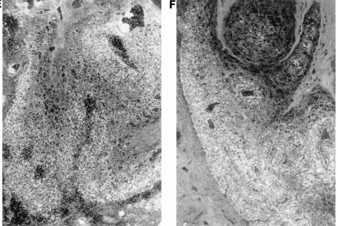
re-gressing papillomas.Previous investigations have shown that spontaneous papilloma regression was associated transiently with an in vitro proliferation response of PBMCs to E2 and some infiltration of the epidermis with leukocytes during re-gression (30). To identify immune cells potentially involved in papilloma regression and to evaluate the effect of regression on viral mRNA expression, thin sections were prepared from spontaneously regressing and growing papillomas. Immune cells were identified by immunohistochemical staining with antibodies to CD4 and CD8, and the presence of viral mRNA was assessed by in situ hybridization. A comparison between regressing and growing papillomas is shown in Fig. 1. One regressing papilloma was removed 4 months after infection, at a time when two of the four papillomas had completely re-gressed (Fig. 1A), and the second was removed from a regres-sor rabbit 6 months after infection, when three of the four papillomas had completely regressed (Fig. 1C and E); the evaluation of growing papillomas is shown in Fig. 1B, D, and F.
on November 9, 2019 by guest
http://jvi.asm.org/
on November 9, 2019 by guest
http://jvi.asm.org/
Few CD81cells were present in the growing papillomas (Fig. 1B
and D), while CD81cells can be seen in the basal and suprabasal
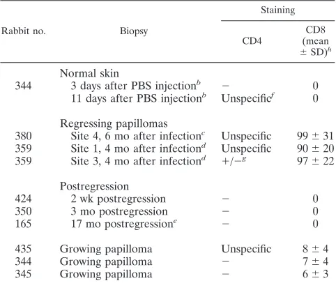
layers of the regressing papillomas (Fig. 1A and C). In contrast, there was no difference in transcription pattern between the re-gressing and growing papillomas as revealed by a probe hybrid-izing to the two major early transcripts (Fig. 1E and F). The results of CD4 and CD8 staining of biopsies from normal skin, from regressing papillomas, from sites after complete regression, and from growing papillomas are summarized in Table 1, and a detailed analysis of transcription in regressing papillomas is pre-sented in Fig. 2. The quantitative evaluation of CD81-cell
infil-tration revealed a highly significant difference between regressing and growing papillomas. Per field of view, all three regressing papillomas contained, on the average, 90 to 100 CD81cells, while
the number in growing papillomas was less than 10. No CD81
cells were detected in normal skin, and sites of regression were already negative at 2 weeks postregression.
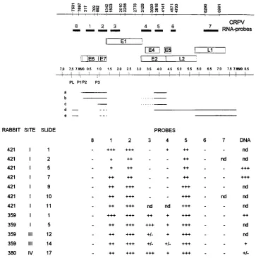
The comparison of the in situ hybridizations (Fig. 1E and F) suggested that there was no difference in viral transcripts be-tween regressing and progressing papillomas. The more exten-sive analyses are presented in Fig. 2 and include analyses of a papilloma from an induced regressor. The analysis of results with mRNA-specific probes revealed a qualitative difference between transcripts in growing papillomas (33, 50) and in re-gressing papillomas (this work). Of the three probes specific for
late transcripts (Fig. 2, probes 6, 7, and 8), none gave a positive signal with the regressing papillomas. In this regard, there was no difference between spontaneously regressing papillomas and the papilloma of an animal in which regression was induced by im-munization with TrpE-E1 (Fig. 2, rabbit 421). Since only one papilloma from an induced regressor was analyzed, it is not clear if the paucity of E1- and E2-specific transcription is related to induced regression. Overall, the immunohistochemical analyses and in situ hybridization of regressing papillomas revealed impor-tant differences from growing papillomas. The level of CD81-cell
infiltration was highly increased during regression. Late tran-scripts detected in the upper layers of growing papillomas were absent from regressing papillomas. However, the presence of CD81cells in the basal and suprabasal layers of the epidermis did
not affect the abundance of early viral transcripts. Finally, within 2 weeks after complete regression, CD81cells were no longer
present in the epidermis.
[image:4.612.69.554.73.398.2]Persistence of viral DNA after tumor regression. In the course of our investigation of the immune response of rabbits to CRPV, spontaneous papilloma regression was observed in 24 animals, representing a regression frequency of 12%. Among the animals in which papillomas had regressed, in two animals a papilloma reemerged at one of the four regression sites. This suggested that viral DNA could persist in a latent state. To determine if DNA persistence was a rare event or
FIG. 1. Immunohistochemical staining of CD81cells and in situ RNA hybridization in regressing and growing papillomas. Preparation of thin sections, staining, and in situ hybridization were as described in Materials and Methods. (A, C, and E) Regressing papillomas; (B, D, and F) growing papillomas. (A and B) 100-fold magnification; (C and D) 200-fold magnification; (E and F) in situ hybridization. The general orientation of sections is with the differentiating layers of epithelium toward the top. The arrowheads point to the basal layer of the epithelium in panels A to D. Little of the dermis below the epidermis is shown. The architecture of regressing papillomas in general was less well maintained than that of growing papillomas or normal skin, and this was particularly evident with sections prepared for immunohistochemical staining which could not be fixed with formaldehyde.
on November 9, 2019 by guest
http://jvi.asm.org/
more common, punch biopsies were obtained from regression sites and analyzed by PCR for the presence of CRPV DNA. The Southern blot analyses of regression site extracts hybrid-ized with an E7-specific probe are shown in Fig. 3 and repre-sent a 2-day exposure at276°C without an amplifying screen. As can be seen, in 9 of the 15 biopsy samples an E7-specific band was detected. When the exposure was repeated for 2 days in the presence of an amplifying screen, an additional two samples (Fig. 3, lanes 7 and 15) were positive. In all samples the amplified segment of rabbit cytochrome P-450 was de-tected by ethidium bromide staining (data not shown). This indicated that the absence of an E7-specific band was not due to inadequate amplification of DNA in the biopsy ex-tract. The results of the analysis of 54 biopsies are summa-rized in Table 2. In about two-thirds of the samples, CRPV DNA was present, and in 16 of the 19 animals tested, at least one site was positive. Furthermore, there was no difference between animals in which papillomas regressed spontane-ously and those in which papilloma regression was induced by immunization with TrpE-E1 and -E2 fusion proteins (36). Finally, CRPV DNA was detected as late as 17 months after regression. These results indicate that the persistence of viral DNA after papilloma regression was a rather common feature.
The E7-specific viral band seen after Southern blot hybrid-ization could not be detected by ethidium bromide staining, while a cellular gene-specific band could be. This suggested
that viral DNA in regression sites was present at concentra-tions of less than two copies per cell. To obtain information about the abundance of viral DNA, PCRs were carried out with DNA purified from biopsies by standard DNA extraction rather than with crude biopsy extracts. This permitted us to estimate the amount of cellular DNA present in PCR mixtures. The results of the analysis of the PCR-amplified DNA by Southern blotting and hybridization with a genomic probe are shown in Fig. 4. The PCR mixtures of regression site DNA contained approximately 0.15mg of DNA (Fig. 4, lanes 1 to 5), while those of papillomavirus DNA and CRPV DNA con-tained 0.5 and 5 31026ng of DNA (Fig. 4, lanes 6 and 7,
respectively). The additional lanes contained molecular size marker DNA (Fig. 4, lanes 8), a PCR with no DNA (lanes 9), and 0.5mg of DNA from normal rabbit skin (lanes 10). Figure 4A shows an ethidium bromide-stained gel of PCR-amplified cytochrome P-450, and Fig. 4B and C show autoradiograms of 2- and 40-h exposures, respectively, of a filter with amplified E7 DNA. Of the five regression site samples, four were positive after a 40-h exposure. The intensity of the E7 bands at 2 h was about the same for one of the regression site DNAs and the papilloma DNA, while the CRPV DNA band was slightly weaker. Neglecting this difference, the CRPV DNA content per cell can be estimated for the regression site and the pap-illoma. The amount of DNA input into the PCR was 105times
higher for the papilloma DNA (0.5 ng) than for the CRPV DNA (531026ng), and the ratio of the sizes of the diploid
cellular genome (63109bp) and the viral genome (83103
bp) is 7.5 3 105. Thus, on average the papilloma cells
con-tained about 7.5 copies of viral DNA per cell. Since the amount of DNA input into the PCR for the regression site (0.15mg) was 300 times higher than that for the papilloma PCR (53 1024mg), the regression site contained about one CRPV copy
per 40 cells. A comparison of the 40-h exposure (Fig. 4C), to the 2-h exposure (Fig. 4B) suggests that the weakest positive biopsy sample contained at least 20 times less CRPV DNA. The data presented in Fig. 3 and 4 show that the abundance of viral DNA varies greatly among positive regression sites, from about one copy per 40 cells to about one copy per 1,000 cells, as some samples became positive only after a 2-day autoradio-graphic exposure with an amplifying screen.
The survey of regression sites summarized in Table 2 was per-formed by using detection of the E7 gene as an indicator. To see if retention of E7 was preferential, the additional viral genes E6 (Fig. 4E) and E1 (Fig. 4F), as well as the URR containing also the origin of replication (Fig. 4D), were amplified. As can be seen for the biopsy sample with the strongest signal for E7 (Fig. 4, lanes 5), all three other sequences tested could also be amplified, as could E2 (data not shown). After a 40-h exposure, E6 could also be detected in three of the low-copy biopsies (data not shown), but not the E1 band or the URR band. Most likely this reflects a lower efficiency of PCR amplification of these two segments. Overall, the data imply that more than the E7 gene was retained, possibly the entire viral genome.
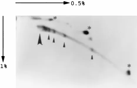
The prolonged persistence of viral DNA at regression sites in the skin, a tissue which under goes continuous renewal, would require at least a low level of replication. If the DNA is maintained extrachromosomally, it would require expression of E1 and E2 (44); alternately, the DNA could be integrated. Because of the low abundance, a direct analysis of the state of the DNA in regression sites is not possible. The reemergence of papillomas from regression sites provided material with which the state of the DNA could be evaluated. One animal in which a papilloma reemerged may have been unusual, since it did not exhibit an antibody response to E2 before complete regression. Such a response was observed in over 80% of
re-TABLE 1. Immunohistochemical staining of CD41and CD81cells
in skin biopsies from growing and regressing papillomas and from postregression sitesa
Rabbit no. Biopsy
Staining
CD4 (meanCD8 6SD)h
Normal skin
344 3 days after PBS injectionb 2 0
11 days after PBS injectionb Unspecificf 0
Regressing papillomas
380 Site 4, 6 mo after infectionc Unspecific 99631
359 Site 1, 4 mo after infectiond Unspecific 90620
359 Site 3, 4 mo after infectiond 1/2g 97622
Postregression
424 2 wk postregression 2 0
350 3 mo postregression 2 0
165 17 mo postregressione 2 0
435 Growing papilloma Unspecific 864
344 Growing papilloma 2 764
345 Growing papilloma 2 663
aBiopsy thin sections were obtained and stained for CD4 and CD8 as
de-scribed in Materials and Methods.
bPBS was injected by high pressure into rabbit skin as described previously
(33).
cAt the time the biopsy was taken, papillomas at three other sites had
com-pletely regressed.
dAt the time the biopsy was taken, papillomas on two sites had completely
regressed.
eA portion of the biopsy taken was analyzed by PCR and was still positive for
CRPV DNA.
fThe staining was not associated with the cellular portion of the thin section. gA signal was present only in a few isolated cells.
hFor the quantitative evaluation of CD81cell infiltration into the epidermis,
the numbers of CD81cells in five randomly chosen areas of approximately 1 by
0.6 mm were counted, the average number was determined, and the standard deviation was calculated.
on November 9, 2019 by guest
http://jvi.asm.org/
[image:5.612.58.299.100.303.2]gressor rabbits surveyed (34). Two-dimensional analysis of the DNA from the reemerging papilloma of this animal is pre-sented in Fig. 5, which shows, from lower right to upper left, the positions of monomeric, dimeric, and tetrameric super-coiled DNAs (asterisks) with larger circular DNA species mov-ing even slower in both dimensions. Although a linear DNA trail is clearly visible, the presence of discrete spots corre-sponding to monomeric to tetrameric linear DNAs (small ar-row heads) suggests that linear DNA was generated from ran-dom degradation of extrachromosomal DNA. At the position where the bulk of the cellular DNA migrated (large arrow-head), as visualized by ethidium bromide staining, only a small portion of the viral DNA was present, and this suggests that at least in this case CRPV DNA was maintained extrachromo-somally after regression.
DISCUSSION
Here we have presented evidence for a regression-associated infiltration by CD81cells into papillomas. The difference in
CD81infiltration between growing and regressing
papillo-mas is much more dramatic than the rather small difference observed by comparing total leukocyte infiltration (30). In the basal and suprabasal layers, where the CD81 cells are
present, there is no evidence of local necrosis, suggesting that the CD81 cells were not massively involved in cell
killing. It is likely that the CD81cells specifically recognize
viral antigen presented on kertinocytes, and this would re-quire a direct contact between the infected cells and CD81
cells. This recognition may then lead to a release of cyto-kines. However, this does not affect viral RNA expression locally, but it may be critical to attract leukocytes into the dermis below regressing papillomas, which were shown to contain TNF-a mRNA (14). The importance of a direct cellular recognition of virus-infected cells is suggested by the finding that E1-induced regression is not always accom-panied by a humoral response to E1 (36). Surprisingly, the reagents used did not reveal any infiltration of CD41cells in
[image:6.612.126.489.71.438.2]the epidermis. Although a technical problem specifically related to staining of CD41cells in the epidermis cannot be
FIG. 2. Summary of the in situ hybridization with sections from biopsies. (Top) Positions of the probes previously described (33), of the open reading frames (indicated by open boxes) (13), and of the major transcripts mapped in domestic and cottontail rabbit papillomas (28, 46). The vertical lines in the open reading frame boxes indicate the positions of the translation start sites. Dashed lines denote exons with unknown 59boundaries, and dotted lines represent alternate splicing patterns. The early promoters are designated P1, P2, and P3, and the late promoter is designated PL (8, 47). (Bottom) Hybridization with eight antisense RNA probes and a DNA probe with sections of a regressing papilloma (site I on rabbit 421, sites I and III on rabbit 359, and site IV on rabbit 380). Adjacent serial sections were placed on one slide for hybridization with the eight different probes for RNA and the probe for DNA.1, weak hybridization;11, moderate hybridization;111, strong hybridization;1/2, signals in only a few isolated cells; nd, not done.
on November 9, 2019 by guest
http://jvi.asm.org/
ruled out, this appears unlikely, since CD41-cell staining
was strong when tested on spleen sections.
The cytotoxic T-cell response inducing papilloma regression appears to be relatively weak compared to a response in a systemic viral infection. This is indicated by the finding that a specific cytotoxic T-cell response could be demonstrated with PBMCs from vaccinia virus-infected rabbits but not with PBMCs from regressor rabbits (35). The data thus suggest the following events in papilloma regression. Virus-infected cells are recognized by infiltrating CD81cells. The effect of CD81
cells on papilloma development is strongly amplified by TNF-a-secreting leukocytes attracted by CD81cells to the dermis
below papillomas. This results in abrogation of the virus-stim-ulated growth in the suprabasal layer (14), directly or indirectly eliminates late viral gene expression, and results in necrosis in the upper layer through apoptosis (14).
Regression, surprisingly, does not result in elimination of all cells which harbor viral DNA. One possibility is that the DNA in these cells was integrated. Clearly, the two-dimensional gel analysis of the DNA from a papilloma which reemerged from a regression site showed that it contained almost exclusively extrachromosomal DNA, and this strongly suggested that the DNA was maintained extrachromosomally after regression. It is unlikely that the DNA changed from an integrated to an extrachromosomal state as cells became competent for papil-loma development. Although such a process can be inferred from experiments with simian virus 40 where cocultivation of transformed cells with permissive cells resulted in the recovery of infectious virus, the process seems to be quite inefficient (45). No such observations have been reported for PVs, where the opposite process is often observed. The findings that spon-taneous regression is associated with an immune response to
E2 (34) and that immunization with E1 and E2 (36) as well as with E6 (23) greatly increases the regression frequency suggest that the cells surviving regression do not express high levels of these viral proteins. Since both E1 and E2 are required for PV DNA replication (44), the residual DNA may reside in cells which are in a resting stage. Attempts to directly identify viral mRNA by reverse transcription-PCR in biopsies from regres-sion sites were negative. A regresregres-sion site with a relatively high DNA content (one viral gene copy per 40 cells) was compared to a growing papilloma (75 viral DNA copies per cell). The negative results for the detection of E6 and E7 mRNAs sug-gested that viral transcription relative to the DNA content at the regression site was at least an order of magnitude lower than that in the papilloma (35). Continued production of some viral protein by CRPV DNA-containing cells after regression, however, was suggested. When biopsies of regression sites and normal skin were explanted into interleukin-2-containing me-dium, CD81cells were detected by fluorescence-activated cell
sorter analysis only in lymphocytes growing out of regression site biopsies. Since CD81cells were not detected in the
[image:7.612.330.544.67.387.2]epi-FIG. 3. Southern blot analysis of PCR-amplified viral DNA from extracts of regression sites. Biopsy extraction, PCR amplification, and Southern blot hybrid-ization with an E7-specific probe were performed as described in Materials and Methods. The PCR amplification mixtures contained the following: lane 1, no DNA; lane 2, extract from normal skin; lanes 3 and 19 molecular size markers; lanes 4 to 7, extracts from spontaneous regression sites; lanes 8 to 18, extracts from induced regression sites; lane 20, 0.5 ng of papilloma DNA. The position of the E7-specific band is indicated by an arrowhead on the left and those of the markers in the marker lanes and their sizes (in base pairs) are given on the right. The exposure shown was for 2 days at276°C without an amplifying screen. After a 2-day exposure with an amplifying screen, E7 bands were also detected in lanes 6 and 14 but not in lanes 8 and 9.
FIG. 4. PCR amplification of different segments of the early CRPV region of DNA isolated from biopsies and CRPV. DNA was isolated from regression site biopsies from a papilloma and from CRPV. DNA isolation, PCR amplification of the different segments, and Southern blot hybridization with a genomic probe were as described in Materials and Methods. Autoradiography was for 2 h (B, D, E, and F) or 40 h (C). (A) Ethidium bromide-stained gel shown also in panel D. The primers present in the PCR were for P-450 and the URR (A and D), E7 (B), E6 (E), and E1 (F). The PCR mixtures contained about 0.15mg of DNA isolated from regression sites (lanes 1 to 5), 0.5 ng of papilloma DNA (lane 6), or 53 1026ng of CRPV DNA (lane 7). Molecular size markers were present in lane 8, and the positions and sizes of the markers (in base pairs) are indicated on the right. Lane 9, PCR without DNA; lane 10, PCR with 0.5mg of normal rabbit skin DNA. The positions of relevant PCR products are indicated by arrowheads on the left.
TABLE 2. Persistence of viral DNA after spontaneous and induced papilloma regressiona
Regression No. with DNA detected/total no. (%)
Sites Animals
Spontaneousb 14/22 (64) 7/9 (78)
Inducedc 20/32 (62) 9/10 (90)
aPCR was performed with extracts from regression sites, and viral DNA was detected by Southern blot hybridization as described in Materials and Methods. bBiopsies were obtained 6 to 20 months after infection or about 3 to 17 months after regression.
cBiopsies were obtained 9 to 13 months after infection or about 6 to 10 months after regression from animals immunized with TrpE-E1 or -E2 or both before infection.
on November 9, 2019 by guest
http://jvi.asm.org/
[image:7.612.63.299.69.127.2] [image:7.612.58.298.616.670.2]dermis by immunohistochemistry after regression, the out-growing CD81 cells may have resided in the dermis.
Alter-nately, their number may have become significant only after growth stimulation with interleukin-2. That cells containing latent CRPV DNA may become competent to express viral genes at a high level is indicated by the spontaneous reemer-gence of papillomas after regression and by the finding that in rabbits infected with a low dose of virus which did not permit papilloma development, papilloma growth could be induced by skin irritation (2). Finally, it is possible that after macroscopic regression there is a continuous equilibrium between the im-mune response killing virus-infected cells and virus-infected cells escaping elimination. At present, we cannot exclude this possibility. However, it is less likely, since immune suppression with cyclosporine of two CRPV DNA-positive regressor rab-bits did not result in a reemergence of papillomas (35).
Our experiments have provided new insights into papilloma regression, with important implications for therapeutic vaccine development. It appears that it will be critical to elicit a strong CD81-cell response, and special consideration has to be given
to the prevention of latent infection.
ACKNOWLEDGMENTS
We thank Yannan Lao and Xiatao Deng for excellent technical assistance and Julie Kim and Andy Serrano for typing the manu-script.
This work was supported by Public Health Service grants CA18151 and CA50339 to F.O.W., grant 89.042.3 from the Wilhelm Sander-Stiftung to T.I., and a grant from the Ria Freifrau von Fritsch Sander-Stiftung to T.I.
REFERENCES
1. Aiba, S., M. Rokugo, and H. Tagami. 1986. Immunohistologic analysis of the phenomenon of spontaneous regression of numerous flat warts. Cancer
58:1246–1251.
2. Amella, C. A., L. A. Lofgren, A. M. Ronn, M. Nouri, M. J. Shikowitz, and
B. M. Steinberg.1994. Latent infection induced with cottontail rabbit pap-illomavirus. Am. J. Pathol. 144:1167–1171.
3. Breitburd, F., R. Kirnbauer, N. L. Hubbert, B. Nonnenmacher, C.
Trin-Dinh-Desmarquet, G. Orth, J. T. Schiller, and D. R. Lowy.1995. Immuni-zation with viruslike particles from cottontail rabbit papillomavirus (CRPV) can protect against experimental CRPV infection. J. Virol. 69:3959–3963. 4. Campo, S. M., G. J. Grindlay, B. W. O’Neil, L. M. Chandrachud, G. M.
McGarvie, and W. F. H. Jarrett.1993. Prophylactic and therapeutic vacci-nation against a mucosal papillomavirus. J. Gen. Virol. 74:945–953. 5. Christensen, N. D., J. W. Kreider, N. C. Kan, and S. L. DiAngelo. 1991. The
open reading frame L2 of cottontail rabbit papillomavirus contains antibody-inducing neutralizing epitopes. Virology 181:572–579.
6. Christensen, N. D., C. A. Reed, N. M. Cladel, R. Han, and J. W. Kreider. 1996. Immunization with viruslike particles induces long-term protection of rabbits against challenge with cottontail rabbit papillomavirus. J. Virol. 70: 960–965.
7. Coleman, N., H. D. L. Birley, A. M. Renton, N. F. Hanna, B. K. Ryait, M.
Byrne, D. Taylor-Robinson, and M. A. Stanley.1994. Immunological events in regressing genital warts. Am. J. Clin. Pathol. 102:768–774.
8. Danos, O., E. Georges, G. Orth, and M. Yaniv. 1985. Fine structure of the cottontail rabbit papillomavirus mRNAs expressed in the transplantable VX2 carcinoma. J. Virol. 53:735–741.
9. De Smet, W., M. Vaeck, E. Smet, L. Brys, and R. Hamers. 1989. Rabbit leukocyte surface antigens defined by monoclonal antibodies. Eur. J. Immu-nol. 13:919–928.
10. Donnelly, J. J., D. Martinez, K. U. Jansen, R. W. Ellis, D. L. Montgomery,
and M. A. Liu.1996. Protection against papillomavirus with a polynucleotide vaccine. J. Infect. Dis. 173:314–320.
11. Evans, C. A., L. R. Gormann, Y. Ito, and R. S. Weiser. 1962. A vaccination procedure which increases the frequency of regression of Shope papillomas of rabbits. Nature (London) 193:289–290.
12. Evans, C. A., and Y. Ito. 1966. Antitumor immunity in the Shope papilloma-carcinoma complex of rabbits. III. Response to reinfection with viral nucleic acid. J. Natl. Cancer Inst. 36:1161–1166.
13. Giri, I., O. Danos, and M. Yaniv. 1985. Genomic structure of the cottontail rabbit (Shope) papillomavirus. Proc. Natl. Acad. Sci. USA 82:1580–1584. 14. Hagari, Y., L. R. Budgeon, M. D. Pickel, and J. W. Kreider. 1995. Association
of tumor necrosis factor-alpha gene expression and apoptotic cell death with regression of Shope papillomas. J. Invest. Dermatol. 104:526–529. 15. Han, R., F. Breitburd, P. N. Marche, and G. Orth. 1994. Analysis of the
nucleotide sequence variation of the antigen-binding domain of DRa and DQa molecules as related to the evolution of papillomavirus-induced warts in rabbits. J. Invest. Dermatol. 103:376–380.
16. Han, R., F. Breitburd, P. N. Marche, and G. Orth. 1992. Linkage of regres-sion and malignant converregres-sion of rabbit viral papillomas to MHC class II genes. Nature (London) 356:66–68.
17. Hoepfl, R. M., N. D. Christensen, M. G. Angell, and J. W. Kreider. 1993. Skin test to assess immunity against cottontail rabbit papillomavirus antigens in rabbits with progressing papillomas or after papilloma regression. J. Invest. Dermatol. 101:227–231.
18. Jansen, K. U., M. Rosolowsky, L. D. Schultz, H. Z. Markus, J. C. Cook, J. J.
Donnelly, D. Martinez, R. W. Ellis, and A. R. Shaw.1995. Vaccination with yeast-expressed cottontail rabbit papillomavirus (CRPV) virus-like particles protects rabbits from CRPV-induced papilloma formation. Vaccine
13:1509–1514.
19. Jarrett, W. F. H. 1985. The natural history of bovine papillomavirus infec-tion. Adv. Viral Oncol. 5:83–102.
20. Kadish, A. S., S. L. Romney, R. Ledwidge, R. Tindle, G. J. P. Fernando, S. Y.
Zee, M. A. Van Ranst, and R. D. Burk.1994. Cell-mediated immune re-sponses to E7 peptides of human papillomavirus (HPV) type 16 are depen-dent on the HPV type infecting the cervix whereas serological reactivity is not type-specific. J. Gen. Virol. 75:2277–2284.
21. Kotani, M., Y. Yamamura, T. Tamatani, F. Kitamura, and M. Miyasaka. 1993. Generation and characterization of monoclonal antibodies against rabbit CD4, CD5, and CD11a antigens. J. Immunol. Methods 157:241–252. 22. Kreider, J. W. 1963. Studies on the mechanism responsible for the sponta-neous regression of the Shope rabbit papilloma. Cancer Res. 23:1593–1599. 23. Lathe, R., M. P. Kieny, K. Dott, C. Gautier, P. Clertant, F. Cuzin, F.
Breitburd, G. Orth, and G. Meneguzzi.1989. Vaccination against polyoma-and papilloma-induced tumors using vaccinia recombinants expressing non-structural proteins, p. 166–176. In A. Mehens and R. E. Spier (ed.), Vaccines for sexually transmitted diseases. Butterworths, London, United Kingdom. 24. Lin, Y.-L., L. A. Borenstein, R. Ahmed, and F. O. Wettstein. 1993. Cottontail rabbit papillomavirus L1 protein-based vaccines: protection is achieved only with a full-length, nondenatured product. J. Virol. 67:4154–4162. 25. Lin, Y.-L., L. A. Borenstein, R. Selvakumar, R. Ahmed, and F. O. Wettstein.
1992. Effective vaccination against papilloma development by immunization with L1 or L2 structural protein of cottontail rabbit papillomavirus. Virology
187:612–619.
26. Manos, M. M., Y. Ting, D. K. Wright, A. J. Lewis, T. R. Broker, and S. M.
[image:8.612.61.294.69.219.2]Wolinsky. 1989. Use of polymerase chain reaction amplification for the
FIG. 5. Two-dimensional agarose gel analysis of DNA from a papilloma which reoccurred at a regression site. An untreated rabbit infected with CRPV at four sites developed papillomas at all four sites. Two months after infection, all four papillomas had regressed. After an additional 2 months, a papilloma started to reemerge at one side and was removed a year later. No papillomas developed at the three other regression sites. The DNA was isolated essentially as described for DNA isolated for PCR (see Materials and Methods). Two micrograms of DNA was electrophoresed overnight in 0.5% agarose in the first dimension and again overnight in 1.0% agarose in the second dimension. South-ern blot preparation and hybridization with a genomic probe were as described for PCR samples. The position of the bulk cellular DNA visualized with ethidium bromide staining is indicated by the large arrowhead. The identification of linear CRPV DNAs (small arrowheads) and of supercoiled DNAs (asterisks) is based on previous analyses (48). The asterisks, from lower right to upper left, indicate the positions of supercoiled monomeric, dimeric, and tetrameric DNAs. The small arrowheads, from lower right to upper left, indicate the positions of linear monomeric, dimeric, trimeric, and tetrameric DNAs.
on November 9, 2019 by guest
http://jvi.asm.org/
detection of genital human papillomaviruses. Cancer Cells 7:209–214. 27. Mueller, M., L. Gissman, R. J. Christiano, X.-Y. Sun, I. H. Frazer, A. B.
Jenson, A. Alouso, H. Zentgraf, and J. Zhou.1995. Papillomavirus capsid binding and uptake by cells from different tissues and species. J. Virol.
69:948–954.
28. Nasseri, M., and F. O. Wettstein. 1984. Differences exist between viral transcripts in cottontail rabbit papillomavirus-induced benign and malignant tumors as well as non-virus-producing and virus-producing tumors. J. Virol.
51:706–712.
29. Okabayashi, M., M. G. Angell, L. R. Budgeon, and J. W. Kreider. 1993. Shope papilloma cell and leukocyte proliferation in regressing and progress-ing lesions. Am. J. Pathol. 142:489–496.
30. Okabayashi, M., M. G. Angell, N. D. Christensen, and J. W. Kreider. 1991. Morphometric analysis and identification of infiltrating leukocytes in regress-ing and progressregress-ing Shope rabbit papillomas. Int. J. Cancer 49:919–923. 31. Pfister, H. 1992. Human papillomaviruses and skin cancer. Semin. Cancer
Biol. 3:263–271.
32. Pilacinski, W. P., D. L. Glassman, K. F. Glassman, D. E. Reed, M. A. Lum,
R. F. Marshall, C. C. Muscoplate, and A. J. Faras.1986. Immunization against bovine papillomavirus infection. CIBA Found. Symp. 120:136– 148.
33. Schmitt, A., A. Rochat, R. Zeltner, L. Borenstein, Y. Barrandon, F. O.
Wettstein, and T. Iftner.1996. The primary target cells of the high-risk cottontail rabbit papillomavirus colocalize with hair follicle stem cells. J. Vi-rol. 70:1912–1922.
34. Selvakumar, R., R. Ahmed, and F. O. Wettstein. 1995. Tumor regression is associated with a specific immune response to the E2 protein of cottontail rabbit papillomavirus. Virology 208:298–302.
35. Selvakumar, R., F. O. Wettstein, and R. Ahmed. Unpublished results. 36. Selvakumar, R., L. A. Borenstein, Y.-L. Lin, R. Ahmed, and F. O. Wettstein.
1995. Immunization with nonstructural proteins E1 and E2 of cottontail rabbit papillomavirus stimulates regression of virus-induced papillomas. J. Virol. 69:602–605.
37. Seto, A., K. Notake, M. Kawanishi, and Y. Ito. 1977. Development and regression of Shope papillomas induced in newborn domestic rabbits. Proc. Soc. Exp. Biol. Med. 156:64–67.
38. Stanley, M. A. 1994. Virus-keratinocyte interactions in the infectious cycle, p. 116–145. In P. L. Stern and M. Stanley (ed.), Human papillomaviruses and cervical cancer. Biology and immunology. Oxford University Press, Oxford, United Kingdom.
39. Stevens, J. G., and F. O. Wettstein. 1979. Multiple copies of Shope virus
DNA are present in cells of benign and malignant non-virus-producing neoplasms. J. Virol. 30:891–898.
40. Suzich, J. A., S. J. Ghim, F. J. Palmer-Hill, W. I. White, J. K. Tamura, J. A.
Bell, J. A. Newsome, A. B. Jenson, and R. Schlegel.1995. Systemic immuni-zation with papillomavirus L1 protein completely prevents the development of viral mucosal papillomas. Proc. Natl. Acad. Sci. USA 92:11553–11557. 41. Syverton, J. T. 1952. The pathogenesis of the rabbit papilloma-to-carcinoma
sequence. Ann. N.Y. Acad. Sci. 54:1126–1140.
42. Tagami, H., A. Ogino, M. Takigawa, S. Imamura, and S. Ofuji. 1974. Re-gression of plane warts following spontaneous inflammation: a histopatho-logical study. Br. J. Dermatol. 90:147–154.
43. Tay, S. K., D. Jenkins, P. Maddox, N. Hogg, and A. Singer. 1987. Tissue macrophage response in human papillomavirus infection and cervical intra-epithelial neoplasia. Br. J. Obstet. Gynaecol. 94:1094–1097.
44. Ustav, M., and U. M. Stenlund. 1991. Transient replication of PBV-1 re-quires two viral polypeptides encoded by the E1 and E2 open reading frames. EMBO J. 10:449–457.
45. Watkins, J. F. 1975. The SV40 rescue problem. Cold Spring Harbor Symp. Quant. Biol. 39:355–62.
46. Wettstein, F. O. 1990. State of viral DNA and gene expression in benign vs. malignant tumors, p. 155–174. In H. Pfister (ed.), Papillomaviruses and human cancer. CRC Press, Boca Raton, Fla.
47. Wettstein, F. O., M. S. Barbosa, and M. Nasseri. 1987. Identification of the major cottontail rabbit papillomavirus late RNA cap site and mapping and quantitation of an E2 and minor E6 coding mRNA in papillomas and carcinomas. Virology 159:321–328.
48. Wettstein, F. O., and J. G. Stevens. 1982. Variable-sized free episomes of Shope papilloma virus DNA are present in all non-virus-producing neo-plasms and integrated episomes are detected in some. Proc. Natl. Acad. Sci. USA 79:790–794.
49. Wilkinson, J. M., J. Galae-Lauri, and H. W. Reid. 1992. A cytotoxic rabbit T cell line infected with a t-herpes virus which expresses CD8 and class II antigens. Immunology 77:106–108.
50. Zeltner, R., L. A. Borenstein, F. O. Wettstein, and T. Iftner. 1994. Changes in RNA expression pattern during the malignant progression of cottontail rabbit papillomavirus-induced tumors in rabbits. J. Virol. 68:3620–3630. 51. zur Hausen, H. 1994. Molecular pathogenesis of cancer of the cervix and its
causation by specific human papillomavirus types, p. 131–156. In H. zur Hausen (ed.), Human pathogenic papillomaviruses. Springer Verlag, New York, N.Y.