STUDY OF FACIAL ARTERY AND ITS BRANCHES WITH SPECIAL
REFERENCE TO SUBMENTAL AND PERIORAL BRANCHES IN
SOUTH INDIAN SUBJECTS
Dissertation submitted for
M.S. ANATOMY- BRANCH - V
DEGREE EXAMINATION
THE TAMIL NADU DR.M.G.R. MEDICAL UNIVERSITY
CHENNAI
CERTIFICATE
This is to certify that the dissertation work on STUDY OF FACIAL
ARTERY AND ITS BRANCHES WITH SPECIAL REFERENCE TO
SUBMENTAL AND PERIORAL BRANCHES IN SOUTH INDIAN
SUBJECTS is the bonafide work done by Dr.M.Vijayalakshmi in the
Institute of Anatomy, Madras Medical College, Chennai – 600003 during the
year 2004-2007 under my supervision and guidance in partial fulfillment of the
regulation laid down by The Tamil Nadu Dr. M. G. R. Medical University,
for the M.S., Anatomy branch V examination to be held in March 2007.
Dr. Kalavathy Ponniraivan B.Sc.,M.D., Dr.Christilda Felicia M.S.,
Dean Director,
Madras Medical College, Institute of Anatomy, Chennai – 600003 Madras Medical College, Chennai - 600003
ACKNOWLEDGEMENT
It is an overwhelming experience of getting the support and guidance
from large number of people in completing this study and I would like to take
this opportunity to thank each one of them.
I wish to express my sincere and profound gratitude to Dr. Christilda
Felicia Jebakani M.B.,B.S.,M.S., Director and Professor, Institute of
Anatomy, Madras Medical College for her valuable guidance, persistant
support and suggestion in conducting and completing this dissertation work, in
spite of her other engagements in the department.
I am thankful and extend my gratitude to Dr. Kalavathy Ponniraivan
B.Sc.,M.B.,B.S.,M.D., Dean, Madras Medical College, Chennai–3 for
permitting me to avail the facilities in this college for doing this study.
I acknowledge my sincere thanks to Mrs.M.S.Thenmozhi who
extended her support by her encouragement throughout my dissections.
My sincere thanks to Dr.B.Chezhian, Dr.P.Thangamani,
Dr.N.M. Pickthal and Dr. V. Lokanayagi who have all extended their support
for the study.
I acknowledge my thanks to Dr. V. Madhini M.D., D.G.O., The
director, Institute of Obstetrics and Gynaecology, Chennai, for permitting me
I extend my thanks to Dr. T. S. Swaminathan and Dr. Sundaresan of
Bernard Institute of Radiology and other faculty members for extending their
support in angiographic studies.
I acknowledge my thanks to Dr. M. Alamelu M.S., M.Ch, Department
of Plastic Surgery, Madras Medical College for providing valuable materials
and suggestions regarding this study.
I would like to extend my thanks to Dr.V.Sathialakshmi,
Dr. P. Murugesan and Dr. S. Sumathilatha, my helpful juniors and other
members of faculty who have been supportive and encouraging throughout the
study. I would like to thank Mrs. Maheshwari and her colleagues who helped
me in preparing the histology slides.
I would like to thank my sisters Mrs. Rajakumari M and
Mrs.Banumathi M. I also thank my nephew Mukesh B, my sons
Harnessh. G and Thyanesh G who have been tolerant throughout and gave all
their suggestions and their support in carrying out search on the internet and in
the preparation of the manuscript.
Above all I thank the ALMIGHTY who blessed me with this life and
CONTENTS
Page No.
1. AIM OF THE STUDY 1
2. REVIEW OF LITERATURE 6
3. DEVELOPMENTAL ANATOMY 30
4. MATERIALS AND METHODS 32
5. OBSERVATION 37
6. DISCUSSION 49
7. CONCLUSION 69
AIM OF THE STUDY
Beauty given is ‘Beauty Glowing’
The age of ‘Beauty by Birth’ is changing into the age of ‘Beauty of
choice’. The beauticians give only the outside ‘touch up’. But scientific
persons like plastic surgeons give permanent beauty, repairing the defects and
remodelling the existing structure.
The act of sharing beauty itself is a beautiful event. This is the one
which has led to the achievement of cosmetic and reconstructive facial
surgeries.
In plastic surgeries of face like cleft-lip, palate, etc, or reconstructive
surgery of nose with Abbe’s flap and other lip flaps, the procedures involve
surgical manipulation of one of the branches of the facial artery in the face. Of
these, the superior labial artery takes its credit in being the artery supplying the
superficial aspect of the dangerous area of the face.
In a condition called ‘caliber persistent labial artery`, the labial artery
enlargement with constant diameter was made out which was either vertical or
oblique from the depth of the lip to the surface of the mucosa. This ‘caliber
persistent labial artery` was mistaken for cystic tumours. When it presented
with surface disintegration of the mucous membrane, it was mistaken for
squamous cell carcinoma with ulcers. When the surgeon operating on such
patient is not aware of it, this may lead to severe bleeding and sometimes even
incidental finding by the pathologist who reports the operated specimen.
Knowledge of this will avoid unnecessary surgery and intra operative bleeding.
Diagnostic and therapeutic radiological methods and development in
surgical methods like embolisation of the tumour- feeding vessel in case of
tumours including angiomas and arteriovenous malformations need a thorough
knowledge of the vascular pattern of that particular area.
In facial artery, the main trunk or one of its branches is being used in
diagnostic radiology and therapeutic interventions like embolisation or ligation.
Variations in the branching of facial artery are as frequent as its anastamoses
with the major branches of the carotid arteries.
Commendable are the works done by plastic surgeons and anatomists all
over the world. Plastic surgeons and anatomists of China, Korea, Iran, Turkey,
West Indies, U.K, etc. have come out with much more details of this arterial
anatomy in the recent years.
This inculcated an interest in doing this Study of Facial Artery in
Human in South Indian subjects.
Isolated dissection of facial artery was done in human cadavers to gain
knowledge of the location of this artery and its branches with respect to easily
identifiable landmarks and thus helping to avoid complications.
The aim of this study is to equip both the anatomists and the surgeons to
surgeons in particular, to have a more confident approach to reconstructive
procedures in this region.
This study about the Facial Artery with reference to its origin and
branching pattern is done under the following headings:
Facial Artery (Main Trunk)
Mode of origin
- Separate trunk from external carotid
- Common linguofacial trunk
Level of origin
- With reference to the greater cornu of the hyoid bone and to the
angle of the mandible.
Distance of entry of facial artery into the face from the angle of the jaw.
Branches of Facial Artery:-
1. Sub-mental artery:
- Distance of origin of submental artery from the origin of facial
artery.
- Distance of the submental artery from the mandibular border.
- Distance of origin of submental artery from the angle of the
mandible.
- Length of the submental artery.
(The above parameters are useful in planning the submental musculocutaneous
2. Inferior labial artery:
- Number of inferior labial arteries (Single Inferior labial artery or
Two Inferior labial arteries).
- Origin of infra (sublabial) labial artery when present.
- Length of the infralabial (sublabial) artery.
- Distance of origin of infralabial artery from the angle of the mouth.
- Length of the inferior labial artery.
- Distance of origin of inferior labial artery from the angle of the
mouth and the level of origin of inferior labial artery with reference
to the angle of the mouth.
(The above and the following are the parameters useful in raising labial flaps.)
3. Superior labial artery:
- Length of superior labial artery.
- Distance of origin of superior labial artery from the angle of the
mouth.
- Distance between the origin of inferior and superior labial arteries.
- Distance of origin of superior labial artery from the mandibular
margin.
4. Septal and Alar branches:
- Origin of septal branch.
- Length of septal branch.
- Distance between the origin of septal branch and the origin of
5. Inferior alar branch:
- Origin of the inferior alar branch.
- Length of the inferior alar branch.
- Distance of origin of superior labial artery and inferior alar branch.
- Distance between the origin of lateral nasal branch and the
inferior alar branch.
(The above parameters are useful in raising nasolabial island flaps in
reconstruction surgeries of nose.)
6. Other branches of facial artery
- Normal branches.
- Unusual branches.
Branching Pattern and Mode of Termination of facial artery
The above may pave way for construction of new flaps based on the
abnormal arteries and also to avoid unforeseen failures when usual methods are
REVIEW OF LITERATURE
HENRY GRAY (1858) in the Text Book of Gray’s Anatomy, names
the facial artery as ‘external maxillary artery’ which branches from the external
carotid artery above the lingual artery, immediately above the greater cornu of
hyoid bone. It takes an upward course beneath the skin and platysma to reach
deep to the posterior belly of the digastric and stylohyoid muscles, where it is
crossed by the hypoglossal nerve. It grooves the posterior part of the
submandibular gland. It lies first on the middle constrictor and may reach the
lateral surface of the styloglossus where it is separated from the tonsil by the
lingual fibres of the superior constrictor. Then it descends down to the lower
border of the mandible in a lateral groove on the submandibular gland, where it
lies between the gland and the medial pterygoid muscle. Then, it curves round
the inferior border of the mandible anterior to the masseter to reach the face. It
is just under the skin and platysma and its pulse is felt well at this place. In the
face, it ascends forward across the mandible, the buccinator, traversing a cleft
in the modiolus, near the buccal angle, ascends to the side of the nose to reach
the medial palpebral commissure. In the face, deep to the facial artery are the
buccinator and levator anguli oris. The artery passes within or superficial to the
levator anguli superioris, to get embedded in the levator labii superioris alaeque
nasi at its termination.
He also says that the facial artery gives out the ascending palatine,
tonsillar, glandular, muscular and the submental artery in the neck, and in the
face the inferior labial, superior labial and the lateral nasal branches. It ends as
the angular artery distal to the superior nasal branch.
He says that the facial artery pulsation is the most palpable where it
crosses the mandibular base and between the thumb and the finger near the
He also says that the tonsillar branch may sometimes arise from the
ascending palatine artery. The submental artery is the largest branch in the
neck, and the superior labial artery give off a septal and an alar branch. He
mentions about the numerous anastamoses not only with the corresponding
contralateral branches but also in the neck with the sublingual branch of the
lingual, ascending pharyngeal and palatine branch of the maxillary and on the
face, with the mental branch of the inferior alveolar, transverse facial branch of
the superficial temporal, infra orbital branch of the maxillary and dorsal nasal
branch of the ophthalmic arteries. The anastamoses in the lips are by the main
trunks, which is an important fact in the labial injuries.
Variations quoted by him are:–
- The facial artery may arise with the lingual as a common linguofacial
trunk.
- Facial artery varies in size and supply to the face.
- It may end as the submental artery and often extends only to the
buccal angle. The deficiency is filled by the branches of the
neighbouring arteries.
- In the latest editions of Gray’s Anatomy – The Anatomical Basis of
Clinical Practice (2005), it is said about a small inconstant
premasseteric artery in the face that passes along the anterior margin
of the masseter.
G.J. ROMANES (1902) in Cunningham’s Text Book of Anatomy
describes the normal course of facial artery in the neck and face. He mentions
about the four branches in the neck, namely, the ascending palatine, the
tonsillar, the glandular and the submental branches, and in the face the inferior
He mentions that the facial artery also gives many unnamed branches in
the neck, in addition to the named four branches. He says that there are
frequently two inferior labial arteries on each side and the superior labial artery
gives a septal branch and the labial arteries are readily palpable when the lips
are held between the finger and the thumb. He mentions about the lateral nasal
as a constant branch which ramifies on the side of the nose.
GRONROOS (1902) in the Annals of Anatomy, published a case in
which, the facial artery ended as submental artery, sending tiny branches along
the anterior border of the masseter muscle. In this case, he observed that the
buccinator branch of the internal maxillary (maxillary) artery was very much
enlarged and after appearing on the cheek, turned up to the usual position of the
facial artery to supply the buccal, nasal and the angular branches.
GEORGE A. PIERSOL (1907) refers the facial artery as external
maxillary artery arising from the anterior surface of the external carotid artery,
a short distance above the lingual artery. He describes its course in the neck as
grooving the submaxillary (submandibular) gland, vertical at the border of the
mandible anterior to the masseter as it enters the face. In the face, it has an
oblique and sinuous course across the face to the nasolabial angle and almost
vertical course beyond this. This terminal vertical portion of the vessel
according to him is usually termed as angular artery. This anastamoses with the
nasal branch of ophthalmic artery. He mentions about the ascending palatine,
tonsilar, submandibular and submental branches from the cervical portion of
the artery.
From the facial portion, the masseteric branches arise from the posterior
surface of the artery, directed upwards to supply the muscle and to anastamose
with the branches of internal maxillary and transverse facial arteries. The
inferior labial artery passes along the outer surface of the horizontal ramus of
the mandible and supply the muscles and integument there and anastamose
according to him, the inferior labial arteries are two in number. Also, the
inferior labial (coronary) artery passes between the mucosa of the lip and
orbicularis oris supplying them and anastomosing with its fellow of the
opposite side. The superior labial artery has the same course as the inferior
coronary artery and it gives a septal branch. Lateral nasal branch arises from
the facial artery as it enters the nasolabial angle and passes forward over the ala
of the nose.
The angular artery is the terminal portion of the external maxillary artery
beyond the nasolabial angle. It passes directly upward in the angle between the
nose and the cheek, giving branches to the adjacent structures, anastomoses
with the nasal branch of the ophthalmic artery and with infraorbital branch of
internal maxillary artery.
He says that the variations in the facial artery may be:
The external maxillary artery (facial artery) arising by a trunk common
to it and lingual artery. It may arise above the level of angle of jaw. Quite
frequently, the facial artery does not extend upon the face beyond the angle of
the mouth replaced by the branches of the transverse facial or internal
maxillary artery in the upper part of its course. The ascending palatine branch
arises directly from the external carotid or from the ascending pharyngeal or
the occipital artery. Tonsillar artery is said to be usually arising from the
ascending palatine artery. He says that the submental may be greatly reduced in
size or even absent, being replaced by whole or in part by the sublingual artery.
He also says that these two arteries are inversely proportionate so far as their
development is concerned.
He advises that in case of injury, as in division of labial branch,
ligation of the wounded vessel is advisable if it is possible, since when the
external maxillary artery is ligated, recurrence of the haemorrhage is likely to
occur due to very free anastomoses between the branches of the opposite sides.
the external maxillary (facial artery) or the main vessel where it runs between
the posterior belly of digastric and styloglossus may be involved. But as the
blood may be furnished by the ascending pharyngeal artery, ligation of the
external carotid would be more likely to be efficient rather than ligating the
facial artery.
TOLDT (1921) in Anatomischer Atlas illustrated the presence of a
branch from the facial artery which ascends along the anterior edge of the
masseter but he did not name it.
BEUNTARO ADAICHI (1928) was the first person to describe the
premasseteric branch of the facial artery accurately, which arises at the lower
border of the mandible. He found it as strong as or even stronger than the facial
artery in 3% of the cases and to exist as a small vessel in unspecified large
number of cases.
He has also found in his study of 1000+ hemifaces, the presence of
strongly developed transverse facial artery which even supplied the labial
branches and ended as angular artery.
Dr. GRANT (1943) studied the variations in the origin of lingual artery
in 211 specimens. He found that:
- in 80%, the superior thyroid, lingual and facial arteries arose
separately;
- in 20%, the lingual and facial arteries arose from a common stem,
inferiorly or high on the external carotid artery
- in one specimen, the superior thyroid and the lingual arteries arose
from a common trunk.
J. PARSONS SCHAFFER (1952) in Morri’s Anatomy, uses the term
‘external maxillary artery’ for the facial artery. He says that either the facial
carotid or sometimes in common with lingual artery. He calls it as angular
artery at the medial angle of the eye. According to him, among the branches of
the external maxillary artery (facial artery) in the neck the ascending palatine
artery may arise often as a distinct branch of external carotid. Tonsillar branch
(ramus tonsillaris) and the ascending palatine artery tend to vary inversely in
size and either may be small or absent when the ascending pharyngeal is large.
He also says that there is also compensatory adjustment between the size of
these three arteries and that of the minor palatine of the descending palatine
artery. He mentions about a small twig from the glandular branches supply the
Wharton’s duct separately.
In the face, he says that the inferior labial artery has frequently an
additional branch, the ‘sublabial artery` passes from the external maxillary to
the region just below the lower lip.
He says that the superior labial artery is usually larger than the inferior
labial artery. It courses tortuously in the upper lip, 1.2 cm from the junction of
the mucus membrane and the skin. It gives off a septal branch. He says that
the compression of this vessel will sometimes control the haemorrhage from
the nose.
He also mentions that the angular artery is the terminal branch of
external maxillary, anastomosing with dorsal nasal branches of ophthalmic and
with infra-orbital artery.
Of the clinical aspects of the facial artery, he says that the external
maxillary artery, when divided at the margin of the mandible just in front of the
masseter both ends must be ligated here. He also says that the facial artery can
be felt a little behind the angle of the mandible or just beneath the mucus
membrane as it gives off the labial branches and also that the labial branches
can be felt lying deeply when the lip is taken between the finger and the thumb.
medial angle of the eye. The small angular branch is always troublesome to
secure from its position.
F.WOOD JONES ET AL (1953) in Buchanan’s Manual of Anatomy
said that the facial artery (external maxillary artery) arises from the anterior
aspect of external Carotid in the Carotid triangle immediately above the lingual
artery, sometimes common with that vessel. He describes the normal course of
facial artery in the neck and its tortuous and superficial course in the face from
the anterior border of masseter to the angle of the mouth, angle of the nose and
the medial angle of the eye. He mentions about the ascending palatine,
tonsilar, glandular and the submental branches in the neck. In the face he
mentions about the muscular branches distributed to the structures in the
masseteric, buccal, and infraorbital regions, where they anastamose with the
buccal, transverse facial and infraorbital arteries. He describes the other
branches in the face as follows:
- Mental branch (inferior labial artery) runs between the mandibular
base and lower lip anastomose with inferior labial, mental and
submental arteries.
- Inferior labial artery (inferior coronary artery) arises inferior to the
angle of the mouth runs between the mucous membrane and
orbicularis oris in the lower lip, anastomoses with the opposite side
and also the previous branch.
- Superior labial artery (Superior coronary artery), larger than the
inferior labial arises from the facial above the angle of the mouth and
has a similar course as the inferior labial. Near the midline of the
upper lip, the superior labial is said to give the septal branch.
R.J. LAST & CHUMMY S. SINNATAMBY (1954) – Last’s
Anatomy – ‘Regional and Applied’ cites that sometimes there may be a
pulsation of the facial artery can be felt at the anterior border of the masseter,
as it crosses the inferior border of the mandible. He says that the submental
branch is given from the facial artery before it passes to the face and this
branch is accompanied by the myelohyoid nerve into the submandibular fossa.
It is said by him that the submental branch sends perforating branches through
myelohyoid to anastomose with a sublingual branch of lingual artery.
ERNEST GARDNER, DONALD J. GREY, RONAN O’ RAHILLY
(1960) in their Text ‘Anatomy – A regional study of human structure’ mentions
the facial artery as external maxillary artery. He also mentions about the
frequent linguofacial trunk. He describes the normal course of facial artery in
the neck as well as in the face and says that it takes part in numerous
anastomoses, including some across the median plane, the latter aiding in the
collateral circulation after ligation of the common or external carotid artery on
one side.
He classifies the branches in the neck as ascending palatine, tonsillar,
submandibular and the submental arteries, the last one being the longest in the
neck.
He also says that among the branches in the face, the inferior labial
artery is usually two on each side. The other branches in the face the superior
labial artery which is larger and more tortuous and gives the septal and alar
branches, the lateral nasal artery supplying the ala and dorsum of the nose and
the terminal angular artery. He also mentions that haemorrhage is controlled
by compressing both parts of a cut lip between index fingers and thumbs.
HENRY HOLLINSHEAD (1961) refers the facial artery as external
maxillary artery. The chief branches of the facial artery described by him in the
neck are ascending palatine, tonsillar, submandibular and submental artery and
in the face, the labial, lateral nasal and the angular branches. He mentions that
the tortuosity of the labial arteries is more marked in the aged and it is found
arteries, in which case the lower arises from the facial below the level of the
alveolar border of the mandible and courses farther from the free margin of the
lip and less close to the mucosa. He says that the superior labial artery gives the
nasal septal branch which is the common source of bleeding from the nose.
According to him, the facial artery has anastomoses with buccinator branch of
internal maxillary, transverse facial artery (from superficial temporal) and with
the infraorbital artery from the internal maxillary while the angular portion
anastomoses broadly with the dorsal nasal branch of ophthalmic artery. The
anastomoses of the two facial arteries across the midline of the face, especially
the labial vessels is mentioned by him as the probable important part of
collateral circulation available after ligation of external or common carotid
artery on one side.
The abnormalities of the facial artery, mentioned by him are
- A deficient facial artery, its place being taken in part by the other
branches.
- He has cited a case in which the facial artery ended with the labial
branches, its place above the mouth being taken by enlarged
branches from transverse facial artery and by similarly enlarged
dorsal nasal branch from the ophthalmic.
RUSSELL T. WOODBURNE A.M. (1961) describes the normal
branching of the facial artery from the external carotid artery and its course in
the neck and face. He describes the branches in the neck as ascending palatine
artery, the tonsillar artery, branches to the submandibular gland and muscles of
the region, and the submental artery. He says that the neck branches do not
reach the face. The inferior labial artery and the larger superior labial artery, the
lateral nasal artery and the terminal angular artery are the branches in the face
described by him. He mentions about the frequently occurring ‘second inferior
labial’ or ‘infra labial’ artery that runs in the sulcus between the lower lip and
SIR SOLLY ZUCKERMAN C.B. (1961) in ‘A New System of
Anatomy’ says that the facial artery and the lingual artery, the antertior
branches of the external carotid arise at about the level where they are crossed
by the hypoglossal nerve just above the greater horn of hyoid bone. He
mentions about the single trunk of origin of the above arteries. He says that the
submental artery runs forwards underneath the submandibular gland.
RICHARD S. SNELL (1963) in ‘Clinical Anatomy of Medical
Students’ mentions the facial artery as arising from the anterior surface of the
external carotid, above the level of greater cornu of hyoid bone and says that it
is the highest of the three branches in the anterior aspect of the neck. He gives
the normal branches of facial artery in the neck as ascending palatine, tonsillar,
glandular and the submental arteries, and in the face as the inferior labial,
superior labial and the lateral nasal arteries, the septal branch being from the
superior labial artery.
MITZ et al (1973) says that in 80% of cases, the facial artery terminates
as lateral nasal branch at the ala of the nose.
W.J. HAMILTON (1976) in his Text Book of Human Anatomy, says
that the facial artery loops upwards deep to the mandible, grooves the
submandibular gland, turns round the lower border of the mandible where its
pulsation is felt at the anterior border of the masseter and has an oblique course
on the face to end at the inner canthus of the eye.
He says that the labial branches are relatively large vessels which are
readily felt on holding the lip between the thumb and the finger, particularly
near the angle of the mouth.
He concludes that the tortuosity of the artery in the neck and face
possibly facilitates stretching of the soft tissue and free movement of the
KOZIELAC and JOZWA (1977) in Gray’s Text Book of Anatomy
cited their findings in a study of 110 human fetuses. They have found the
occurrence of common linguofacial trunk in 43% and the facial artery not
reaching the medial orbital angle in 42%. In the latter cases, it ended as
superior labial artery in 20% and inferior labial artery in 22%.
HERBERT (1978) has confirmed the presence of a strongly developed
transverse facial artery, which gave the labial branches and ended as angular
artery.
KEITH L. MOORE (1980) in ‘Clinically oriented Anatomy’ says that
the facial artery is the major arterial supply to the face and it arises from the
external carotid artery. He says that the facial artery is superficial at the lower
margin of the mandible as it crosses the mandible to the face and it is one
finger’s breadth lateral to the angle of the mouth near its termination of its
sinuous course through the face. He also mentions that the terminal part of the
facial artery beyond its superior branch is the angular artery. He says that the
facial artery can be occluded by pressure against the mandible where the vessel
crosses it. But he refers that because of the numerous anastomoses between the
branches of the facial artery and other arteries of the face, compression of the
facial artery on one side does not stop all bleeding from a lacerated facial
artery, or one of its branches. He says that the pressure should be applied on
both sides of the cut end of a lacerated lip to stop the bleeding. He also
indicates that the anaesthesiologist standing at the head of the operating table
may palpate the facial artery as it winds round the inferior border of the
mandible, when the pulse of the other arteries are inconvenient to be palpated.
MIDY D., MAURUC B., VERGNES P. & CALIOT P. (1986) present
the result of their dissection of 40 facial arteries and their collaterals. Three
segments of the facial portion were studied and they present it as 4 types,
RONALD A. BERGMAN Ph.D., ADEL K. AFIFI M.D. ET AL
(1988) Illustrated, Encyclopedia of Human Anatomic Variations - Opus II-
Cardiovascular system - Arteries, Head, Neck, Thorax.
He says the following about facial artery.
Facial artery may be frequently rudimentary, may terminate as
submental artery (not reaching the face, or as a labial or alar nasi (lateral nasal
artery) and not as the angular in 43% of cases studied. In this case it was
replaced by nasal branch of the ophthalmic artery at the medial side of the orbit
or by the transverse facial or by the maxillary artery.
Facial artery when larger than usual may replace the frontal branches of
the ophthalmic or the nasal artery. Submental artery may arise from the lingual
artery instead of from facial artery. Unusual branches of facial artery observed
are ascending pharyngeal, superior laryngeal, tonsillar, sternocleido mastoid,
maxillary or sublingual. Facial artery may replace the lingual artery and supply
the sublingual gland. Superior and inferior labial branches of the facial artery
may be poorly developed or absent in which case they are replaced by the
contra lateral vessel which is usually enlarged. Facial artery may arise by a
common trunk with lingual. Occasionally facial artery arises above its usual
position. Then it descends beneath the angle of the jaw to assume its ordinary
course. The arch thus formed above the submandibular gland may extend for
some distance beneath the ramus of the jaw, lying between internal pterygoid
and styloglossus muscle. The arteria anguli nasi is usually the terminal branch
of the facial artery. It is frequently small and is variable in its distribution.
Ascending palatine artery may:-
- directly arise from the external carotid (20%),
- arise from the facial artery in 70%,
- arise from the ascending pharyngeal in 8%,
- arise from the lingual in 1%,
NIRANJAN N.S. (1988) has dissected 25 adult preserved cadavers and
he has come out with the following findings:
He presents that the facial artery was symmetrical in 17 out of 25 (68%)
of the dissected specimens. In his study, the facial artery terminated as an
angular artery in 34 sides (68%), as a lateral nasal vessel in 13 (26%) and as a
superior labial vessel in 2 (4%) and in 1 (2%) as the alar base. A longer course
of the facial artery was noted in 5 (10%) of the specimens.
SOI KONNEN K., WOLF J., HIETANEN J. MATTILA K. (1991)
have studied the anatomy and the tortuosity of the facial, transverse facial and
infra orbital arteries in 69 human cadavers, age ranging between 18 – 95 years.
They have given 4 categories of facial vascularisation according to the
gradually diminishing relative dominance of the facial artery.
The type of vascularisation was not dependant on age or sex. The
tortuosity of all three arteries showed a statistically significant increase with
age. A weak correlation was found between the relative dominance and the
tortuosity of facial artery.
N. ANTHONY MOORE (1993) in Mosby’s ‘Success in Medicine’
says that the facial artery arises from the external carotid just distal to the
lingual artery but may arise in common as a linguofacial trunk. He describes
the normal course of facial artery in relation to the submandibular gland, deep
to the mandible, its ascent to the face and its termination at the medial angle of
the eye as the angular artery which anastomoses with the dorsal nasal artery
(from ophthalmic artery), forming an anastomosis between the external and the
internal carotid arteries.
RENAN UFLACKER (1993) of South Carolina in ‘Atlas of Vascular
Anatomy – an angiographic approach’ says that facial artery originates from
the anterior aspect of external carotid artery as the third branch just above the
border of the mandible becoming superficial and subcutaneous. He says that at
this point, the main facial artery can have two different courses, a more
posterolateral (or) jugal course a more anteromedial (or) labial course.
The facial artery turns cranially to the side of the nose ending at the
medial palpebral commissure, anastomosing with the dorsal nasal branch of the
ophthalmic artery. He mentions about the abundant anastomoses of the facial
artery of one side with its contralateral branches in the face and neck and also
with the sublingual branch of the lingual artery and with the palatine branch of
the maxillary artery and also in the face with the mental branch of the inferior
alveolar artery, the infra orbital branch of maxillary and dorsal nasal branch of
ophthalmic artery. He has said that the territory vascularised by the facial artery
is in haemodynamic equilibrium with the adjacent arteries that may be part of
the facial territory. As he mentions about the branches of the facial artery, he
indicates that the ascending palatine artery which is the branch given off from
the facial artery close to its origin may directly arise from the external carotid
artery, from the ascending pharyngeal or from the accessory meningeal artery.
After mentioning about the tonsillar and glandular branches, the submental
artery is described as the largest cervical branch which sometimes replaces the
entire facial trunk when it is hypoplastic. He says that among the inferior and
superior labial, the septal and an alar branch are usually from the superior labial
artery. The lateral nasal branch also called the angular artery ascends on the
side of the nose and inferior masseteric artery arises from the facial artery after
it has passed under the mandible.
LAMBERTY B G H CORMACK (1994) describes the course and
branches of the facial artery in the neck and in the face.
He says that the facial artery approaches the medial angle of the eye as
the angular artery, where it passes beneath the medial palpebral ligament and
anastomoses with the branches of the ophthalmic artery. He also says that the
mandible and the angle of the mouth unlike the portion of the artery above the
angle of the mouth which is fixed and passes superficial to or through the facial
muscles. He also says that this artery gives a number of named and many small
unnamed cutaneous branches.
He mentions about the premasseteric branch as the one arising from the
facial artery at the lower border of the mandible and ascending along the
anterior border of the masseter in the company of facial vein.
He says that the superior labial branch from the facial artery gives off
the septal and alar branches. He calls the terminal part of facial artery as
angular artery, beyond the levator anguli oris, where it runs towards the medial
canthus of the eye, just beneath the skin, giving many significant unnamed
cutaneous branches.
The variations of the facial artery mentioned by him are:–
(i) It may be weak in 10% of the cases, reaching only the angle of the mouth
and giving only labial branches.
(ii) It may fail to reach the face at all in 1% of the cases, being represented
only by the submental branch. He says that in these cases, the territory of
absent facial is ‘taken over’ by the contralateral facial and ipsilateral
transverse facial from the superficial temporal artery or sometimes aided
by the infra orbital and buccinator arteries from the maxillary artery.
Cormack reveals that 78% of the facial arteries end as lateral nasal (alar)
artery supplying the alar skin. He illustrates that in the remaining 22% cases,
the facial artery may end as follows:-
- It may end as superior labial artery without giving the inferior labial
- The facial artery may divide into lateral nasal (alar) and the angular
artery at the level of the angle of the mouth itself without giving both
labial branches.
- It may stop short with the lower jaw, ending as inferior labial artery.
He also says that the superior labial branch of the facial artery gives the
septal and alar branches. The alar branch passes around the ala deep in the
groove between the ala and the upper lip and then in the groove between the
nose and the cheek where it divides into two branches, one of which passes
superiorly and anastomoses with the external nasal and the other passes
towards the tip of the nose.
He says that the alar branch may arise from the facial artery directly
instead of from its superior labial branch. He mentions about a significant
anastomoses between the superior and inferior labial arteries at the angle of the
mouth.
PARK C., LINEA WEAVER WC, BUNCKE H.J. (1994) studied the
vascular anatomy of the perioral region by dissecting fresh cadavers. They have
confirmed the presence of a septal branch and an alar branch to the upper lip
and a vertical labiomental branch to the lower lip. They have opined that new
regional flaps with deep septal or alar branch can be raised to correct the defect
of the lower lip or a composite flap with the labiomental branch can be used to
correct combined defects of the upper lip and nose or partial defects of the
lower lip.
LOVAS JG, RODU B., HAMMOND HL, ALLEN CM, &
WYSOCKI G.P (1998) have seen Caliber Persistent Labial Artery in 187
cases clinically and an additional 23 cases through surgical oropathological
services. This is usually mistaken for a squamous cell carcinoma. They
reported that the Caliber Persistent Labial Artery presents as soft bluish
lateral pulsation which only the artery can exhibit. Incidence of this condition
was reported to be more in upper lip, with a ratio of 2:1. Males and females
were almost equally affected with the ratio of 76:86, being in clinical cases and
9:13 in histopathologic cases.
This is usually mistaken for a tumour like haemangioma, varix, vascular
malformation, phlebolith or artery itself. None of the cases in the above study
manifested as an ulcer nor as carcinoma. They stress that the clinician should
look for a lateral pulsation in the lip mucosal papules, to avoid unnecessary
surgery and intra-operative bleeding and the pathologists should recognize this
as a clinical lesion biopsied, rather than mistaking it for an incidental finding.
RAN W, NiS. & FAN X (1998) – The Plastic Surgeons of China
dissected 8 adult cadavers and studied the relation between the superior labial
artery and the skin of nasolabial groove. They have revealed the following
results: Superior labial was one of the branches of facial artery and could be
found in every cadaver. The diameter of the artery was 0.8 (+/–) 0.1 mm and
the length was about 90 mm. The right and left facial arteries were connected
in the midline in the formation of arcuate artery. The arcuate artery lay within
the submucous tissue beyond the vermilion border about 6 mm. They also
confirmed the presence of concomitant veins. When the facial artery sectioned
was beyond the site of superior labial artery the skin of nasolabial groove on
the same side could receive blood supply from contralateral superior labial
artery.
This anatomical research concludes that the insular skin flap of the
nasolabial groove with retrograde superior labial artery could be used in
patients.
ONDEROGLU S. (1999) has examined the facial artery with the
surrounding tissue in the modiolus from 15 formalin fixed human cadavers.
Each specimen of 1 cm – 3 cubic mass from the region where facial artery
and studied under light microscope. He evaluated that the facial artery was
surrounded by only adipose tissue except in one specimen. He says that there
was not a fibrous canal nor cleft at the angle of the mouth in the modiolus.
A.S. MONI (1999) describes the facial artery as the chief artery of the
face, arising from the external carotid in the carotid triangle and terminating
near the medial angle of the eye as angular artery, after its tortuous facial
course. He gives its branches in the neck as ascending palatine, tonsillar,
glandular and submental artery and in the face the inferior labial, superior
labial, lateral nasal and muscular branches to the muscles of facial expression.
He says that the pulsation of the artery can be felt along the lower border of the
mandible near the antero inferior angle of the masseter and during injury of
face, bleeding from the facial artery can be stopped by compressing the artery
against the lower border of the mandible.
JUNG DH, KIM HJ, KOL HS, OH CS, KIM KS, YOON J.H., &
CHUNG IH. (2000) studied 51 cadavers and their corresponding nasal
sections after injecting red latex. The blood vessels that supply the nasal tip
were examined with naked eye in these cadavers, with reference to their size
and distribution of the vessels. The subdermal layer in which the vessels lie and
the course of the vessels were also investigated.
They have come out with the following results:- Blood supply to the
nasal tip was proved to be lateral nasal artery in 78% (80/102 cases). It was by
dorsal nasal arteries in 22% (42/102 cases). Columellar branches were narrow
and varied in number and hence appeared insufficient as a main blood supply.
These arteries passed through musculoaponeurotic layer, but they were also in
close proximity to the main surgical plane in the dome of the lower lateral
cartilage, which is important in external rhinoplasty.
SCHULTE DL, SHERRIS DA, KASPERBAUER JL. (2001) did
detailed anatomical dissection of mid and lower face of injected, 9 cadaver
arteries and 15 inferior labial arteries. According to them, the superior labial
artery was single in all the cases.
At the angle of the mouth or labial commissure this artery was superior
to the vermilion border in 94% and within 10 mm of the free margin of the lip.
In the midline, the superior labial artery was within the vermilion border in
75%. It was within orbicularis oris in 19% and between the mucosa and
orbicularis oris in 81%. Inferior labial artery was single in all dissections. Its
course was variable in position relative to the vermilion border or to its take off
from facial artery. In the central position it was found within orbicularis oris in
13% and between the mucosa and orbicularis in 87%. It was found within 15
mm of free margin of the lip.
ZHAO YP, ARIJI Y., GOTOH M, KURITA K, NATSUME N., MA
XC. & ARIJI E. (2002) have published the Doppler sonographic features of
facial artery in the anterior face in 46 healthy volunteers and 3 patients with
haemangioma. Detection of main trunk by doppler in the anterior face, and the
superior and inferior labial arteries was 100%. Detection of branches in the
buccinator was 92.4%. No significant difference was made between the
indexed values of right and left artery. Significant correlation was found
between the right and left main trunk with reference to the flow diameter and
minimum velocity and the flow diameter and pulsatility index of the superior
labial artery. Doppler sonographic study of the haemangiomas was
characterized as a hypoechoeic area with internal and surrounding blood flows.
This method of study appears to be useful in the follow-up examination of
haemangioma in this area.
NAKAJIMA H, IMANISHI N, & AISO S. (2002) have studied 25
facial arteries radiographically in 19 fresh cadavers that had been injected with
lead oxide gelatin mixture. Anatomical variations of the major branches of the
facial artery in the upper lip and nose were investigated and they classified into
determined as an artery running towards the alar base. In 22 cases (88%), the
facial artery bifurcated into superior labial artery and lateral nasal artery at the
angle of the mouth. In 2 cases (8%), the facial artery became the angular artery
after giving superior labial and lateral nasal sequentially. In 1 case (4%), the
facial artery became an angular artery after branching off into superior labial
artery. The lateral nasal branch arose from the superior labial artery. Branches
from the superior nasal and lateral nasal arteries were observed. Vascular
anastomosis between these two branches were created in the upper lip,
columellar base and nasal tip and an intimate vascular network was formed.
This study is useful in creating a bilobed upper lip flap for a clinical case with a
full thickness defect of the ala.
EDIZER M., MAGDEN O., TAYFUR V., KIRAY A., ERGUR I., &
ATABEY A. (2003) investigated the arterial anatomy of the lower lip in 14
adult male preserved cadaver heads. And in one cadaver head that was used for
silicon rubber injection to fill the regional arterial tree. They have published
that the inferior labial artery was the main artery of the lower lip and it
branched off from facial artery in all cases. The mean length of inferior labial
artery was 52.3 mm (range 16 – 98 mm). Mean distance of origin of inferior
labial artery from the labial commissure was 23.9 mm. The mean external
diameter of the inferior labial artery at its origin was 1.2 mm.
The sublabial artery was present in 10 cases (71%) of the cadavers.
Mean diameter of this artery was 1 mm, length was 23.4 mm and it was at 27.6
mm from the labial commissure. They have also told that the sublabial artery
may originate from the facial artery or inferior labial artery. They found that
this region of the face does not have a constant arterial distribution, the inferior
and sublabial artery (if it exists) can be in different locations unilaterally or
bilaterally and their diameter and length may vary.
KOH KS, KIM KJ, OH OS, & CHUNG IH. (2003) investigated the
said that the final branch of the facial artery was the lateral nasal branch in 44%
of cases and the angular branch in 36.3% cases. In 54.5% of the cases, the
facial artery ended symmetrically. They found that there were only individual
variations, but not racial difference. They also said that the superior and
inferior labial arteries on the right side were more dominant than those on the
left. The distance between the branching points of inferior alar branch and the
lateral nasal branch was 15.9mm and it was 25.2mm between the superior
labial and inferior alar branch. The branching point of the inferior labial was
30.9mm apart on average from the superior labial branch. The courses of the
facial arteries showed no significant differences based on either laterality or
gender.
MAGDEN ‘O’, EDIZER M, TAYFEN V, ATABEY A, &ERGUR I.
(2004) the anatomists of Turkey have published their findings from dissection
of 14 preserved cadavers and another cadaver head used to show the arterial
tree by colored silicone injection technique.
They have found that the superior labial artery was the main artery of
the upper lip and it always branched from the facial artery. Its length was 45.4
mm (the range being 29 to 85 mm). The mean distance of origin of the superior
labial artery from the labial commissure was 12.1mm. It was 1.3 mm in
external diameter at its origin. The mean distance of origin of superior labial
artery from the lower border of the mandible was 46.4 mm. The alar branch of
superior labial artery was single in 82%. The mean length of this branch was
14.8 mm and mean diameter being 0.5 mm at its origin. The distance between
the origins of superior labial artery and the septal branch was 33.3 mm. Septal
branch was single in 90 % of the cases. The mean length of the septal branch
was 18.0 mm and it was 0.9 mm in diameter at its origin.
They have concluded that the arterial distribution of the upper lip was
not constant as the superior labial artery can occur in different locations,
MAGDEN O. along with EDIZER M, TAYFUR V, & ATABEY A.
(2004) dissected the submental regions of 13 formalin-fixed cadavers
bilaterally, in order to study the feasibility of submental artery island flap
which is a versatile option in reconstruction of perioral, intraoral and other
facial defects, leaving an acceptable donor site scar. They have come out with
the following findings with the mean values of measurements. The facial artery
was 2.7 mm in diameter at its origin.
It crossed the mandibular border 26.6 mm from the mandibular angle.
The origin of submental artery was 27.5 mm from the origin of the facial artery
5 mm from the mandibular border and 23.8 mm from the angle of the
mandible. The diameter of the submental artery was 1.7 mm at the origin.
The submental artery was found to course superficial to the
submandibular gland. In one case, the artery passed through the gland. The
total length of the submental artery was 58.9 mm. The artery anastomosed with
the contralateral artery in 92% of the cadavers. The submental artery was deep
to the anterior belly of digastric muscle in 81%.
This detailed anatomical data regarding the location, dimension and
relationship of facial artery, the submental artery and the submental vein may
be useful in the dissection of submental artery island flap.
PINAR YA, BILGE ‘O’, GOVSA F. (2005) studied the perioral
branches of facial artery, as the use of flaps to reconstruct lip defects require
detailed knowledge of local vasculature. New flaps for the surgery around the
mouth can be devised by the surgeons with such detailed knowledge of the
facial artery. They have confirmed the consistent presence of septal and alar
branches in the upper lip and a labiomental branch in the lower lip. Vascular
anatomy of perioral regions was studied in 25 cadavers, fixed in 10%
formaldehyde solution and red latex injected into common carotid arteries
Facial artery was symmetrical in 17 out of 25 heads (68%). It terminated
as angular facial vessel in 11 halves (22%), as a nasal facial vessel in 30 halves
(60%), as an alar vessel in 6 halves (12%) and as a superior labial vessel in 2
(4%) facial halves. It terminated as hypoplastic type of facial artery in one
(2%) facial half. The average external diameter of the superior labial artery was
1.6 mm (with a range of 0.6 to 2.8 mm) at its origin. The origin of superior
labial artery was superior to the angle of the mouth in 34 of 47 specimens
(72.3%) and at the angle of the mouth in 13 of 47 specimens (27.7%). In 2 of
the remaining 3 specimens, superior labial artery was the continuation of facial
artery and the other was hypoplastic.
The columellar branches were supplied by the Superior labial artery in
all the specimens except the hypoplastic type. The columellar branches were
classified according to their number and type. Inferior labial artery was not
found in 5 specimens (10%). In other specimens the site of origin of the
inferior labial artery varied between lower margin of the mandible and the
angle of the mouth. The external diameter of the above was 0.5 to 1.5 mm.
The inferior labial artery arose from the facial artery above the angle of
the mouth in 4 specimens (8%), inferior to the angle of the mouth in
11specimens (22%) and at the angle of the mouth in 30 specimens (60%).
They observed that the labiomental arteries which formed anastomoses
between the facial artery, inferior labial artery and submental artery showed
variations in their course in the labiomental region.
VAZQUEZ L, LOMBARDI T, GUINAND M, KINSI H,
SAMSON J. (2005) used ultra sonography including pulsed and color doppler
analysis as a non-invasive method to illustrate the Caliber Persistent Labial
Artery (CPLA) in the lip. Three patients with suspected CPLA were examined
with the above method, and localized and determined the extension of the
infralabial artery. The sonograms were compared to clinical and
artery in the 3 cases examined. The course of this Constant Diameter Artery
was either vertical or oblique from the depth of the lip to the surface of the
mucosa. This abnormality was confirmed histopathologically. Hence,
ultrasonography and doppler study are the non-invasive tools for the diagnosis
and pre-operative s evaluation, as well as follow-up of labial lesions related to
CPLA and to distinguish CPLA from other vascular lesions of the lip such as
aneurysm.
LOUKAS M, HULLETT J, LOUIS RG Jr, KAPOS T, KNIGHT J,
NAGY R, & MARYOZ Z (2006) of West Indies examined 284 hemifaces
from 142 formalin fixed cadavers. They recognized and categorized their
observations regarding distribution pattern of facial artery as follows:
Type ‘A’ (47.5%) in 135 hemifaces -- Facial artery bifurcates into superior
labial artery and lateral nasal artery. The lateral nasal divided into superior and
inferior alar and ends as angular;
Type ‘B’ (38.7%) in 110 hemifaces -- Same as type ‘A’ but lateral nasal ends
as superior alar and angular was absent.
Type ‘C’ (8.4%) in 24 hemifaces -- Facial artery terminates as superior labial
artery.
Type ‘D’ (3.8%) in 11 hemifaces -- Angular artery arises from the facial trunk
rather than as termination of lateral nasal, and facial ending as superior alar
artery.
Type ‘E’ (1.4%) in 4 cases -- Facial artery ended as rudimentary twig without
DEVELOPMENTAL ANATOMY OF
FACIAL ARTERY IN HUMAN BEING
Development of facial arteries and great vessels involve three major
phases. During the initial phase at 3½ weeks the visceral arches serve a little
more than a conduits carrying blood from the heart to the rest of the embryos.
By 6 weeks, the first two arches have lost their connection with the
heart. The first aortic arch vessel has been essentially replaced. The dorsal end
of the second aortic arch artery (hyoid artery) adjacent to the internal carotid
artery (dorsal aorta) persists as a stem of the stapedial artery, virtually
supplying the entire facial region.
The external carotid artery first appears as a sprout which grows
headward from the aortic sac close to the ventral end of the third arch artery.
The proximal portion of the third aortic arch vessel that is adjacent to the
internal carotid (6 weeks) grows forward and upward and by 9 weeks, has
fused with the stapedial artery. Only a small portion of the stem of the stapedial
artery persists at this stage (i.e.) at 9 weeks. The external carotid and its
branches form the definitive vascular system for the most of the face.
In early embryo, the embryonic aortic arch arteries are however become
surrounded by neural crest very early, although there is initially no expression
of either smooth muscle or elastin antigen by these cells. Rosenquist and Beall
(1990) have shown that the original smooth muscle cells disappear along the
great vessels to their first branching point.
Ablasion of cardiac neural crest leads to changes in the embryonic aortic
arch vessels. They may be absent, too large, too small or aberrant in their
connection and there is loss of bilateral symmetry.
The source of blood supply to the territory of the trigeminal nerve varies
arteries begin to regress at 4mm stage by the time the third arch appears, the
supply to the corresponding arch is derived from a transient ventral pharyngeal
artery which grows from aortic sac and terminates by dividing into mandibular
and maxillary branches. A pair of mandibular arteries grows into the region
occupied by the first pair of aortic arches. Later on, the stapedial artery
develops and passes through the condensed mesenchymal sight of the future
ring for stapes and replaces second pair of arches in the hyoid branchial arches.
It anastomoses with the cranial end of the ventral pharyngeal artery and thereby
annexing its terminal distribution. The fully developed stapedial artery possess
three branches namely the maxillary, mandibular and the supraorbital, which
follow the division of trigeminal nerve. The mandibular and maxillary diverge
from a common stem. When the external carotid artery emerges from the base
of the third arch, it incorporates the stem of the mental pharyngeal artery and
its maxillary branch communicates with the common trunk of origin of
maxillary and mandibular branches of the stapedial artery and annexes these
vessels.
There is evidence suggesting that the fusion between the external carotid
and the stapedial artery constitute a developmental weak point predisposed to
haemorrhage which may be responsible for some aspects of cranio facial
MATERIALS AND METHODS
STUDY MATERIAL
The study material consists of:
a) 21 adult cadaveric heads (42 hemifaces).
b) 4 full term foetuses (8 hemifaces).
c) 2 clinical cases.
METHODS OF STUDY
1. Conventional Dissection Method
a) In adult cadavers
b) In foetal cadavers
2. Dye injection Method
a) Red latex predissectional injection of the facial artery.
b) Red Oxide in fat medium injection of the facial artery prior to
dissection.
3. Histological study
4. Clinical Study
1. Conventional Dissection Method
Adult Cadaveric study:-
Adult human Cadavers were selected from those allotted to the first
M.B., B.S., and first B.D.S., students at the Institute of Anatomy, Madras
In 11 Cadavers (22 hemifaces) the dissection was carried out as
follows:-
The skin, platysma, and the superficial fascia over the carotid triangle on
each side of the neck and also the face on each side were carefully reflected
laterally from a midline incision. The margins of the sternocleidomastoid and
the superior belly of omohyoid were defined after reflecting the deep cervical
fascia. The submandibular gland was mobilised, after defining the margins of
the posterior belly of digastric and stylohyoid above. The carotid sheath thus
exposed in the carotid triangle was opened and the external carotid artery and
its branches were tried and identified. The third branch from the external
carotid in the neck, the facial artery, was identified and it was traced distally
deep to the mandible.
The level of origin and the mode of origin of facial artery was noted and
recorded. The tiny ascending palatine and tonsillar branches were identified
after clearing the dense plexus of veins in this area. The submandibular
branches which were always more than 2 in number were identified.
The submental artery which arose from the facial artery, as the artery
separated from the submandibular gland was identified. This branch was
traced as it turned forwards on the myelohyoid muscle to the anterior belly of
digastric and to the chin. The distance of origin of the submental artery from
the origin of facial artery, its distance from the mandibular margin and its
length were measured in each specimen and recorded.
The facial arterial trunk was traced on the face to its termination. The
mode of termination was noted. The inferior, and superior labial branches were
traced upto their anastomoses in the midline. The lengths of these branches,
their distance from the angle of the mouth, the mandibular margin, and the
The septal and alar (inferior alar) branches of the superior labial artery
and the lateral nasal branch of the facial artery were traced to their termination.
The length of the septal and inferior alar branches and the distance of their
origin from superior labial and lateral nasal arteries were measured in each
specimen and recorded.
Foetal Cadaveric Study:-
Full term foetuses 4 in number, obtained from the Institute of Obstetrics
and Gynaecology, Egmore, were embalmed and preserved in formalin solution.
About 200-300 ml of 10% formalin solution was used in each foetus, and it
was injected into the anterior fontanalle, pleural, pericardial and peritoneal
cavities followed by subcutaneous injections in some places like limbs,
thoracic wall and the abdominal wall.
The red latex mixture was injected into the external carotid, prior to the
dissection. Careful and meticulous dissection was carried out in the neck and
the face, following the same method as done in the adult cadavers.
2. Dye Injection Method
Latex used for commercial purpose, mixed with red ink was injected, as
a method for better identification of the branches.
Linear skin incision about 1inch length was made along the anterior
border of the sternocleidomastoid muscle, at the level of the upper border of the
thyroid cartilage. Carotid sheath was identified and opened, and the external
carotid artery was traced at its origin. A small caliber rubber canula was
introduced through a linear incision on the arterial wall and advanced to 3cms
into the external carotid artery. The external carotid artery was ligated
proximal to the portal. About 3 ccs of the latex-ink mixture was injected and
the specimen was left for 24 hours for the dye to settle. The above method was
In another 10 hemifaces the same procedure was carried out, but
redoxide with molten bull’s fat was used as injection medium. This medium
was prepared by mixing the bull’s fat, turpentine oil and vegetable oil in the
proportion of 2:1:1. Redoxide powder was added for colouring. First the bull’s
fat was melted in a thick bottomed vessel to which red oxide powder was added
and thoroughly mixed. The turpentine oil and vegetable oil were added to the
above mixture to prevent the bull’s fat from solidifying quickly. This mixture
was loaded in a metal syringe when it was still hot and in liquid form and
injected into the external carotid quickly with force. The specimen was
allowed to settle down for at least 6 hours before carrying out the dissection on
it. Dissection was carried out in the usual conventional method already
described.
During the above dissections, variations in the facial artery and its
branches were photographed and documented.
3. Histological Study
As described by Onderoglu.S (1999), chunks of tissue at the modiolus,
measuring about 1 cubic centimetre were taken from fresh cadavers and
processed for histological study. The tissue sections were stained with eosin
and haematoxylin and studied under light microscope for the presence of
artery, its structure, and the tissue surrounding the artery. Verhoeff's staining
method also was used to differentiate various tissue structures.
4. Clinical Study
3 Cases from plastic surgery and vascular surgery departments,
Government General Hospital, Chennai-3, have been selected for the study
S.No Name Age/Sex Diagnosis
1) Sudha 14/Female Mandibular tumour – Left cheek 2) Palayam 25/Male A-V malformation – Lower jaw - Left
cheek
3) Vasantha 16/Female A-V malformation (? Angioma) - Left cheek
In the above three cases angiogram was done as described below to
identify the facial artery, which was suspected to be the tumour feeding vessel.
Transfemoral approach was followed. By Seldinger’s method, the femoral
artery was catheterised under aseptic precautions. The catheter was advanced
proximally and manipulated to the left external carotid artery. Urograffin
(contrast) was injected. Periodic angiogram pictures were taken and studied.
OBSERVATION
Facial arteries in 42 hemifaces in human cadavers preserved in formalin
were studied. Of these, conventional dissection method was carried out in 22
hemifaces. Predissectional red latex injection in 10 sides and red oxide with
bull’s fat mixture injection in 10 more sides were carried out to study the
branches of the facial artery.
The main facial trunk was studied for its mode of origin from the
external Carotid artery as separate trunk or common trunk with lingual artery –
Level of origin with reference to the greater cornu of hyoid bone or the angle of
mandible. Point of entry into the face in relation to the angle of the mandible.
The branches of the facial artery were observed with specific reference to the
submental, labial, septal and alar branches, as they are very important for the
facial reconstructive surgeries and in raising new cutaneous flaps.
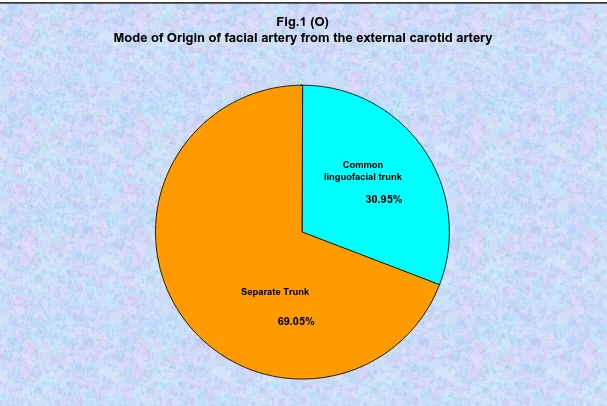
FACIAL ARTERY
Mode of origin:
- Separate origin from external carotid was observed in 29/42
hemifaces (69.05%).
- Common linguofacial trunk was observed in 13/42 hemifaces
(30.95%).
- Common linguofacial trunk was found to be present bilaterally in
5/21 cadavers (23.8%) and unilaterally in 3/21 (14.28%).
High origin of facial artery:
The facial artery arose just above the angle of the mandible in 1 case
(1/42), i.e., 2.38%. Here it was found that the submental artery was almost in