A STUDY OF CLINICAL
BIOCHEMICAL, SONOLOGICAL
PROFILE OF HEPATIC STATUS IN
TYPE 2 DIABETES MELLITUS IN
TERTIARY CARE SETTING
Dissertation submitted in partial fulfillment of regulation for the award of M.D. Degree in General Medicine (Branch I)
The Tamilnadu
Dr. M.G.R. Medical University March 2010
2
Certificate
Certified that this is the bonafide dissertation done byDR. MOHAMED
SHIHAB.P and submitted in partial fulfillment of the requirements for the
Degree of M.D., General Medicine, Branch I of The Tamilnadu Dr. M.G.R.
Medical University, Chennai.
Date : Professor and Chief of Medical Unit IV
Date : Professor & Head
Department of Medicine
Date : Dean
3 DECLARATION
I solemnly declare that the dissertation titled “A STUDY OF
CLINICAL,BIOCHEMICAL,SONOLOGICAL PROFILE OF HEPATIC
STATU IN TYPE 2 DIABETES MELLITUS IN TERTIARY CARE
SETTING” was done by me from January 2008 to August 2009 under the
guidance and supervision of Professor Dr.NEDUMARAN.MD. DM
This dissertation is submitted to the Tamilnadu Dr. MGR Medical
University towards the partial fulfillment of the requirement for the award of MD
Degree in General Medicine (Branch I).
Dr.MOHAMED SHIHAB .P
Place: Coimbatore
4 ACKNOWLEDGEMENT
I wish to express my sincere thanks to our respected Dean Dr.V. Kumaran
M.S,MCh for having allowed me to conduct this study in our hospital.
I express my heartfelt thanks and deep gratitude to the Head of the Department of
Medicine Prof: Dr.K.Umakanthan.M.D for his generous help and guidance in the
course of the study.
I owe a great debt of gratitude to our respected Prof and unit chief
Dr. Nedumaran MD DM ,and Prof.Dr.M.Ramaswamy without whose help and
advice this work would not have been possible.
I also extend my gratitude to Dr T Ravishankar.M.D,D.M, Prof:of Medical
gastroenterology ,Dr.Umashankar ,DMRD.,Asst:Prof of Radiology for their
expert guidance.
I sincerely thank all professors and Asst:Professors
Dr.K.Swaminathan.M.D,Dr.Balamurugan M.D,,Dr.V.Arulselvan M.D, for their
guidance and kind help.
5 CONTENTS
SECTION NO: TITLE PAGE NO:
1 INTRODUCTION 6
2 AIMS&OBJECTIVES 10
3 REVIEW OF LITERATURE 12
4 MATERIALS &METHODS 39
5 ANALYSIS OF RESULTS 43
6 DISCUSSION 55
7 CONCLUSIONS 64
8 BIBLIOGRAPHY 67
9 APPENDIX 1) MASTERCHART 78
2) PROFORMA 81
6
7 INTRODUCTION
The prevalence of diabetes is increasing world over and is expected to affect 57
million adults in india by 2025.
Apart from kidney, eye, heart and blood vessels, liver is also indirectly related
with diabetes mellitus. Virtuallythe entire spectrum of liver disease is seen in
patients withtype 2 diabetes. This includes abnomal liver enzymes, non alcoholic
fatty liver disease (NAFLD), cirrhosis, hepatocellular carcinoma,and acute liver
failure. In addition, there is an unexplainedassociation of diabetes with
hepatitisC.
Finally, the prevalenceof diabetes in cirrhosis is 12.3–57% . Thus, patientswith
diabetes have a high prevalence of liver disease and patientswith liver disease
have a high prevalence of diabetes
NAFLD
Ludwig introduced the term Nonalcoholic steatohepatitis (NASH) to describe a
form of liver disease that is histologically indistinguishable from alcoholic
hepatitis but occurs in people who do not consume excess ethanol.1 There is
8
increased prevalence in diabetes. It has been shown to be a predisposing factor
for insulin resistance and hyperinsulinemia, a major cause of cryptogenic
cirrhosis and may even lead to hepatocellular carcinoma.2,3,4
Nearly 70-80% of the diabetic subjects have been reported to have hepatic fat
accumulation, referred to as NAFLD.5 There are not enough studies done on the
hepatic status of diabetic patients in our country. Hence this study aims to
describe the hepatic profile of type 2 diabetic patients.
NAFLD represents a spectrum of diseases from simple fatty liver (steatosis),to
steatosis with inflammation, necrosis, and possible cirrhosis, thatoccurs in people
who drink little or no alcohol.
NAFLD affectsmore women than men and can be found in all age groups
diabetes, by most estimates, is now the most common cause ofliver disease in the
U.S. Cryptogenic cirrhosis, of which diabetesis, by far, the most common cause,
has become the third leadingindication for liver transplantation
.The liver helps maintain normal blood glucose concentrationin the fasting and
postprandial states. Loss of insulin effecton the liver leads to glycogenolysis and
an increase in hepaticglucose production. So Insulin resistance is the main
9
. The precise genetic,environmental, and metabolic factors and sequence of
eventsthat lead to the underlying insulin resistance, however, isnot fully
understood.
Despitedown-regulation of the insulin receptor substrate-2-mediatedinsulin
signaling pathway in insulin-resistant states, the up-regulationof
SREBP-1c(sterol regulatory element protein 1 c) and subsequent simulation of de novo
lipogenesisin the liver leads to increased intracellular availability oftriglycerides,
promoting fatty liver. This also increases VLDLassembly and deposition in liver.
.The insulin-resistant state is also characterized by an increasein pro
inflammatory cytokines such as tumor necrosis factor-(TNF-), which may also
contribute to hepatocellular injury.
10
11 AIM OF STUDY
This study aims to describe clinical, biochemical, sonological profile of hepatic
status in type 2 diabetes mellitus in tertiary care setting in relation with non
12
13 RIVEW OF LITERATURE
The Role of the Liver in Glucose Homeostasis
An appreciation of the role of the liver in the regulation of carbohydrate
homeostasis is essential to understanding the many physical and biochemical
alterations that occur in the liver in the presence of diabetes and to understanding
how liver disease may affect glucose metabolism. The liver uses glucose as a fuel
and also has the ability to store it as glycogen and synthesize it from
noncarbohydrate precursors (gluconeogenesis). Mann and Magath demonstrated
that a total hepatectomy in a dog results in death within a few hours from
hypoglycemic shock , underscoring the important role the liver plays in
maintaining normoglycemia.
Glucose absorbed from the intestinal tract is transported via the portal vein
to the liver. Although the absolute fate of this glucose is still controversial, some
authors suggest that most of the absorbed glucose is retained by the liver so that
the rise in peripheral glucose concentration reflects only a minor component of
postprandial absorbed glucose. Therefore, it is possible that the liver plays a more
significant role than does peripheral tissue in the regulation of systemic blood
14
taken up by the liver but is rather metabolized via glycolysis in the peripheral
tissues.
Many cells in the body, including fat, liver, and muscle cells, have specific
cell membrane insulin receptors, and insulin facilitates the uptake and utilization
of glucose by these cells. Glucose rapidly equilibrates between the liver cytosol
and the extracellular fluid. Transport into certain cells, such as resting muscle, is
tightly regulated by insulin, whereas uptake into the nervous system is not
insulin-dependent. Glucose can be used as a fuel or stored in a macromolecular
form as polymers: starch in plants and glycogen in animals. Glycogen storage is
promoted by insulin, but the capacity within tissues is physically limited because
it is a bulky molecule.
Glucose can be used as a fuel or stored in a macromolecular form as
polymers: starch in plants and glycogen in animals. Glycogen storage is
promoted by insulin, but the capacity within tissues is physically limited because
it is a bulky molecule. Insulin is metabolized by insulinase in the liver, kidney,
and placenta. About 50% of insulin secreted by the pancreas is removed by
first-pass extraction in the liver. Insulin promotes glycogen synthesis (glycogenesis) in
the liver and inhibits its breakdown (glycogenolysis). It promotes protein,
very-low-15
density lipoprotein cholesterol. It also inhibits hepatic gluconeogenesis,
stimulates glycolysis, and inhibits ketogenesis. The liver is the primary target
organ for glucagon action, where it promotes glycogenolysis, gluconeogenesis,
and ketogenesis . The formation of glucose from lactate and various
noncarbohydrate precursors is known as gluconeogenesis and occurs mainly in
the liver and kidneys.
The liver, kidney, intestine, and platelets contain the enzyme
glucose-6-phosphatase, which produces glucose from glucose-6-phosphate and is the final
step in the production of glucose via gluconeogenesis. This enzyme is absent in
other tissues. Glucose that is metabolized peripherally may therefore be
converted back to glucose or to hepatic glycogen via gluconeogenesis with
lactate as the primary substrate, this is known as Cori cycle.
In type 2 diabetes, excessive hepatic glucose output contributes to the fasting
hyperglycemia. Increased gluconeogenesis is the predominant mechanism
responsible for this increased glucose output, while glycogenolysis has not been
shown to be increased in patients with type 2 diabetes. Hyperglucagonemia has
been shown to augment increased rates of hepatic glucose output, probably
16 Key signaling pathways involved in glucose homeostasis in the liver
Binding to the glucagon receptor on hepatocytes activates the
serine/threonine kinase , this kinase causing phosphorylation and activation of
glycogen phosphorylase kinase (GPK) and subsequently glycogen
phosphorylase (GP), thus activating glycogenolysis. An increase in intracellular
cyclic AMP also induces gluconeogenesis enzymes (phosphoenolpyruvate
carboxykinase [PEPCK] and glucose-6-phosphatase [G6Pase]) via induction of
peroxisome proliferator activated receptor-γ co activator 1α (PGC-1α) It should
be emphasized, however, that under fasting conditions (potentially
hypoglycemic conditions, in particular), non-hormonal mechanisms (principally
hepatic auto regulation by glucose itself) are capable of supplying a significant
proportion (up to 50%) of the body’s glucose requirements via enhancement of
both glycogenolysis, glucose cycling and eventually gluconeogenesis) .
The pathways involved in insulin signalling in the liver are highly complex
involving hundreds of signaling molecules38,39 ,and thus a myriad of potential
points for modulation and interaction with other pathways, such as those
involved in glucose auto regulation. Insulin signaling processes also appear to
17
The first key component in the signaling process is the insulin receptor
itself and the associated intracellular insulin receptor substrate (IRS) proteins39 .
The IRS-2 subtype appears to play a more prominent role in the liver, whereas
the IRS-1 subtype may be more important in skeletal muscle40 and these two
proteins have different capacities to interact with downstream signaling
elements41. Within the liver, IRS-1 has been more closely linked with glucose
homeostasis, whereas IRS-2 may be more closely linked with lipid metabolism42
although surprisingly, liver-specific knockout of IRS 2 in mice does not appear
to impair hepatic glucose and lipid metabolism43
The second key component involves the activation of the phosphatidyl
inositol 3-kinase (PI3K) pathway, which appears to be crucial for insulin’s
metabolic actions in vivo in the liver.44 After PI3K activation, the specific
regulation of glucose and lipid homeostasis by insulin in the liver diverges PI3K
dependentactivation of Akt (also known as protein kinase B [PKB]) appears to
regulate factors involved in gluconeogenesis, whereas PI3K-dependent activation
of atypical forms of protein kinase C appears to regulate factors involved in
lipogenesis . For instance, pathway downstream of Akt leads to inactivation of
phosphorylase, activation of glycogen synthase, and stimulation of glycogen
18
In addition to acute effects on metabolic processes, insulin can also induce
changes in genetranscription in the liver down-stream of the PI3K pathway45,46.
Insulin can influence the expression of over 150 genes — this occurs via key
transcription factors, such as FOXO1 that inhibits expression of PEPCK and
G6Pase and inhibits gluconeogenesis), sterol-response element binding proteins
(SREBPs) that primarily regulate genes involved in lipid synthesis), and
specificity protein 1 (Sp1) that regulates genes for insulin receptors and leptin).
Hence the liver may play a much more important role than the
peripheral tissues in regulating the normal blood glucose. Liver also removes
about 50% of the insulin secreted by the pancreas during its first pass through the
liver.
NON ALCOHOLIC FATTY LIVER DISEASE
Nonalcoholic fatty liver disease (NAFLD) includes a wide spectrum of
liver injury ranging from simple steatosis to steatohepatitis, fibrosis, and
cirrhosis. Whereas simple steatosis has a benign clinical course, steatohepatitis is
a recognized cause of progressive liver fibrosis and can develop into cirrhosis.
NAFLD and nonalcoholic steatohepatitis (NASH) are the two most common
chronic liver diseases in United States general population with a prevalence of
19
type 2 diabetes, and hyperlipidemia with insulin resistance as a key pathogenic
factor.
PREVALENCE AND NATURAL HISTORY
Little is known about the prevalence or natural history of NASH. Biopsy
evidence is available primarily from the relatively few symptomatic patients.
Sequential biopsies in patients with NASH are particularly uncommon. If biopsy
specimens had been obtained, it is often difficult to determine from the
morphological descriptions whether fatty changes or steatohepatitis had been
present. Furthermore, NASH may be missed because by the time biopsies are
done, most fatty changes may have disappeared so that the hepatitis or cirrhosis
is considered cryptogenic. Nevertheless, some rough estimates can be made. For
example, in a study of 4613 male Japanese company employees, 534 were
moderately obese and almost half had hepatic steatosis as judged by computed
tomography. Twenty-four per cent of these obese patients had abnormal alanine
aminotransferase (ALT) activities. A subsequent study revealed ultrasonographic
evidence of fatty livers in 14% of 2574 patients from Okinawa. Fatty change was
most common in persons between 40 and 49 years of age. Obesity was the
20
strongly associated factor48. In an autopsy study NASH was found in 18.5% of
markedly obese patients and in 2.7% of lean patients.
As stated, most cases of NASH have been described in women with or
without diabetes, but recent studies47 suggest that the condition is also common
in men and that obesity, hyper lipidaemia and glycemia are not prerequisites.
Non-alcoholic fatty liver without appreciable inflammation or fibrosis appears to
be the most common manifestation of NASH, although by strict criteria it is not a
hepatitis. Thus, in a recent study of 14 patients with obesity and diabetes-related
NASH and a median follow-up of 11 years (range 7-16 years), none developed
evidence of progressive liver diseases48 . However, transition from the
uncomplicated non-progressive fatty liver to slowly progressive NASH may be
difficult to discern because biopsy samples are often reviewed without the use of
the tell-tale connective tissue stains and because sampling variations exist, as in
most other liver diseases.
The development from NASH to steatohepatitic cirrhosis was clearly
documented in a study of 47 patients who had been observed for 1.5-21.5 years
(median 4.5 years); two patients developed cirrhosis that, in one instance, was
complicated by hepatocellular carcinoma (HCC). The degree of obesity, hyper
lipidaemia and hyperglycaemia did not correlate with the severity of the
21
rare and, if it occurs, very slow, the Mayo Clinic experiences with NASH suggest
a less favorable scenario.
NAFLD and associated conditions
NAFLD is associated with various conditions, which may be considered
while diagnosing it. It is mainly associated with:
Obesity (69 - 100%)
Diabetes mellitus (36 - 75%)
Hyperlipidaemia (20 - 81%)
These conditions are associated with insulin resistance and metabolic
syndrome, which is frequently observed with NAFLD.
Obesity: More than 70% of patients with NASH are obese. Body weight ranging
from 10 - 40% higher than ideal is associated with 4 - 6 fold higher incidence of
NAFLD. There is direct correlation between the severity of obesity and
severity of NAFLD.
Diabetes: Upto 75% patients with NASH have diabetes mellitus. Obese,
middle-aged females with DM are more likely to have fatty liver changes on
ultrasonography
Hyperlipidaemia: 20 - 80% of patients with NASH have hyperlipidaemia in the
form of high blood cholesterol level and/or high triglyceride levels.
22
Total parenteral nutrition for prolonged periods.
severe insulin resistance.
Significant and rapid weight loss in obese subjects.
Familial lipid disorders, e.g., aβ-lipoproteinaemia, hypo β- lipoproteinaemia.
Limb lipodystrophy.
Weber-Christian disease.
Drugs: corticosteroids, methotrexate, tamoxifen,
PROGRESSION OF DISEASE
The progression from steatosis to steatohepatitisto cirrhosis and, in some
patients, to hepatocellular carcinoma over a period of many years is well
established . The prognosis worsens with each stage of disease. Why some
patientsprogress while most do not is not known. The only reliable way,to date,
of determining this progression is liver biopsy, which may have significant
economic implications (good or bad) for the management of patients with type 2
diabetes.
It is tempting and perhaps deceptively intuitive to think that, in some
people, simple fatty liver progresses to steatohepatitis and then to fibrosis and
cirrhosis. However, an equally plausible alternate hypothesis is that individuals
23
In fact, the limited long-term follow-up studies support the latter paradigm more
than the former.
The factors that determine whether a patient with NAFLD also develops necro
inflammatory changes and fibrosis are not known.
Possibilities include genetics, dietary composition, and concomitant forms of
other liver disease (e.g., chronic hepatitis C). There may be important racial and
ethnic predispositions, but these remain poorly characterized at this time.
Predictors of NASH and advanced fibrosis:
HAIR score
1. Hypertension
2. Alanine transaminase (ALT) > 40 IU/l
3. Insulin resistance (IR) index > 5
Presence of 2 or all 3 factors predict NASH.
BAAT score
1. Body mass index (BMI) > 28 kg/m2
2. Age > 50 yrs
3. ALT > 2-fold rise
4. TG > 1.7 mmol/l
24 PATHOGENISIS
The pathogenesis of NASH is unknown. In 1998,James first proposed the‘two
hit’ hypothesis for pathogenesis NASH. Fatty liver,the earliest and most
prevalent stage of NAFLD is thought to sensitize the liver to additional necro
inflammatory insults, thus promoting disease progression to steatohepatitis,
cirrhosis and hepatic failure. A number of factors point to multi factorial nature
of this disease, including derangement in metabolic parameters,
endotoxin-induced cytokine release and oxidative stress. After absorption from the
intestines, fat is carried to the adipose tissue for storage in the form of
triglycerides. It is released as free fatty acids (FFA) when the body is deprived of
food or under the effect of certain hormones/ drugs (such as epinephrine,
corticosteroids). FFA are carried to the liver bound to albumin. After entering
25
Insulin resistance the first hit in f NAFLD
This is the first hit hypothesis in the pathogenesis of NAFLD
The association between the severity of insulin resistance/ presence of
NIDDM, and the risk of NASH can be explained by peripheral insulin resistance
increasing the supply of FFA to the liver and by hepatic insulin resistance
favouring the development of oxidative stress. A central abnormality in the
26
increases circulating free fatty acids which are then taken up by the liver as an
energy source. The fatty acids overload the hepatic mitochondrial ß-oxidation
system, leading to accumulation of fatty acids in the liver Indeed, some
investigators suggest NAFLD to be the hepatic manifestation of the insulin
resistance syndrome29. NAFLD does not universally progress to NASH, and the
precise pathogenesis of steatohepatitis is yet to be determined.
However, dysregulation of peripheral lipid metabolism seems to be
important. There is a strong association between non alcoholic fatty liver and
features of the metabolic syndrome, suggesting a simultaneous insulin resistance
and decreased sensitivity to leptin.Leptin may have a role in the regulation of fat
deposition, fibro genesis, and inflammation in patients with NAFLD.6 Obese
patients with insulin resistance have decreased serum adiponectin and increased
serum resistin .
Cytokines and NASH
Cytokines are attractive candidates for the ‘second hit’ in the pathogenesis
of NASH. They are capable of producing all the classical histological features of
NASH, including hepatocyte death/apoptosis (TNF-a), neutrophil chemotaxis
(IL-8) and hepatic stellate cell activation (TNF-a, TGF-b) . There is evidence
that endotoxin-mediated cytokine release is important in the occurrence of
27
prevent or reverse its development. In addition, it has been shown that patients
with NASH had an increased expression of TNF-a mRNA both in their liver and
adipose tissue compared to obese controls, and this over-expression correlated
with histological severity. Lipid metabolism is, in part, regulated by adipokines,
including tumor necrosis factor (TNF) and adiponectin. TNF-, which interferes
with insulin signaling thereby favoring steatosis, is elevated in fatty liver disease
albeit not specific to type 2 diabetes29. TNF- is also pro inflammatory and, thus,
may play a role in the pathogenesis of the inflammation in NASH30 .
Adiponectin, in contrast to TNF-, is anti lipogenic and anti-inflammatory and,
thus, may protect the liver from lipid accumulation and inflammation.
Adiponectin levels are decreased in conditions associated with NAFLD,
including insulin resistance28 obesity29,type 2 diabetes , and NAFLD29.
Adiponectin and TNF- therefore have opposing effects. The net effect of
increased TNF- and decreased adiponectin is prosteatotic and pro inflammatory.
This low level of adiponectin expression may predispose patients to the
progressive form of NAFLD
Oxidative stress and lipid per oxidation
There is growing evidence implicating FFA in the production of oxidative stress
28
acid oxidation can both lead to increase in reactive oxygen species generation
and subsequent lipid per oxidation. In the fasting state, patients with NAFLD
have increased plasma levelsof b-OH butyrate49
Under normal conditions, hepatic aerobic metabolism involves a
steady-state production of pro-oxidants such as reactive oxygen species (ROS) and
reactive nitrogen species (RNS), which are balanced by a similar rate of their
consumption by antioxidants. Imbalance in the pro-oxidant/ antioxidant
equilibrium in favour of pro-oxidants constitutes the oxidative stress
phenomenon, a condition that may induce a number of patho physiological
events in the liver. Hepatotoxicity by oxidative stress may be achieved through a
direct attack of ROS and RNS on essential biomolecules with loss of their
biological functions and cell viability .
Alternatively, ROS may indirectly activate redox sensitive transcription
factors such as nuclear factor kB (NF-kB)50 or activator protein-1 (AP-1)51 , thus
triggering the production of cytotoxic, pro inflammatory and/or fibrogenic
mediators by Kupffer cells and other non parenchymal cells. . These studies
suggest that chronic oxidative stress may be important in the progression of fatty
liver Pessayre et al52 have shown that excess fat deposition in the liver is
29
related to the severity of steatosis. The end-products of lipid per oxidation,
4- hydroxynoneal and malondialdehyde, covalently bind to hepatic proteins, and
act as potent agents for neutrophil chemotaxis and stimulating pro-inflammatory
cytokines. Malondialdehyde also activates hepatic stellate cells to produce
collagen, leading fibrosis
Other factor
In addition to obesity and insulin resistance, some other environmental or
genetic factor(s) is required for the progression of NASH. Studies in
leptin-deficient ob/ob mice which have profound insulin resistance and dramatic
hepatic steatosis without steatohepatitis or fibrosis, suggests that leptin may in
fact have a role in promoting hepatic fibrogenesis, directly by an autocrine effect
on hepatic stellate cells and indirectly by up-regulating the production of TGF-b
from sinusoidal endothelial cells and Kupffer cells . The association of hepatic
iron accumulation and NAFLD continues to be debated. While some studies have
found that 22 to 62% of individuals with fatty liver disease have evidence of iron
overload, other have failed to show such relationship53 .In another study, a higher
30 Candidate genes
NASH and cryptogenic cirrhosis study suggest that genes might play an
important role in NAFLD. Different types of candidate genes of NAFLD as
follows: genetic factors related to insulin resistance, FFA supply and lipid
metabolism.
Apo lipoprotein E, a regulator of lipoprotein metabolism, was included and
considered to be of great importance. Genes associated with the ‘second hit’,
include (i) genes encoding proteins involved in the severity of oxidative stress
such as HFE (haemochromatosis gene), CYP2E1, CYP4A; (ii) genes encoding
cytokines and their receptors; (iii) genes related to adverse effects of FFA such as
transcription factors, peroxisome proliferator-activated receptors (PPARs).
Among these candidate genes are: (a) leptin and its receptor, which are related to
obesity, insulin resistance, increased FFA synthesis and reduced FFA oxidation;
(b) PPAR regulating a variety of genes encoding enzymes involved in FFA
oxidation and oxidative stress; and (c) PPAR which up regulates UCP2
(un-coupling protein C) and inhibits leptin gene expression and macrophage function.
NASH is now conceptualized as encompassing at least three components
among the tetrad of steatosis, hepatocellular injury, focal mixed cell-type
31
steatosis with ballooned hepatocytes and Mallory bodies, zone-3 peri-cellular and
peri-venular fibrosis with or without bridging fibrosis, and lobular inflammatory
cell infiltration6 Liver disease has not been associated with type 1 diabetes
mellitus, which reflects the current understanding that insulin resistance, not
insufficiency, is associated with this type of liver disease.7,8
Further proof for the association of liver disease with diabetes comes from the
Insulin Resistance Atherosclerosis Study (IRAS), which showed that liver
function markers like the Aspartate aminotransferase (AST) and Alanine
aminotransferase (ALT) are predictors of incident diabetes9 An Italian study
showed that 10.5% of the subjects who had elevated AST and ALT had diabetes.9
A similar study in Cleveland showed that nearly 33% of subjects with NASH had
diabetes.10
There are several histological stages in the progression of NAFLD to
cirrhosis. The earliest stage is a simple fatty liver alone. Over time,
steatohepatitis may become associated with increasing fibrosis. Eventually,
cirrhosis may develop. Cirrhosis secondary to NASH may also be complicated by
32 CLINICAL FEATURES
Most patients of NAFLD (45 - 100%) have no symptoms or signs of liver
disease at the time of diagnosis .
In these patients, abnormal liver function tests are often discovered
incidentally. When symptoms occur, they are non-specific – like
persistent fatigue (50 - 73%), pruritus (0 - 6%), oedema (2 - 10%), malaise, and
right upper quadrant discomfort or pain8.
Other features like GI bleeding (0 - 3%), jaundice (0 - 5%), ascites (0 -
3%), pruritus, and oedema point towards severe liver disease. Ascites, hepatic
encephalopathy, and variceal bleeding indicate cirrhosis of liver due to
progressive NASH.
When the disease is not advanced, diffuse non-tender smooth
hepatomegaly is present in 25 - 53% of patients. Such patients are usually obese
and/or diabetic.
Advanced disease may present with right hypochondrium tenderness,
jaundice, palmar erythema, spider angioma, portal hypertension, ascites, varices,
and splenomegaly.
33 DIAGNOSIS
Diagnosis of NAFLD is based on two criteria: (i) establishing the presence
of a fatty liver or steatohepatitis, and (ii) establishing the nonalcoholic nature of
the disease process. Radiologic imaging of the liver with sonography, compute
tomography (CT), or magnetic resonance imaging (MRI) has an adequate
threshold for detection of fatty infiltration of the liver, used either singly or in
combination. Each of these modalities has its own pitfalls and cannot distinguish
steatosis from steatohepatitis. These methods are also insensitive in detecting
steatosis of less than 25%
Liver biopsy is the gold standard for diagnosis of NAFLD/NASH for the
following important reasons: (i) to confirm diagnosis and establish severity of
fibrosis and presence of cirrhosis, and (ii) to exclude other coexisting conditions
that can result in hepatitic steatosis. However, ethical consideration as well as
inherent risk associated with this procedure limit its widespread applicability.
Histological diagnosis of steatohepatitis relies on a constellation of
lesions that include steatosis (mainly macrosteatosis, occasionally
microsteatosis), ballooning of hepatocytes (hepatocyte injury), perisinusoidal
34
histological criteria required for diagnosis are the presence of steatosis and intra
lobular necrotic inflammatory reactions54.
Tri chrome stain shows blue staining fibrosis around swollen hepatocytes.
The ALT/AST ratio is usually less than one. Imaging studies may help with
diagnosing fatty infiltration of the liver, but they do not help in distinguishing
35
Grading and stages of NAFLD56 .
Grade of NAFLD
Macro vesicular steatosis
Grade 0: No steatosis
Grade 1: < 33% steatosis
Grade 2: < 33–66% steatosis
Grade 3: > 66% steatosis
Necro inflammatory activity
Grade 1 (mild) steatosis up to 66%; occasional ballooned hepatocyte
(mainly zone 3); scattered intra-acinar neutrophil (PMN) lymphocytes, no or mild
portal inflammation.
Grade 2 (moderate) steatosis of any degree; obvious zone 3 ballooning
degeneration; intra-acinar PMNs; zone-3 peri sinusoidal fibrosis may present
mild to moderate, portal and intra-acinar inflammation.
Grade 3 (severe) pan acinar steatosis; widespread ballooning; intra-acinar
inflammation; PMNs associated with ballooned hepatocyts, mild to moderate
portal inflammation.
Stage of NAFLD
36
Stage 2: zone 3 peri sinusoidal/peri cellular fibrosis with focal or
extensively peri portal fibrosis.
Stage 3: zone 3 peri sinusoidal/peri cellular fibrosis and portal
fibrosis with focal or extensive bridging fibrosis.
Stage 4: cirrhosis
TREATMENT STRATEGIES
Currently, there are no effective therapies for NASH, as its natural history and
prognosis are not well understood. Treatment of patients with non-alcoholic fatty
liver has typically been focused on the management of associated conditions such
as obesity, diabetes mellitus, and hyper lipidemia as well as discontinuation of
potentially hepatotoxic drugs
. Appropriate metabolic control for patients with diabetes mellitus or hyper
lipidemia is recommended, but is not always effective in reversing non-alcoholic
fatty liver associated with obesity may resolve with weight reduction,, although
the benefits of weight loss have been inconsistent. On the other hand, striking
weight losses have also been associated with progression of the disease.
Moderate and gradual weight loss can safely improve in chronic liver disease
associated with obesity and diabetes. Rapid weight loss may aggravate the
37
children and 1600 g per week in adults is recommended, although the most
appropriate rate of weight loss is still to be established in fatty liver.
A number of pharmacologic agents have been shown to be promising in
the treatment of NASH . Promising results of pilot studies evaluating
ursodeoxycholic acid, gemfibrozil, betaine, N-acetylcysteine and
alpha-tocopherol suggest that these medications may be of potential benefit in the
treatment of patients with nonalcoholic fatty liver, but need further study in
controlled trials. The association of hyperinsulinemic insulin resistance has
provided a target for treatment. Metformin, a biguanide that reduces
hyperinsulinemia and improves hepatic insulin resistance has been shown to
greatly reduce hepatomegaly and steatosis in mice and may potentially be useful
in the treatment of NASH in humans.
Study by Department of clinical biochemistry (2008) by sandya
Sharma,dharmveer et all in SMS college Jaipur showed that (Indian) journal
clinical biochemistry,2008/23) Subject with NAFLD were more obese,
dyslipidemic and glucose intolerant. Almost 70% subjects with NAFLD had
metabolic syndrome which is five and half fold higher than those without
NAFLD. Results of the present study supports the hypothesis that the insulin
38
physiology of NAFLD57 as subjects with NAFLD were insulin resistant and
prevalence of NAFLD was significantly higher in those with metabolic
syndrome.Thus,fatty liver can be considered as hepatic consequence of
39
40
Materials and methods
This study was done at the Diabetology Clinic of Coimbatore Medical College
and Hospital, Coimbatore.
One hundred and eighteen type 2 diabetic patients diagnosed according to the
American Diabetic Association criteria, newly diagnosed or on follow- up were
included in the study.
.Random selection was done using random number charts.
Patients with history of any chronic drug intake other than oral hypoglycemic
drugs, jaundice or alcohol intake, HBsAg positive were excluded from the study.
An informed consent was taken. The study protocol was approved by the Ethics
committee of Coimbatore Medical College Hospital.
The type of oral hypoglycemic drug intake, height and weight were recorded and
Body Mass Index (BMI) calculated.
Patients were subjected to biochemical investigations to detect the liver enzyme
levels, serum bilirubin, serum albumin, serum globulin, serum total proteins and
2-41
3.5mg%, AST of 0 – 35 IU/L, ALT of 0 – 35 IU/L, Bilirubin levels of 0.3-1
mg%, total cholesterol less than 200mg%, serum total proteins of 5.5-8mg%,
fasting glucose less than 130mg% and a 2hr post prandial glucose of less than
180mg% were taken as normal . Their most recent fasting and post-prandial
blood glucose values were recorded to asses the control of diabetes.
Fifty two patients were subjected for ultrasonographic examination by a qualified
radiologist who was masked from the patient’s diagnosis or the indication for
ultrasound, to assess the liver parenchyma, liver size, gall bladder, biliary and
portal system.
The echo texture of the liver parenchyma was graded as follows
Grade 1: A slight diffuse increase in fine echoes in the hepatic parenchyma with
normal visualization of the diaphragm and intrahepatic vessel borders.
Grade 2: A moderate diffuse increase in fine echoes with slightly impaired
visualization of the intrahepatic vessels and diaphragm.
Grade 3: A marked increase in fine echoes with poor or no visualization of the
intrahepatic vessel borders, diaphragm and posterior portion of the right lobe of
the liver.12
We used ultrasonography to detect liver changes since this method is sensitive in
detecting fatty liver, is cheap, minimally invasive and easy to perform, and
42
liver on ultrasound increases as the amount of fat in the liver increases, though
the same is not true for fibrosis. However, this method does not allow for
quantification of fat infiltration.13
The data was analyzed and statistical conclusions drawn using the SPSS 13.0
43
44
ANALYSIS OF RESULTS
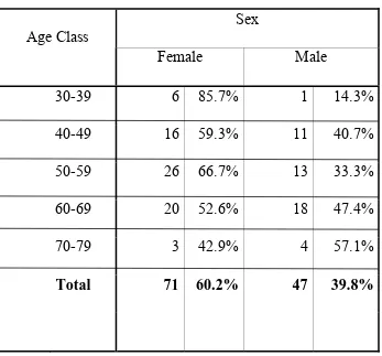
A total of one hundred and eighteen patients were included in the study,
71(60.2%) females and 47(39.8%) males. The age and sex distribution is shown
[image:44.612.139.486.323.646.2]in table no: 1.
Table No: 1 Age and Sex distribution
Sex Age Class
Female Male
30-39 6 85.7% 1 14.3%
40-49 16 59.3% 11 40.7%
50-59 26 66.7% 13 33.3%
60-69 20 52.6% 18 47.4%
70-79 3 42.9% 4 57.1%
45
Eleven (9.3%) cases were newly diagnosed during the course of the study.
Sixty six (55.9%) cases had diabetes for duration less than 5 years. Forty one
(34.7%) cases had diabetes for a duration more than or equal to 5 years.
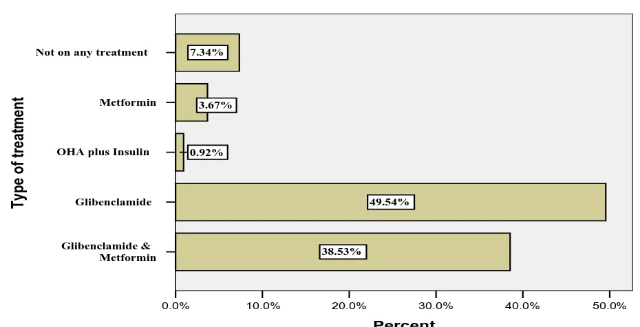
One hundred and eleven patients were on oral hypoglycemic drugs,
including 2 patients who were additionally on plain insulin injections. Majority
of the patients were either on glibenclamide alone or combination of both
glibenclamide and metformin. Eight patients though diagnosed to have diabetes
46
ranged from 2.5mg to 10 per day for glibenclamide and 500 mg to 3000 mg per
day for metformin.
Fig 1 Type of Antidiabetic treatment
Not on any treatment Metformin OHA plus Insulin
Glibenclamide Glibenclamide & Metformin Typ e of treat ment 50.0% 40.0% 30.0% 20.0% 10.0% 0.0% Percent 7.34% 3.67% 0.92% 49.54% 38.53%
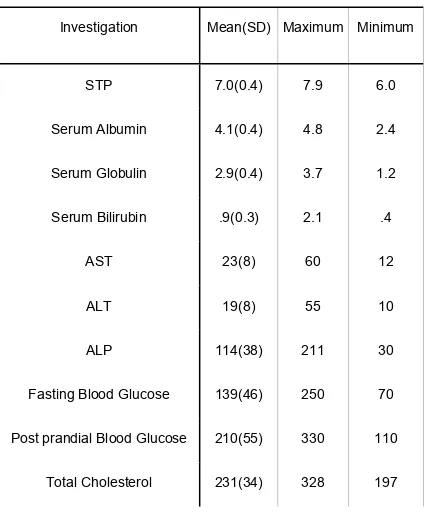
[image:46.612.88.553.221.459.2]47
Table No: 2 Biochemical Investigations
Investigation Mean(SD) Maximum Minimum
STP 7.0(0.4) 7.9 6.0
Serum Albumin 4.1(0.4) 4.8 2.4
Serum Globulin 2.9(0.4) 3.7 1.2
Serum Bilirubin .9(0.3) 2.1 .4
AST 23(8) 60 12
ALT 19(8) 55 10
ALP 114(38) 211 30
Fasting Blood Glucose 139(46) 250 70
Post prandial Blood Glucose 210(55) 330 110
Total Cholesterol 231(34) 328 197
The total serum proteins were within the normal range for all the patients
48
mg% were found in 4 (3.4%) patients and low globulin level, taken as less than 2
mg% in 2 (1.7%) patients.
Liver function tests revealed an elevated AST levels taken as more than 35
IU/L in 8 (6.8%) patients and an elevated ALT levels taken as more than 35 IU/L
in 5 (4.2%) patients. An AST/ALT ratio of more than 1 was found in 101
(85.6%) patients. Alkaline phosphatase levels were above 120 IU/L in 37
(31.4%) patients.
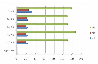
Age-wise and Sex-wise comparison of the liver function tests (Tables
3&4) revealed no significant difference between the various age classes or
49
Table No: 3 Age wise comparison of mean value of Liver function tests
Age class Test
30-39 40-49 50-59 60-69 70-79
STP 7.0 7.0 7.0 7.1 7.0
S.Albumin 4.0 4.3 4.0 4.0 4.3
S. Globulin 2.9 2.9 2.9 2.9 2.9
S. Bilirubin .9 .9 .8 1.0 1.2
AST 23 22 22 23 31
ALT 19 19 19 20 25
50
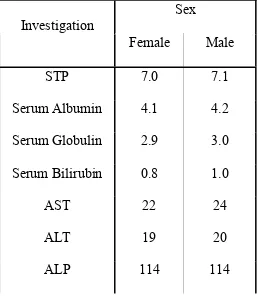
Table No: 4 Sex wise comparison of Liver function tests
Sex Investigation
Female Male
STP 7.0 7.1
Serum Albumin 4.1 4.2
Serum Globulin 2.9 3.0
Serum Bilirubin 0.8 1.0
AST 22 24
ALT 19 20
ALP 114 114
51
Among the patients whose recent fasting and 2 hr post prandial blood
glucose values were available, 47 percent had a fasting glucose more than 130
mg% and 61% had a 2 hr post prandial glucose more than 180mg%. There was
no significant difference between mean values across gender or age classes.
Body mass index measurements revealed that 25 (35.2%) women were
over weight (BMI>25) and 5 (7.0%) were obese (BMI>30). The numbers of
52
Ultra sonographic examination was done in 52 patients, fatty liver was
found to be more common in females. Overall 23 patients (42.3%) had fatty liver
out of the 52 patients screened. Sex wise and BMI class wise distribution is given
in tables 5 & 6. Hepatomegaly was identified in 5 (9.6%) patients of whom 4
were males. Asymptomatic gall stones were found in 5 (9.6%) patients, 3 females
and 2 males. Bile duct was found to be dilated (>5mm) in 6 patients, portal vein
was normal (<12mm) in size in all the patients
Table No: 5 Sex distribution of fatty liver
Sex Fatty Liver
Female (%) Male (%)
Grade 1 fatty liver 13(50) 8(30.8)
Grade 2 fatty liver 2(7.7) 0(0)
53
Table No: 6 BMI Class wise distribution of fatty liver
BMI Class
Fatty Liver
Underweight Normal Overweight Obese Obesity Morbid
Grade 1 fatty liver .0% 25.0% 31.3% 100% .0%
Grade 2 fatty liver .0% .0% 6.3% .0% .0%
55
56
DISCUSSION
Since clinical symptoms of fatty liver are nonspecific or silent this study does not
attempt to define the clinical symptoms of fatty liver. Fatty liver most commonly
affects middle-aged women with obesity, altered glucose metabolism, hyper
lipidemia, and hypertension.
Age, Gender and Obesity
As reported by Kelly et all28 there was no difference in the mean age of
patients with fatty liver as compared to those with normal liver. Sixty five
percent of the patients with fatty liver in this study were females but no
significant difference(p value>0.05) in proportion based on gender was found in
those with grade 1 fatty liver compared to those without evidence of fatty liver.
Obesity was found to have a significant association with fatty liver, in the current
study 70% of patients with grade 1 fatty liver had a BMI more than 25 and both
57 Liver enzymes
Reid etal14 and Dixon et al15 found elevated AST levels in patients with
NASH. Laboratory abnormalities identified included a 2-4-fold elevation of
serum amino transferase levels while other liver function test results were
normal. Agarwal etal16 and Kelly et al28 documented elevation of ALT as the
biochemical abnormality in patients with NASH. A recent study found that
patients with NASH and those with higher grade of histological inflammation
had increment of transaminases and albumin levels . The same study showed a
correlation of fibrosis with AST and ALT levels. Elariny etal showed that while
ALT was associated with NASH and advanced fibrosis, the majority of the
patients with either NASH or advanced fibrosis had normal AST.13 An
AST/ALT ratio >1.0 was yet another finding in a study on NASH15.
Contrary to all these a study in 2003 found liver enzymes to be insensitive
and unreliable to confirm the diagnosis or stage the extent of fibrosis. Older age,
obesity, and diabetes were shown to be predictive of fibrosis.
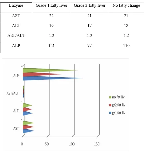
Our study also did not find a significant elevation of any of the liver
enzymes. There was no statistically significant difference (p value >0.05)
between the parameters among patients with grade 1 fatty liver and those without
58
AST/ALT ratio more than one, but it was not found to have any association with
[image:58.612.76.544.209.703.2]fatty liver.
Table No: 7 Liver enzymes in patients with fatty liver
Enzyme Grade 1 fatty liver Grade 2 fatty liver No fatty change
AST 22 21 21
ALT 19 17 18
AST/ALT 1.2 1.2 1.2
59 Insulin resistance
Obesity, insulin resistance, and increased concentrations of plasma fatty
acids are considered to increase the risk for fatty liver, and each of these
metabolic factors is also characteristic of type 2 DM29. It has been reported that
fatty liver in turn influences severity of hepatic insulin resistance in type 2 DM.
Among nonobese men without type 2 DM, fatty liver was found to correlate with
hepatic insulin resistance independently of obesity and intra-abdominal
adiposity28. Volunteers with type 2 DM and fatty liver were substantially more
insulin resistant than those with type 2 DM but without fatty liver and had
higher levels of plasma free fatty acids and more severe dyslipidemia30. The
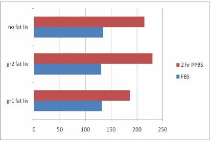
present study did not measure insulin resistance but comparing the mean blood
glucose values between those with or without fatty liver did not reveal any
60 Table No: 8 Mean Fasting and Post prandial blood glucose in Patients
with fatty liver
Blood Glucose
Grade 1 fatty
liver
Grade 2
fatty liver
No fatty change
Fasting
132 130 134
61 Hyperlipidemia
Fatty acid flux to the liver has been postulated as an important factor in the
pathogenesis of fatty liver and is also an important determinant of the synthesis
and secretion of triglyceride-rich lipoproteins29. It is possible, therefore, that
hepatic steatosis may influence the severity of dyslipidemia in type 2 DM.
Hypertriglyceridemia is more severe in individuals with fatty liver. Only the
total cholesterol levels were assayed in our study. A comparison of the mean total
cholesterol levels between the fatty liver group and the rest did not reveal any
statistically significant difference.
Treatment
Patients with diabetes should have their disease controlled appropriately.
Since NAFLD is associated with insulin resistance, the use of insulin-sensitizing
agents may be logical. The thiazolidinediones (e.g. pioglitazone) improve
peripheral insulin sensitivity. A small study of patients treated with troglitazone
showed improvement in mean ALT levels and in hepatocellular inflammation.
Metformin has been shown to improve serum aminotransferase levels.
However, there are no definitive data on the use of these drugs in the
treatment of NAFLD12 .The risks of hepatotoxicity associated with these agents
62
controlled trial, metformin was found to be superior to vitamin E in terms of
normalization of ALT31.
A significant reduction in all the liver enzymes was observed after
Essentiale treatment32 Essentiale is prepared from Soya beans, and has
phosphatidylcholine as its active ingredient. Some studies have explored the
efficacy of glitazones, vitamin E, probucol, atorvastatin and alternative therapies
like betaine, and have shown some beneficial results33,34,35,36
Based on the results of the study by Osei-Hyaiman et al, the hepatic
Endocannabinoid system may be a target for the treatment of nonalcoholic
steatohepatitis (NASH)37
Currently, treatment is focused on modifying risk factors such as obesity,
diabetes mellitus, and hyperlipidemia. Antioxidants such as vitamin E,
63
64 CONCLUSION
The increasing prevalence of fatty liver in diabetes is well established.
There is an increasing understanding about its aeti pathogenesis, and its various
pathological stages have been well defined.
It is important to acknowledge that the increased incidence of steatohepatitis and
hepatic fibrosis in type 2diabetes may translate into increased incidence of hepato
cellular carcinoma.
Liver biopsy though the gold standard for the diagnosis and staging of the
disease, cannot be used for large scale screening. More non invasive methods are
the need of the hour for early and wide screening to detect this disease.
Liver enzymes were thought to be a potential non invasive strategy for early
detection of this disease, but the present study did not find any correlation of the
65
so the conclusion is less expensive non invasive USG of liver will be the ideal
diagnostic tool which is also well trained operator dependant can definitely help
to detect early fatty liver in patients with diabetes
The moral of study is force is needed for all physicians to study and practice with
USG as bed side diagnostic tool for management and follows up study of type 2
66
67 REFERENCES
1 Ludwig J, McGill DB, Lindor KD. Review: nonalcoholic steatohepatitis.J
Gastroenterol Hepatol 1997;12:398-403.
2 G, Gordon FD, Lewis WD, Pomfret E, Pomposelli JJ, Jenkins RL, et al.
Cryptogenic cirrhosis: clinicopathologic findings at and after liver
transplantation. Hum Pathol 2002; 33:1098-104
3 Choudhury J, Sanyal AJ. Insulin resistance and the pathogenesis of
nonalcoholic fatty liver disease. Clin Liver Dis 2004;8:575-94.
4 Shimada M, Hashimoto E, Taniai M, Hasegawa K, Okuda H, Hayashi N,
et al. Hepatocellular carcinoma in patients with non-alcoholic steatohepatitis. J
Hepatol 2002; 37:154-60
5 Gupte P, Amarapurkar D, Agal S, Baijal R, Kulshrestha P, Pramanik S, et
al. Non-alcoholic steatohepatitis in type 2 diabetes mellitus. J Gastroenterol
Hepatol 2004;19:854-8
6 Sanyal AJ, Campbell-Sargent C, Mirshahi F, Rizzo WB, Contos MJ,
Sterling RK, et al . Nonalcoholic steatohepatitis: association of insulin resistance
68
7 Sanyal AJ, Campbell-Sargent C, Mirshahi F, Rizzo WB, Contos MJ,
Sterling RK, et al . Nonalcoholic steatohepatitis: association of insulin resistance
and mitochondrial abnormalities. Gastroenterol 2001;120:1183-
8 Marchesini G, Bugianesi E, Forlani G, Cerrelli F, Lenzi M, Manini R, et al
. Nonalcoholic fatty liver, steatohepatitis, and the metabolic syndrome. Hepatol
2003;37:917-23
10 Loguercio C, De Simone T, D'Auria MV, de Sio I, Federico A, Tuccillo C,
et al; Italian AISF Clinical Group.Non-alcoholic fatty liver disease: a multicentre
clinical study by the Italian Association for the Study of the Liver. Dig Liver Dis
2004;36:398-511
11. Younossi ZM, Gramlich T, Matteoni CA, Boparai N, McCullough AJ.
Nonalcoholic fatty liver disease in patients with type 2 diabetes. Clin
Gastroenterol Hepatol 2004;2:262-5.
12 Diabetes Care, Vol 15, Issue 3 430-441
13 Reid AE.Nonalcoholic steatohepatitis.Gastroenterology. 2001
Sep;121(3):71014
14 Dixon JB,Bhathal PS, O'Brien PE. Nonalcoholic fatty liver disease:
predictors of nonalcoholic steatohepatitis and liver fibrosis in the severely obese.
69
15 Agarwal SR, Malhotra V, Sakhuja P, Sarin SK. Clinical, biochemical and
histological profile of nonalcoholic steatohepatitis. 2001 | Volume : 20 | Issue :
5 | Page : 183-6
16 Neuschwander-Tetri BA, Brunt EM, Wehmeier KR, Sponseller CA,
Hampton K, Bacon BR. Interim results of a pilot study demonstrating the early
effects of the PPAR-gamma ligand rosiglitazone on insulin sensitivity,
aminotransferases, hepatic steatosis and body weight in patients with
non-alcoholic steatohepatitis.Hepatology 2003;38:434-435
17 Helmalek MF, Angulo P, Jorgensen RA, Sylvestre PB, Lindor KD.
Betaine, a promising new agent for patients with nonalcoholic steatohepatitis:
results of a pilot study. Am J Gastroenterol 2001;96:2711-7.
18 Kiyici M, Gulten M, Gurel S, Nak SG, Dolar E, Savci G, et al.
Ursodeoxycholic acid and atorvastatin in the treatment of nonalcoholic
steatohepatitis. Can J Gastroenterol 2003;17:7133
19 Osei-Hyiaman D, Depetrillo M, Pacher P, et al. Endocannabinoid
activation at hepatic CB(1) receptors stimulates fatty acid synthesis and
70
20 Mehta K.; Van Thiel D.H.; Shah N.; Mobarhan S. Nonalcoholic Fatty
Live Disease: Pathogenesis and the Role of Antioxidants. Nutrition Reviews,
Volume60, Number 9, 1 September 2002, pp. 289-293(5)
21 Trombetta M, Spiazzi G, Zoppini G, Muggeo M: Review article: type 2
diabetes and chronic liver disease in the Verona diabetes study. Aliment
Pharmacol Ther 22(Suppl. 2):24–27, 2005
22 Belcher G, Schernthaner G: Changes in liver tests during 1-year treatment
of patients with type 2 diabetes with pioglitazone, metformin or gliclazide.
Diabet Med 22:973–979, 2005[
23 Lebovitz HE, Kreider M, Freed MI: Evaluation of liver function in type 2
diabetic patients during clinical trials: evidence that rosiglitazone does not cause
hepatic dysfunction. Diabetes Care 25:815–821, 2002
Hultcrantz R, Glaumann H, Lindberg G, Nilsson LH: Liver investigation in 149
asymptomatic patients with moderately elevated activities of serum
aminotransferases
24 Mann FC, Magath TB: Studies on the physiology of the liver. II. The effect
of the removal of the liver on the blood sugar level. Arch Intern Med 30:73-84,
71
25 Angulo P: Nonalcoholic fatty liver disease. N Engl J Med 346:1221–1231,
2005
26 Chitturi S, Abeygunasekera S, Farrell GC, Holmes-Walker J, Hui JM, Fung
C, Karim R, Lin R, Samarasinghe D, Liddle C, Weltman M, George J: NASH
and insulin resistance: insulin hypersecretion and specific association with the
insulin resistance syndrome. Hepatology 35:373–379, 2002
27 Crespo J, Cayon A, Fernandez-Gil P, Hernandez-Guerra M, Mayorga M,
Dominguez-Diez A, Fernandez-Escalante JC, Pons-Romero F: Gene expression
of tumor necrosis factor alpha and TNF-receptors, p55 and p75, in nonalcoholic
steatohepatitis patients. Hepatology 34:1158–1163, 2001
28 Kelley et al. Fatty liver in type 2 diabetes mellitus: relation to regional
adiposity, fatty acids, and insulin resistance. Am J Physiol Endocrinol Metab 285
(4): E906-E916, 20033
29 Day C and Saksena S. Nonalcoholic steatohepatitis: definitions and
pathogensis. J Gastroenterol Hepatol 17: S377–S384, 20023
30 Seppala-Lindroos A, Vehkavaara S, Hakkinen AM, Goto T, Westerbacka J,
Sovijarvi A, Halavaara J, and Yki-Jarvinen H. Fat accumulation in the liver is
associated with defects in insulin suppression of glucose production and serum
fatty acids independent of obesity in normal men. J Clin Endocrinol Metab 87:
72
31 Bugianesi E, Gentilcore E, Manini R, Natale S, Vanni E, Villanova N, et al .
A randomized controlled trial of metformin versus vitamin E or prescriptive diet
in nonalcoholic fatty liver disease. Am J Gastroenterol 2005;100:1082-90. 3
32 Poongothai S, Karkuzhali K, Prakash GS, Sangeetha T, Saravanan G, Deepa
R, Gopalakrishnan S, Mohan V. Effect of essentiale in diabetic subjects with Non
- Alcoholic fatty liver. Int J Diab Dev Ctries 2005;25:12-19
33 Liangpunsakul S, Chalasani N. Treatment of Nonalcoholic Fatty Liver
Disease. Curr Treat Options Gastroenterol 2003;6:455-63.
34 Neuschwander-Tetri BA, Brunt EM, Wehmeier KR, Sponseller CA,
Hampton K, Bacon BR. Interim results of a pilot study demonstrating the early
effects of the PPAR-gamma ligand rosiglitazone on insulin sensitivity,
aminotransferases, hepatic steatosis and body weight in patients with
non-alcoholic steatohepatitis. J Hepatol 2003;38:434-40.
35 Abdelmalek MF, Angulo P, Jorgensen RA, Sylvestre PB, Lindor KD.
Betaine, a promising new receptors stimulates fatty acid synthesis and contributes
to diet-induced obesity. J Clin Invest 2005; 115: 1298-1305.agent for patients
with nonalcoholic steatohepatitis: results of a pilot study. Am J Gastroenterol
73
36 M, Gulten M, Gurel S, Nak SG, Dolar E, Savci G, et al. Ursodeoxycholic
acid and atorvastatin in the treatment of nonalcoholic steatohepatitis. Can J
Gastroenterol 2003;17:713-8.
37 Osei-Hyiaman D, Depetrillo M, Pacher P, et al. Endocannabinoid activation at
hepatic CB(1) receptors 1305.
38 Le Roith D, Zick Y. Recent advances in our understanding of insulin action
and insulin resistance. Diabetes Care 2001; 24: 588-597.
39 Taniguchi CM, Emanuelli B, Kahn CR. Critical nodes in signalling pathways:
insights into insulin action. Nat Rev Mol Cell Biol 2006; 7: 85-96.
40 Kido Y, Burks DJ, Withers D et al. Tissue-specific insulin resistance in
mice with mutations in the insulin receptor, IRS-1, and IRS-2. J Clin Invest 2000;
105: 199-205
.41 Sesti G, Federici M, Hribal ML, Lauro D, Sbraccia P, Lauro R. Defects of
the insulin receptor substrate (IRS) system in human metabolic disorders. Faseb J
2001; 15: 2099-2111.
42 Taniguchi CM, Ueki K, Kahn R. Complementary roles of IRS-1 and IRS-2
74
43. Simmgen M, Knauf C, Lopez M et al. Liver-specific deletion of insulin
receptor substrate 2 does not impair hepatic glucose and lipid metabolism in
mice. Diabetologia 2006 49: 552-561.
44 Taniguchi CM, Kondo T, Sajan M et al. Divergent regulation of hepatic
glucose and lipid metabolism by phosphoinositide 3-kinase via Akt and
PK.Clambda/zeta. Cell Metab41 Aiston S, Hampson LJ, Arden C, Iynedjian PB,
Agius L. The role of protein kinase B/Akt in insulin-induced inactivation of
174-182. 41 Aiston S, Hampson LJ, Arden C, Iynedjian PB, Agius L. The role of
protein kinase B/Akt in insulin-induced inactivation phosphorylase in rat
hepatocytes. Diabetologia 2006;
45,46 . Mounier C, Posner BI. Transcriptional regulation by insulin: from the
receptor to thegene. Can J Physiol Pharmacol 2006;
47 . Powell BE, Cooksley WGE, Hanson R, Searle 3, Halliday J\V, Powell LW.
The natural history of nonalcoholic steatohepatitis: A follow-up study of
forty-two patients up to 21 years. Hepawlogy 1990; 11: 74-80.
48 . Nomura H) Kashiwagi S) Hayashi 33 Kajiyama W3 Tani S) Goto M.
Prevalence of fatty liver in a general population of Okinawa, Japan. Jpn. Y. Med.
75
49 Kaplowitz, N., Mechanisms of liver cell injury. J. Hepatol., 2000, 32, 39–47.
50 Tilg, H. and Diehl, A. M., Cytokines in alcoholic and nonalcoholic
steatohepatitis. N. Engl. J. Med., 2000, 343, 1467–1476.
51 Marchesini, G. et al., Non-alcoholic fatty liver disease. A feature of the
metabolic syndrome. Diabetes, 2001, 50, 1844–1850
52 Crespo, J. et al., Gene expression of tumour necrosis factor a an
TNF-receptors, p55 and p57, in non-alcoholic steatohepatitis patients. Hepatology,
2001, 34, 1158–1163. .
53 Day, C. P., The genetic basis for non alcoholic and alcoholic
steatohepatitis.In Steatohepatitis (NASH and SH), Kluwer, Dordrecht, Falk
Symposium No 121, 2001, pp. 43–5359
54 Jiang G, Zhang BB. Glucagon and regulation of glucose metabolism. Am J
Physiol
Endocrinol Metab 2003; 284: E671-678
55 . Simmgen M, Knauf C, Lopez M et al. Liver-specific deletion of insulin
receptor substrate 2 does not impair hepatic glucose and lipid metabolism in
76
56. Angelico F, Del Ben M, Conti R, Francioso S, Feole K,
Maccioni D, Antonini TM, Alessandri C. Non-alcoholic fatty
liver syndrome: a hepatic consequence of common metabolic disease 2004
57. Taniguchi CM, Kondo T, Sajan M et al. Divergent regulation of hepatic
glucose and lipid metabolism by phosphoinositide 3-kinase via Akt and
PKClambda/zeta. Cell Metab41 Aiston S, Hampson LJ, Arden C, Iynedjian PB,
Agius L. The role of protein kinase B/Akt in insulin-induced inactivation of
174-182. 41 Aiston S, Hampson LJ, Arden C, Iynedjian PB, Agius L. The role of
protein kinase B/Akt in insulin-induced inactivation phosphorylase in rat
77
.
.
.