Study of anatomical position of appendix
in normal population and inflamed cases
Dissertation submitted in partial fulfillment of regulation for the award
of
M.S. Degree in General Surgery (Branch I)
The Tamilnadu
CERTIFICATE
Certified that this is the bonafide dissertation done by Dr.ARAVINDH. R and submitted in partial fulfillment of the requirements for the Degree of M.S., General
Surgery, Branch I of The Tamilnadu Dr. M.G.R. Medical University, Chennai.
Date: Guide and Chief of Surgery Unit SI
Professor & Head of the Department of General Surgery
Dean
Coimbatore Medical College
Date: Coimbatore - 641 010
DECLARATION
I solemnly declare that the dissertation titled Study of anatomical position of the appendix in normal population and in inflamed caseswas done by me from November
2009 to December 2011 under the guidance and supervision of Professor
Dr. P.V.VASANTHA KUMAR M.S.,
This dissertation is submitted to the Tamilnadu Dr. MGR Medical University
towards the partial fulfillment of the requirement for the award of MS Degree in General
Surgery (Branch I).
Place: Coimbatore
ACKNOWLEDGEMENT
I am privileged to express my thanks to Dr Vimala M.D.,Dean of Coimbatore
Medical College and Hospital in granting me permission to utilize the hospital facilities to
undergo this Dissertation.
I am grateful to Dr P V Vasanthakumar M S., Professor and Head of the
Department of Surgery for allowing me to carry out this Dissertation work in this
department.
I would like to thank my previous Chiefs Rtd. Prof. Dr.P.Govindaraj M S, M ch.,
Rtd. Prof Dr A Ramamoorthy M S., Rtd. Prof. Dr G Mohan M S., Rtd. Prof Dr P M
Nanjundappan M S., for their guidance and support.
I would also like to express my thanks to all surgery unit chief for allowing me to
use their unit patients to indulge in the study.
I am happy to convey my thanks to all the Registrar and Assistant Professors for
their advice and guidance.
I wish to thank all my co-Post Graduates and Interns for their help.
I am also happy to extend my thanks to the nursing faculties and other
paramedical staffs for their support.
Finally with all happiness I thank all my patients for their kind co-operation all
TABLE OF CONTENTS
S.No Particulars Page No.
1. Introduction 1
2. Objectives 2
3. Review of Literature 3
4. Methodology 44
5. Results 49
6. Discussion 60
7. Conclusion 63
8. Summary 66
9. Bibliography 68
10. Annexures 71
LIST OF TABLES
Sl.No Particulars Page
No.
1 Position of the appendix according to various authors 7
2 Alavarado Score 27
3 Teicher scoring system 28
4 Distribution of Age & position of Appendix in inflamed cases 51
5 Length of appendix in inflamed and normal cases 51
6 Distribution of sex inposition of appendix 53
7 Typical and atypical presentation of appendix 54
8 Type of surgery in appendix 55
9 Positon of appendixin normal population and in inflamed cases 56
LIST OF FIGURES
Sl.No Particulars Page No
1 Artery supply of appendix 4
2 positions of appendix 6
3 ultrasound finding of appendix 24
4 Distribution of age in the position of appendix 52
5 Age of onset of acute appendicitis 52
6 Length of appendix 53
7 Typical and atypical presentation of appendix 54
8 Type of incision for appendecectomy 55
9 Position of appendix in inflamed cases and normal group 56
10 Inflamed appendix with gall stones 58
11 Pelvic appendicitis 58
12 Thickened,elongated and perforated appendix 59
INTRODUCTION
Appendix is a vestigial organ present only in man, certain anthropoid apes and
wombat. It is a mysterious structure, being the frequent site of inflammation, with no
function. Acute appendicitis is most common surgical emergency. The diagnosis and
position of appendix is elusive and needs high index of suspicion in preventing serious
complication
The appendix present in various positions and each position has varying clinical
signs making hard to diagnose.
There are few studies saying that position of appendix is related to the cause of
inflammation like Varshney et-al have concluded that the retrocaecal position of the
appendix is less prone for infection and Collins et-all has stated that perforation and
inflammation is seen in retrocaecal cases. Collins et-al had told that pelvic as common
position (78.5%)1 and Pickens G et-al2 as postileal and Wakeley et- al as retrocaecal
(65.3%)3 . Guidry SP4 and Poole GV5 et al have concluded that anatomic variations of
the location of appendix are often responsible for delay in the diagnosing appendicitis.
Hence this study was conducted to know whether position of appendix has any
relationship to inflammation.
Our study is performed in clinical cases representing the inflamed appendix group
and other laporatomy group, which represents the normal appendix group. In the inflamed
group the relation between the various positions of the appendix, the clinical presentation
OBJECTIVES OF THE STUDY
To study the position of the appendix in normal population and compare it with
the position of inflamed cases.
To determine the relation of clinical presentation and management of appendix to
that of various position of the appendix.
To analyse the age incidence, sex incidence, duration of onset, symptomatology,
Review of Literature
REVIEW OF LITERATURE
HISTORICAL NOTE
To understand the present and foresee the future we must turn always to the past. Existence of appendicitis in remote times is evidenced by the acutely inflamed and
perforated appendix found preserved in the mummy of a young royal princess of
Egypt (Spencer).
BERENGE – ARIL – DE CARPI in 1521 first described the appendix. JANE
FERNIL described it as ‘Caecum Intestinum’ in 1554. LORNEZ HEISTER described
Appendicular Abscess in 17106. CLAUDIUS AMYAND, surgeon to Westminster in
St.George's Hospitals performed the first Appendicectomy in 1736 on a boy aged 11 years
who had a right scrotal hernia accompanied by a fistula. In 1824, LOYER
VILLERMAY described two examples of acute appendicitis leading to death.
Description of symptoms of Appendicitis and appendicular perforation was published in
the textbook by Bright and Addison in1839. It was Fitz, Professor of Medicine at Harvard
who in 1886 gave a lucid and logical description of the clinical features and pathological
changes of the disease and termed “Appendicitis”.
In 1880, Tait of Birmingham operated and removed a gangrenous appendix with
recovery of the patient. McBurney in New York pioneered early diagnosis and early
operative intervention and devised the muscle splitting incision.
The operative death rate for the later cases of perforated appendix with peritonitis
was distressingly high. Oschner in chicago and Sherren at the London Hospital were
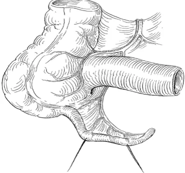
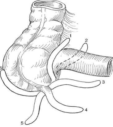
DEVELOPMENT AND ANATOMY
Appendix is the undeveloped distal end of caecum. It is pushed to the
posteromedial aspect of caecum by about 2.5cm below the ileocacecal valve, by excessive
growth of the right wall of caecum. It arises at the site where the three taeniae coli
coalesce. It occupies various positions more commonly the Retrocaecal position,
the rest being Pelvic, Postileal, Preileal, Paracaecal and Subcaecal.
It has the same coats as large gut. Its lumen which can admit a match stick is
irregular encroached upon by multiple longitudinal mucosal folds. It contains an
excess of lymphoid tissue in the sub mucosal layer beneath the columnar celled mucosa
of colonic type with few crypts on the base of which lies the Kulchitzky cells. The
mesentry of the appendix is contiguous with the lower leaf of mesentry of the ileum and
posterior to it. An additional bloodless fold of peritoneum named after Treves
connecting the terminal ileum to caecum and the mesentry of Appendix may be
present. The Appendicular Artery runs in the free border of the mesentry and is a branch of
the ileocolic Artery. An Accessory Appendicular artery may be present. The veins of the
appendix drain into the ileocolic vein, then into the superior mesentric vein. Slender
[image:14.595.216.402.559.732.2]lymphatics drain into ileocaecal lymph nodes8.
INCIDENCE LENGTH OF APPENDIX
Length of appendix varies between 1-20cms. The length of appendix gets
apparently modified by the inflammatory process9.
POSITION OF APPENDIX
The relation of the base of the appendix to the caecum is always constant
where as the tip of it may lie in any of the following positions as per the study of Cecil
Wakeley - Retrocaecal 74%, Pelvic 21%, Paracaecal 2%, Subcaeccal 1.5%, Prelieal 1%,
and postileal 0.5%.
These are the various position of appendix as described by Sir Frederich Treaves 8,9,10
: Retro-caecal or Retro-colic or12 O’ clock position - Appendix lying behind the caecum or the ascending colon and can be intraparietal or extraparietal
Splenic or 2 O’ clock position - Appendix directs towards the spleen this has two types i.e. it may pass either in front of the ileum (Pre-ileal) or behind the terminal part of the ileum (Post-ileal)
Para-colic or Para-caecal or 11 O’ clock position - Appendix directs upwards & to the right to caecum. It can lie behind peritoneum or lie in front of kidney into the
peritoneal cavity
Promonteric or 3 O’ clock position - Appendix directs transversely inwards towards the sacral promontory.
Midinguinal or 6 O’ clock position or Sub-caecal position: Appendix passes downward towards the middle of the inguinal ligament.
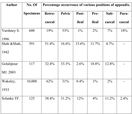
TABLE - 1
Position of the appendix according to various authors1
Author No. Of Specimens
Percentage occurrence of various positions of appendix. Retro-
caecal
Pelvic Post- ileal Pre- ileal Sub- caecal Para-caecal Varshney S. 1996
600 19% 53% 1% 2% 7% 18%
Shah &Shah,
1942
591 51.4% 16.6% 15.6% 11.7% 4.7% -
Golalipour
MJ. 2003
117 32.4% 33.3% 2.6% 18.8% 12.8% -
Wakeley,
1933
10,000 62% 31% 0.4% 1% 2% -
Solanke TF. 125 38.4% 31.2% 12% 4% 11.2% 2.4%
Normal variations in appendix10,11
1 - Extra-peritoneal retro-caecal, para-caecal fossa present
2 - Extra-peritoneal retro-caecal, para-caecal fossa absent
3- Extra-peritoneal retro-caecal, lying anterior to the right kidney & associated with
subhepatic caecum
4 - Intra-peritoneal
5 - Intra-peritoneal retro-caecal with in paracaecal fossa
[image:17.595.91.521.124.494.2]AGE INCIDENCE
12,13Appendicitis is rare before the age of 2, becoming increasingly common
during childhood and adolescence. The maximum incidence between the ages of 20 and
30 years. Thereafter, there is a gradual decline but no age is exempted. The patient
often gives a history of previous slight attacks. The amount of lymphoid tissue
in the appendix and the incidence of acute appendicitis, the peak for both occuring in
the middle teens. About 72% of patients are under the age of 30 yrs and 4% are 50 yrs
or above. With increasing age, the percentage of patients with simple appendicitis
diminishes whereas the percentage of perforated appendicits increases. In preschool age
children, the rate of perforation is over 60% due to anatomical differences,
difficulties in diagnosis and the habit of giving a purgative to any child with abdominal
pain and fever.
SEX INCIDENCE
The male-female ratio of appendicitis is about 1:1 prior to puberty, according to
Edward.H.Storer. At puberty the frequency increases, so that the male to female ratio is
about 2:1 between 15-25 years after which the male incidence gradually declines until
the sex related incidence are again equal1 4. Acute appendicitis is generally more
common in males and the mortality rate is also higher in males than females. The
AETIOLOGICAL FACTORS
The aetiology has not been completely clarified. The following factors are purely
contributory.
I a) Anatomical factors15
The Appendix is a blind tube with a narrow lumen with bacteria teamed contents.
Its vestigeal character predisposes to troubles. Obstruction is (1) by the action of valve of
Gerlach especially (2) When foreign bodies chiefly faecolith are present, (3) because
of the mobility of appendix to the position of caecum (4) the effects of kinks, bands,
adhesions and other causes of angulation. The presence of muscular hiatus in
which areas, the mucosal tissue is in juxta position with serosal tissue, explains the
possible spread of infective process once it has developed. Anamolous positions can
cause considerable diagnostic confusions.
b) Race and Diet16
Appendicitis is particularly common in the highly civilized European,
American, and Australian countries while it is less common in Asians, Africans and
Polynesians. If the latter races migrated to the former, they soon acquire the local
susceptibility to the disease. This is due to the departure from a simple diet rich in
cellulose to one relatively rich in meat, and it is not the whole explanation, for it occurs
in lifelong vegetarians.
c) Social Status
Acute appendicitis is more common in the upper and middle classes than in the
d) Familial Susceptibility
This unusual but accepted fact is accounted for by a hereditary abnormality
in position of the organ which predisposes to infection.
e) Obstruction to the lumen of Appendix17
Some form of obst ruction to its lum en is usuall y demonstrated in 80% of cases.
i) Faecolith : Faecal material commonly present in the normal and inflamed
appendix is differentiated from a faecolith which is ovoid, laminated about
1-2 cm in length and faecal coloured. It is composed of inspissated faecal
material, Ca and Mg PO4s, carbonates, bacteria and epithelial debris. Rarely an
incorporated foreign body. They are Radio-opaque. Worms, and other intestinal
parasites can injure the appendicular mucus membrane and occasionally,
block its lumen, or act as a nidus for bacterial infection. If worms were
causative of appendicitis then, the disease should be more prevalent in children
while it is so in adolescents and young adults.
ii) Other foreign bodies include vegetable seeds, cherry, stones, inspissated Barium,
etc. In older patients a caecal carcinoma and in the young a carcinoid are
occasional causes. Metastasis to the appendix from carcinoma breast especially
may cause acute appendicitis. Our present study did not demonstrate any of these.
f) Trauma
Accepted when Appendicitis follows trauma almost immediately.
g) Acute respiratory infection
Acute Bronchitis, tonsillitis and acute sinusitis appear to be impressive
h) Diverticula
Efforts of Appendicular musculature to empty the occluded lumen due to previous
inflammation, leads to marked increase of intraluminal pressure with occasional herniation
of mucosa through the areas of least resistance.
i) Primary Bacterial origin17
As appendix lodges mixed bacterial flora its exact relationship could not be
clearly made out. It has been reported in literature that the onset of ileocaecal
tuberculosis is from the appendix. It is rare to find isolated involvement of
Appendix in tuberculosis.
j) Infection via Blood stream.
Infection may be hematogenous, probably when it occurs during the course of
measles or has followed upon acute tonsillitis or surgery.
k) Amoebiasis
This condition may mimic or be the cause of acute appendicitis as evidenced by
PATHOLOGY
18Like any inflammatory process elsewhere, appendicitis is a reaction to an injury,
but the exact manner with which the injury occurs is not known. Transient circulatory
disturbances due to the normal efforts of caecum to expel its contents may change the
position of Appendix and cause a pull or a twist of its mesentry, thereby temporarily
impeding its blood supply. At the site of injury to the mucosa a minute ulcer occurs and
fibrin, red cells, leucocytes and plasma oozed out into the lumen. A focal inflammation
thus set in proceeds in succession or resolves even without treatment.
The menace of acute appendicitis lies in the frequency with which the peritoneal
cavity is infected from this focus, by (1) perforation (2) transmigration of bacteria through
the appendicular wall. The greater omentum, ‘the abdominal policeman’, attempts to
arrest the spread of peritoneal infection, whilst violent peristalsis from ingested purgatives
tends to spread it. Obviously the inflamed appendix lies dangling amidst coils of small
intestine, the threat of peritonitis is increased, should early perforation occur, diffuse
peritonitis is inevitable.
It is of great importance to recognise the following types of appendicitis:
1. Acute appendicitis without perforation.
2. Acute appendicitis with perforation
a) With local peritonitis
b) With local abscess
c) With appendicular mass
Examination of a series of fresh specimens of acutely inflamed appendices
will show that these fall into two groups. It first demonstrates a "catarrhal" inflammation of
beyond which there is acute inflammation, distension with pus, and in later cases progression
to gangrene and eventually perforation.
1. CATERRHAL APPENDICITIS
Is initially a mucosal and sub mucosal inflammation. In early cases the appendix
may appear quite normal externally or merely shows hyperemia. On slitting it open, the
mucosa will be seen thickened, odematous and reddened; latter it becomes studded with
dark brown haemorrhagic infarcts, patches of grey-green gangrene or small ulcers.
Eventually the whole appendix becomes swollen and turgid and the serosa becomes
roughened, loses its healthy sheen and is coated with a fibrinous exudate. The probable etiology of this condition is bacterial invasion of the lymphoid tissue in the appendix wall
and indeed some cases are probably localised manifestation of generalised enteritis.
Because the lumen of the appendix is not obstructed; these cases rarely progress to
gangrene and in many instances the acute inflammatory attack resolves spontaneously.
In other cases, however the swelling of the lymphoid tissue in the appendix wall may lead
to obstruction of the lumen and the condition may then proceed to obstructive
appendicitis and gangrene. Even when the acute inflammation process subsides; the
appendix probably never regains its pristine state, adhesion formation and kinking of the
appendix may lead to a final episode of acute obstructive appendicitis.
2. OBSTRUCTIVE APPENDICITIS19
It is a dangerous type where appendix becomes a closed loop bowel containing
decomposing faecal matter. The changes after obstruction depend upon the amount and
character of the factor causing obstruction. Faecolith is the common cause of
obstruction. When obstructed, there is accumulation of mucus that proceeds to
bacterial access to deeper tissue planes and continues with vessel thrombosis which
leads to gangrene and then perforation. On other occasions, bacterial invasion occurs through
pressure erosion of attained faecolith which may discharge into the peritoneal cavity through
perforation.
MICROSCOPICAL EXAMINATION
In the early state there is scanty neutrophilic exudation throughout the musoca,
submucosa and muscularis. As a later stage, the neutrophilic exudation is more advanced
through the wall and serosa. Still later, there is abscess formation within the wall along
with foci of suppurative necrosis leading to Acute Suppurative Appendicitis. Further
worsening produces green black gangrenous necrosis throughout the wall extending to the
serosa – termed acute gangrenous appendicitis.
EFFECTS OF PERFORATION
The appendices may rupture at any spot, but most frequently the site of
perforation is along the antimesenteric border. Following perforation a localized abscess
may form in the right iliac fossa or in the pelvis or diffuse peritonitis may ensue. Whether
the peritonitis remains localised or becomes generalised depends on many factors,
including the age of the patient, the virulence of the organisms, the rate at which the
inflammatory condition has progressed within the appendix and the position of the appendix.
It is usually stated that poorer localisation of the infection occurs in infants, due to the
fact that the omentum of the child is flimsy and less able to form a protective sheath
around the inflamed appendix. A more likely explanation is that delays in diagnosis are
more prone to occur in infants. A similar state of affairs occurs in the elderly. In the non
obstructed type of acute appendicitis, the disease is comparatively limited in its course and
the obstructive form, the rapidity of the process gives little time for defensive adhesions to
develop before the sudden flood of infected contents. An appendix situated in the
retrocaecal or pelvic location is more likely to form an abscess than the one in the
preilealor subcaecal position.
BACTERIOLOGY
17The bacteriology of the inflamed appendix is that of the normal bowel flora,
suggesting secondary invasion of damaged tissue from the lumen of the bowel. A
detailed study by Pieper and colleagues (1982) gave both aerobic and anaerobic
isolates from all cases. The most common organisms present were Escherichia coli among
the aerobic group. Other aerobic gram negative rods, including Klebsiella, Proteus and
Pseudomonas. Less common were Enterococci and streptcocci. Of the anaerobic
group were Bacteroides fragilis, gram positive cocci, and clostridium
perfringens. But in the present study regular study of the swabs taken from the appendix
was not done. Knowing the bacteriology of appendicitis is important to control wound
infection. Anaerobic wound infections were much reduced by the routine use of
prophylactic antibiotic of 500mg of i.v Metronidazole about half an hour before surgery.
This was evidenced by the report in the British Journal of surgery (August 1980) by
Bates Touguet Tutoon in the article, Prophylactic Metronidazole in Appendicectomy.
Broad spectrum antibiotics namely Ampicillin which has got effect on both gram
positive and gram negative organisms and Gentamycin against gram negative organisms
is also used with Metronidazole.
CLINICAL MANIFESTATIONS
17A) SYMPTOMS 1 .. PAIN
Usually the first symptom is pain around umbilicus, in the epigastrium or generalized. This
is the vague visceral pain. It is due to distension of the appendix. It is constant in catarrhal
type and colicky in obstructive appendictis. After 6 - 1 2 hours, it localizes to the point
where the inflamed appendix irritates the parietal peritoneum. This is the accurately
localized constant somatic pain. The site and character of pain varies with the variable
positions of Appendix i.e Retrocaecal appaendix may cause loin pain, pelvic
appendix may cause hypogastric pain and the pre and postileal appendices cause testicular
pain from irritation of the spermatic vessels and ureter.
2. NAUSEA AND ANOREXIA
Anorexia nearly always accompanies appendicitis. It is so constant that the
diagnosis should be questioned if the patient is not anorectic. Nausea invariably
accompanied the onset of appendicitis.
3. VOMITING
Vomiting occurred in the early stages of appendicitis because reflex pyloro-spasm. So
the characteristics are there are two to three hours of vomiting till the stomach empties
and no more vomiting until the appendix perforates and causes peritonitis, paralytic
ileus which occurs in late cases.
4. DIARROEA
Diarrhoea may be a result or cause for appendicitis. It is invariably occurs in
post-ileal and pelvic position of the appendix where it irritates bowel and there is passage of
5.CONSTIPATION
Most of the patients with acute appendicitis have this form of symptom, even
before the onset of pain. Approximately 7% of patients in Western countries develop this
"Gas Stoppage Sign" in acute appendicitis has been described by Englebert Iphy and
Lawrence.
6. FEVER
According to Smith (1965) 60% of patients had a temperature 37.2 °C. During
the first six hours there is rarely any alteration in the temperature. In severe cases, as time
passes the temperature rises to about 38.3 C but seldom more.
7. BURNING MICTURITION
Urinary symptoms and signs, chiefly dysuria,increased frequency, pain burning
micturition and haematuria occur in small proportion of cases in acute appendicitis.
These symptoms are due to the appendix impinging on the ureter or bladder.
B) SIGNS
1.ELEVATED TEMPERATURE
Temperature elevation of about 1o C is characteristic of acute appendicitis but if
there is any complication the temperature shoots up fast. Pulse rate is greater than
100/min in most of the cases.
2. TENDERNESS
SHERREN'S TRIANGLE
This is a triangle extending from the anterior superior iliac spine and pubic
symphysis and converging toward the umbilicus. Tenderness anywhere in the region
TENDERNESS OVER MCBURNEY'S POINT
This is a point in the spino-umbilical line at the junction of the medial 2/3rd and
lateral l/3rd. This tenderness is the most constant physical sign in acute appendicitis,
though at times it is masked by generalized pain which was not yet localised. The
tenderness is due to cutaneous hyperaesthesia or due to local peritoneal irritation from
the stimulation of the sub-serous nerve plexus.
REBOUND TENDERNESS (PLUMBERG'S SIGN)
Deep palpation and sudden release of pressure elicited this sign. In late stages and
in obese patients, it is difficult to elicit this sign.
ROVSING'S SIGN
Pressure over the left iliac fossa causes pain in the right iliac fossa. This is due to
sudden shifting of coils of ileum to the right impinging upon the inflamed appendix.
BALDWIN'S TEST
It is another test for retrocaecal appendicitis. While maintaining the finger tip
pressure over the flank, the patient is asked to raise the right lower limb off the bed
keeping the knee extended. The test is positive if the patient complains of an increase of
pain or drops the limb with an expression of agony on the face. This test is useful in
patients with heavy muscle abdomen in which the tenderness is always difficult to elicit.
ZACHARY COPE'S PSOAS TEST
In retrocaecal appendicitis, since the inflamed organ lies on the psoas major,
the patient experiences pain when this muscle is stretched by causing extension of the
ZACHARY COPE'S OBTURATOR TEST
In pelvic appendicitis, as the inflammed organ lies on the obturator internus, the
patient complains of pain if this muscle is stretched by flexing and medially rotating the
thigh.
3. GUARDING AND RIGIDITY
Guarding was due to the irritation of the parietal peritoneum. So to prevent
the pain occuring this protective reflex in the muscle was present. Rigidity is due to
peritonitis setting in. The rigidity of the muscle vary from the degree of sensitivity of
nervous system; degree of peritonitis, localisation of infection to the abdominal wall and
the involvement of the nerve endings. The muscles cannot maintain a constant
contraction beyond a certain point at which the neuromuscular reflex becomes fatigued.
So the absence of rigidity in some cases can be explained on this basis. This reflex may
also be affected by absorption of bacterial toxin. A patient with peritonitis often being
intoxicated that tenderness and rigidity are both absent.
4. ABDOMINAL DISTENSION
Whether localised or generalised it is a constant feature in early cases of acute
appendicitis and it is not seen in advanced cases, though it may be prominent in
ileus or diffuse peritonitis or localised distension.
5. COUGH SIGN
Cough produces sharp localizing pain in cases of acute appendicitis.
6. RECTAL AND PELVIC EXAMINATION
In rectal examination - the right index finger is introduced into the rectum and
tenderness on bimanual examination, the appendix is not inflamed, unless it lies in
such a position over the pelvic brim that is missed entirely by the exploring finger. If
peritonitis developed there was generalised tenderness in rectal examination. Pelvic
examination was useful chiefl y to differentiate from gynaecological causes of
pain.
SPECIAL FEATURES
ACUTE APPENDICITIS IN INFANTS & CHILDREN
In infants under 3 years the chances of perforation is over 80% and the mortality
considerably high as the greater omentem is short and undeveloped and unable to localize
the infection.
In children, they usually have complete aversion to food. They do not sleep
during the attack and bowel Sounds are absent in the early stages. Early appendicectomy
or exploratory laparotomy in doubtful cases is advised.
ACUTE APPENDICITIS IN THE ELDERLY
Gangrene and perforation occurs more frequently. The higher morbidity in the
elderly is due to both delay by the patient in seeking medical care and delay in appropriate
treatment, because of paucity of findings in the presence of severe disease. Impaired blood
supply and structural weakness of the appendix are said to produce earlier perforation
in older patients.
ACUTE APPENDICITIS IN PREGNANCY
The Appendix shifts to the upper abdomen thus favoring peritonitis.
cases it is best to perform early appendectomy due to 10 times greater mortality in over
6 months than in the first trimester.
DIAGNOSTIC STUDIES
Acute appendicitis is more a clinical diagnosis, to be supported by the
following investigations.
WHITE BLOOD CELL COUNT20
A polymorph leucocytosis is stressed by American authors as an important feature
of acute appendicitis. The differential count and total white blood cell count usually
are abnormal in appendicitis, but the degree of abnormality does not correlate with
the degree of abnormality in the appendix. Upto 1/3 of patients particularly older
patients have a normal total count with a shift to the left in the differential count even
when the total count is normal (Neutrophilia).
1. C reactive protein levels: It is an acute phase reactant. Eriksson et al (21)
, in a study of 227 patients, found that CRP has a sensitivity of 87% and a specificity of 50%.This
protein is persistently elevated unlike leukocyte count.
2. Phospho-lipase A2 levels: an acute phase reactant. Whereas leukocyte count is the investigation of choice in acute uncomplicated appendicitis, C-reactive protein and
phospho-lipase A2 correlates better with protracted inflammation and appendicular
perforation. Increased phospholipase A2 values did not unequivocally indicate diagnosis
but when all the values were normal, appendicitis could be excluded with 100% certainty.
URINE EXAMINATION
This of course, should be a routine in every patient with acute abdominal pain.
The presence of haematuria or pus cells in the urine point to a urinary tract infection
but by no means exclude acute appendicitis, when found adherent to right ureter or bladder
RADIOGRAPHY
22Plain x-ray of Abdomen in the supine and erect postures are of value in the
differential diagnosis of acute abdominal pain. Brooks and Killen (1965) list the
radiological signs that may be evident in the patients with acute appendicitis as follows:
• Fluid levels localized to the caecum and terminal ileum indicates local
inflammation.
• Localized ileus with gas in the caecum, Ascending colon and terminal ileum is
called Sentinel loop sign.
• Decreased soft tissue density in the right lower quadrant.
• Blurring of the preperitoneal radiolucent fat line.
• A faecolith in the right iliac fossa (differential diagnosis - Ureteric stone, gall stone,
or a calcified mesentric lymphnode).
• Blurring of the psoas shadow on the right side.
• A gas filled appendix
• Free intraperitoneal gas.
• Deformity of caecal gas shadow due to an adjacent inflammatory mass.
Most of our patients 76% acute appendictis showed localized ileus with or without
fluid levels. Few patients (40%) showed absence of properitoneal fat line and
decreased soft tissue density in the right lower quadrant. Faecolith was evident in 18%
BARIUM ENEMA
23, 24Its use as an emergency is almost confined to USA and UK. Smith et al 1979
enumerated the following findings.
1. Persistent non-visualisation of the appendix (although it can
occur in 5 - 10 % of normal appendices).
2. Partial visualisation of the appendix.
3. Pressure defect on the caecum.
4. Irritability of the caecum and terminal ileum on screening.
ULTRASONOGRAPHY
25, 26Pearson (1988) reviewed the use of high resolution ultrasonography with
graded compression. The ultrasonographic appearences of acute appendicitis are of a
non-compressible, aperistaltic tubular structure with a central dilated lumen surrounded by
an inner echogenic mucosal layer and an outer oedematous wall that shows few
echoes. Ultrasound has 93% accuracy in diagonsing appendicitis if a skilled radiologist
is available.
The diagnostic criteria for the diagnosis of acute appendicitis by ultrasound :
(After Jeffrey) (25)
1. Sonographic Mc Burney’s sign of maximum tenderness by the probe. This sign is
lost in appendicular perforation.
2. Blind ending, immobile, non compressible, aperistaltic, tubular structure.
Measuring the distance from the echogenic mucosa to the outer oedematous wall
that shows few echoes assesses mural thickness
4. Faecolith in the lumen.
5. Peri-appendiceal collection.
6. Hypo or hyper – peristaltic loops in the right iliac fossa.
7. Bull’s eye or target lesion visualized in the transverse plane with diameter > 6mm.
8. Miscellaneous signs: Cockade around target lesion. Tubular structure > 50 mm in
length.
Poor results had been reported in the diagnosis of appendicular perforations. Reasons for
it are:
1. Loss of localizing rebound tenderness.
2. Decompression of the target lesion and decrease in the diameter.
[image:34.595.195.421.400.627.2]3. Ileus with dilated bowel loops within the right iliac fossa.
DIAGNOSTIC LAPAROSCOPY
27Minimal invasive diagnostic procedure that can visualize the appendix and other
pathological sites in the abdomen. Previous laparotomy is considered a contraindication.
The advantage of laparoscopy was positive visualisation and the exclusion of the
differential diagnosis such as salphingitis, terminal ileitis, ectopic pregnancy,
endometriosis, ruptured corpus-luteal cysts, tumour infiltrates, Para ovarian cysts and
Mittelschemerz syndrome.
Disadvantage are ascitis , pregnancy ,previous laparotamy. Signs of acute appendicitis on laparoscopy.
1. Partial or complete visualisation of inflamed appendix.
2. Pus in the right iliac fossa.
3. Omentum adherent to structure of the right iliac fossa.
4. Inflammation of the peri-caecal tissues.
CT SCANNING
28,29It is accurate in diagnosing the advanced cases but inaccurate in early appendicitis.
They described the common findings in acute appendicitis on C.T as:
1. Peri-caecal inflammation. (58%)
2. Abscess formation. (65%)
3. Calcified appendicolith. (23%)
RADIO ACTIVE ISOTOPE IMAGING
A patient’s leucocytes may be incubated with radioactive isotope. After
re-injection these leucocytes may be detected in an inflamed appendix. This investigation is
unreliable in women. Navvaro et al (30)uses Indium 111 and found a sensitivity of 93%
with an overall accuracy rate of 91%.
It was unreliable in women, as gynecological conditions may mimic the scan
appearance of appendicitis.
DIAGNOSTIC PERITONEAL ASPIRATION CYTOLOGY
(Stewart et al, 1986)
31, 32, 33Simple, Cheap, Safe and effective method. The aspirate is obtained by a fine
umbilical catheter and placed on a glass slide, smeared, air dried and stained by
Romanofsky method. On light microscopy, if neutrophils are accounted for more than
50% of the nucleated cells, the test was positive. False positive results may be
Apart from these investigations there are many scoring system. The important two are
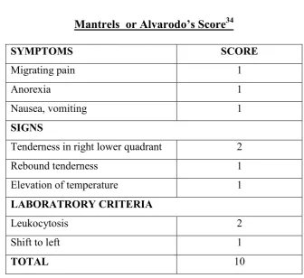
TABLE NO - 2
Mantrels or Alvarodo’s Score
34SYMPTOMS SCORE
Migrating pain 1
Anorexia 1
Nausea, vomiting 1
SIGNS
Tenderness in right lower quadrant 2
Rebound tenderness 1
Elevation of temperature 1
LABORATRORY CRITERIA
Leukocytosis 2
Shift to left 1
TOTAL 10
a score of less than or equal to 6 to be clinically dubious and more than or equal to 7 to be
typical.
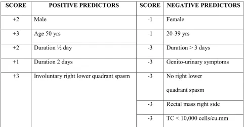
Teicher et al (35)
described a scoring system, after retrospectively studying 100 cases of
TABLE NO – 3
TEICHER SCORING SYSTEM
A score greater than 3 was taken as a positive predictor of acute appendicitis.
These scoring system are used to prevent negative laparotomy in emergency situations
without radiological investigation
SCORE POSITIVE PREDICTORS SCORE NEGATIVE PREDICTORS
+2 Male -1 Female
+3 Age 50 yrs -1 20-39 yrs
+2 Duration ½ day -3 Duration > 3 days
+1 Duration 2 days -3 Genito-urinary symptoms
+3 Involuntary right lower quadrant spasm -3 No right lower
quadrant spasm
-3 Rectal mass right side
DIFFERENTIAL DIAGNOSIS
It is wise to consider carefully possible diseases of the chest, the
abdomen, the pelvis, the genitourinary system, the central nervous system, and
the spines because although acute appendicitis is the commonest abdominal
emergency the diagnosis at times can be extremel y difficult.
A) Nasopharynx and Thorax
1. TONSILLITIS
In children abdominal colic may arise from swallowed exudate (tonsil
tummy).
2. PNEUMONIA AND PLEURISY
Especially at the right base, gives rise to right sided abdominal pain but they are
associated with an increased respiration rate and the pain prevents deep inspiration.
B) Diaphragm to the level of umbilicus
I. PERFORATED PEPTIC ULCER
Contents pass from the perforated area to the right paracolic gutter. This
may mimic appendicitis but the differentiating feature may be origin of pain
and other localising signs.
2. ACUTE CHOLECYSTITIS
Murphy's sign and radiation of pain through to the right scapula are
important features.
3. CYCLICAL VOMITING
C) Umbilicus to the Brim of pelvis 1. ENTERO-COLITIS
Intestinal colic together with diarrhoea and vomiting but localised tenderness does
not usually occur.
2. NONSPECIFIC MESENTRIC LYMPHADENITIS
Shifting tenderness, when the child turns is very characteristic and other cervical
lymph nodes may be enlarged.
3. INTESTINAL OBSTRUCTION
Colicky pain and vomiting may be present. Plain X-ray shows fluid levels.
4. REGIONAL ILEITIS
A history of diarrhoea with a doughy mass of inflamed ileum can be felt.
5. CARCINOMA CAECUM
It is rare. H/O discomfort, altered bowel habits or unexplained anaemia. On
examination mass is palpable in RIF.
6. MECKEL'S DIVERTICULITIS
It is difficult to differentiate from acute appendicitis. However signs may be
central or left sided. Occasionally history of lower gastro-intestinal bleeding may be
present.
7. INTUSSUCEPTION
Median age of onset is around 18 months. Presence of Psoas mass in Right Lower
8. HENOCH-SCHONLEIN PURPURA
It is preceded by sore throat or respiratory tract infections.
9. ACUTE PANCREATITIS
Ruled out by serum amylase measurement.
10. RECTUS SHEATH HEMATOMA
Localised pain without gastro-intestinal symptoms.
D. The Pelvis 1. SALPINGITIS
Vaginal discharge, menstrual irregularities, and dysmenorrhoea and burning
micturition are the differentiating points. Per vaginal examination reveals adnexal and
cervical tenderness.
2. MITTLESCHMERZ PAIN
Mid-cycle abdominal and pelvic pain
3. ECTOPIC GESTATION
Tubal abortion resembles acute appendicitis. Pain commences on the right side
and stay there. Cervix is softened. Referred pain to shoulder is present. H/O missed
menstrual periods and positive pregnancy test are present.
4. RUPTURED OVARIAN FOLLICLE
It is indicated by the history and ultrasound finding..
5. TWISTED RIGHT OVARIAN CYST
Pain is severe, often referred to the loin, and is made worse when the patient rolls
6. SIGMOID DIVERTICULITIS
Long sigmoid loop with diverticulitis lies to the right of midline. Impossible to
differentiate. Abdominal CT is particularly useful.
E) The Retroperitoneal Structure
1.RIGHT URETERIC COLIC
It commences in the loin and spreads to groin. Plain X-ray may show stone and
urine analysis may be helpful.
2. RIGHT SIDED ACUTE PYELONEPHRITIS
There is increased frequency of micturition, fever, rigor and pyuria.
F) Central Nervous system
1. POST-HERPETIC PAIN
Marked hyperesthesia and herpetic eruption.
2. TABETIC CRISIS
Pain and vomiting and other signs of tabes are present.
3. SPINAL CONDITIONS
Pott’s disease, carcinomatous deposits, osteoporosis and myelomatosis causes
compression of the nerve roots. Other conditions include porphyria, and diabetic crisis
TREATMENT
36The correct treatment of appendicitis in all its aspects is one of the most important
subjects in abdominal surgery because it is the most common major abdominal condition
calling for emergency operation. The treatment of acute appendicitis is appendicectomy
and the sooner done the better. There are four exceptions to this excellent rule.
1. The patient is moribund with advanced peritonitis.
2. The attack has already resolved. Here appendicectomy can be done as an elective
procedure to prevent recurrence.
3. Where circumstances make operation difficult or impossible for example in a
small boat at sea. This condition deserves conservative line of treatment.
4. An appendicular mass has formed without evidence of general peritonitis.
CONSERVATIVE TREATMENT: OSCHNER SHERRAN’S REGIME
This method of treatment is applicable to all types of acute appendicitis in which
operation is withheld due to one reason or the other. The details of the method may be :
1.
Patient is nursed in a propped up position, in order to encourage anyperitoneal exudate to gravitate towards the pelvis. This is not widely practiced
nowadays.
2.
Intensive chemotherapy is instituted.3.
First 24 to 48 hours, nothing is administered by mouth except sips of water. Gastricsuction is advocated. After 24 to 48 hours, provided that all the symptoms are
subsiding, fluid diet is commenced and gradually supplemented.
4.
Careful recording of pulse rate and temperature.5.
Sedatives are given.infection.
7.
No aperient until pulse, temperature and abdominal condition settles.The majority of patients react favorably to this treatment.
INDICATION FOR OPERATION
1. The most dangerous sign is a rising pulse rate. If this sign is present the conservative
treatment should be discontinued and surgery adopted. A steady high pulse rate is also
an indication for surgery.
2. High swinging temperature is also another indication.
3. Continuous pain, vomiting, diarrhoea are other features.
4. If there is spreading tenderness or resistance, surgery is preferred.
5. In certain group of patients, the abscess formation occurs which although is localised
needs drainage if it becomes adherent to anterior abdominal wall and is pointing
towards it.
In the present series, most of the cases were taken as emergency. Elective surgery
was advocated in patients who had formation of mass or an abscess which was drained or
treated conservatively due to other operative risk.
PRE-OPERATIVE PREPARATION
In surgery, the preoperative preparation is very important for excellent operation
and short convalescence. The steps include:
(1) Re-expansion of intra vascular volume
(2) Restoration of electrolyte balance
(3) Resolution of fever
(5) Continuous Ryle tube suction with no oral feeds
(6) No enema to be given.
A useful guideline to postpone surgery is till (1) temperature becomes less than
100o F (2) Pulse rate less than 120/mt. (3) The patient has voided urine atleast once. All
these corrections can be done within 4 to 6 hours. The patients vital signs and clinical
response form the ultimate test of satisfactory response.
In the present study, 80% of the cases were operated within 2-4 hours after
admission; with initial pre-opeartive preparation like I.V. fluids, Ryle's tube aspiration;
and antibiotics. The antibiotics commonly used were cefotaxime, Garamycin and
Metronidazole injections. Bates in 1980 and Simons et al and Devitt et al indicate the
potency of Metronidazole.
ANAESTHESIA
The type of anaesthesia for appendicectomy is determined by the preference of
surgeon and anaesthetist, whether general anaesthesia or spinal.
INCISIONS
There is no one special incision for appendicectomy. Experience should enable the
surgeon to determine with a fair degree of accuracy the position and the pathological
changes in the appendix before operation. When the patient is fully anaesthetised the
surgeon should once again systematically palpate the abdomen to locate the appendix,
which may be felt as a circumscribed lump, diffuse thickening or as a movable tumor.
the appendix lies immediately beneath the abdominal wall in the right iliac fossa, then a
McBurney's grid iron incision is the choice. If required, the incision may be extended
inward through the sheath of the rectus muscle called the Fowler Weir incision.
Morrison's modification or more correctly Kocher's modification of the McBurney’s
incision in which all the muscles are divided transversely or obliquely in line with the
incision and is useful to gain access to a hidden adherent retrocaecal appendix in
an obese patient. When appendix is thought to be central in position or is lying in the
pelvic cavity a median or paramedian subumbilical incision is chosen. Battle's pararectal
incision is preferred by some surgeons though not widely used as there are more chances
of post operative wound dehiscence and incisional hernia. Transverse or Rockey-Davis
incision gives rapid access to the right lower portion of abdomen, If the incision is
appropriately placed centered in the midinguinal point 1-2cm below umbilicus. However,
there was a theoretical objection to transverse incision. The medial end of the incision is
relatively close to the midline, so that when localized pus was present and spillage occurs,
there was a danger of dissemination.
In the present series three main incisions were chosen in the cases. They are (1)
Lanz incision (2) McBurney s 3) Right Paramedian 4) Laproscopic
LANZ INCISION
It is a skin crease Langer's line incision. It is made more or less transversely and
curves so that it lies in the interspine crease. Thereafter the muscles are divided as in
McBURNEY'S INCISION
It is an incision perpendicular to the spinoumblical line on the McBurney;s point,
2/3 of the length below and 1/3 of the length above the line. The McBurney’s incision
was undoubtedly the most popular, but it is the Transverseor Rockey-Davis incision that
meets the criteria for an appropriate incision most closely.
RIGHT MID OR LOWER PARAMEDIAN INCISION:
Doubts as to the correctness of diagnosis could be tackled safely with this
incision.
PROCEDURE
By any of the above said incisions the abdomen is opened. Retractors are inserted
beneath the peritoneum and the wound margins separated to permit inspection. Omentum
will be seen and is a good evidence of acute appendicitis. The caecum is picked up with
fingers or turtle’s forceps. The appendix is lifted and coaxed gently on to the surface of
the skin. The flimsy adhesions to the surrounding structures can often be atraumatically
separated by dissection with a small swab or dissecting forceps this enables us to note
the position of appendix.
The freed appendix can be brought out, with a Babcock’s forceps. If the
mesoappendix is long, they are ligated with silk. When the mesoappendix is gangrenous,
careful ligature application is needed. It is essential to insert a single interrupted suture
close to the base of the appendix at its mesenteric border in order to secure the intramural
branch of the posterior caecal artery, a branch of appendicular artery. A purse string
crushed. After removal of the forceps, the crushed area is ligated with chromic catgut.
Appendix is cut and the stump cauterised and invaginated by pulling the purse string. Few
interrupted suture in the caecal wall as Z stitch can also be done. When the caecal wall is
swollen and oedamatous the purse string is omitted, instead two strong ligatures are
applied to the base of the appendix, the sprouting mucous membrane is cauterised. When
the base is inflamed, it should not be crushed for fear of distributing infection. It should
be ligated close to the caecal wall just tight enough to occlude the lumen after which
appendix is amputated.
The methods of treating the appendix stump:
1.
Simple ligation of the stump.2.
Ligation and inversion of the stump.Wound closure:
After removal, the operated area has been irrigated (in gangrenous or perforated
appendix) each fascial and muscular layer was closed with an absorbable suture material.
Skin was closed with non-absorbable sutures.
RETROGRADE APPENDICECTOMY
It is indicated when the appendix base is autoamputated or when appendix base is
well visualised but the remaining portion is either in the paracolic or retrocaecal in
position. The base of the appendix is held between finger and thumb and fine
hemostat is passed between the caecum and appendix to create space and two
similar instruments are applied across the appendix and divided. The
mesoappendix is clamped and divided by traction on the distal clamp. The purse
string is introduced and then the appendix is freed. Then a regular method of
PERITONEAL TOILETING
This is done in cases of perforation, peritonitis, using normal saline and
metronidazole.
DRAINAGE
Drainage of the peritoneal cavity is not always necessary especially so in an
imperforated appendix. Drainage is not advised even after excision of an
inflammatory mass.
Aird felt that the mortality rate of the abscess could be reduced by drainage of the
abscess. Campbell and Me.Phail are of the same view. Drainage is required when
1. Following the removal of a gangrenous appendix in a walled off abscess.
2. After appendicectomy for diffuse appendiceal peritonitis.
3. When appendicectomy could not be done and when in doubt, drain is kept,
Free peritoneal drains may be detrimental perpetuating the inflammatory process by
foreign body reaction but also serving as a nidus for intra-abdominal adhesions.
LAPAROSCOPIC APPENDICECTOMY
37, 38Performed through three small incisions of 10 mm, 5 mm, and 10 mm, at the level
of umbilicus through which the telescope is introduced, at the right costal margin in the
mid axillary line and the left iliac fossa for operating instruments.
Advantages
1. Procedure is done under direct vision.
2. Less pain and early recovery.
4. Minimal handling of the small intestines which minimizes serosal damage which can
lead on to adhesion formation.
5. Less tissue trauma because of small operating instruments.
6. Associated gynecological problems can be managed easily.
7. Short hospital stay.
Disadvantages:
1. Operative time required is more compared to open appendectomy.
2. Setting up of instruments and team is difficult for emergency appendectomy.
3. It is expensive compared to conventional method.
4. Expertise in technique is needed.
5. Procedure is not useful if perforation or peritonitis is present.
In summary, laparoscopic appendectomy is a safe alternative to open appendectomy.
COMPLICATIONS AND THEIR MANAGEMENT
181.PERITONITIS
The symptoms and signs of peritonitis could be grouped into two. Reflex
symptoms which occurs early and toxic which occurs late. Reflex symptoms and signs are
pain, vomiting, anxious facies, abdominal muscular rigidity, collapse, fever etc. There is
an increased relationship between the toxic and reflex symptoms. Severe toxemia
diminishes the sensitivity of the reflex one. The progress of peritonitis is altered by the
treatment and it may get localised in (1) Right iliac fossa (2) Cul-de-sac of Douglas (3)
‘walling off ' of appendix by omentum and coils of intestines. Certain cases do develop
generalised peritonitis with typical Hippocratic facies etc.
TREATMENT
The preoperative preparation is similar. A right subumbilcal paramedian incision is
made. Verify the positions and condition of the appendix. Caecum is gently freed from
adhesions. Appendix delivered and appendicectomy done. If caecal wall is inflamed the
purse string burial may be difficult. Peritoneal cavity toileted and drained.
2. MECHANICAL INTESTINAL OBSTRUCTION AND ILEUS
Ileus may be due to local or generalised peritonitis. Mechanical obstruction is due
to formation of adhesions which are part of protective process. These adhesions are light
and flimsy. Incorporation of a bowel in an appendiceal abscess is a frequent cause of
ileus.
The distinction between mechanical obstruction and ileus is very important because
the former needs surgical treatment where as the later can be treated conservatively.
1. PRIMARY PERIAPPENDICULAR ABSCESS
A ruptured appendix in which the pathological process has localised or a diffuse
peritonitis with signs of localisation is the formation of an abscess. Signs and symptoms
are widely varied depending upon the site of the appendix and location of the abscess.
There are two types of abscesses:-
i) Recent Abscess:
This is a local collection of pus pooling around perforated appendix that is
completely shut off by the greater omentum, the abdominal parietes and adjacent coils of
ii) Establised abscess:
This is a chronic abscess and the surrounding structures densely adherent.
1. PELVIC ABSCESS:
Easiest to diagnose once it is suspected. A repeated rectal examination every 48
hours is diagnostic. Before the formation of abscess the patient has bowel and bladder
irritation due to pus tracking down due to gravity. Pelvic abscess if it points towards
vagina, a posterior colpotomy can be done. It is always preferable to drain rectally.
2. ILEOCAECAL ABSCESS
Presents as a primary peri-appendiceal abscess.
Technique of Drainage
After anaesthesia, incision is made over the prominent and fluctuant area. Peritoneum
is carefully entered as the intestines may be adherent. Index finger inserted and the pus
evacuated. Drainage tube is placed. Tube is left in situ for about 72 hours. Abscesses are
best approached by an incision immediately medial to the anterior superior iliac spine.
The lateral edge of the peritoneum exposed and by stripping it medially with the finger
and the mass is reached retro-peritoneally.
OTHER POST-OPERATIVE COMPLICATIONS 1. Early Complications
a) Local 1.Woundsepsis, 2.Abscess, 3.Infected sinus, 4.Dehiscence,
5. Faecal fistula, 6.Keloid, 7.Haematoma.
b) Pulmonary : 1.Basal pneumonitis, 2. Atelectasis
c) Coronary : Thrombosis
1. Unresolving appendicular abscess rupturing into urinary bladder, ureter, rectum,
small intestine, vagina.
2. Peritonitis.
3. Adhesive ileus.
4. Pyelophlebitis.
5. Thrombosis of major vessels.
2.
Late complications1. Incisional hernia
2. Right inguinal hernia.
3. Adhesive obstruction.
4. Pyelophlebitis, Liver abscess
METHODOLOGY
STUDY DESIGN:
This study is a prospective study.
FOR NON INFLAMMED CASES:
These cases represents the normal population .These are patients admitted in
Coimbatore medical college during the period of Nov 2009 to Dec 2011 for which
laparotomy done for other conditions than appendectomy. All of these cases are opened
as in any other laparotomy and appendix position and length noted first before disturbing
any of the structure. This is recorded in the profoma.
Any patients with pathology in the caecum and terminal ileum were excuded in
this study.
FOR CLINICAL CASES:
All cases with diagnosis of appendicitis admitted in Coimbatore medical college
during Nov 2009 to Dec 2011. All cases are subjected to clinical evaluations like signs,
symptoms, and investigations like ultrasound if clinical findings are doubtful. History is
elicited thoroughly regarding any atypical presentation. Clinical examination included
head to foot examination and to look for clinical signs of appendicitis like Rovsing sign,
Obturator sign, Baldwin sign, Psoas sign and per rectal examination. These are noted in
the proforma .
During the surgery by any method appendix position, length, with or without
complications like perforation and abscess formations were noted. The appendix
specimen was sent for histopathological examination to the qualified pathologist. Those
PROFORMA
For clinical cases (representing inflamed cases)
Serial No. Age: Sex
Name: Ward: Unit:
I.P.No. Occupation:
Address:
Date of admission:
Date of operation:
Date of discharge:
I. Presenting complaints:
a) Pain: Duration: Nature: Typical /Atypical Radiation :
b) Vomiting: Duration: Frequency:
Projectile / Regurgitated / bile stained / Not bile stained /
Pain relief after vomiting / contents of vomitus.
c) Fever: Duration: Intermittant / continuous
Associated with chills & rigors.
d) Diarrhoea / Constipation / Tenesmus / Blood & mucous.
e) Urinary complaints: Duration: Frequency:
Dysuria / Burning / Haematuria / Radiation of pain.
II. Family history
III. Past history: Number of similar attacks in the past: Previous hospitalization & follow-up:
IV. Menstrual history: Regular / Irregular Spotting ______/_____days
Deliveries:
V. Per - Abdomen
Inspection: Normal / Fullness Pointing test: Umbilicus / RIF
Palpation:
Site of maximum tenderness: RIF / Flank / Mc Burney’s point.
Rebound tenderness
Rovsing’s sign /Psoas sign /Baldwin’s sign /Obturator sign:
Local temperature
P.R: Tenderness - Yes / No Mass : Yes / No
VI. Investigations:
Hb:_____gm% TC: DC: ESR:
Urine: Albumin_____ , Sugar______ , Microscopy______
Ultrasound Abdomen:
RIF: APPENDIX - Length
- Diameter
- Mass / Abscess
- Probe tenderness
Pelvis:
Other findings:
VII. SURGERY:
Date: Time: Emergency/Elective
Preoperative diagnosis:
Postoperative diagnosis:
Incision:
Type of anaessthesia:
Operative findings: - Position: Preileal / Postileal / Retrocaecal / Paracaecal / Subcaecal / Pelvic / Promonteric
- Length
- Appearance
- Intraperitoneal / Extraperitoneal
- Fixed / Mobile
Caecum:
Ileum:
PROFORMA
For OTHER LAPAROTOMY cases (representing normal population)
Serial No: Age : Sex:
Name IP. No.
Address :
Date of surgery:
Diagnosis:
Position of the appendix
- Retro-caecal
- Para-caecal
- Sub-caecal
- Pelvic
- Promonteric
- Post-ileal
- Pre-ileal
Length of the appendix
RESULTS
Total number of cases studied were 125 cases of which 75 cases were normal
population i.e. cases for which laparotomy done other than for appendicitis. Remaining
50 cases are inflamed group, patients with appendicitis whose histopathology report is
positive.
In our series appendicitis is more common during the 2 rd
decade (36%), followed
by the 3 nd
decade (30%). About 78% are male patients with appendicitis. Hence there is
male dominance.
The chief complaints of most patients are fever and abdominal pain. Fever is
present more than half the patients with appendicitis. Abdominal pain is severe in
intensity in 72% of the population,68% of patient shows typical right iliac pain and
tendeness rest of them showed vague pain. About four cases (case no.16,22,24,44)
presented with signs of diffuse peritonitis but peroperatively only two cases had
perforation (Fig No.12) and two had abscess collection in right iliac fossa.
Anorexia is present only in 20% of patients while vomiting and nausea
predominates the history in 48% of patients.
8 cases had atypical presentation and the position of those appendix are mostly
retrocaecal and then comes ileal position in this 8 cases four had emergency surgery (Fig
No.10).
Psoas test was positive in 2 case both had retrocecal position ,obturator test is
examination is present in 5 cases in which 3 were pelvic (Fig No.11) and other 2 were
gangrenous. No other test was positive in our patients.
Total number of emergency cases done where 33 nearly 67%. Two cases
(no.14,27) presented with appendicular mass and interval appendecectomy was done after
2 months. The position of appendix in these cases were retrocaecal (Fig No.13).
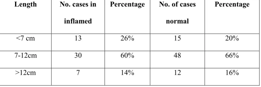
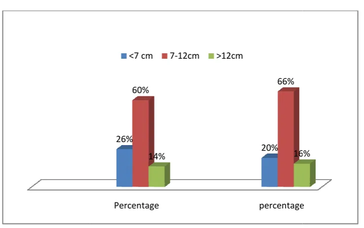
Length of appendix was variable and did not influence the position. Smallest was
6 cm and the lengthiest was 19 cm in our study. The average length was 10.3cm.
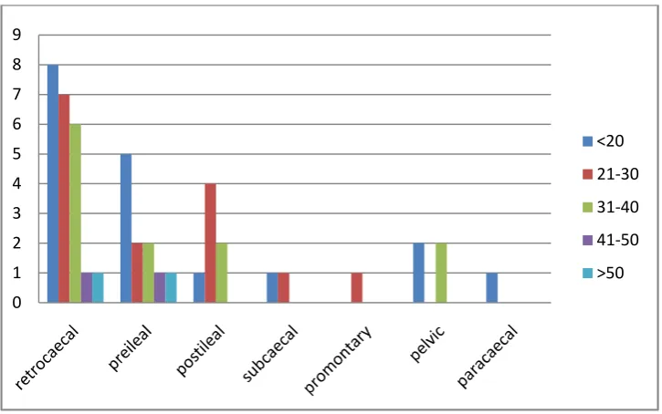
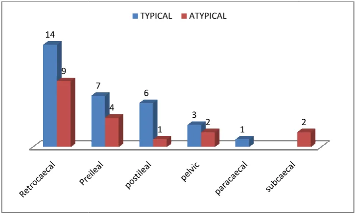
The position of the appendix influences the clinical presentation of the
appendicitis with the retro-caecal position (30% of cases), post-ileal position (14% of
cases), and the Pelvic position (20% of cases) presenting with atypical symptoms. 22%
of cases presented atypically of