ROLE OF NT- PRO BNP LEVELS IN PREDICTING THE
PROGNOSIS IN ACUTE CORONARY SYNDROMES
Submitted in partial fulfilment of Requirements for
M.D.DEGREE EXAMINATION BRANCH-I INTERNAL MEDICINE
THE TAMILNADU DR. M.G.R MEDICAL UNIVERSITY CHENNAI
INSTITUTE OF INTERNAL MEDICINE MADRAS MEDICAL COLLEGE
CERTIFICATE
This is to certify that the dissertation entitled “ROLE OF NT -
PRO BNP LEVELS IN PREDICTING THE PROGNOSIS IN ACUTE
CORONARY SYNDROMES” is a bonafide work done by
DR ANOOP C HARIDAS, Post Graduate Student, Institute of Internal Medicine, Madras Medical College, Chennai-3, in partial
fulfillment of the University Rules and Regulations for the award of
MD Branch – I Internal Medicine, under our guidance and
supervision, during the academic period from April 2008 to April
2011.
Prof. C. RAJENDIRAN, M.D., AssociateProf. S TITO, M.D.,
Director & Professor, Institute of Internal Medicine, Institute of Internal Medicine, MMC & GGH, MMC & GGH, Chennai – 3. Chennai – 3.
Prof . J. MOHANASUNDARAM, M.D.,
Dean,
Madras Medical College, Government General Hospital,
I solemnly declare that the dissertation entitled “ROLE OF NT -
PRO BNP LEVELS IN PREDICTING THE PROGNOSIS IN ACUTE
CORONARY SYNDROMES” is done by me at Madras Medical College,
Chennai-3 during May 2010 to November 2010 under the guidance and
supervision of Associate Prof. S. TITO, M.D., to be submitted to The
Tamilnadu Dr M.G.R Medical University towards the partial fulfillment
of requirements for the award of M.D DEGREE IN GENERAL
MEDICINE BRANCH-I.
Place: Chennai
Date:
Dr. ANOOP C HARIDAS M.D. GENERAL MEDICINE Postgraduate Student,
I thank Prof. J.MOHANASUNDARAM, M.D., Dean, Madras Medical College, for having permitted me to conduct the study and use
the hospital resources in the study.
I express my heartfelt gratitude to Prof. C. RAJENDIRAN, M.D.,
Director, Institute of Internal Medicine for his inspiration, advice and
guidance in making this work complete.
I express my deep gratitude to my chief Associate Prof. S. TITO,
M.D., Institute of Internal Medicine for his comments and guidance to complete the study.
I am extremely thankful to Assistant Professors of Medicine
Dr. G. SUBBARAGHAVALU, M.D., and Dr. C SRIDHAR, M.D., for guiding me with their time, corrections and prompt help rendered
whenever approached.
I also thank all the postgraduate students and paramedical staff for
their cooperation which enormously helped me in the study. I am also
indebted to thank all the patients and their caring relatives. Without their
BNP - Brain Natriuretic Peptide
NT ProBNP - N-Terminal Pro Brain Natriuretic Peptide
ACS - Acute Coronary Syndrome
CAD - Coronary Artery Disease
HF - Heart Failure
UA - Unstable Angina
MI - Myocardial Infarction
NSTEMI - Non ST Elevation Myocardial Infarction
STEMI - ST Segment Elevation Myocardial Infarction
AF - Atrial Fibrillation
ANP - Atrial Natriuretic Peptide
LV EF - Left Ventricular Ejection Fraction
TIMI - Thrombolysis in Myocardial Infarction
SK - Streptokinase
PCI - Percutaneous coronary intervention
TIMI - Thrombolysis in Myocardial Infarction
INC - Including
SOB - Shortness of breath
LV EF - Left Ventricular Ejection Fraction
MILD LV DYS - Mild Left ventricular dysfunction.
MOD LV DYS - Moderate Left Ventricular Dysfunction.
SEVERE LV DYS - Severe Left ventricular Dysfunction
INC - Including
REC - Recurrent
pg/ml - Pico Gram per milli litres
S. No PARTICULARS PAGE
1. INTRODUCTION 01
2. AIM OF THE STUDY 04
3. REVIEW OF LITERATURE 05
4. MATERIALS AND METHODS 35
5. OBSERVATION & RESULTS 40
6. DISCUSSION 59
7. LIMITATIONS 62
8. CONCLUSION 63
9. RECOMMENDATIONS 64 10. BIBLIOGRAPHY 65 11. ANNEXURES 77
I. PROFORMA
II. MASTER CHART
INTRODUCTION
Cardiovascular diseases contribute 29.3 % of all deaths
worldwide according to WHO statistics. Out of these around half of the
deaths are due to ischemic heart diseases. In India it is the leading cause
of death (15%) followed by respiratory infections (11 %) and
cerebrovascular diseases (7 %).
Cardiovascular disease continues to be the major cause of death
despite theuse of new pharmacological strategies to lower blood lipids,
more aggressive therapy of hypertension, and changes in lifestyle. Acute
coronary syndromes include acute myocardial infarction (MI) with
ST-segment elevation, non ST elevation myocardial infarction (NSTEMI)
and unstable angina (UA). This syndrome is a serious health problem
because it is responsible for 20% of all medical emergency department
admissions with the highest risk for adverse events and death.
In recent times lot of advances have been made in the field of
biomarkers for the diagnosis of acute myocardial infarction like
Ischemia-Modified Albumin, Myeloperoxidase, Phosphorylase
Isoenzyme BB, IL- 6 , PAPP-A and many more are under trial.
But in the field of prognostic biomarkers only very few are
available which includes high sensitivity CRP and initial Troponin I
In an ideal scenario patients with acute myocardial infarction
brought immediately to a specialist care center with good expertise in
primary percutaneous coronary intervention (PCI) as well as in
thrombolysis, often achieve good initial results with a fully reperfused
heart, asymptomatic patient, fully normalized ECG, early resolution of
enzymes and a good left ventricular function in echocardiography. Even
in such ideally treated patients on regular follow up, quality of living is
often guaranteed but the risk for another coronary event is still many
fold higher than the general population.
Often the questions from patients are, will I be symptom free ?,
will I get chest pain again even if I take the medicines regularly, is my
heart working well ? Will there be any other complications?
Here in this study use of N- Terminal pro Brain Natriuretic
Peptide (NT-proBNP) which is an inactive remnant of a hormonally
active Brain Natriuretic peptide (BNP) is evaluated in predicting the
prognosis after an acute coronary syndrome. This study intends to see
whether its initial level of NT-proBNP taken during the first episode of
an ACS, correlates with the risk of recurrent symptoms like angina,
dyspnea, palpitations, recurrent hospital admissions for ACS including
unstable angina, NSTEMI, STEMI or death in a period of 6 months after
Measurement of NT proBNP is indeed an indirect measurement
of Brain Natriuretic Peptide itself. The former is measured as it has a
longer half-life than hormonally active BNP. (1)(2)(3)
BNP is a natriuretic peptide that is mainly released from the
cardiac myocytes in the left ventricular wall in reaction to stretch and
tension of the myocardial wall. The pro hormone proBNP splits into
BNP and the hormonally inactive remnant N-terminal proBNP by
proteolytic cleavage and both peptides will be secreted in equimolar
amounts into the circulation. It protects the body from plasma overload
by inducing diuresis, natriuresis, vascular dilatation and inhibition of the
sympathetic nervous system.
The half-life of BNP is around 20 minutes and the half-life of
NT-proBNP is around 120 minutes. (1)
At present for predicting the prognosis in an acute coronary
syndrome we rely on scoring systems like Thrombolysis in Myocardial
Infarction (TIMI) score and initial Troponin I levels. Few studies have
come up with use of BNP in predicting the occurrence of cardiac failure
in ACS patients in western population.(13)(14)(15)
As Brain Natriuretic Peptide is one of the most sensitive
biomarker for cardiac muscle strain, this study intends to evaluate its use
as a prognostic biomarker in patients admitted with Acute Coronary
Aims & Objectives
1. Study whether NT ProBNP is elevated in Acute Coronary
Syndromes
2. To Study whether NT proBNP levels during an Acute Coronary
Syndrome correlate specifically with symptoms like recurrent
angina, exertional dyspnoea, palpitations, recurrent hospital
admissions for ACS including Unstable Angina, NSTEMI and
STEMI or death during the follow up.
3. To Study whether it predicts a deteriorating left ventricular
function, and renal function.
REVIEW
OF
REVIEW OF LITERATURE
Brain natriuretic peptide (BNP), also known as B-type natriuretic
peptide is a 32 amino acid polypeptide secreted by the ventricles of the
heart. BNP is named as such, because it was originally identified in
extracts of porcine brain. In humans it is mainly produced in the cardiac
myocytes of the left ventricular wall. Unlike ANP, whose major storage
sites are in both the atria and ventricles, the major source of plasma
BNP is the cardiac ventricles, suggesting that BNP may be a more
sensitive and specific indicator of ventricular disorders than other
natriuretic peptides. (1)(2)
The release of BNP appears to be in direct proportion to
ventricular volume and pressure overload. In reaction to stretch and
tension of the myocardial wall the pro hormone proBNP splits into BNP
which is hormonally active and the hormonally inactive remnant, a 76
amino acid N-terminal proBNP (NT-proBNP) by proteolytic cleavage.
This process occurs under the influence of integrins, structures at the
Z-disc of sarcomeres. After the stretch of these sarcomeres both peptides
will be secreted in equimolar amounts into the circulation.
Circulating BNP acts as an antagonist of the renin angiotensin
aldosterone system, and protects the body from plasma overload by
inducing diuresis, natriuresis, vascular dilatation and inhibition of the
sympathetic nerves system. BNP has antiremodeling and antifibrotic
activity (2).
The half life of BNP is around 20 minutes and the half life of
NT-proBNP is around 120 minutes (1).
Natriuretic Receptors :
1. Guanylyl cyclase-A (GC-A) also known as natriuretic peptide
receptor-A (NPRA/ANPA) or NPR1
2. Guanylyl cyclase-B (GC-B) also known as natriuretic peptide
BNP binds to and activates the atrial natriuretic factor receptors
NPRA, and to a lesser extent NPRB, in a fashion similar to atrial
natriuretic peptide (ANP) but with 10-fold lower affinity. Both atrial
natriuretic peptide and brain natriuretic peptide bind and activate GC-A,
whereas CNP binds and activates GC-B. The biological half-life of BNP
however is twice as long as that of ANP and that of NT-proBNP even
longer, making these peptides better targets than ANP for diagnostic
blood testing. (7)
BNP is cleared from the circulation by receptor-mediated
endocytosis via the C-type natriuretic peptide receptor, as well as by
enzymatic degradation via zinc-containing endopeptidases located on
the vascular endothelial cells and in the renal tubules. Little is known on
the exact clearance mechanism of NT-proBNP, although it has been
suggested that the kidneys play a major role in this clearance (1)(2).
Effect of BNP
Relationship of BNP/NT-proBNP and gender
In healthy adults, gender differences in plasma levels of
natriuretic peptides are found, with females having higher plasma levels.
However, some of the studies found a gender difference for
ANP/NT-ANP and not for BNP/NT-proBNP. This might reflect the age difference
between men and women; because BNP/NT - proBNP were shown to be
more affected by ageing as compared to ANP/NT-ANP, this age
difference could be more powerful in BNP/NT-proBNP. (22)
BNP in Geriatric Age Group
This relationship between age and natriuretic peptide levels is a
consequent to age-related changes in left ventricular compliance as well
as a decreasing GFR (1)(2)(7). Age stratification improves the ability of
NT-proBNP to identify a high likelihood for acute heart failure (HF).
The confirmation and exclusion cut-points for NT-proBNP will help the
clinicians more confidently to utilize the marker in the evaluation of the
dyspnoeic patient, preserving sensitivity for younger patients with
elderly non-systolic HF below the threshold for acute HF, >97% had an
NT-proBNP value above the „rule out‟ cut-point of 300pg/mL(7)
.
NT-proBNP levels are higher in older female subjects when
compared with age-matched male subjects, possibly due to a higher
prevalence of diastolic abnormalities or more significant age-related
reductions in GFR in women. There were no gender differences in
younger age groups. The cut-off points proposed by PRIDE study is
1800pg/mL for those >75 years (20). Thejus et al Study on NT proBNP
and Atrial Fibrillation takes cutoff value of NT-proBNP for diagnosis of
heart failure as 125 pg/ml in the below 75 years age group and 450
pg/ml in the age group above 75 years (19). The addition of this cut-point
is relevant, as the age-related effects on NT-proBNP results are
significant as the average age of patients with acute heart failure is
rising.
Anemia and NT-proBNP levels
Anemia is a common phenomenon in HF and is related to the
severity of the disease. Hb was related to BNP and NT-proBNP levels
independent of severity of HF as measured by left ventricular ejection
suspected coronary artery disease showed an independent association
between Hb and BNP levels (21)(1). Anaemia results in elevated plasma volume, independent of the severity of Heart Failure.
Since BNP and NT-proBNP are released in response to ventricular
volume overload, it is conceivable that BNP and NT-proBNP levels are
higher in anaemic HF patients compared to non-anaemic HF patients.
Additionally, patients with anaemia and renal dysfunction showed
higher BNP and NT-proBNP levels when compared to anaemic patients
without renal dysfunction (21). Renal dysfunction was found to be a
major cause of anaemia in HF patients, mediated by an erythropoietin
production deficiency in the kidneys.
BNP in Acute Coronary Syndromes :
BNP has usefulness as a prognostic marker among patients with
acute myocardial infarction. BNP has prognostic value across the full
spectrum of patients with ACS, including those with UA/NSTEMI.
(1)(13)(14)(15)
In OPUS-TIMI 16, patients with elevated levels of BNP (>80
pg/ml) or NT-proBNP had a two to threefold higher risk of death by 10
TACTICS-proBNP in patients presenting with UA/NSTEMI adds importantly to
our current tools for risk stratification (29).
Both the European Society of Cardiology and HFSA/AHF
guidelines suggest that BNP assays are a useful adjunct to clinical
assessment (29).
“Breathing Not Properly” a multinational studydemonstrated that
BNP was able to distinguish dyspnoea caused by heart failure from that
caused by pulmonary disease with a high degree of predictive power and
it was the strongest predictor in multiple logistic regression analysis of
the diagnosis of heart failure (9). In addition, this study was able to
demonstrate the use of a cut-off 100 mg/dl for a normal NT-proBNP.
The BNP test alone had a diagnostic accuracy of 81.2 percent, compared
to 74.0 percent for clinical judgment alone. Although it may seem
disconcerting that the addition of the physician assessment to the BNP
measurement only barely improved diagnostic accuracy to 81.5 percent,
the standard for diagnosis in this trial remained the clinical assessment
by cardiologists, reviewing the totality of the clinical information. This
trial established the role of BNP testing in the assessment of dyspnea in
the emergency setting, and in general a BNP value of less than 100pg/ml
concentrations greater than 400pg/ml have high positive predictive
value for heart failure as the etiology of the dyspnoea (8)(9)(10).
Given the larger mass of ventricle rather than atrial myocardium,
the total amount of mRNA for BNP is higher in the ventricles than the
atria. Natriuretic peptides are released early after STEMI, peaking at
about 16 hours (13)(14). Evidence exists that natriuretic peptides released
from the left ventricle during STEMI originate both from the infarcted
myocardium as well as the viable noninfarcted myocardium (1)(18). The
rise in BNP and N-terminal pro-BNP levels after STEMI correlates with
infarct size and regional wall motion abnormalities(14). Patients with
anterior infarction, lower cardiac index and more significant congestive
heart failure after STEMI have higher levels of N-terminal pro-BNP and
BNP and such elevations correlate with a worse prognosis (18 ) .
Measurement of natriuretic peptides can provide useful
information both early and late in the course of STEMI (19). Patients with
elevated levels 6 hours after the onset of symptoms have a marked
increase in mortality even after adjusting for other known prognostic
indicators.Conversely, patients with persistently elevated levels at 3 to 4
weeks after STEMI have an increased risk of cardiac-related mortality
Several studies have shown that BNP and NT-proBNP levels
have powerful prognostic value for death and MI in patients with Non
ST Elevation Acute Coronary Syndromes , independent of markers of
myocardial necrosis or inflammation(1)(14)(15) . The FRISC-II trial (144)
found that BNP levels predicted the benefit of revascularization, but
there was no such association in TACTICS - TIMI-18(29). Serial BNP
measurements can be used for dynamic risk profiling. Patients with
normal troponin levels and low BNP levels are at very low risk of
Clinical significance of BNP in Heart Failure
Both BNP and NT-proBNP levels in the blood are used for
screening of acute CHF. The plasma concentrations of both BNP and
NT-proBNP are also typically increased in patients with asymptomatic
or symptomatic left ventricular dysfunction. BNP accurately reflects
current ventricular status. BNP is an independent predictor of high LV
end-diastolic pressure and is more useful than ANP or norepinephrine
levels for assessing mortality risk in patients with heart failure.
For patients with Congestive Cardiac failure, BNP levels of more
than 100 pg/mL have better than a 95% specificity and greater than a
98% sensitivity when comparing patients without heart failure to all
patients with heart failure(1).
For patients with Congestive Heart Failure, BNP values will be
above 100 pg per millilitre (11). The Achilles heel of the NT proBNP
molecule is the overlap in kidney disease in the heart failure patient
population.
The BNP test is used as an aid in the diagnosis and assessment of
severity of congestive heart failure. Natriuretic peptide concentrations
correlate with filling pressures and both admission and pre-discharge
BNP concentrations are predictive of outcomes (24). The shorter half-life
of BNP and the substantial renal clearance of NT-proBNP support the
selection of the BNP assay for this potential diagnostic use. Changes in
serial measures of BNP are predictive of outcomes in patients with
AHF, beyond clinical and echocardiographic assessments and clinical
studies are currently ongoing to assess the utility of serial inpatient BNP
measures.
Hullsman-Berger et al found that using NT-proBNP measurement
outcome than multidisciplinary care alone in patients after
hospitalization for heart failure (26). In a multi-institutional trial, 278
patients were randomized to NT-proBNP guided intensive
management (BM), multidisciplinary care, or usual care. After 12
months, patients in the BM group had fewer days of heart failure
hospitalization (488 d) than patients in the multidisciplinary care group
(1,254 d) or usual care group (1,588 d) (p < 0.0001 for both). In
addition, first heart failure rehospitalization was lower with BM than
multidisciplinary care (28% versus 40%, respectively and p value was
0.06. The combined end point of death or heart failure rehospitalization
was lower in the BM group (37%) than in the multidisciplinary group
(50%; p < 0.05). The death rate was 22% in both the BM and
multidisciplinary groups, but 39% in the usual care group (p < 0.02) (26).
Pleural Effusion is common in cardiac failure. A pleural fluid
N-terminal pro-brain natriuretic peptide more than 1500 pg/mL is virtually
Acute Dyspnoea Clinical Stratification with BNP levels
Diagram showing clinical stratification according to BNP levels.
Reference: Topol Textbook of Cardiology 3rd Edition (3).
NT-proBNP is cost-effective in the diagnosis and management of
dyspnoeic patients in the emergency room (3).
Dyspnoea Rales Raised JVP
Rales Pedal Oedema
S3
Blood Test ECG Chest X Ray Echocardiography
BNP < 100 pg/ml
Unlikely to be Acute Heart Failure
Evaluate for other Etiologies (ie. pneumonia )
BNP 100 - 500 pg/ml
Rule Out Pulmonary Embolism Pulmonary Hypertension
Renal Failure
Further imaging and testing
warranted
BNP > 500 pg/ml
High likelihood of Acute Heart Failure
Cardiac Examination
S3 gallops or third heart sounds are detected in approximately 11
to 34 percent of patients admitted with Acute Heart Failure. An S3 is
fairly specific as an indicator of LV systolic dysfunction, and correlates
BNP and Incidence of Atrial Fibrillation (AF)
Framingham Heart Study showed elevated BNP levels to be
predictive of atrial fibrillation (AF), cardiovascular outcomes, and death.
Just 68 subjects developed AF during the longitudinal study of men and
women >65 years selected from four US communities. The group notes
that NT-proBNP remained the strongest predictor of incident AF after
adjustment for other variables including age, sex, medication use, blood
pressure, echocardiographic variables, diabetes mellitus, and heart
failure. Asselberg et al found that in the general population, elevated
NT-proBNP levels at baseline predicted the development of AF when
reassessed at 4 years (27). The baseline median level was 62.2 pg/ml, in
those who eventually developed atrial fibrillation compared to 35.7
pg/ml in those who did not. The difference was found to be highly
significant statistically (p = 0.001). Values above the 80th percentile (97
pg /ml in women and 60 pg/ml in men) were associated with an
odds ratio of 2.65 for the occurrence of AF. Mollmann et al
found that baseline NT-proBNP above 900 pg/ml significantly predicts
(p<0.05) persistence of AF at 4 weeks after DC version of lone atrial
NT ProBNP in Pulmonary Embolism
Elevations of NT-proBNP and BNP indicate myocardial stretch
caused by right ventricular pressure overload. These patients also have
an increased risk of a complicated hospital course, with a higher
likelihood of recurrent pulmonary embolism, respiratory failure
requiring mechanical ventilation, hypotension requiring vasopressors
and death (41)(43)(51).
BNP in Cardiomyopathy
BNP levels were proposed as a test to discriminate between
restrictive cardiomyopathy and constrictive disease, with concentrations
approximately five times greater in the former compared with the latter.
(2)(3)(67)
BNP in Aortic Stenosis
BNP and NT-proBNP are elevated in proportion to severity of AS
and symptomatic status (1)(23). NT proBNP decreases after successful
aortic valve replacement surgery. BNP serum level above 66 pg/mL
Renal function and BNP/ NT- proBNP levels
Elevated levels of BNP were also independently related to renal
dysfunction (32).This difference may be explained by differences in
clearance as NT-proBNP is mainly cleared from the blood by the
kidneys, while BNP is cleared mainly by neutral endopeptidases and
natriuretic peptide clearance receptors.
This implies that the influence of renal dysfunction should be
more pronounced on NT-proBNP levels compared to BNP levels.
Implications for clinical practice indicate that haemoglobin and renal
function should be taken into account when interpreting the elevated
levels of BNP and NT-proBNP.
CKD influences the levels of B-type natriuretic peptide. In
general, when the eGFR is less than 60 ml/min/1.73 m2, a higher BNP
cut off point of 200 pg/ml should be used in the diagnosis of heart
failure. It is now well recognized that CKD (eGFR < 60 ml/min/1.73
m2), when present in patients with HF, independently predicts poor
outcomes (32)(51). Brain natriuretic peptide (BNP) antagonizes the renin
angiotensin system and angiotensin II, and may serve as a biomarker of
Silva et al. measured NT ProBNP in patients with uncontrolled
hypertension and RAS (70 percent diameter stenosis), hypertension
improved in 77 percent of those with elevated BNP levels, compared
with group of five patients with a baseline BNP level less than or equal
to 80 pg/ml (P = 0.001). If the BNP level fell more than 30 percent after
successful stent placement, 94 percent (16 of 17 patients) had
improvement in their blood pressure control (51).
But BNP with its physiological effects of vasodilators, diuretic,
and natriuretic action do not appear to be sufficient to prevent the
disease progression to Cardiorenal Syndrome Type II.
NT proBNP in severe sepsis and septic shock
NT-proBNP values are frequently increased in severe sepsis and
septic shock. Values are significantly higher in nonsurvivors than
survivors. NT-proBNP on day 3 in the intensive care unit is an
independent prognostic marker of mortality in severe sepsis (68).
NT-proBNP Elevations in Adult Respiratory Distress Syndrome
NT-proBNP levels are elevated among patients with ARDS in a
range typically considered consistent with heart failure. NT-proBNP
NT - proBNP Predicts Stroke Risk
NT-proBNP is an independent marker of stroke risk(34)(35), while
other traditional risk factors like hypertension, left ventricular
dysfunction were found not to be independent predictors. NT-proBNP
was much better to point out which of the patients had a high risk for
these cardiovascular diseases. According to Pedersen F, et al. (34)(35),
potential prognostic factors for stroke, including age, gender, systolic
and diastolic blood pressure, atrial fibrillation, and NT-proBNP, were
evaluated using Cox proportional hazards analysis. The investigators
showed that NT-proBNP was a strong independent predictor of stroke
risk in this population with hazard ratio 4.1.
Atrial Natriuretic Peptide
Atrial Natriuretic Peptide (ANP) is closely related to BNP and
CNP (C-type natriuretic peptide). All share the same amino acid ring.
ANP was discovered in 1981 by a team in Kingston, Ontario, Canada.
ANP or atrial natriuretic factor (ANF) or atrial natriuretic hormone
(ANH), or atriopeptin, is a powerful vasodilator, and a polypeptide
control of body water, sodium, potassium and adipose tissue. ANP is
produced, stored and released by the cardiac myocytes in the atria of the
heart (1)(2)(3).
ANP is secreted in response to
1. Atrial distension or stretching of the vessel walls as in myocardial
infarction or aortic stenosis.
2. Sympathetic stimulation of β-adrenoreceptors
3. Hypernatremia
4. Angiotensin-II
5. Endothelin
6. Exercise
ANP secretion increases in response to immersion of the body in
water, which causes atrial stretch due to an altered distribution of
intravascular fluid.
Surface receptors of natriuretic peptides.
1. Guanylyl cyclase-Also known as natriuretic peptide receptor-A
(NPRA/ANPA) or NPR1
2. Guanylyl cyclase-B (GC-B) also known as natriuretic peptide
The vast majority of natriuretic peptide-dependent effects are
mediated by elevations of intracellular cGMP concentrations. NPR-C
functions mainly as a clearance receptor by binding and sequestering
ANP from the circulation. All natriuretic peptides are bound by the
NPR-C. Atrial natriuretic peptide and brain natriuretic peptide bind and
activate GC-A
Physiological effects
ANP cause a reduction in blood volume and therefore a reduction
in cardiac output and systemic blood pressure. Lipolysis is increased and
renal sodium reabsorption is decreased. The overall effect of ANP on
the body is to counter increases in blood pressure and volume caused by
the renin-angiotensin system (1).
Renal Effects
Dilates the afferent glomerular arteriole, constricts the efferent
glomerular arteriole, and relaxes the mesangial cells. Thus
increasing the glomerular filtration rate , resulting in greater
excretion of sodium and water.
Increases blood flow through the vasa recta which will wash the
solutes out of the medullary interstitium. The lower osmolality of
the medullary interstitium leads to less reabsorption of tubular
Decreases sodium reabsorption in the proximal convoluted tubule
and cortical collecting duct of the nephron via cGMP dependent
phosphorylation of Epithelial sodium Chanel (ENaC).
Inhibits renin secretion, thereby inhibiting the renin-angiotensin
system.
Reduces aldosterone secretion by the adrenal cortex.
Vascular
Relaxes vascular smooth muscle in arterioles and venules.
Cardiac
Inhibits maladaptive cardiac hypertrophy
Adipose tissue
Increases the release of free fatty acids from adipose tissue.
Plasma concentrations of glycerol and nonesterified fatty acids
are increased by IV infusion of ANP in humans.
Degradation
Regulation of the effects of ANP is achieved through gradual
degradation of the peptide by the enzyme neutral endopeptidase (NEP).
C – Type Natriuretic Peptide:
C – type Natriuretic Peptide (CNP), encoded by a gene
symbolized NPPC. The biologically active CNP consists of 22 amino
venodilator compared to ANP. This is because CNP is a selective
agonist for the B-type natriuretic receptor (NPRB) whereas ANP and
BNP are selective for NPRA(1).
Other natriuretic factors
In addition to the mammalian natriuretic peptides (ANP, BNP,
CNP), other natriuretic peptides with similar structure and properties
have been isolated elsewhere in the animal kingdom. Tervonen (1998)
described a salmon natriuretic peptide known as salmon cardiac peptide,
while dendroaspis natriuretic peptide (DNP) can be found in the venom
of the green mamba, a species of African snake
Pharmacological modulation
Neutral endopeptidase (NEP) is the enzyme that metabolizes
natriuretic peptides. Several inhibitors of NEP are currently being
developed to treat disorders ranging from hypertension to heart failure.
Most of them are dual inhibitors. Omapatrilat (dual inhibitor of NEP and
angiotensin-converting enzyme) developed by BMS did not receive
FDA approval due to angioedema safety concerns. Other dual inhibitors
of NEP with ACE/angiotensin receptor are currently being developed by
Nesiritide
B-type natriuretic peptide, discussed above as a diagnostic tool, is
also be used in pharmacological doses for the treatment of AHF (1)(2).
Nesiritide (recombinant human BNP) is identical to the endogenous
peptide and causes potent vasodilation in the venous and arterial
systems including coronary vasculature. It also produces significant
reductions in venous and ventricular filling pressures and a mild
increase in cardiac output, with subsequent improvement in symptoms
of dyspnoea. As with other vasodilators, nesiritide may reduce diuretic
requirements. Nesiritide is indicated for treatment of patients with
acutely decompensated congestive heart failure who have dyspnoea at
rest or with minimal activity. It should not be administered for the
indication of replacing diuretics, enhancing diuresis, protecting renal
function, or improving survival. A bolus of 2 mg/kg followed by a 0.01
mg/kg/min infusion is the recommended starting dose for nesiritide.
Nesiritide has clear effects on hemodynamics and is readily
administered with limited need for frequent dose adjustments and an
absence of tolerance, but its high cost, lack of clear clinical benefit
beyond other less expensive and more readily titratable agents, and
Evaluation Left Ventricular Systolic Function
Left ventricular size can be assessed from M – mode
measurements of the left ventricular internal diameter (LVID). The cube
formula is used to calculate left ventricular volume (1)(2)(3).
Left Ventricular Volume = π/3 x LVID3
Calculation of Ejection fraction
Ejection fraction (EF) is the fraction of blood pumped out of
ventricles with each heartbeat. The end-diastolic (EDV) and end-systolic
volumes (ESV) are measured by the cube formula in M mode
echocardiography (1)(2). Stroke volume is the difference between the
EDV and ESV.
EF = (EDV – ESV) / (EDV) %
Grading of Left Ventricular Ejection Fraction(1)
Normal : 65 ± 8 %
Satisfactory : 45% – 55 %
Mild LV dysfunction : 35% – 45%
Moderate LV dysfunction : 25% - 45 %
Killip classification
Killip classification is a system used in acute myocardial
infarction in order to risk stratify them. Individuals with a low Killip
class are less likely to die within the first 30 days after their myocardial
infarction than individuals with a high Killip class (42).
Killip class I includes individuals with no clinical signs of heart failure.
Killip class II includes individuals with rales or crackles in the lungs, an S3, and elevated jugular venous pressure.
Killip class III describes individuals with frank acute pulmonary edema.
Killip class IV describes individuals in cardiogenic shock or hypotension (measured as systolic blood pressure lower than 90 mmHg)
and evidence of peripheral vasoconstriction like oliguria, cyanosis or
sweating.
The Killip-Kimball classification has played a fundamental role in
classic cardiology, having been used as stratifying criteria for many
other studies. Worsening Killip class has been found to be
Thrombolysis in Myocardial Infarction risk score for unstable angina or non-ST ( TIMI Score )
Thrombolysis in Myocardial Infarction (TIMI) Risk Score for
unstable angina or non-ST-elevation myocardial infarction (NSTEMI) is
a clinical prediction rule that was originally developed to predict the
likelihood of morbidity in patients with unstable angina or NSTEMI.
(1)(29)(5)
The TIMI Risk Score for unstable angina or NSTEMI has also
been used to detect patients with chest pain who are at increased risk of
acute coronary syndrome. (5)
Calculation
Single point is given to each of the parameters
65 years of age or older
At least 3 risk factors for coronary artery disease (family history
of coronary heart disease, hypertension, hypercholesterolemia,
diabetes, current smoking)
Prior coronary stenosis of 50% or more
ST-segment deviation on electrocardiogram at presentation
At least 2 Anginal events in prior 24 hours
Use of aspirin in the last 7 days
Interpretation
The risk of death, myocardial infarction or urgent myocardial
revascularization is according to the number of points:
0-1 point: 5%
2 points: 8%
3 points: 13%
4 points: 20%
5 points: 26%
6-7 points: 41%
Among patients with unstable angina or NSTEMI, those with a
TIMI Risk Score for ST-Elevation Myocardial Infarction
(STEMI)
The TIMI risk score for ST elevation is based upon data from
15,000 patients with an ST segment elevation myocardial infarction
eligible for fibrinolytic therapy. It is a simple arithmetic sum of eight
independent predictors of mortality (29).
TIMI Risk Score for STEMI
Historical
Age 65-74
75 and above
2 points
3 points
Diabetes Mellitus / Hypertension or Angina 1 point
Examination
Systolic Blood Pressure < 100 3 points
Heart Rate > 100 2 points
Killip II-IV 2 points
Weight < 67 kg 1 point
Anterior ST Elevation or Left Bundle Branch Block 1 point
Time to treatment > 4 hours 1 point
The risk score is a weighted integer score based upon clinical risk
indicators at presentation. For each patient, the score is calculated as the
sum of points for each risk feature present (range 0–14). The risk score
MATERIALS AND METHODS
Study Design:
Prospective Observational Study.
Study Population:
Patients less than 60 years of age admitted with Acute
Coronary Syndromes in Medical Ward, GGH, Chennai, were taken into
study
Inclusion Criteria:
Patients with Acute Coronary Syndromes with no prior
history of cardiac disease.
Unstable Angina , NSTEMI , STEMI.
Age less than 60 years.
Exclusion Criteria:
Age more than 60 yrs.
Patients with Valvular Heart Disease
Patients with Anemia
Patients with Renal Failure
Ethical Clearance: Obtained.
Methodology
A total of 70 patients with acute coronary syndromes were
identified over a period of May 2010 to December 2010, according to
the above criteria and were included in the study. Of these only 52
patients turned up for follow-up and participated in the study. Rest was
omitted from the study.
A questionnaire was prepared. Noted the age, sex, address, phone
number of the patient ,phone number of the relatives and in case the
family had no phone , number of neighbours were recorded. Primary
complaints like angina, dyspnoea, symptoms of cardiac failure were
recorded. Risk factors for coronary artery disease like diabetes mellitus,
systemic hypertension, smoking, hyperlipidemia, renal failure and other
complaints if any were noted. Clinical examination included a detailed
general examination including vital signs and systemic examination of
cardiac, respiratory, gastrointestinal, and nervous systems. Killips Class
was recorded if the patient was in acute MI. In NSTEMI,STEMI and
unstable angina, TIMI scoring was also calculated.
Laboratory Investigations:
Blood urea and serum creatinine were used to rule out frank renal
Electrocardiogram was taken to look for ST elevation or new onset left
bundle branch block identified by comparison with previous ECG if
available.
In all cases an initial Echocardiography was obtained to assess the
left ventricular function and ejection fraction.
Within 2 to 24 hours of the onset of symptoms, NT-proBNP
levels were measured in blood with Rapid NT proBNP Assay Kit.
Description of the NT proBNP Rapid Assay Kit :
The strips are coated with canine antibodies to human NT pro
terminal end of Brain Natriuretic Peptide. It is a semi quantitative assay
and it gives the measurement as < 100 pg / ml , 100 – 500 pg/ml and >
500 pg/ml. Values below 100pg/ml was considered as minimal or
insignificant and values above 100 pg/ml was considered significant.
Significant values in the range of 100 – 500 pg/ml is taken as
moderately elevated and values above 500 pg/ml were taken as
markedly elevated.
During subsequent visits:
All patients were asked for any recurrent symptoms like angina,
palpitations, dyspnoea, any rehospitalization for cardiac problems
All patients were asked to come for a repeat Echocardiogram for
assessing the left ventricular function and compared with the previous
echocardiographic findings at the time of admission. In all patients renal
function tests were repeated during the follow up. If the patients did not
come for the follow up ,the patients or the relatives were contacted
through phone or post to enquire about the further developments and to
get information regarding rehospitalization and death elsewhere.
Investigations :
Parameter Method
ECG 12 lead ECG
Urea GLDH/urease
Creatinine Picrate method
Haemoglobin Acid Haematin Method
Troponin T or CPK -MB Hs method
NT ProBNP Elecsys Rapid Assay
Normal Values
Parameter Normal Values
Urea 7 – 20 mgs/dl
Creatinine M: 0.6 – 1.2mg/dl F 0.5 –.9mg/dl
Echocardiography
Ejection Fraction
Normal > 55 %
Troponin T
MI cut off level
0.0 to 0.01ng /ml
> 0.1 ng / ml
CPK - MB 0 – 5.5 ng / ml
NT ProBNP
Significant Cut Off Level in (< 60 years age ) > 100 pg / ml
Statistical Analysis:
Statistical analysis was done with
1. SPSS software version 19
2. Microsoft Excel 2010.
OBSERVATIONS & RESULTS
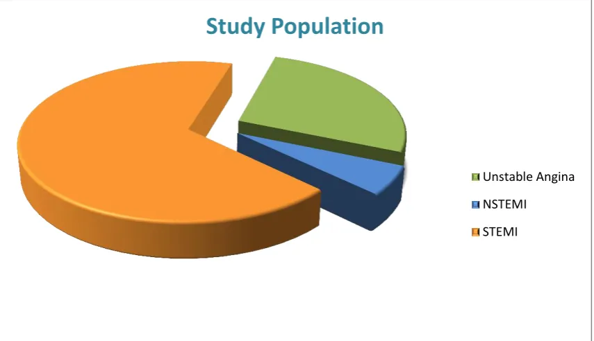
STUDY POPULATION CHARACTERISTICS
Study population included 52 patients admitted Acute Coronary
Syndromes who never had history of any cardiac problems prior. All
patients are under the age of 60. Youngest of them was 39 and the oldest
person was 59 years of age. Average Age was 50.3 and median age was
51. As the levels of NT proBNP in elderly individuals tend to be
elevated physiologically they are excluded from the study. Anemic and
renal failure patients are also excluded. Out of 52, there were 38 male
and 14 female patients. There were 14 Unstable Angina cases, 3
[image:50.595.109.537.467.713.2]NSTEMI and 35 STEMI cases.
Figure 1: Category distribution of ACS patients.
Study Population
Unstable Angina
NSTEMI
Blood was taken 2 to 24 hours after the onset of symptoms for
measurement of NT proBNP. Values were classified according to the
Elecsys rapid assay kit reference value and recorded as below 100 pg/ml
or minimal (not significant), between 100 to 500 pg/ml which was taken
as moderately elevated (significant) and values above 500 pg/ml which
was taken as markedly elevated (significant).
Levels of NT proBNP in various Acute Coronary Syndromes
NT ProBNP Levels
Unstable
Angina % NSTEMI % STEMI %
Normal
(<100pg/ml) 6 42.8 1 33.3 7 20
Significant Elevation (>100pg/ml)
8 57.2 2 66.6 28 80
Total No:
Figure 2: Showing incidence elevated NT proBNP level in various ACS
Here 80 % of patients with STEMI have elevated BNP levels and
those with NSTEMI (66.6 %) and Unstable Angina (57.2 %) had lesser
incidence of elevated NT proBNP. This was correlating with
observations in other studies like James S K, Lindahl et al. GUSTO
IV(13) and Fragmin and fast Revascularisation during Instability in
coronary artery disease (FRISC)-II)(15).
0.00% 10.00% 20.00% 30.00% 40.00% 50.00% 60.00% 70.00% 80.00% 90.00% 100.00%
Unstable Angina NSTEMI STEMI
Significant Elevation (>100pg/ml)
Gender and NT ProBNP levels.
[image:53.595.106.553.240.556.2]Gender and NT ProBNP Levels
Figure 3: Percentage of male and female patients with elevated NT proBNP
As p value is 1.000 (>0.05) there was no correlation between gender and
NT proBNP levels in the study group of patients under the age of 60
years with ACS.
0 20 40 60 80 100
Male Female
Elevated > 100 pg/ml
<100 pg /ml Gender and NT
ProBNP Levels
NT ProBNP Levels
P Value (Fisher's Exact Test) Not
elevated Elevated
Male
(Percentage)
10 28
(26.3%) (73.7%)
1.000 Female
(Percentage)
4
(28.6%)
10
Cardiac Enzymes and NT ProBNP Levels
Cardiac Enzymes and NT proBNP Levels
NT proBNP Levels Total
P Value Not elevated Elevated NT ProBNP Elevated
Count 9 26 35
% within Cardiac
Enzymes (25.7%) (74.3%)
(100.0 %) % within NT proBNP
Levels [64.3%] [68.4%] [67.3%]
1.000 Normal
Count 5 12 17
% within Cardiac
Enzymes (29.4%) (70.6%)
(100.0 %) % within NT proBNP
Levels [35.7%] [31.6%] [32.7%]
Total
Count 14 38 52
% within Cardiac
Enzymes (26.9%) (73.1%)
(100.0 %) % within NT proBNP
Levels [100.0%]
[100.0% ]
[100.0 %]
RESULT
Study showed that there is no significant correlation (as P value is
1.0) between cardiac enzymes and NT proBNP levels. This is expected
as NT-proBNP is elevated in unstable angina in addition to ST elevation
myocardial infarction and NSTEMI. So this support the fact that they
are independent variables even though NT proBNP is elevated in MI
NT proBNP as a Prognostic Biomarker
Main aim of the study was evaluating the role of NT proBNP
levels in an ACS patient for predicting the prognosis of an Acute
Coronary Event in a 6 month follow up for any symptoms of recurrent
angina, palpitations, dyspnea, rehospitalzations including unstable
angina, Non ST elevation MI, STEMI, deterioration of left ventricular
function, deterioration of renal functions and death.
This is the summary of the results in follow up period of 6 months
Recurrent Symptoms in follow up NT ProBNP Levels A n g in a P a lp ita ti o n D y spn o ea R eh o sp it a li za ti o n fo r U A o r N STEM I LV D y sfunct io n R en a l D y sf u n cti o n R ec STE MI D ea th
<100 pg/ml 3 1 0 1 2 0 1 0
100-500 pg/ml 13 5 12 11 12 3 3 2
Figure 4: Showing NT ProBNP levels and occurrence of recurrent symptoms as percentiles in the follow up
All the recurrent symptoms in the follow up like angina,
palpitation, hospital admissions for unstable angina and NSTEMI,
development of left ventricular dysfunction, deterioration of renal
functions, recurrent ST elevation myocardial infarction and death were
more common in patients with initial NT proBNP levels more than 100
pg/ml. There was no mortality in patients with NT proBNP levels below
100 pg/ml. It was noted that none of the patients with NT proBNP level
below 100 pg/ml, progressed to renal failure.
In depth analysis of each outcome is done and the result was
evaluated. 0% 10% 20% 30% 40% 50% 60% 70% 80% 90% 100% An gina Pa lp ita tio n Re h o sp ita liza tio n s U A a n d N ST EM I Re n al In su ff icie n cy Re ccure n t ST EMI 6 m o n th m o rt al ity
Nt Pro BNP >100 pg/ml
NT ProBNP Level Elevation and Recurrent Angina
NT ProBNP Levels Elevation
Angina Total P Value Yes No
< 100 pg/ml (Minimal)
Count 3 11 14
0.002 % within NT ProBNP
Levels Elevation (21.4%) (78.6%) (100.0%) % within Angina [10.0%] [50.0%] [26.9%] 100-500 pg/ml(Moderate)
Count 14 9 23 % within NT ProBNP
Levels Elevation (60.9%) (39.1%) (100.0%) % within Angina [46.7%] [40.9%] [44.2%] >500 pg/ml (Marked)
Count 13 2 15 % within NT ProBNP
Levels Elevation (86.7%) (13.3%) (100.0%) % within Angina [43.3%] [9.1%] [28.8%] Total
Count 30 22 52 % within NT ProBNP
[image:57.595.107.566.402.667.2]Levels Elevation (57.7%) (42.3%) (100.0%) % within Angina [100.0%] [100.0%] [100.0%]
Figure 5: Showing Percentage of incidence of recurrent anginas
0 10 20 30 40 50 60 70 80 90 100
< 100pg/ml 100-500 pg/ml > 500 pg/ml
Angina
NT-ProBNP Levels above 100 pg/ml and Angina
NT ProBNP Levels
Angina Total
P Value Yes No
< 100 pg/ml
3 11 14
>100 pg/ml
.001 27 11 38
60.9 % of the patients with 100-500 pg/ml of NT proBNP level
had recurrent angina while 86.7% of the patients with NT proBNP
values above 500 pg/ml developed recurrent angina. As the P value is
0.001 even a 100-500 pg/ml (Moderate) elevation of NT proBNP
above 100pg/ml itself was a good predictor for this outcome.
Result - There is a significant increase in incidence of recurrent angina
in Acute Coronary Syndrome patients with NT proBNP levels above
Elevated NT ProBNP Levels and Palpitations
NT ProBNP Levels Elevation
Palpitations Total P Value
Yes No
< 100 pg/ml (Min) Count 1 13 14
% within Nt ProBNP
Levels Elevation (7.1%) (92.9%) (100.0%)
.015 % within Palpitations [7.1%] [34.2%] [26.9%]
100-500 pg/ml
(Moderate) Count 5 18 23
% within NT ProBNP
Levels Elevation (21.7%) (78.3%) (100.0%) % within Palpitations [35.7%] [47.4%] [44.2%]
>500 pg/ml (Marked)
Count 8 7 15
% within NT ProBNP
Levels Elevation (53.3%) (46.7%) (100.0%) % within Palpitations [57.1%] [18.4%] [28.8%]
Total Count 14 38 52
% within NT ProBNP
[image:59.595.106.527.116.695.2]Levels Elevation (26.9%) (73.1%) (100.0%) % within Palpitations [100.0%] [100.0%] [100.0%]
Figure 6: Incidence of palpitations in patient with different levels of NT proBNP
Result: There is a significant positive correlation between NT proBNP
0 20 40 60 80 100 120
<100 pg/ml 100-500 pg/ml >500 pg/ml
Without Palpitation
Elevated NT ProBNP Levels and Dyspnoea
NT ProBNP Levels Elevation (pg/ml)
Dyspnoea Total P Value Yes No
< 100 pg/ml (Min)
<100
Count 0 14 14 % within NT ProBNP
Levels Elevation (.0%) (100.0%) (100.0%) % within Dyspnoea [.0%] [46.7%] [26.9%] 100-500 pg/ml(Moderate) 100-500 Pg/ml Count
12 11 23 % within NT ProBNP
Levels Elevation (52.2%) (47.8%) (100.0%)
.001 % within Dyspnoea [54.5%] [36.7%] [44.2%]
>500 pg/ml (Marked )
Count
10 5 15 >500 % within NT ProBNP
Levels Elevation (66.7%) (33.3%) (100.0%) % within Dyspnoea [45.5%] [16.7%] [28.8%] Total Count 22 30 52
% within NT ProBNP Levels
[image:60.595.118.528.120.421.2]Elevation (42.3%) (57.7%) (100.0%) % within Dyspnoea [100.0%] [100.0%] [100.0%]
Figure 7: Chart comparing NT proBNP levels and percentage of incidence of recurrent dyspnoea in the follow up.
Result: Dyspnoeic episodes were increasing with the level of NT
proBNP. In chi square test the P value is 0.001 which is very significant
at 1% level.
0 20 40 60 80 100 120
< 100 pg/ml 100- 500 pg/ml > 500 pg/ml
Dyspnoea
NT ProBNP Levels Elevation and Hospital Admission for Unstable Angina and NSTEMI
NT ProBNP Levels Elevation
Hospital Admissions Total Value P Yes No
< 100 pg/ml (Min)
Count 1 13 14 % within NT ProBNP Levels
Elevation (7.1%) (92.9%) (100.0%) 0.009 % within Hospital Admission [4.8%] [41.9%] [26.9%] 100-500 pg/ml(Moderate)
Count 11 12 23 % within NT ProBNP Levels
Elevation (47.8%) (52.2%) (100.0%) % within Hospital Admission
[52.4%] [38.7%] [44.2%]
>500 pg/ml (Marked)
Count 9 6 15 % within NT ProBNP Levels
Elevation (60.0%) (40.0%) (100.0%) % within Hospital Admission
[42.9%] [19.4%] [28.8%] Total
Count 21 31 52 % within NT ProBNP Levels
[image:61.595.105.561.124.653.2]Elevation (40.4%) (59.6%) (100.0%)
Figure 8: Percentage of Hospital Admissions including UA and NSTEMI in patients with different NT proBNP levels.
As p value is 0.009 (< 0.05) there is a significant correlation between
elevated NT proBNP levels and rehospitalization for unstable angina
0 10 20 30 40 50 60 70 80 90 100
No Hospital Admission
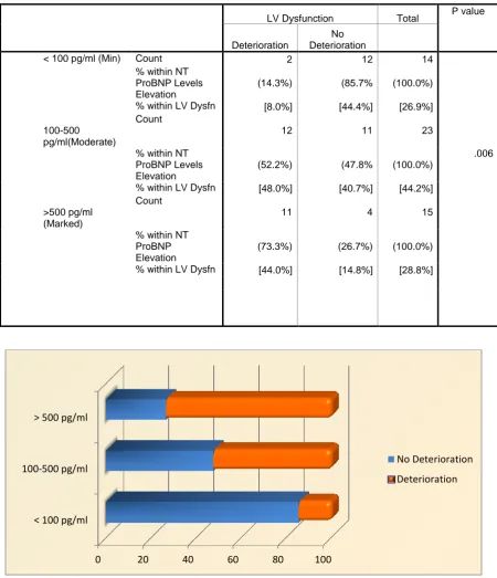
Elevated NT proBNP Levels and LV Dysfunction
LV Dysfunction Total P value Deterioration No Deterioration
< 100 pg/ml (Min) Count 2 12 14 % within NT
ProBNP Levels Elevation
(14.3%) (85.7% (100.0%) % within LV Dysfn [8.0%] [44.4%] [26.9%] 100-500
pg/ml(Moderate)
Count
12 11 23 % within NT
ProBNP Levels Elevation
(52.2%) (47.8% (100.0%)
.006 % within LV Dysfn [48.0%] [40.7%] [44.2%]
>500 pg/ml (Marked)
Count
11 4 15 % within NT
ProBNP Elevation
[image:62.595.103.554.99.623.2](73.3%) (26.7%) (100.0%) % within LV Dysfn [44.0%] [14.8%] [28.8%]
Figure 9: Percentage of development of left ventricular dysfunction in ACS patients with elevated NT proBNP levels
Result:
Elevated NT ProBNP levels correlated significantly with development
of left ventricular dysfunction in the 6 month follow up. P value was
0.006
0 20 40 60 80 100
< 100 pg/ml 100-500 pg/ml > 500 pg/ml
No Deterioration
Elevated NT-ProBNP Levels and Renal Function NT ProBNP Levels
Elevation
Renal Function Total P Value
Normal Raised
< 100 pg/ml (Min)
Count 14 0 14
% within NT ProBNP
Elevation (100.0%) (.0%) (100.0%)
100-500 pg/ml(Moderate)
% within Renal Function [30.4%] [.0%] [26.9%]
Count
20 3 23
.231
% within NT ProBNP
Elevation (87.0%) (13.0%) (100.0%)
>500 pg/ml (Marked)
% within Renal Function [43.5%] [50.0%] [44.2%]
Count 12 3 15
% within NT ProBNP
(80.0%) (20.0%) (100.0%)
[image:63.595.114.526.88.562.2]% within Renal Function [26.1%] [50.0%] [28.8%]
Figure 10: Percentage of patients developing elevated renal parameters in ACS with different NT proBNP levels
Comparing elevated NT proBNP with deterioration of renal
function, the Pearson Chi Square test gives the P value as 0.231. So
there is only a positive correlation of renal failure with elevated NT
proBNP, but the correlation was not statistically significant.
0 20 40 60 80 100 120
< 100 pg /ml 100 - 500 pg.ml > 500 pg/ml
Elevated NT ProBNP Levels and Recurrent Myocardial Infarction
NT ProBNP Elevation
Rec MI Total P value Yes No
< 100 pg/ml (Minimal)
Count 1 13 14 % within NT ProBNP
Elevation (7.1%) (92.9%) (100.0%) % within Rec MI [7.7%] [33.3%] [26.9%] 100-500 pg/ml(Moderate)
Count 3 20 23 % within NT ProBNP
Elevation (13.0%) (87.0%) (100.0%)
.001 % within Rec MI [23.1%] [51.3%] [44.2%]
>500 pg/ml (Marked)
Count 9 6 15 % within NT ProBNP
Elevation (60.0%) (40.0%) (100.0%) % within Rec MI [69.2%] [15.4%] [28.8%] Total
Count 13 39 52 % within NT ProBNP
[image:64.595.108.550.138.620.2]Elevation (25.0%) (75.0%) (100.0%) % within Rec MI [100.0%] [100.0%] [100.0%]
Figure 11: Percentage of MI during follow in various NT proBNP groups
There is a significant correlation between NT proBNP elevation
and recurrent myocardial infarction with levels > 100pg/ml. So NT
proBNP is a good predictor of recurrent myocardial infarction in an
ACS patient.
0% 20% 40% 60% 80% 100%
< 100 pg/ml 100-500 pg/ml > 500 pg/ml
No MI
Predictive Value of NT proBNP
When NT proBNP levels were compared for the correct
percentage of predictions the results were as follows.
T-Test
Group Statistics
NT ProBNP
Levels N Mean Std. Deviation
Std. Error Mean
Risk TIMI Score Not elevated 14 2.07 .917 .245
Elevated NT
ProBNP 38 2.47 .893 .145
0.159
Observed outcome after 6
months
Predicted
Outcome Percentage
Correct
Survived Death
Survived 43 2 95.6
Death 1 6 85.7
Overall Percentage 94.2
When NT proBNP level was evaluated as a risk prediction model,
as per the Hosmer–Lemeshow statistical analysis which was used as
goodness of fit for logistic regression models, the result obtained was
that elevated NT proBNP levels were able to identify the outcome as
death in 85.7 % of cases and it predicted survival in 95.6 % cases
correctly.
Elevated NT-ProBNP Level and Mortality
NT ProBNP Elevation
Outcome Total P Value Survive Death
< 100 pg/ml (Min)
Count 14 0 14 % within NT ProBNP
Elevation (100.0%) (.0%) (100.0%) % within Outcome [31.1%] [.0%] [26.9%] 100-500 pg/ml(Moderate)
Count 21 2 23 % within NT ProBNP
Elevation (91.3%) (8.7%) (100.0%)
.021 % within Outcome [46.7%] [28.6%] [44.2%]
>500 pg/ml (Marked)
Count 10 5 15 % within NT ProBNP
Elevation (66.7%) (33.3%) (100.0%) % within Outcome [22.2%] [71.4%] [28.8%] Total
Count 45 7 52 % within NT ProBNP
[image:66.595.107.544.116.659.2]Elevation (86.5%) (13.5%) (100.0%) % within Outcome [100.0%] [100.0%] [100.0%]
In this study out of 52 ACS patients 7 patients died. It is to be
noted that none of the patients in the group with low NT proBNP level
died in the 6 month follow up. Out of seven patients died, two patients
had NT proBNP level between 100 and 500 pg/ml while 5 had levels
above 500pg/ml. Patients with elevated NT proBNP above 100 pg/ml
[image:67.595.111.503.320.465.2]had significant mortality rate as the P value was 0.021.
Table comparing markedly elevated NT proBNP (above 500 pg/ml) with mortality
NT ProBNP Levels
Mortality Total
P Value Yes No
<500 pg/ml
2 35 37
>500 pg/ml
.003 5 10 15
When the NT proBNP levels were above 500pg/ml there was 33%
mortality rate and P value was even more significant at 0.003. But NT
proBNP level above 100 pg/ml itself was statistically significant. Hence
this study concluded that there is a significant correlation between the
mortality and elevated BNP and the cutoff level can be taken as above
100 pg/ml.
Result: NT proBNP is a good predictor of mortality in patients with
TIMI Scoring and NT proBNP
Although not being the primary objective of the study, it was
interesting to note that risk stratification in TIMI scoring for UA &
NSTEMI and risk stratification by NT proBNP levels were comparable.
TIMI Score and NT-ProBNP Levels
NT ProBNP Levels Total
P Value Not elevated Elevated NT ProBNP 5%
Count 5 8 13 % within TIMI
Score (38.5%) (61.5%) (100.0%) % within NT
ProBNP Levels
[35.7%] [21.1%] [25.0%] 8%
Count 3 6 9 % within TIMI
Score (33.3%) (66.7%) (100.0%) % within NT
ProBNP Levels
[21.4%] [15.8%] [17.3%] .525 13%
Count 6 22 28 % within TIMI
Score (21.4%) (78.6%) (100.0%) % within NT
ProBNP Levels
[42.9%] [57.9%] [53.8%] 20%
Count 0 2 2 % within TIMI
Score (.0%) (100.0%) (100.0%) % within NT
ProBNP Levels
[.0%] [5.3%] [3.8%] Total Count 14 38 52 % within TIMI
Score (26.9%) (73.1%) (100.0%) % within NT
ProBNP Levels
[100.0%] [100.0%] [100.0%]
In patients with ACS, NT proBNP levels did not significantly correlate
with TIMI scoring for risk stratification in UA and NSTEMI. NT
proBNP and TIMI scoring tend to be independent variables. Hence NT
proBNP tends to be an independent predictor for risk and it may be