DISSERTATION ON
“A STUDY ON UPPER AERODIGESTIVE TRACT
FOREIGN BODIES”
Submitted in partial fulfillment of the requirements for
M.S DEGREE BRANCH – IV OTORHINOLARYNGOLOGY of
THE TAMIL NADU DR.M.G.R.MEDICAL UNIVERSITY
UPGRADED INSTITUTE OF OTORHINOLARYNGOLOGY MADRAS MEDICAL COLLEGE
ACKNOWLEDGEMENT
I would like to express my sincere gratitude to Prof.J.MOHANASUNDARAM M.D.DNB.PhD, The Dean, Madras Medical College, for having permitted me to use the hospital material in this study.
I am immensely grateful to Prof.K.BALAKUMAR M.S.D.L.O., The Director and Professor, Upgraded Institute of Otorhinolaryngology, for his valuable guidance, suggestions, encouragement and help in conducting this study.
I express my sincere gratitude to Prof.JACINTH.C.CORNELIUS M.S.D.L.O., Professor, Upgraded Institute of Otorhinolaryngology, for his valuable support in conducting the study.
I am greatly indebted to Prof.A.MURALEEDHARAN M.S.D.L.O., Professor, Upgraded Institute of Otorhinolaryngology, who encouraged and helped me throughout the study.
I express my sincere gratitude to Prof.D.BALAKRISHNAN M.S.D.L.O., Chief, Department of Otorhinolaryngology, Institute of Child Health, Egmore, Chennai , who encouraged and helped me throughout the study.
I express my sincere thanks to The Secretary and Chairman, Institutional Ethical Committee, Government General Hospital, Madras Medical College, Chennai.
I express my sincere thanks to all the Assistant Professors, for their thoughtful guidance throughout the work.
I thank all the Professors, Assistant Professors and Post graduates of the department of Radiology and Anaesthesiology for their valuable support.
I thank all my colleagues and friends for their constant encouragement and valuable criticism.
Last but not the least, I express my gratitude for the generosity shown by all the patients who participated in the study.
I am extremely thankful to my family members for their continuous support. Above all I thank The God Almighty for His immense blessings.
CERTIFICATE
This is to certify that this dissertation entitled “A STUDY ON UPPER AERODIGESTIVE TRACT FOREIGN BODIES” submitted by Dr.ARCHANA BALASUBRAMANIAN, appearing for M.S.E.N.T, BRANCH IV Degree examination in March 2010, is a bonafide record of work done by her, under my direct guidance and supervision in partial fulfillment of regulations of The Tamil Nadu Dr.M.G.R Medical University Chennai. I forward this to The Tamil Nadu Dr.M.G.R. Medical University , Chennai ,Tamil Nadu , India.
THE DIRECTOR AND PROFESSOR THE DEAN
Upgraded Institute of Otorhinolaryngology, Government General Hospital, Madras Medical College, Madras Medical College, Government General Hospital, Chennai –600003. Chennai-600003.
CONTENTS
S.NO CONTENTS PAGE.NO
1. INTRODUCTION 1
2. AIMS OF THE STUDY 3
3. REVIEW OF LITERATURE 4
4. MATERIAL AND METHODS 42
5. RESULTS AND ANALYSIS 45
6. DISCUSSION 55
7. SUMMARY 60
8. CONCLUSION 62
9. BIBLIOGRAPHY 10. PROFORMA 11. MASTER CHART 12. ABBREVIATIONS
13. INSTITUTIONAL ETHICAL COMMITTEE CERTIFICATE
INTRODUCTION
A foreign body is an endogenous or exogenous substance, incongruous with the anatomy of the site where it is found. Chevalier Jackson defined a foreign body as “an object or a substance that is foreign to its location”1. Foreign body ingestion and aspiration can affect persons of any age, but the vast majority of these accidents occur in children under the age of five1. It is estimated that 150 deaths occur annually in children, due to asphyxiation3. Foreign bodies in the airway, pharynx and oesophagus continue to be a diagnostic and therapeutic challenge to practising otolaryngologists. Despite improvements in public awareness and emergency care, death due to aspiration is a leading cause of death in children. A high index of suspicion for foreign body aspiration or ingestion is needed, because a foreign body can mimic other medical conditions, particularly without a witnessed event. Hence there can be a delay in management, that may lead to complications. According to the National safety council, suffocation from foreign body ingestion and aspiration is the third leading cause of accidental death in children younger than one year and the fourth leading cause in children between 1 and 6 years8.
Accidental ingestion or aspiration tends to be twice as common in boys4. In patients with multiple oesophageal foreign body impactions, 80 % have an oesophageal anomaly on further evaluation3
When any patient gives a history of a foreign body, investigation is warranted regardless of their age or apparent absence of signs and symptoms. Rarely serious complications such as recurrent pneumonia, atelectasis, lung / retropharyngeal or mediastinal abscess, or massive hemorrhage due to a vascular fistula may occur before a thorough investigation is launched
.
4
.In patients suspected of having ingested or aspirated a foreign object, appropriate x-rays are taken. Radiographs in airway foreign bodies are frequently normal in the first 24 hours after the initial event, but may become abnormal over time1. The treatment of choice is prompt endoscopic retrieval. It is occasionally possible to retrieve a nasal, oropharyngeal or hypopharyngeal foreign body in a co-operative patient with only local anaesthesia. Rigid endoscopy has proven over time to be the safest and most efficacious therapy3.
AIMS OF THE STUDY
1.To find out the etiology and prevalence of upper aerodigestive tract foreign bodies.
2.To find out the age and gender distribution of aerodigestive tract foreign bodies and the common types and the most common sites of foreign body impaction.
3.To study the various types of presentations of foreign bodies, investigations and treatment modalities available and what were used in our institution.
4.To find out the percentage of foreign bodies causing complications.
REVIEW OF LITERATURE
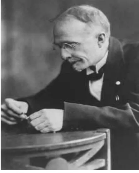
Foreign bodies in the Aero Digestive Tract are as old as mankind itself1. Among the oldest reference is the one cited by the Greek fablist Aesop in 560 BC, the episode of the gluttonous wolf with an impacted bone, which was skillfully removed by the crane, per via naturale. Hippocrates in 460 BC, conceived the intubation as ideal and Verdue in AD 1717, used bronchotomy to remove bone. Before the 20th century emetics, expectorants, purgatives, and bloodletting were practiced as methods of removal. Killian is credited with the first bronchoscopic removal of a foreign body of the airway in 1897 when he removed a bone from the trachea of a man with a 9mm rigid tube32. Chevalier Jackson (Fig 1) in the early 20th century is credited with revolutionizing the field of Broncho-oesophagology with the development of instruments and techniques for foreign body removal33. These have reduced the mortality rate associated with foreign body removal from more than 20% to 2%. Little change in technique occurred until the 1970s when Hopkin’s rod-lens telescopes became available, vastly improving illumination and visualization3
TYPES OF FOREIGN BODIES .
4
1) Organic - cotton , paper, seeds, wool / Inorganic – metallic pieces, plastic, glass, chalk, rubber.
:
2) Vegetative / Non-vegetative
3) Radio-opaque /Radio-lucent
4) Exogenous / Endogenous 5) Annimate / Inannimate
AIRWAY FOREIGN BODIES :
These remain a diagnostic challenge to health care professionals. They become life threatening emergencies that require immediate intervention. Every effort must be made to avoid delay in diagnosis as this may lead to major complications5. Airway foreign bodies still cause significant mortality and anoxic brain damage4. The majority of cases and deaths occur in toddlers younger than 3 years, upto 25% in less than one year age group3. The reasons toddlers are more susceptible are3
1) They lack molars necessary for proper grinding of food.
2) They have less controlled co-ordination for swallowing and immaturity in laryngeal elevation and glottic closure.
3) There is an age related tendency to explore objects by placing it in the mouth.
4) They are often running or playing at the time of ingestion.
Patients with altered mental status are at risk for occult aspiration, which may be difficult to diagnose. Round-shaped foods are the most frequently aspirated objects: ground nuts, grapes, raisins, peanuts, seeds, beans etc. Adults are more likely than children to have non-food items aspirated into the airway1. In adults, 75% of foreign
bodies lodge in the proximal airways (larynx, trachea, main bronchi). In children, bronchus is the most common site1,10.(FIG 3) Foreign body at laryngeal level is often caused by inappropriately executed attempts to finger sweep an oropharyngeal foreign body. Even though asymptomatic on presentation, transient coughing or gagging should raise the index of suspicion for a foreign body. Onset of wheezing in a healthy child or “recurrence” of asthma after discontinuation of therapy and persisting bronchopneumonia despite treatment, should heighten suspicion of a foreign body1,7
When the aspiration of foreign body is witnessed by the care taker, the following characteristic symptoms are described: an early choking or gagging episode followed by a cough spell
.
1,4
. As the object moves distally in the airway the symptoms become less apparent or even disappear. Vegetable matter like peanuts cause rapid, severe chemical bronchitis and granulation tissue4. Sometimes foreign bodies can change position in the airway and cause intermittent/complete airway obstruction3
Foreign body accidents usually involve three distinct stages .
4
. The first is the initial event characterized by an episode of coughing, gagging and choking. Following this, the patient typically experiences an asymptomatic interval, as the reflexes accounting for the symptoms of the initial event are fatigued.This stage leads to the misdiagnosis and frequent
delay in diagnosis. The final stage is characterized by complications due to obstruction, erosion or infection.
Foreign objects can be bilateral, with 3.6% of patients in one series3. Signs of upper airway obstruction are: dyspnoea, drooling, stridor, and cyanosis. Clinical presentation can range from chronic nonspecific respiratory complaints to acute airway obstruction likenoisy breathing, vomiting, and possibly slight hemoptysis. These symptoms, known as the penetration syndrome, occur in half of patients aspirating and include a choking sensation accompanied by respiratory distress with coughing, wheezing, and dyspnoea11. In some cases, coughing impacts it in the subglottic region. Stridor is a frequent component of an acute aspiration episode in patients of all ages. If the object is sharp and thin it may get embedded between the vocal cords or in the subglottic region. The patient may be unaware of the foreign body in cases of penetrating trauma or blast injuries, besides the intubated, tracheostomised or obtunded patients. Discrepancy in breath sounds between sides of the chest and unilateral wheezing are significant since most objects will impact in the one of the main stem bronchi. The classic diagnostic triad of unilateral wheeze, cough, and ipsilaterally diminished breath sounds is observed in less than 50% of cases3,5,39.Flexible fiberoptic laryngoscopy can add valuable information on laryngomalacia or other non traumatic etiologies. Foreign bodies, including many types of fish
FIG 3 : BRONCHIAL FOREIGN BODIES
CT SCAN : RIGHT MAIN BRONCHUS – FOREIGN BODY
FOREIGN BODY-PIN REMOVED
FOREIGN BODY – LEFT MAIN BRONCHUS
bones are radio-lucent. Therefore, the decision to pursue surgical intervention should be based on the patient’s history and a physical examination. Patients with a retained airway foreign object may present with complications such as retropharyngeal/lung abscess and atypical or recurrent pneumonia.
Physical findings depend on the degree of airway obstruction and duration of the object's presence in the respiratory system. Cyanosis is present in 10% of patients, and coughing, audible wheezing, or overt respiratory distress occurs in 25% to 37% of patients with aspirated objects3. Patients with upper airway foreign objects may have stridor and sub-sternal retractions may be noted in patients with intra-tracheal foreign bodies39. Patients with secondary infection may have fever. Clinical signs of complete obstruction include poor air exchange, ineffective cough, severe distress and cyanosis. Assessment of the neck may reveal accessory muscle use. Tracheal palpation may reveal a thud, indicating movement of a mobile foreign body against the tracheal wall9. Abnormal inspiratory sounds may be heard on tracheal auscultation. “all that wheezes is not asthma.” 1,4
OBSTRUCTIVE EMPHYSEMA
4
Occasionally a
:
foreign body acts as a one-way valve, allowing air into the lung during inspiration, but permitting none to exit during expiration. This is because air passages dilate during inspiration and
contract during expiration. Signs: Increased resonance and reduced breath sounds .X ray : emphysema on expiratory film, increased radiolucency of lung distal to the foreign body , mediastinal shift to opposite side and seperation of ribs from each other.(Fig 5)
The right main bronchus is the most common location for an airway foreign body1,4
1. It’s greater diameter and smaller angle of branching from the carina when compared to the left main bronchus.
. (Fig 4) This is due to:
2. There is greater inspiratory air flow to the right lung and 3. The carina is positioned slightly to the left of the midline .
But in young children where the difference is less pronounced, there is more equal distribution of foreign bodies4
Careful auscultation of the chest is the most critical part of the examination
.
5
Laryngeal foreign bodies: They usually cause complete or partial airway obstruction that has the potential to cause asphyxiation if not relieved promptly with the Heimlich manoeuvre
. Investigators have reported a high incidence of normal physical findings (14-45%). A negative examination should not be used to rule out the presence of a foreign body, but a positive finding is a valuable tool in establishing the need for bronchoscopy.
3,40
(Fig 6) or tracheotomy. Partial obstruction at the level of the larynx is usually caused by flat, thin
FIG 4: BRONCHOPULMONARY SEGMENTS
objects that lodge between the vocal folds in the sagittal plane. Symptoms include stridor, cough, hoarseness, dyspnoea and odynophagia.
Tracheal foreign bodies : These are rare but are slightly more common than laryngeal foreign bodies. Three features described by Jackson and Jackson 1,3
Types of bronchial obstruction
which can be noticed on examination are the
audible slap which is best heard at the open mouth during a cough, the
palpatory thud, and the asthmatoid wheeze, heard with the ear kept at the patient's open mouth.
7
1.Bypass valve
:
2.Expiratory check / one way valve (most common ) 3.Inspiratory check / stop valve
Signs of stop valve obstruction9
1.Atelectasis of lung distal to foreign body.2.Shift of mediastinum to the same side.3.Compensatory emphysema of the opposite lung for adequate ventilation 4.Respiratory distress, cyanosis, cardio respiratory failure.5.Absent breath sounds on the affected side. Stop valve type of objects completely obstructs the airway.(Fig 5)
:
Radiology:
In the stable patient, plain radiography of the neck and chest remains the mainstay of airway foreign body imaging. Most foreign bodies are radio-lucent (80%)1. Films taken during expiration can reveal
hyperinflation at the ipsilateral lung. Atelectasis can be seen when the aspirated object completely obstructs the airway. Atelectasis and pneumonia are commonly seen in delayed diagnosis (after 24 hrs)4. Air trapping can be seen when inspiratory and expiratory films are compared, which may show a flat, fixed diaphragm on the involved side and the heart and mediastinum shift to the uninvolved side during expiration10. Bronchiectasis and bronchial stenosis develop later.Radiographs should not be used to rule out the presence of foreign bodies, but to aid in diagnosis5. Over one half of tracheal and 25% of bronchial foreign bodies have normal chest x rays4,5. Compared with history and physical examination, radiography appears to be the least sensitive in predicting the bronchoscopic findings5. Other imaging techniques of potential utility are fluoroscopy, but it may have a lower sensitivity and specificity than chest radiographs15. Computerized tomography, (FIG 3) is useful in evaluating patients with suspected airway foreign bodies when plain films are negative.
Acute airway obstruction (“CAFE CORONARY”)34,40:
Acute airway obstruction is said to cause around 3000 deaths per year. The object, usually a bolus of food, bolted in a restaurant, lodges in the larynx or pharynx, causes acute respiratory embarrassment. If the airway is not restored, irreversible cerebral ischemia occurs within 6 minutes. Survival is based on the actions of passers-by, rather than
trained medical staff. Attempts to revive the foreign body by fingers, are to be avoided, because it only causes further impaction. The Heimlich manoeuvre may be life saving. The rescuer stands behind the subject and places a clenched fist below the xiphisternum(Fig 6). This is followed by rapid subdiaphragmatic upward thrusts, producing artificial cough of some sort. If this step fails, a cricothyrodotomy may have to be done.
Management of an airway tract Management :
foreign body is removal, which generally leads to rapid recovery of the patient. Basic life support manoeuvres to remove a foreign body in children include back blows and chest thrusts in infants and abdominal thrusts in children and adolescents3. Blind finger sweeping has resulted in conversion of partial to complete airway obstruction when objects are displaced into the subglottic space. Coughing and gagging indicate partial obstruction. In children older than one year, the Heimlich manoeuvre or sub diaphragmatic thrusts are used. Emergency needle cricothyrodotomy is a procedure of last resort to access the airway in an obstructed patient who cannot be intubated or ventilated11. A large intravenous catheter (14 to 18 Gauges) is passed through the midline of the inferior edge of the cricothyroid membrane. Laryngeal foreign bodies can be removed by direct laryngoscopy. Tracheal and bronchial foreign bodies are best
removed using rigid bronchoscopes5
Indications for Endoscopy :
. In the rare event of not being able to remove it endoscopically , thoracotomy and bronchotomy may be needed
To prevent a diagnostic delay, a witnessed choking event followed by a period of coughing, should be considered an acceptable indication for bronchoscopy5. A good rule of the thumb is that diagnostic bronchoscopy should be performed, if any one of the three diagnostic tools (history, examination or radiography) is positive. Early bronchoscopy in any patient with a suspected foreign body
It is important that foreign bodies are removed with the least endolaryngeal and endotracheal trauma. It is ideal to use the rigid endoscopes. Telescopes attached to foreign body forceps, make the removal easier. Tomaske and colleagues found in a study that children who underwent bronchoscopy <2hrs fasting, did not have any pulmonary aspirations of gastric contents. But when the child is stable, fasting guidelines should be followed
is the key to reduce morbidity and mortality.
6
Flexible bronchoscopes lack the ability to ventilate, which is afforded by their rigid counterparts. In patients with lesser overall clinical suspicion, fiber optic bronchoscopy may be indicated, but Rigid bronchoscopy(Fig 8) is the optimal first step when clinical suspicion is high.
.
Bronchodilators and postural therapy for dislodgment of airway
foreign bodies is to be condemned, due to the risk of mobilizing the object from its distal position, only to cause its impaction in the narrow subglottis or glottis1
Anaesthesia
. Several situations can be regarded as urgent or an emergency, with endoscopy performed as soon as possible:(1) Actual or potential airway obstruction (2) aspiration of dried beans or peas. With prolonged periods in the airway, the bean or pea absorbs moisture, thus causing swelling and airway obstruction or the obliteration of forceps spaces, making removal more complicated.
57
An important anaesthetic consideration is that the bronchoscope competes for space with the anaesthetic device, in the trachea. Pulmonary ventilation needs to be continued during the procedure and this can be achieved in the following ways:
:
1.Using a small tracheal tube, though the risk is that the bronchoscope may dislodge it.
2.Using the technique of apnoeic ventilation. The patients lungs are ventilated with 100% oxygen until the lung volume is depleted of nitrogen and effectively is full of oxygen and no other gas.
3.Jet ventilation using Sanders injector. This is essentially a pressure relief valve and tubing - one end attached to a high pressure oxygen supply on the anaesthetic machine and the other end to the bronchoscope.(Fig 7)
FIG 7: JET VENTILATION
4.Some bronchoscopes have a side port (Racine adaptor), to which the standard anaesthetic /oxygen tubing can be attached
Complications of jet ventilation are36
Rupture of bulla, pneumothorax, pneumomediastinum/pneumo- pericardium and mediastinal emphysema.
:
An experienced anesthesiologist is usually required. The plan for the procedure should be discussed and the possible complications reviewed with the entire team, before taking the patient to the operating suite. The endoscopist should then spend adequate time selecting the appropriate instrument with which to grasp the object.
The proper size bronchoscope should be prepared with alternate sizes available when planning the retrieval of an airway foreign body. This is most important in children, where laryngeal and tracheal sizes are highly variable and the effects of swelling from use of too large an endoscope and excessive airway trauma are poorly tolerated1
It is preferable to keep the patient in spontaneous breathing, to prevent positive pressure ventilation, which may induce distal migration of the foreign body
. The use of too small a scope can compromise removal and cause excessive leak with ventilation.
4
15 . Further more, during natural inspiration the airway
Laryngoscopy is initially performed to evaluate the larynx and hypopharynx, to expose the larynx for atraumatic bronchoscope insertion, and application of topical anaesthetic3
The bronchoscope is introduced into the right side of the mouth with thumb and index finger held like a pen
. It is also imperative that 4% lignocaine is sprayed to anaesthatise the glottis, trachea and carina before instrumentation. Age appropriate instrumentation is essential to prevent trauma. The patient is placed supine, with head initially placed on the head ring, then removed when the scope has passed through the cords. Draping of the body is avoided to aid in visualizing the respiratory movements. Classically the boyce position is used, with flexion of all cervical joints except the atlanto-occipital joint, that is extended. Injury to the teeth and tongue is to be avoided while introducing and manipulating the scope. The preoperative state of the teeth is to be noted and patient warned of the possible damage.
4
.The beak of the scope lifts the epiglottis, and is then advanced through the cords, where it is rotated 90 degrees, so that the beak passes sideways. At this stage the pillow or head ring is removed. The scope is rotated back into position. Examination of the bronchial tree is done in a systematic manner. Rotating the head to left, right main and lower lobe, right upper and right middle lobe bronchi are visualised. Then rotating head to right, left main bronchus is seen. The entire tracheobronchial tree should be inspected,
beginning with the non-affected segments to assure adequate respiratory function, while attempts at removal are made. Occasionally, there is the unexpected discovery of an additional foreign body. Once the foreign body is located, all secretions and debris should be cleared from around the object using suction.
The object is then addressed with the previously chosen forceps. The blades of the forceps should be placed around the object with care to avoid driving the object further to the periphery. There should be adequate space around the foreign body needed for application of the forceps (forceps space)1
Advantages of Rigid bronchoscopy (Fig 8)
. Foreign bodies which are prone to fragmentation should be grasped, only firmly enough to assure adequate grip. Once the forceps are secure on the object, the scope, forceps and foreign body are removed as a single unit.
1.Removal of foreign body is easier
2.Anaesthesia is easier and visualisation is better. Oxygenation can be maintained, reliably.
Advantages of flexible bronchoscopy: 1.Done under local anaesthesia
2.Video connection and viewing possible
Many endoscopists have been troubled by the slipping of the object from the grasp of the forceps, most commonly at the narrow glottis with
the possibility of complete airway obstruction. If this situation occurs, it is imperative that the obstruction be relieved immediately. This may be accomplished by completing the removal of the object. When this is not feasible, the object should be pushed distally in order to relieve the obstruction, or occasionally, it is necessary to fragment the object. Multiple foreign bodies are said to occur in 5-19% of cases3
Airway foreign bodies must be grasped in a secure manner and controlled during their removal. Slipping of a foreign body during its passage through the trachea or in the larynx may convert partial airway obstruction to total obstruction with an inability to ventilate the patient. In children, this risk is increased in the subglottis because of its intrinsic relative narrowness. Attempts to remove a foreign body with a sharp end may cause additional trauma: “Advancing points perforate, trailing points do not”
.
1
The obstructed bronchus is suctioned to remove secretions, which may aid in more rapid re-inflation of the lung
.In unusual circumstances, selected foreign bodies may be removed through a tracheostomy incision or a thoracotomy.
12
. Granulation tissue can be removed and bleeding controlled with topical vasoactive agents on cotton pledgets1,8. In some institutions, Fogarty catheters/Dormia baskets are used to remove the object. In cases of sharp objects and open safety pins, special forceps may be needed, and practice on a dummy may help. Thoracotomy may be required in failed cases.
Principles of removal33
1.Selection of adequate size foreign body forceps :
2.Achieving best exposure of foreign body
3.Bronchoscope positioned close to foreign body without touching it and keeping adequate forceps space.
4.The distal end of the forceps used, should pass beyond the midpoint of the foreign body.
5.Small objects are removed through the scope, while larger ones are removed by trailing mechanism.
Smooth foreign bodies in the peripheral bronchus can be removed by passing a Fogarty balloon catheter distal to the object, gently inflating it and withdrawing it33
Telescopes attached to the endoscopes, aid in better visualization. Pointed foreign bodies such as nails, hooks and pins are almost always situated with the point directed superiorly. The point must be enclosed in the blades of the forceps to prevent perforation of the bronchus. Clerf Arrowsmith safety pin closing forceps is used to close open safety pins and to remove them. Disengagement from the mucous membrane and closure of the pin with closing forceps and then removal through the scope is undertaken. The tip faces downwards and then removed through the scope (retroversion)
.Hollow foreign bodies can be removed by placing one blade of the alligator forceps inside and one outside
33
.
Following removal : a second look is needed to ensure that another foreign body has not been overlooked and to remove any fragments, secretions and mucus to speed up resolution of atelectasis or pneumonia4
Complications :
. If the procedure was prolonged or a tight fit is noted in the subglottis, steroids are indicated to reduce postoperative laryngeal oedema. Chest physiotherapy may help to mobilise secretions and to prevent infection.
Delay in diagnosis increases the perioperative morbidity8. Total or near total main stem bronchial obstruction, leads to poor alveolar aeration and shunting of pulmonary perfusion away from the affected lung. When the foreign body migrates to the other lung, abrupt respiratory decompensation occurs32. Delayed diagnosis also causes pneumonia, atelectasis and granulation tissue formation, which can lead to significant bleeding on removal4,8
Complications of bronchoscopy are : .
Haemorrhage
Post operative stridor, laryngospasm , bronchospasm
Hypoxia, laryngeal oedema, subglottic oedema ( over sized bronchoscope , prolonged endoscopy ,extensive manipulation, trauma during extraction)
Arytenoid dislocation
Transient arrhythmias and bradycardia, aspiration
NASAL FOREIGN BODIES :
The nose is perhaps the most common site for the insertion of foreign bodies by children. Children put foreign objects into their nostrils or into their siblings nasal orifices. Usually the child or its friend will tell the parent or the caretaker that they have put something into the nose46. The foreign bodies most often found include beans, sponge pieces, pebbles(FIG 9), rubber(Fig 10) plastic toy fragments, and other small round objects like cell batteries. Perhaps because most people are right-handed, more than two thirds of nasal foreign bodies were right-sided in one series. Children with nasal foreign bodies tend to be younger, most commonly under 5 years of age4,46. Intranasal alkaline button battery may cause electrical or chemical burns with liquefaction necrosis46
Most patients seek medical attention within 24 hour. Nasal
. (Fig 11)
foreign bodies may be asymptomatic, sometimes identified as incidental findings on radiographs. In most of the cases, the patient admits or is seen to be placing an intranasal object. Other signs and symptoms are Unilateral, Purulent, malodorous nasal discharge or features of nasal obstruction or even persistent epistaxis48. These patients often are misdiagnosed and treated with antibiotics for supposed sinusitis. When the history suggests a foreign body, but none is identified on examination, imaging is required55. Because of risks of iatrogenic movement of the foreign body
FIG 9: CHOANAL FOREIGN BODY-STONE
[image:33.595.201.448.616.806.2]CHOANAL FOREIGN BODY REMOVED
FIG 11 :BUTTON BATTERY
further posteriorly (Fig 9)or into the airway, children may need to be restrained to permit the examination. Necrosis of the nasal mucosa and septum may accompany button battery impaction.
A short burst of air blown into the mouth of a child, with finger occlusion of the non-obstructed nasal cavity, may force the foreign object out of the nose. The insufflation is preferably applied as a “kiss” from a parent, but also can be provided by a manual ventilation bag57. Rarely, computerised tomography or magnetic resonance imaging may be indicated to visualize suspected foreign bodies or their complications. Adequate illumination is essential. Necessary instruments include a blunt-tipped right-angle probe, suction catheter and alligator forceps. The forceps are used when the foreign body is to be directly grasped, and the right-angle probe is used in an attempt to reach proximal to the foreign object and displace it. Suction is primarily necessary for removing purulent secretions and blood that may obscure the field. Occasionally removal may require general anaesthesia, Endoscopy, Caldwel-Luc or lateral rhinotomy46,55. A Rhinolith - can form around an endogenous or exogenous foreign body, which is a partially or totally calcified mass of tissue in the nasal cavity, where layers of mucin aggregate around the object.57
FOREIGN BODY INGESTION:
Before the mid 1850s, the most common management for suspected oesophageal foreign body impaction was to attempt to push the object into the stomach. The first oesophagoscope used in 1890 by Mackenzie (Fig 2)was later improved by Jackson, Ingals and Mosher3 Oesophageal foreign bodies are considered less precarious than airway foreign bodies. Even so they occur more frequently and are responsible for over 1500 deaths per year. The normal oesophagus has four anatomical sites of narrowing – the cricopharynx, aortic arch, left main bronchus and lower esophageal sphincter. Anatomically these are commonly found at cricopharynx, at the cross over of aortic arch, at mid oesophagus and at lower oesophageal sphincter
.
3,14
Most cases of foreign-body ingestions occur in the paediatric population, with a peak incidence at the ages between 6 months and 6 years
.(FIG 12)
5
. Young children explore their environments with their mouths and are thus at risk for the ingestion and aspiration of non-food items. In this age group the second molars are not well developed and the grinding and swallowing mechanisms are poor and glottic closure is immature57. In adults some people are at higher risk to have a foreign body, such as neurologically impaired patients, edentulous individuals, patients with certain psychiatric illness, mental retardation, impairment caused by alcohol, pica, those seeking some secondary gain with access to a medical
FIG 12 : LEVELS OF NORMAL ANATOMICAL OESOPHAGEAL CONSTRICTIONS
FIG 13 : METAL OESOPHAGEAL FOREIGN BODY
[image:37.595.54.299.493.792.2]FIG 14 : CHICKEN BONE
FIG 16 : BUTTON BATTERY - CRICOPHARYNX
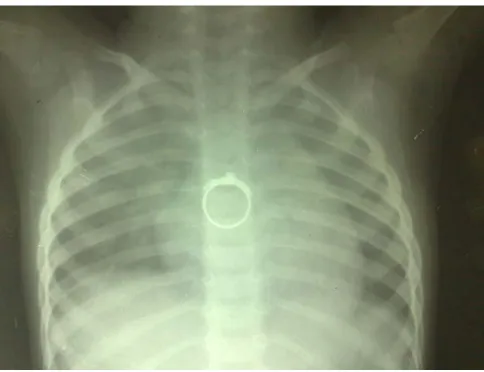
facility and individuals at the extremes of age3. Foreign bodies are grouped according to their size and shape. They can be classified as blunt and sharp objects3. 80 % of foreign bodies occur in children and cervical oesophagus is the commonest site. The object most often encountered in children is a coin1,5.(Fig 17),in adults – chicken bones(FIG 14) and fish bones.Other common objects are foods, toys, bones, batteries, wood, and glass3,5
Clinical features :
. In children they may get lodged in the tonsils, as these (the tonsil) tend to be bigger at this age. Most obstructions in adults were food bolus impaction and generally occurs in older patients.
The signs and symptoms of foreign body ingestion are quite diverse and non-specific5
The usual symptoms in children are – irritability, poor feeding, drooling, increased work of breathing, vomiting, pain and cough. Many of these can be misdiagnosed as gastrointestinal disorder or viral illness. Respiratory symptoms are more common in children as their tracheal lumen is narrow and easily compressible and hence include stridor and choking
. Adults tend to intensely describe the event and acknowledge the potential for a foreign body. Children can be much more vague, and in 7 – 35 % they present with no symptoms.
16
. In general, symptoms are more common if foreign body is at cricopharynx, than if it is lower down in the oesophagus. In adults – dysphagia, pain, cough, vomiting, increased salivation and persistent
foreign body sensation are the symptoms. Spicules of bone are most commonly lodged in the tonsil, tongue base or vallecula5,21
Physical examination:
. History of any known oesophageal anatomic abnormality or prior instrumentations should be asked.
Symptoms referable to the oesophagus whether the complaint is dysphagia or odynophagia must be taken seriously5
Oropharyngeal examination also may provide indirect clues; for example, a missing dental plate on examination should lead to suspicion. Base of tongue, vallecula, supraglottic area, and pyriform sinus should be examined. The presence of pooling of saliva may indicate a foreign body obstruction lower down
. Hypersalivation or drooling is a concerning symptom and can be a sign of complete obstruction. In a child respiratory distress, pulmonary infection, wheezing, or stridor should prompt to think of an oesophageal foreign body as a possibility.
4
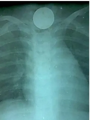
The button battery (Fig 16) commonly used in hearing aids, watches, calculators and other portable electronic devices
. This is called Jackson’s sign. Subcutaneous emphysema found by neck palpation, indicates probable oesophageal perforation.
4
, can cause oesophageal rupture. The peak incidence of ingestion occurs at 1-2 yrs. In one hour they cause mucosal damage. In 4 hours, leakage of caustic
contents cause erosion through the muscular wall and within 6 hours, an oesophageal perforation with mediastinitis, tracheo-oesophageal fistula or death may occur. Most batteries are of smaller diameter (< 15mm )and usually traverse the gastrointestinal tract with minimal injury20. These batteries cause pathologic changes through direct pressure, electrical current, corrosives leakage, heavy metal poisoning or liquefaction necrosis due to leakage of caustic alkaline3,4
Sharp objects may perforate the oesophagus (15-35%)(FIG 20 ) Even coins can cause stridor, oesophageal erosion, aortoesophageal or tracheoesophageal fistula, mediastinitis, or paraoesophageal abscess. Other complications of oesophageal foreign bodies include esophageal oedema, laceration or erosion, hematoma, granulation tissue, retropharyngeal abscess(FIG 27,28), migration of the foreign body into the fascial spaces of the neck, strictures, and proximal oesophageal dilation. Signs of mediastinitis indicate oesophageal perforation
.
4,16,20
Unlike airway, the diagnostic tool in oesophageal foreign bodies is radiography
. Perforation of the oesophagus with erosion into the vasculature or pulmonary tree can result in presentations ranging from hemoptysis to pulmonary abscess to life-threatening haemorrhage.
5
. Foreign bodies in the oesophagus are much more likely to be radio-opaque. The initial step is generally a chest radiograph and lateral cervical spine x-ray study using soft tissue. The primary utility of
plain radiography lies in detection of radio-opaque objects. Plain radiography has identified metal foreign objects(FIG 13) missed on direct (including endoscopic) examination. Anteroposterior and lateral films are required to localise the foreign body. Lateral films are more superior in identifying radiolucent objects by identifying more subtle findings such as tracheal compression, tracheal deviation and air trapped within the oesophagus(FIG 18). In children, a “mouth-to-anus” film is usually obtained, to see the entire oesophagus as well as the abdomen, in case the foreign body has passed into the stomach or beyond4. In adults, if neck or chest films are negative, abdominal films are sometimes obtained for reassurance of the presence of the foreign body
Calcified airway cartilages are misleading and contribute to false-positive rates as high as 25%. (cricoid, thyroid and stylohyoid calcifications and osteophytes)
in the stomach.(Fig 22)
52,5
. Normal ossification of airway cartilages begins in the third decade and progresses with age. The typical curvilinear contour and well-defined margins of bony fragments may help distinguish them from normal laryngeal calcifications. Oesophageal foreign objects usually align themselves in the coronal plane and are posterior to the tracheal air column on lateral view. Oesophageal foreign bodies align parallel to the spine and laryngeal ones align perpendicular to it. Coins in the oesophagus lie in the coronal position in
FIG 17 : OESOPHAGEAL FOREIGN BODY - COIN
virtually all cases, because the opening into the oesophagus is much wider in this orientation(Fig 14).
Plain films of the neck and chest have sensitivity of 25% for impacted fish bones.When plain films fail to visualize foreign bodies and suspicion remains high, one option is contrast oesophagography, which can be useful with radiopaque and sometimes with radiolucent foreign bodies19,21. If perforation is not a concern, barium may be used as the contrast medium because it provides higher quality images. It yields better results, but risks aspiration and coats the object and oesophagus, reducing effectiveness of subsequent endoscopy. Computerised tomography with coronal and sagittal reconstructions are useful in identifying foreign bodies or characterising further objects seen on plain films, as it can give information about foreign body size, type, location, complications and orientation with respect to other anatomic structures5,26.(FIG 24) A relatively inexpensive and non invasive modality reported to be useful in detection and characterization of metal foreign bodies is the hand-held metal detector33
Management :
.
A specialist in the examination through the orifices above the clavicles, should become a master of both rigid and flexible techniques. Early endoscopy should be considered in cases of potential toxicity (e.g., button battery ingestion), altered anatomy (e.g., prior abdominal surgery),
or sharp foreign bodies3,4,5. Cocaine can kill a body packer and an impacted button battery can cause fatal electrochemical tissue damage. In luminal obstruction, the foreign body may become lodged and may exert pressure on the adjacent tissue, causing necrosis and perforation. Pharyngeal foreign bodies
Since the first report in 1972, on the removal of a
visualized by direct or indirect laryngoscopy usually can be removed with a forceps or clamp with caution. For sharp objects, displaced oesophageal stents, or impacted button batteries, efficient and urgent management is required.
foreign body with a flexible endoscope by Mackenzie et al, there has been an increasing application of this method, because of its advantages, such as avoidance of operations or surgeries for most patients, reduced cost, accessible technical facility, excellent visualization, simultaneous diagnosis of other diseases, and a low rate of morbidity3. In cases of impacted food bolus, pharmacological manoeuvres may be tried to move the bolus into the stomach. Glucagon (0.5 to 2 mg) given intravenously has been used to relieve distal food obstructions. It lowers the smooth muscle tone at the lower oesophageal sphincter without inhibiting normal oesophageal peristalsis. Glucagon, if given too rapidly, may cause vomiting and risk rupture of an obstructed oesophagus and should not be used in patients with sharp-edged foreign bodies1.
Gas forming agents have been rarely used. Two other agents used for distal food bolus impaction are nitroglycerine and nifedipine.Both of these agents have a relaxing action on the lower oesophageal sphincter, and are safe (if only marginally effective) manoeuvres for therapy of impacted food bolus. Effervescent agents are sometimes effective in accelerating the passage of an obstructing food bolus3,4. The administration of carbonated beverages (including soft drinks) results in the passage of the obstructing food bolus in 60% to 80% of patients treated. The use of enzymatic meat tenderizer (papain) to soften a food bolus, a traditional method, is not recommended. Some authorities advocate a period of observation in stable patients. The goal of observation is spontaneous passage into the stomach. This is not indicated in patients who present more than 24 hours after ingestion, or who have pooling and intolerance to oral secretions. The period of observation should not be more than 24 hours, a period of 8 to 16 hours is generally acceptable in asymptomatic children who have oesophageal foreign bodies53. Patients with sharp-edged, distal foreign bodies
Endoscopy should be performed immediately for patients experiencing significant distress and for children with impaction of an alkaline button battery. Batteries that pass into the stomach should be , those who have contraindications to use of the aforementioned agents, and those who do not respond to treatment should be evaluated with endoscopy.
followed radiographically and clinically to ensure passage50.(FIG 22) Urgent intervention is also indicated for sharp objects, button batteries, coins in the proximal oesophagus, and impactions that impair the handling of secretions. Although balloon and magnet techniques have been used by radiologists to extract foreign bodies
Oesophagoscopy and foreign body removal :
, endoscopy has become the treatment of choice at most institutions. Uncontrolled coagulopathy, cervical spine instability or rigidity, trismus or hypertrophic changes in the cervical spine may exclude the use of Rigid oesophagoscopy, although flexible oesophagoscopy may still be applicable.
Oesophagoscopy is considered a safe procedure with excellent retrieval rates. The choice of either flexible or rigid endoscopy, depends on the experience of the endoscopist and the equipments available. An essential benefit of both are the ability to examine the oesophageal wall after removal of the foreign body.
Flexible endoscopes 33
These are available in a wide range of diameters, beginning at 5.0 mm. In general, these have two or three channels in addition to an optical channel - one for suctioning secretions or insufflation of the oesophageal lumen and the others for introducing instruments. The main advantage is that it can be done under local anaesthesia, under conscious sedation, in
:
patients who are a significant risk for general anaesthesia, and also used in patients who have cervical spine diseases. Disadvantages: the diameter of the instrument port is 2.0 to 4.0 mm limiting the size of instruments that can be introduced through it. Hence the size and nature of the foreign bodies that can be removed is limited by the size of the foreign body graspers and suction catheters that can be introduced through it. Furthermore, since the foreign body cannot be retracted into the scope during removal, injury to the mucosa is more. Finally, the post cricoid, pyriform areas and cricopharynx are not well visualized using flexible scopes.
Rigid Oesophagoscopes 1,3,45
Rigid scopes are available in various sizes, lengths and shapes, making this technique amenable to various situations. The oval open rigid scopes (Robert Jasberg ) are suited for foreign body removal. The round open ones (Jackson style ) are more suitable for negotiating obstructions and strictures. It has one central channel and one or two smaller channels. The large central channel accommodates a variety of instruments. The distal tip is thick and smooth increasing the ease of introduction of the instrument and decreasing the likelihood of mucosal trauma. Telescopes attached to the oesophagoscope aid in better visualization during manipulation and removal of the foreign body.
:
In the case of any foreign body, preliminary rehearsal of the intended procedure using a duplicate coin or button, as proposed by Chevalier Jackson, facilitates a subsequent manoeuvre and is more likely to produce successful results1,33
The major disadvantage of rigid oesophagoscopy is that it requires general anaesthesia, increasing the cost and morbidity of the procedure and it is also associated with a higher incidence of complications, such as dental trauma and oesophageal perforation. It is also not amenable to patients who have trismus and cervical spine problems
.
If the foreign body is lodged high in the oesophagus, shorter cervical oesophagoscope may be used. Especially in children, to prevent the slippage of foreign body into the oesophagus from the cricopharynx when the muscle relaxant is given during anaesthesia, the patient is put in reverse Trendelenberg position. Longer oesophagoscopes are needed for objects that are present distally. The largest instrument that will pass easily is chosen for maximum visualization and ease of instrumentation. The patient should be positioned in a neutral sniffing position, with the cervical spine straight to allow easy passage over the cervical kyphosis. The scope is passed through the right side of the mouth and directed towards the pyriform fossa and angled towards the sternal notch. If the foreign body is too large to be withdrawn through the lumen
FIG 19 : FOREIGN BODY THROAT - BLADE
FIG 20 : OPEN SAFETY PIN
[image:50.595.45.287.510.696.2]the oesophagoscope is advanced to shield the foreign body4
During manipulation of the scope, the left hand is used to advance the scope, and the right hand stabilizes the instrument. Injury to the posterior wall is common, if too much pressure is applied by the advancing tip of the scope. Lifting the distal tip with the right thumb as the fulcrum, will avoid injury
. Sharp objects should be sheathed or rotated, so that the point trails.
4
Sharp objects
.
1,33
: extraction of such objects is extremely challenging. Locating the point is crucial. The endoscope is aligned parallel to the long axis of the airway or oesophagus to minimize the likelyhood of mucosal injury. The open safety pin (FIG 20) or other sharp or pointed objects should be removed with the dangerous edge ensheathed in the endoscope or with the points trailing. This often requires such techniques as endogastric version or inward rotation. A number of specialized instruments have been designed for such occasions, including pin bending forceps, broad staple forceps, rotation forceps and safety pin closing forceps (Clerf Arrowsmith forceps)1,4
Fish bone : Fish bones are a common Upper Aerodigestive and oesophageal foreign body found in adults
. If the sharp object is deeply impacted, then open surgical procedure may be the safest approach.
47
. Fish bones are sharp objects and they can get lodged in the aerodigestive tract(FIG 23) and cause complications, although this is rare - about 1% to 3%. But the associated
FIG 23 : FISH BONE
complications are potentially catastrophic, including cervical abscess, mediastinitis, esophageal aortic fistula, Oesophago-carotid fistula and lung abscess1,51. Fish bones are translucent on physical examination and often radiolucent. They usually lodge in the tonsils, due to the presence of many crypts in which it gets caught1,4. It may be present in the posterior one third of tongue or the vallecula. It very rarely crosses these sites. Hence examination of the oral cavity and using an indirect laryngoscopic mirror after spraying an anaesthetic agent will identify the fish bone in majority of instances. The patient most often points to a site of irritation in the throat. If not found, then an endoscopic examination can be carried out and radiological investigation resorted to. Patients complain mostly of a foreign body sensation51. A sharp pricking sensation is highly predictive. A complete oral examination is mandatory51. Plain films may exhibit poor sensitivity when the bone is lodged in the area of maximum soft tissue overlap. There is also poor specificity because of thyroid, cricoid and hyoid calcifications, which can be misleading. One should not rely on a negative radiograph to rule out a retained bone . All patients who complain of a foreign body in the throat should be taken seriously. The current thinking is that in the absence of a proven retained foreign body, the sensations described are due to minor trauma of the digestive tract that are produced when the bone is swallowed1,51. Most of the time, these are removed by forceps under
direct vision, some times requiring endoscopic removal and rarely Rigid endoscopy 56
Disc battery ingestion .
4,49
The peak incidence of ingestion occurs between 1 – 2 years of age. This requires immediate action. Radiography will locate the object. An immediate oesophagoscopy is performed to remove it and assess the state of the oesophageal mucosa. Mercuric oxide containing batteries can cause systemic mercury poisoning, if they open in the stomach. Prompt radiographic confirmation may show a double density shadow produced by a bilaminar disc battery, and the child should be prepared for endoscopic removal. The removal is difficult because of associated inflammation and the fact that it slips. Following removal, follow up radiography should be performed at regular intervals to exclude late development of stricture.
:
Pill ingestion1,3
Inadvertent swallowing of Dental prosthesis
: medications in pill form may lodge within the oesophagus because of increased transit time, dry swallow, adherent swallow or supine swallow. They may cause caustic injury due to prolonged contact time with the mucosa.
1
can occur in a variety of circumstances to any edentulous person, but the stroke patient is at particular risk. Swallowing dysfunction and impaired oral sensation
X-RAY SHOWING DENTURE
ENDOSCOPIC VIEW OF DENTURE FIG 24 : DENTURES IN THE OESOPHAGUS
DENTURES REMOVED
are the most common cause. A small prosthesis can be managed as a sharp foreign body(FIG 24), but an impacted dental plate may require an open approach.
In the illicit practice of body packing 3,4
Complications encountered include perforation of the oesophagus with resultant mediastinitis and erosion into vascular structures. Negative endoscopy may also represent the migration of the object from the aero digestive tract necessitating further radiographic studies such as computerized tomography or magnetic resonance imaging in order to define its position better. In such situations, removal of the object may require thoracotomy.
(to smuggle heroin or cocaine hidden in swallowed latex bags) oesophageal impaction may occur. Any endoscopic manipulation can cause release of the contents into the gastrointestinal tract resulting in grave morbidity and death.
Post operative care is usually straightforward and antibiotics or corticosteroids are necessary only for the treatment of complications.
Another removal strategy, best suited for smooth, non-impacted and blunt objects, employs a contrast-filled balloon catheter and fluoroscopy. It was first described in 1966. Contraindications to foley’s catheter include total oesophageal obstruction, which prevents passage of the catheter tip distal to the foreign body, oesophageal perforation, sharps and multiple oesophageal foreign bodies. A Foley’s catheter is introduced
FIG 27: RETROPHARYNGEAL
ABSCESS FIG 28 : FOREIGN BODY WITH
RETROPHARYNGEAL ABSCESS FIG 25 : X-RAY NECK LATERAL
VIEW-COIN - CRICOPHARYNX
FIG 26 : X-RAY CHEST LATERAL VIEW SHOWING OPEN SAFETY PIN IN
[image:57.595.47.257.482.809.2] [image:57.595.287.559.537.777.2]into the oesophagus, and the balloon is passed beyond the foreign body, inflated with radiographic contrast material, and withdrawn under fluoroscopic monitoring. Another strategy for active foreign body removal is Bougienage. Bougienage has been found in one study to be equally safe, more efficient, and much less expensive than endoscopy33. After the removal of an oesophageal foreign body
Oesophageal perforation
, regardless of the method used, a search is made for a second foreign body as multiple foreign bodies are present in 5 % cases. A follow-up oesophagogram is frequently necessary to evaluate oesophageal anatomy and patency.
3,4
The commonest complication of oesophagoscopy is perforation due to : 1.absence of serosal layer .2.negative intrathoracic pressure.
:
Oesophageal perforation is a potentially life-threatening condition. It may be caused by the foreign body itself, the length of time it is present,or during attempts to retrieve it .Most iatrogenic injuries occur at the pharyngoesophageal junction because the wall in this area is thin and there is no serosal layer to reinforce it. Another site is the oesophagogastric junction. Other factors predisposing to iatrogenic perforation include anterior cervical osteophytes, Zenker's diverticulum, oesophageal strictures, achalasia, patients on long term steroids, corrosive poisoning and malignancies. The mortality rate for cervical and abdominal perforations are 23 to 25 % and for thoracic is 40 to 45 %. If
identified within 24 hrs of occurrence, the mortality drops from 45 to 25 %.3
Patients with an upper oesophageal perforation usually present with neck or chest pain, dysphagia, respiratory distress and fever. Odynophagia, nausea, vomiting, hoarseness, or aphonia may also result. Patients with perforation of the lower esophagus may present with abdominal pain, pneumothorax, hydropneumothorax, and pneumomediastinum. The pain often radiates into the back, to the left side of the chest, and to the left or both shoulders.
When a perforation occurs, saliva and gastric contents can enter the mediastinum.
Most patients have mediastinal or cervical emphysema, with a “crunching” sound heard during auscultation (Hamman's sign)33
Pain or fever following oesophageal instrumentation should be considered an indication of perforation until proved otherwise. The patient should be observed for atleast 8 – 12 hours, when they are kept nil oral and on intravenous fluids. Antipyretics and analgesics are not given during this period as they may mask the symptoms of perforation. Chest radiograph and an upright abdominal radiograph are usually obtained first. Radiographic abnormalities may be detected in up to 90% of . Abdominal examination may reveal epigastric or generalized abdominal tenderness, often with guarding and involuntary rigidity. Patients with severe mediastinitis may present in fulminant shock.
patients such as subcutaneous emphysema, pneumomediastinum, mediastinal widening, pleural effusion, or pulmonary infiltrate. Radiographic changes may not be present in the first few hours after the perforation. Barium sulfate is superior in identifying small perforations; however, it may incite an inflammatory response in tissues. For this reason, water-soluble agents (e.g., Gastrograffin) should be used first. Candidates for emergency management are clinically unstable patients, patients with perforations that contaminate the mediastinum or pleura, patients with intra-abdominal perforations, or patients with perforations with associated pneumothorax1,3
Complications of oesophagoscopy :
. Broad-spectrum intravenous antibiotics should be initiated early. Patients should be kept nil oral, and a ryle’s tube feeding advocated. Either a low cervical incision or a thoracotomy is then required to repair the perforation if possible and drain the site. Some iatrogenic perforations can be managed conservatively, with close observation in certain low-risk patients who are clinically stable.
Oesophageal perforation , haemorrhage , trauma to lips and tooth, laryngeal or oesophageal edema secondary to manipulation in the postcricoid area or esophagus is usually transient and resolve within 48hrs, arrhythmias , aspiration pneumonia , pneumothorax , cervical spine injury and aortic aneurysm rupture26,29.
Sharp pointed objects are associated with greater mortality. Vascular accidents and diffuse suppurative processes are the most common cause of mortality. Any foreign body that causes fever, vomiting, abdominal pain, or significant symptoms and Objects that remain in the location for more than 1 week should be considered for surgical removal .The longer the foreign body is present in the aerodigestive tract, the more difficult it is to remove.
MATERIAL AND METHODS Study design : Prospective
Study period : July 2007 To September 2009
Study place : The study was conducted in Upgraded Institute of Oto- rhinolaryngology, Government General Hospital, Madras Medical College, Chennai – 03 and in The Institute of Child Health, Egmore, Chennai 08.
Study population :All patients with upper aero digestive Tract Foreign body who reported to the department of otorhinolaryngology of Madras Medical College and Institute of Child Health, during the study period. Inclusion criteria : 1. All age groups with history of foreign body aspiration / ingestion.
2.Patients with complications of foreign bodies even without a history.
Exclusion criteria : 1.Patients not willing for study.
2.Animate foreign bodies were excluded from the study. Patients of all ages, including children and adults were included in the study. A total of 350 patients were studied, of which 115 were adults and 235 were children < 12 yrs. A total of 185 were males and 165 were females. A detailed history including situation in which the foreign
elicited by a pre-structured questionnaire. A thorough examination of vitals,ear,nose and throat, abdomen and respiratory system were done in all cases.
In foreign bodies throat, x-ray soft tissue neck antero-posterior and lateral views were taken. x-rays were not taken if the foreign body was visible on clinical examination. Chest x-ray and plain x-ray abdomen were taken for all patients with ingested foreign bodies and computerised tomography of neck and chest were taken if found necessary. x-ray and computerised tomography of nose and nasopharynx were done if needed. Endoscopic assessment was done if x-rays did not reveal a foreign body, in patients with a strong history of foreign body. Appropriate lab tests were done . Procedures done were,
1. Office procedure: It was used for foreign bodies lodged in the faucial tonsils, base of tongue and nose. It was done under local anesthesia. Appropriate instrument was used to remove the foreign body.
2. Nasal endoscopic removal: this method was used for foreign bodies in the nose and nasopharynx under local/general anaesthesia.
3. Rigid endoscopy: Depending on the location of the foreign body, the appropriate endoscope was used; namely, direct laryngoscope, oesophagoscope, and bronchoscope.
All patients who underwent procedure under general anaesthesia, were observed postoperatively for 24 hours and repeat or
check x rays were done in airway foreign bodies after 48 hours .In case of complications, endoscopic/open procedures were performed. They were :
1. Endoscopic drainage: for retropharyngeal abscess. Using rigid oesophagoscopy, the site of maximum bulge was palpated and a linear incision was made to drain the abscess. A ryle’s tube was inserted and patient started on antibiotics and analgesics.
2.Tracheotomy : for patients with central airway foreign bodies, with difficulty in removal by routine bronchoscopy.
3.Thoracotomy: For failed bronchoscopic retrieval of obstructing foreign body. A standard thoracotomy incision was used.
All data including age of presentation, the types of foreign bodies, clinical features, radiological and endoscopic findings, procedures done, outcome, hospital stay and complications were noted, tabulated and analysed.
ETHICAL COMMITTEE APPROVAL :
Institutional Ethical Committee, Government General Hospital, Madras Medical College, Chennai reviewed the experimental design and protocol as well as the letter of information and consent form. Full approval of the board was granted. All patients were given information outlining the experimental protocol and all patients signed a consent form prior to entering the study.
RESULTS AND ANALYSIS :
In this study on aerodigestive tract foreign bodies, among 350 patients, foreign bodies were more common in children (67%)than adults(33%).{CHART 2}163 were in the airway and 187 in the digestive tract{GRAPH 1}
Airway foreign bodies were more common in children(98%) and digestive tract foreign bodies in adults (60%){TABLE 3} .The most common age group for nasal foreign body was 2-3 yrs and for tracheobronchial foreign body was 1-2 yrs.{TABLE 1 and 2}
In general, males (52.85%) had a higher prevalence than females (47.14%) {CHART 3}, {GRAPH 3}. In nasal foreign bodies, the child putting the object into its nose was more common (96.66%), than put by another child (3%){CHART 4}
The most common incident leading to digestive tract foreign body was careless eating and in children the second most common was accidental slippage. {GRAPH 4 and 5}
History of foreign body ingestion/aspiration:
The presence of history of foreign body was more accurate in digestive tract (97%)and nasal (95%) than tracheobronchial foreign bodies (86%){CHART 5}.
CHART 1: Overall distribution of foreign bodies:
CHART 2 : Distribution among adults and children :
34%
11% 12%
43%
Nasal foreign bodies
Fish bone
Tracheobronchial foreign bodies
Oesophageal foreign bodies
33% 67%
TABLE 1: Age:
AIRWAY(163) DIGESTIVE TRACT(187)
TABLE 2: Age :
Age in years Nasal Tracheobronchial
Number % Number %
0-1 3 2.5 8 18.6
1-2 25 20.8 15 34.88
2-3 42 35 9 20.9
3-4 28 23.3 4 9.3
4-5 10 8.3
[image:67.595.80.523.124.602.2]>5 12 10 7 16.27
TABLE 3: Distribution between adults and children:
Airway(163) Digestive tract(187)
CHILDREN 159(97.54%) 76(40.64%)
ADULTS 4(2.4%) 111(59.35%)
Age(Years) Number %
0-12 159 97.54
13-20 3 1.84
21-30 31-40
41-50 1 0.61
51-60 >61 Age(Years)
Number %
0-12 76 40.64
13-20 9 4.8
21-30 15 8.02
31-40 27 14.43
41-50 21 11.2
51-60 25 13.36
>61 14 7.4
GRAPH 1: Overall distribution between airway and digestive tract :
GRAPH 2 : Sex distribution in various foreign bodies :
150 155 160 165 170 175 180 185 190
airway tract digestive tract 163
187 number of
patients
site of foreign body
0 50 100
Nasal Tracheobronchial Fish bone Oesophageal
number of patients
si
tes of
f
or
ei
gn body
CHART 3: Over all sex distribution:
GRAPH 3 : Sex distribution in adults and children :
52.85% 47.14%
Male(185) Female (165)
0 20 40 60 80 100 120 140
adults children
60
125
number of patients
m
a
les
0 20 40 60 80 100 120
adults children
55
110
number of patients
F
e
m
al
e
CHART 4:Incident causing nasal foreign bodies:
GRAPH 4:Incident causing digestive tract foreign bodies – adults:
GRAPH 5:Incident causing digestive tract foreign bodies in children:
97% 3%
Nasal foreign body - Incident
put by the child
put by other child
0 20 40 60 80 100
Talking while eating Accidental slippage Careless eating
Percentage
Incident
0 10 20 30 40 50 60
Talking while eating Accidental slippage Careless eating
Percentage
In
cid
en