A STUDY OF 50 CASES OF FOCAL
SEIZURES WITH CT SCAN CORRELATION
Dissertation submitted in partial fulfilment of regulation
for the award of
M.D. Degree in General Medicine
(Branch I)
THE TAMILNADU
DR. M.G.R. MEDICAL UNIVERSITY
CHENNAI
CERTIFICATE
This is to certify that this dissertation titled “A STUDY OF 50 CASES OF
FOCAL SEIZURES WITH CT SCAN CORRELATION” submitted by
Dr.D.K.SIVAKUMAR to the Tamil Nadu Dr. M.G.R. Medical University Chennai,
in partial fulfilment of the requirement of the award of M.D. Degree Branch I
(GENERAL MEDICINE) is a original research work carried out by him under our
direct supervision and guidance.
Date : Professor Dr S Veerakesari. M.D.,
Head of the department Department of Medicine
Date : ProfessorDr.S. USHA. M.D.,
Guide & Professor Department of Medicine
Date : Dr.R Vimala, M.D., The Dean,
Coimbatore Medical College Coimbatore - 641 014
DECLARATION
I solemnly declare that the dissertation titled “A STUDY OF 50 CASES OF FOCAL SEIZURES WITH CT SCAN CORRELATION” was done by me from April 2009 to September 2010 under the guidance and supervision of
Professor Dr. S.USHA. M.D.,
This dissertation is submitted to The Tamilnadu Dr.M.G.R. Medical University, Chennai, towards the partial fulfilment of the requirement for the award of M.D. Degree Examination, Branch-I (General Medicine) to be held in APRIL 2011.
Place: Coimbatore DR.D.K.SIVAKUMAR
ACKNOWLEDGEMENT
It is my proud previlage to express my sincere thanks to
Dr. R.VIMALA, M.D., Dean, Coimbatore Medical College and
Dr. MATHIVANAN, M.S., Medical Superintendent, for permitting me to utilize the clinical materials of this hospital.
I express my heartfelt thanks and deep gratitude to the Head of the Department of medicine, Professor Dr. S.VEERAKESARI, MD., for giving me inspiration Guidance and help in preparing this dissertation
I also thank Professor Dr. S.USHA, M.D., Dr. NEDUMARAN, M.D., Dr. M.RAVEENDIRAN, M.D. for their kind help.
I am also very grateful to professor DR.K. GOVINDARAJAN., MD., D.M., Prof. of Neurology for the help rendered in evaluating the study cases.
I also express my thanks to Dr. T. GEETHA, M.D,
Dr. S.AVUDIAPPAN, M.D., Dr. S.SELVAMANI, M.D. Asst. Professors in the Department of Medicine in giving me full support During my study.
A STUDY OF 50 CASES OF FOCAL
SEIZURES WITH CT SCAN
CONTENTS
S.NO. TITLE PAGE NO.
1. INTRODUCTION 1 2. AIM OF THE STUDY 4 3. REVIEW OF LITERATURE 5 4. MATERIALS & METHODS 49 5. OBSERVATION AND RESULTS 51 6. DISCUSSION 67 7. SUMMARY AND CONCLUSIONS 73 8. ANNEXURES
INTRODUCTION
Epilepsy has been important medical problem of Mankind since ancient times. About 3000 years ago a secondarily generalized major seizure was fully described in Akkadian, the oldest written language, written in Mesopatomia ( Now Iraq) (1). Epileptic seizures continue to cause significant morbidity even in this computer age and our understanding of its etiology, pathophysiology and management is still in its infancy.
Hippocrates first extensively studied epilepsy. His monograph “On the sacred diseases” stated a basic fact about epilepsy- that it originates in the brain. Unfortunately for many centers superstition and ignorance prevailed over rational inquiry. In fact in India, Epilepsy is still considered a mental disorder and legislation continues to reflect this view. Julius Caesar, Alexander the Great and Napoleon Bonaparte are a few of the famous personalities who are said to have suffered from this disorder.
I - Partial ( focal, local) seizures II - Generalized seizures
III - Unclassified seizure
With sub division under each categories
Partial seizures are those in which in general the first clinical and Electroencephalographic changes indicate initial activation of a system of Neurons limited to part of one cerebral hemisphere.
Many Investigations have suggested that people with partial seizures are more likely to have recurrence than generalized seizures(2)
In the evaluation of partial seizure we the physician utilize various tools. First and foremost is the history of illness and then EEG and Neuro imaging. The incidence of structural abnormality in partial seizure is relatively high when compared to generalized seizure, and it is about 79.3% in a study by S. Misra et
al, done at Banaras Hindu University, Varanasi.(3)
AIM OF THE STUDY
1. To study the age and sex distribution of Focal seizures.
2. To study the etiological factors responsible for Focal seizures
3. To study the value of CT Scan of the brain in the diagnosis of the etiology of Focal seizures.
REVIEW OF LITERATURE
Epilepsy is a disease with a long history and lot of myths and stigma to its name.
Broadly seizures are classified into partial, generalized, unclassified by International league against epilepsy 1981. Partial seizure is defined as a one in which in general the first clinical and electroencephalographic changes indicate initial activation of a system of Neurons limited to a part of the cerebral hemisphere. Partial Seizure is further classified as simple partial and complex partial by the presence or absence of impairment in the level of consciousness.
I Simple partial II Complex partial,
III Partial evolving to generalized seizure
Partial Seizure is further categorized according to the presence of symptoms into
(Source: Modified from Penfield and Jasper )
CLASSIFICATION OF PARTIAL SEIZURES
1. WITH Motor Signs
a. Focal Motor without march
b. Focal Motor with March (Jacksonian) c. Versive
d. Postural
e. Phonatory (Vocalization or arrest of speech) 2. With Somato – Sensory or special sensory signs
Simple Hallucination eg. : tingling, light flashes, buzzing a. Somato Sensory b. Visual
c. Auditory d. Olfactory e. Gustatory f. Vertiginous
3. With Autonomic symptoms or sings (including epigastric sensation, pallor, sweating, flushing, piloerection and papillary dilation)
4. With psychic symptoms (disturbance of higher cerebral functions) These symptoms rarely occur without impairment of consciousness and are more commonly experienced as complex partial seizures. a) Dysphasic
c) Cognitive (Dreamy states, distortion of time sense) d) Affective (fear, anger etc)
e) Illusions (e.g. macropsia)
f) Structural hallucinations (e.g. Music, scenes)
B. COMPLEX PARTIAL SEIZURES (With impairment of
consciousness,may some time being with simple symptomatology) 1. Simple Partial onset followed by impairment of consciousness
a. With simple partial seizures (A1-A4) followed by impaired consciousness
b. With Automatism
2. With Impairment of consciousness at onset a. with impairment of consciousness only b. with automatism
C. PARTIAL SEIZURES EVOLVING TO SECONDARILY
1. Simple partial seizures (A) evolving to generalized seizures 2. Complex partial seizures (B) evolving to generalized seizures 3. Simple partial seizures (A) evolving to complex partial seizures and then to generalized seizures.
CONTINUOUS SEIZURE TYPE
Generalized status eiplepticus
Generalized tonic-clonic status epilepticus Clonic status epilepticus
Absence status epilepticus Tonic status epilepticus Myoclonic status epilepticus Focal status epilepticus
Epilepsia Partialis continua of Kojevnikov Aura continua
Limbic status epilepticus (psychomotor status) Hemiconvulsive status with hemiparesis
Precipitating stimuli for reflex seizures Visual stimuli
Patterns
Other visual stimuli
Thinking Music
Eating Praxis Somatosensory Proprioceptive Reading
Hot Water Startle
Charcot used to name simple partial seizure a Bravais-Jackson seizure.The incidence of simple partial seizure among epileptic population is calculated to be around 17%(4). Partial seizure may run in families. Recent evidence points to linkage of partial epilepsy to chromosome 10q. (5) Patients
with history of febrile seizure have the chance of developing later seizure with temporal lobe focus in the adult life in about 5%. Most of the time correct history elicitation helps us to get in to the proper diagnosis and thereby effective management. Because proper history first helps us to localize the side of lesion then further probing regarding the type of seizure whether motor, sensory, psychic helps us to localize to a particular lobe. Then if we still go further in to the history the pattern of movements and the site of involvement and type of progression helps us to further localize to particular area of the lobe. But type of seizure may not always necessarily correspond to location of lesion as evidenced by the study conducted
Common seizure patterns and their localization; (7)
Clinical type Localization
Somatic motor
Jacksonian ( focal motor) Pre rolandic gyrus
Masticatory, Salivation, speech arrest Amygdaloid nuclei, opercular Contra versive Frontal
Head and eye turning associated with Arm movement or athetoid – dystonic Posture
Supplementary motor cortex
Somatic sensory
Somato sensory Post rolandic Unformed images, lights, pattern Occipital Auditory Heschl’s gyrus Vertiginous Superior temporal Olfactory Mesial temporal Gustatory Insula
Visceral autonomic Insular-Orbital-frontal cortex Formed hallucination Temporal Neo cortex or
Amygdaloid-hippocampal complex Dyscognitive experience Temporal
(Adopted from ILAE commission Report – Epilepsia, volume 42, No.6, 2001; 796 – 803, 2001)
EPILEPSY SYNDROMES AND RELATED CONDITIONS. Benign familial neonatal seizure
Early myoclonic encephalopathy Ohtahara syndrome
Migrating partial seizures of infancy West syndrome
Benign myoclonic epilepsy in infancy Benign familiar infantile seizures Benign infantile seizures (Nonfamilial) Dravet’s syndrome
HH syndrome
Myoclonic status in nonprogressive encephalopathies Benign childhood epilepsy with centrotemporal spikes
Late onset childhood occipital epilepsy (Gastaut type) Epilepsy with myoclonic absences
Epilepsy with myoclonic – astatic seizures Lennox – Gastaut syndrome
Landau – Kleffner syndrome (LKS)
Epilepsy with continuous spike and waves during slow wave sleep (other than LKS)
Childhood absence epilepsy Progressive myoclonus epilepsy Progressive myoclonus epilepsies
Idiopathic generalized epilepsies with variable phenotypes Juvenile absence epilepsy
Juvenile myoclonic epilepsy
Epilepsy with generalized tonic-clonic seizures only
Reflex epilepsies
Primary reading epilepsy Startle epilepsy
Autosomal dominant nocturnal frontal lobe epilepsy Familial temporal lobe epilepsies
Generalized epilepsies with febrile seizure plus Familial focal epilepsy with variable foci
Symptomatic (or probably symptomatic) focal epilepsies
Limbic epilepsies
Mesial temporal lobe epilepsy with hippocampal sclerosis Mesial temporal lobe epilepsy defined by specific etiologies Other types defined by location and etiology
Neocortical epilepsies
Rasmussen Syndrome
Other types defined by location and etiology
Conditions with epileptic seizures that do not require a diagnosis of epilepsy Benign neonatal seizures
Reflex seizures
Alcohol withdrawal seizures
Drug or other chemically induced seizures Immediate and early posttraumatic seizures Single seizures or isolated clusters of seizures Rarely repeated seizures (Oligoepilepsy)
Based on seizure and electroencephalographic ( EEG) characteristics, age, and evidence of brain pathology, a patient with localization – related / Symptomatic epilepsy often can be classified into one of four groups of epilepsy Syndromes, according to the presumed cerebral lobe in which seizures originate :
temporal lobe, frontal lobe, parietal lobe, or occipital lobe. Extensive and sometimes conflicting literature on cerebral localization using clinical and EEG data exists. This chapter contains a summary of features agreed by the Commission on Classification and Terminology of the International League Against Epilepsy and some relatively recently described syndromes.
Epilepsies and epilepsy syndromes with onset at all ages and
Accompanying seizures types, Localization – related / symptomatic ( focal,
Temporal lobe epilepsy syndromes ( SPS, CPS, TCS) Frontal lobe epilepsy syndromes ( SPS, CPS, TCS) Pareital lobe epilepsy syndromes ( SPS, CPS, TCS) Occipital lobe epilepsy syndromes 9SPS, CPS, TCS)
I. Temporal Lobe Epilepsies:
A. General Characteristics
Simple partial, complex, or secondarily generalized seizures may occur with onset frequently in childhood or young adulthood. Seizures may occur randomly at intervals, or in clusters. Simple partial seizures are characterized by autonomic or psychic symptoms, or both, and by certain sensory phenomena, such as olfactory and auditory illusions or hallucination. The most common sensation is a rising epigastric discomfort.
B. Routine EEG characteristics: Routine EEGs may shows
a) no abnormality
b) slight or marked asymmetry of the background activity or
C) Sub Types:
1. Amygdala – Hippocampal Seizures
Amygdala-hippocampal seizures are the most common form of temporal lobe epilepsy and generally conform to the general description. Seizures are characterized by rising epigastric discomfort, nausea marked autonomic signs, and other symptoms including borborygmi, belching, pallor, fullness of the face, flushing, arrest of respiration, pupil dilation, fear, panic, and olfactory gustatory hallucinations, Scalp EEG often shows unilateral or bilateral spikes most prominent in the anterior temporal leads.
One variant of amygdale - hippocampal seizures is called the mesial temporal lobe epilepsy syndrome. Such patients demonstrate mesial temporal sclerosis on imaging studies. They typically have a strong family history of epilepsy showing an autosomal dominant inheritance with incomplete penetrance. The patient has seizures (often completed) during infancy or childhood. After a silent period lasting 2 to 15 years, unprovoked partial seizures being in late childhood or early adolescence. The seizures are refractory to medical treatment in 20% to 30% of patients.
2. Lateral Temporal Seizures
language disorders (dominant- hemisphere focus). These may progress to complex partial seizures if propagation to mesial temporal or extra temporal structures occurs. Lateral temporal seizures usually lack several of the features typical of mesial temporal seizures, including automatisims, controlateral dytonia, Swerving head movements, body shifting, hyperventilation, and postictal cough or sigh. The scalp EEG often shows unilateral or bilateral spikes most prominent in the middle or posterior temporal leads.
A special subtypes termed autosomal dominant lateral temporal epilepsy ( also called autosomal dominant nocturnal epilepsy) has been reported. Onset is in the second or third decade of life. The subtype is characterized by rare partial seizures, usually secondarily generalized, arising mostly in sleep, simple partial sensory phenomena of visual ( lights, colors, simple figures) or auditory ( buzzing or humming) sense may occur. Paroxysmal activity may be seen in the EEG interictally in the temporal or occipital leads. The condition responds to antiepileptic drugs but may require prolonged administration. Genetic analysis has found linkage to chromosome 10q, locus EBN1, gene KCNQ2.
II- Frontal Lobe Epilepsies
A. Clinical Characteristics.
a) Frequent seizures often in sleep b) Short seizure duration
c) Minimal or no postictal confusion after complex partial seizure d) Rapid secondary generalization
e) Prominent motor manifestations that are tonic or postural f) Complex gestural automatisms ( may be sexual) at onset; g) Frequent falling during seizure; and
h) Frequent episodes of status epilepticus
B) EEG Characteristics
The interictal EEG of frontal lobe epilepsy patients may show a) no abnormality
b) back ground asymmetry and
c) spikes, sharp waves or paroxymal fast activity that can be
III PARIETAL LOBE SEIZURES:
A) General characteristics:
Parietal lobe epilepsy syndromes usually are characterized by simple partial and secondarily generalized seizures. Most seizures remain simple and exhibit sensory phenomena. Most frequently, seizures are of the anterior parietal subtype.
B). EEG Characteristics:
Interictal EEGs may show a) Normal results b) focal slowing or c) focal spikes and sharp waves that are unilateral or bilateral synchronous or asynchronous. Slow and sharp activity spreading beyond parietal leads is not common. Vertex or midline epileptic form abnormalities can be seen with somatosensory seizures arising from the mesial surface of the parietal lobe.
IV- OCCIPITAL LOBE SEIZURES
A. General Characteristics:
controlateral to the side of the seizure and may remain stationary or move across the field. Persistent (hours) amaurosis can be a postictal phenomenon.
Other occipital seizure manifestations include tonic and clonic eye deviation, head deviation, blinking, a sensation of eye movement, and nystagmoid eye movements. Eye and head movements usually are controlateral to the side of the seizure focus in occipital seizures ( this may not be the case for seizures arising in other areas)
B. ECG Characteristics
Surface EEGs most often demonstrate extensive posterior temporal occipital paroxysmal activity. This pattern may be difficult to distinguish from temporal lobe epilepsy of posterior temporal origin.
ETIOLOGY
COMMON CAUSE OF SEIZURES OF NEW ONSET
A. PRIMARY NEUROLOGIAL DISORDERS
1. Benign febrile convulsions of childhood 2. Idiopathic epilepsy
3. head trauma 4. Stroke of vascular malformation 5. Mass lesions 6. Meningitis or encephalitis
B. SYSTEMIC DISORDERS
1. Hypoglycemia 2. Hyponatremia 3. Hyperosmolar states like hyperosmolar non ketotic coma 4. Hypocalcemia 5. Uremia 6. Hepatic encephalopathy 7. Porphyria
8. Drug overdose 9. Drug withdrawal 10. Global cerebral ischemia
11. Hypertensive encephalopathy 12. Hyperthermia
CAUSE MAINLY RELATED TO PARTIAL SEIZURES
1. CEREBRAL TRAUMA
b. Head injury – cerebral contusion and hemorrhage 2. STRUCTURAL LESIONS
a. Vascular malformation
b. Aneurysms c. Cerebral tumors d. Cysts e. Hydrocephalus 3. INFECTIONS
a. Meningitis b. Encephalitis c. Abscess d. Empyema e. Syphilis f. Tuberculosis g. HIV h. toxoplasmosis 4. INFLAMMATION
a. Sarcoidosis b. Multiple sclerosis c. Systemic lupus erythematosis
THE MAJOR KNOWN CAUSES OF EPILEPSY ARE:
1. PERINATAL FACTORS
lesion in the hippocampal region known as ammon horn sclerosis may be caused by anoxia sustained during protracted labor. Excessive moulding of the head during prolonged labour may lead to herniation of the medical part of the temporal lobe over the free edge of the tentorium in the New born. This may compress the posterior cerebral artery. Such herniations may spontaneously reduce, but the ischemic area under gliosis (incisural sclerosis) and may give risk to temporal lobe seizures in the later life.
2. HEAD INJURIES
Post – traumatic seizures are of two types:
a. ‘Early seizures’ occur within 7 days of the head injury b. ‘Late Seizures’ occur after the first 7 days.
3. BRAIN TUMORS
Both primary and secondary brain tumors can cause epilepsy. Tumors located in the Rolandic areas are more likely to cause seizures. The most common primary tumor that causes seizures is meningioma. Tumors are more likely to cause seizures in adults than in children. In adults with partial seizures the chances of detecting a brain tumor may be as high as 30%.
4. INTRA CRANIAL INFECTIONS
5. CEREBROVASULAR DISEASES
Epilepsy occurs in about 10% of patients with cerebral infarction. Epilepsy, especially partial seizures often occurs in patients with cortical venous thrombosis. Vascular malformations cause epilepsy in adulthood. Though the malformations are present from birth, seizures sometimes do not occur until the fifth decade of life.
Cerebrovascular diseases and stroke become increasingly common cause of epilepsy in the later years of life. A community based study of stroke showed an incidence of seizures by one year of 4% of cases with cerebral infraction, in 18% of patients with intra cerebral hemorrhage and in 28% of patients with subarachnoid hemorrhage. Other studies have emphasized that embolic or hemorrhagic strokes carry the highest risk. However, asymptomatic carotid occlusion and cerebral infraction may be found in patients presenting with epilepsy later in life and seizures may also precede a stroke. Overall 16% of acutely precipitated seizures caused by cerebrovascular events and this percentage increases to 40 in the elderly. Epilepsy is also common manifestation of AV malformations. It may occur in upto 40-50% of patients and most commonly occur in those who have had episodes of hemorrhage or who have been treated surgically.
CNS involvement have seizures. Other vasculitis disorders with the CNS involvement can also cause seizures e.g. PAN, Behcet’s disease and MCTD, Hypertensive encephalopathy and bacterial endocarditis also can cause seizures.
6. INTRA CRANIAL TUBERCULOMAS
A tuberculoma is a tumor like mass of typical tubercular granulomatous tissue that produces symptoms of space occupying lesion (SOL).
A Higher incidence of intra-cranial tuberculoma has reported from India. Intra cranial tuberculoma are due to hematogenous spread of infection and are caused by human type of tubercle bacilli. The initial lesion is solitary or a cluster of microscopic lesions generally in the subcortical region consisting of a central area of necrosis surrounded by characteristic epitheloid and giant cell reaction. The lesions enlarge as a result of expansion of individual foci that later coalesce. Caseous necrosis, vasodilation and typical tuberculous cellular infiltration along with surrounding edema add to the mass effect.
CLINICAL PICTURE: The disease primarily affects younger subjects. Fever is rarely present. Usual presentation is that of a slowly growing ICSOL. The symptoms and signs are those of raised ICT, focal or generalized seizures and focal neurological deficit either alone or in combination.
The Focal neurological signs are determined by the location of the lesions in the brain. In children, the cerebellum is more frequently affected whereas in adults the common site is the cerebral hemisphere, the commonest site being the fronto parietal region, Nearly 20-30 % of patients have multiple lesions, which can cause confusion in clinical localization.
Lab Diagnosis: ESR is raised in only a small number of patients. CSF examination may reveal a slight to moderate pleocytosis (10-15 cells) and increase in proteins. No reliable immunodiagnostic test is available as yet. Calcification is uncommon, occurring in only 4-6% and X-ray skull is usually non-contributory.
7. NEUROCYSTICERCOSIS
According to recent literature cysticercosis is the most common cause of symptomatic epilepsy in the world. The disease is cause by the larval form of the pork tapeworm. It is acquired by feco-oral transmission.
T solium is the tapeworm for which man can be the intermediate host harboring the larval form of the worm. They pass through the lungs and then embolise lodging in skeletal muscle, eyes and the CNS. In the CNS the oncospheres may lodge in the gray matter, at the junction of the gray and white matter or in the subarachnoid space. In tissues the embryos develop into encapsulated larval forms called cysticerci which are filled with clear fluid and contain a visible scolex.
The CNS is involved in 90% of cases of human cysticercosis. The presence of single cysticercus in the CNS in unusual. In more than 80% of cases multiple cysts are found. Cerebral lesion typically evolve from an active to a transitional form and then to an inactive form.
Four types of CNS cysts are encountered in cysticercosis. Parenchymal
cysts, Meningeal cysts, Ventricular cysts , Spinal cord cysts .
The final stage of this process is characterized by the presence of calcified nodule, presumably the result of dystrophic calcification of the necrotic larva.
Seizures are the most common clinical manifestation at all stages of intra parenchymal infestation, although headaches and focal symptoms are common during the active and transitional stages. Meningeal and intraventricular cysts can result in hydrocephalus.
The mechanism of development of seizures in neurocysticercosis is not known. One hypothesis is that the lesion disturbs the microenvironment of the surrounding neurons either by affecting neurotransmitters or by stimulating axonal reorganization in ways that favour excitation or inhibition.
Serological tests may be useful in the diagnosis, but negative results do not rule out cysticerocis. Indeed, when inflammation is absent even the most accurate test the Enzyme-linked Immuno Transfer Blot is negative in 60-80% of patients and probably is more than 80% of cases involving a single lesion. Sensitivity of serological testing decreases considerably late in the course of the diseases.
Treatment with the antihelminthic drugs-praziquental or albendazole may be beneficial during the active and transitional stages and may even help control seizures, but this type of treatment is unlikely to be effective when the disease is inactive. When epilepsy is proved to be resulting from a single lesion, seizure control may be effected by complete resection of the lesion.
8. METABOLIC AND SYSTEMIC DISORDERS may be associated with seizures that abate with correction of the underlying abnormality. These patients are not considered to have epilepsy.
1. Hypoglycemia can produce seizures especially when serum glucose levels fall to 20-30 mg.
2. Hyponatremia may be associated with seizures at serum sodium levels below 120 meq/1 or at a higher level following a rapid decline.
3. Hyperosmolar states including both hyperosmolar nonketotic hyperglycemia and hypernatremia may lead to seizures when the plasma osmolality rises above 330 mosm/I.
4. Hypocalcemia with serum calcium levels in the range of 4.3 – 9.2 mg/dl can produce seizures with or without tetany.
6. Hepatic encephalopathy is sometimes associated with generalized or focal seizures.
7. Porphyria is a disorder of Heme biosynthesis that produces both neuropathy and seizures.
8. Drug overdosage can exacerebrate epilepsy or can cause seizures in non-epileptics.
9. Drug withdrawal especially withdrawal from ethanol, sedatives or anticonvulsants may be accompanied by one or more GTCS that usually occur within 48 hours.
10. Global cerebral ischemia may be associated with spontaneous myoclonus, action myoclonus, and partial or generalized seizures.
11. Hypertensive encephalopathy can present with GTCS or partial seizures.
12. Eclampsia develops seizures or coma. Eclampsia usually occurs in the third trimester, near term, but can occur upto 2 weeks post partum.
infarction. Clinical features include raised ICT, altered sensorium, seizures, focal neurological deficts and cranial nerve palsies. Diagnosis can be established by CT /MRI/Angiogrpahy. The most frequent direct sign in CT scan is the empty delta sign (triangular rim of contrast surrounding a clot within the superior sagittal sinuses) which is seen in 30% of case. Treatment includes control of infection, cerebral edema and other supportive measures. Heparinisation has been found to be useful in improving the outcome in recent studies.
14. Hyperthermia - causes seizures, confusion or coma, shock and renal failure.
15. Hypomagnesemia - Serum Magnesium levels less than 1.3 mmo1/1 can cause seizures.
16. Hypophosphatemia – tonic clonic seizures occur with serum phosphate levels less than 1 mg/d1.
17. Thyroid disease – Seizures occasionally occur in hyperthyroidism. Seizures are more common with hypothyrodism and may occur in about 25% of patients with myxoedema coma.
In an Indian study of 100 consecutive surgical specimen from cases
patients had Ammon horn sclerosis, corpora amylacea deposition in 54 patients, 6 patients had neoplasm, 31 patients had non specific changes. (8)
CLINICAL FEATURES
The clinical symptoms and signs of partial seizures (simple and complex) are discussed here.
A. SIMPLE PARTIAL SEIZURES:
Any part of the body may be involved in a focal seizure depnding on the site of epileptic discharge in the motor cortex. It may the form of jacksonian march, speech arrest or partial dysphasia or epileptic pallilalia. Todd’s paralysis may follow a focal seizure and lasts for minutes to honour.
PARTIAL SEIZURES WITH AUTONOMIC SYMPTOMS: Rarely partial seizures may manifest by autonomic disturbance such as vomiting, pallor, flushing, sweating and papillary dilatation.
PARTIAL SEIZURES WITH SOMATOSENSORY OR SPECIAL
B. COMPLEX PARTIAL SEIZURES:
The most common type of complex partial seizures, occurring in over 90% of patients, is that with psychomotor symptomatology. In about half of the patients the seizure is preceded by an aura. Then a phase of motionless total unresponsiveness begins lasting for about 10-15 seconds. Facial grimace and head turning often precede this phase. The head turning has not localizing value.
The violence and aggression that are said to characterize patients with temporal lobe epilepsy. Unprovoked assaults or outbursts of intense rage or blind fury are unusual. Rarely laughter or roaming may be the most striking feature of an automatism (Gelastic epilepsy and Epilepsia procusiva respectively); or simply wander aimlessly either as an ictal or postictal phenomenon (Poriomania). Dystonic posturing of the arm and leg contralateral to the seizure focus is found to be a frequent accompaniment. After the attack, the patient usually has no memory about what was said or done. Any type of CPS may proceed to tonic spasm or other forms of secondary generalized seizures.
The third or last phase in CPS is the phase of partial responsiveness with reactive automatism. This phase can last for 5 minutes, the patient being confused usually for two to three minutes with an individual patient tending to have seizures of the same duration on each occasion.
C. PARTIAL STATUS EPILEPTICUS:
Inhibitory factors particularly of the cortex and the thalamus usually terminate focally originating motor seizures after one to two minutes. In other circumstances, excitation overwhelms inhibition, allowing extensive propagation and secondary generalization. When neither side wins this excitatory-inhibitory struggle, a prolonged re-iterative partial motor seizure results. Several varieties have been described.
The first variety described by Kojevnikov and termed Epilepsia partialis continua, consists of repeated jacksonian seizures characterized by a motor march. The Second variety consists of persistent, stereotyped, focal or regional, periodic or quasi – periodic myoclonus without march. At the other extreme are repetitive attacks or Jacksonian march.
Epilepsia partialis continua also may be associated with the chronic focal encephalitis of Rasmeussen
of alternating phases of total unresponsiveness and speech arrest with stereotyped automatisms and partial responsiveness, with partial speech arrest and quasi-purposive automatisms. At times, a complex partial status may result in wandering, a condition termed “fugue state”. Seizures may be confused with psychiatric disease or metabolic or other encephalopathies with delirium, i.e. Delirum tremens. A sudden alteration of behaviour, particularly in patients with a previous history of epilepsy should raise this possibility. The usual site of origin is mesial temporal and the limbic structures or the frontal areas.
INVESTIGATIONS IN PARTIAL EPILEPSY:
Electroencephalography:
Next thing which helps us to localize in the evaluation of seizures is Electroencephalography, EEG is complementary to CT & MRI.
Focal spikes, focal polymorphic delta waves, frequency difference between 2 hemisphere > 1 Hz, Focal slow waves (FIRDA), occipital sharp waves, phase reversal, onset of spike wave in EEG have localizing value.
Polymorphic delta waves - Superficial space occupying lesion Rhythmical slow waves - Deep SOL
Parietal sharp waves - Versive or sensory Anterior Temporal sharpwave - Complex partial Midline sharp waves - Simple partial
PLEDS - Epilepsia partialis continua
If EEG of a person shows anterior temporal spike or sharp wave it is strongly associated with the occurrence of clinical focal onset of seizure. When this pattern is seen on EEG likely hood of the person developing seizure is 98%.
Converse is not true – proved in study by Joseph F. Hylihan
M.D. mentioned in his article focal EEG wave form abnormalities.(9)
Focal polymorphic delta waves were often (68%) associates with focal structural lesion in CT brain. Stroke being the most frequent etiology.(10)
In patients with normal CT brain in a study of 100 cases of focal delta activity convulsion itself was the most common cause with the exception of Mesial Frontal lobe epilepsy. Ictal EEG recordings are very useful in the
In many situations EEG in partial seizure may be totally Normal. Hence if it is abnormal above mentioned wave forms may help us to localize. On the contrary Normal EEG doesn’t rule out focal onset. Many a time the surface EEG is normal in partial seizure, because a critical area of 6 cm² has to be involved for the scalp electrode to pick up focal abnormality in the EEG.
When a patient has got only partial motor seizure the chance of EEG being abnormal is only 33%, if he has pure simple partial seizure with only sensory, the chance of EEG being abnormal is only 15% (13)
When the EEG of a patient with partial seizure is abnormal the chance of getting an abnormal CT brain is about 57.8% (16). Limitations of EEG with
evaluation of seizures;
1. Standard scalp electrodes record from as little as 1/3 of cortex 2. Deep cortex are after too distant for scalp electrodes.
3. Scalp and skull serve as special averages further hindering EEG interpretation.
Wicket rhythm may be mistaken for epileptiform changes in temporal lobe.(17)
Magneto encephalogram and EEG complement each other for the detection of interictalepileptform discharges. EEG offers the advantage of long term recording significantly increasing its diagnostic yield which is not feasible with MEG. MEG is more sensitive for the detection of Neocortical spike sources and can clarify the spatial relationship between the irritative zone and structural lesion.
In the evaluation of focal seizure one interesting feature in EEG is mirrorfocus. Mirror focus does not alter the prognosis after the epileptic surgery.
NEURO IMAGING:
CT SCAN
There were many studies with CT scan brain with regard to epilepsy both Nationally and internationally.
In one study by Hussein et al published in the year 2004, CT brain was abnormal in 68% of their study population ie., patients with partial seizure. (18).
In their study 75/102 positive CT lesion causes showed single ring enhancing lesion, Parietal lobe being the commonest site for SCECTL in their study. Partial seizures with or without secondary generalization being the commonest seizure type in patients with single contrast enhancing granuloma. This is shown by Chopra et al 1992. (19)
Vedhantham Rajasekar, Chandy et al has proposed following CT criteria to diagnose Neuro cysticercosis.
TUBERCULOMA
Parenchymal tuberculosis. (Contrast
bilateral ring-enhancing lesions ( tuberculomas ) in the frontal and parietal lobes
Parenchymal tuberculosis. Axial contrast
demonstrates multiple enhancing caseating and non predominantly within the left frontal and parietal lobes.
Parenchymal tuberculosis. (Contrast-enhanced CT scan shows multiple enhancing lesions ( tuberculomas ) in the frontal and parietal lobes
Parenchymal tuberculosis. Axial contrast-enhanced T1-weighted MR image demonstrates multiple enhancing caseating and non-caseating tuberculomas, predominantly within the left frontal and parietal lobes.
enhanced CT scan shows multiple enhancing lesions ( tuberculomas ) in the frontal and parietal lobes)
NEUROCYSTICERCOSIS
Enhanced CT scans of the brain in a patient with neurocysticercosis show multiple ring-enhancing lesions with perifocal edema
Nonenhanced CT scan of the brain demonstrates the multiple calcified lesions of inactive parenchymal neurocysticercosis.
NEUROCYSTICERCOSIS
Enhanced CT scans of the brain in a patient with neurocysticercosis enhancing lesions with perifocal edema.
nced CT scan of the brain demonstrates the multiple calcified lesions of inactive parenchymal neurocysticercosis.
Enhanced CT scans of the brain in a patient with neurocysticercosis
MENINGIOMA
POST
ICH WITH MIDLINE SHIFT
POST STROKE SEIZURE-
ICH WITH MIDLINE SHIFT
INTRACEREBRAL INFARCT
CORTICAL VENOUS THROMBOSIS
CT venogram showing a filling defect in the sagittal sinus (black arrow)-“Empty Delta sign”
Follow up CT scan of the head demonstrated hemorrhagic conversion of the infarct
May not be present but if present it must be without midline shift.
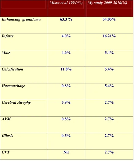
With thin slice contrast CT scan brain sensitivity of detecting such lesions is about 98%. Hence he concluded contrast CT is a reliable and cost effective modality to diagnose one of the commonest cause of seizure in this part of world. He even, quotes that “use of CSF for Immunological test to diagnose NCC is not recommended when clinical and CT features are generally straight forward (20). Misra et al in the year 1994, in their study of CT observation in partial seizures ,they found CT was abnormal in 79.3% of patients with partial seizures done at BHU – Varanasi, Commonest lesion being focal disc or ring enhancing lesion (63.3%) followed by calcification (11.8%) (21). Zee et al in the year 1980 were the first to report solitary contrast enhancing granuloma on CT scan brain. SCG are classified as disc enhancing, ring enhancing, and doughnut lesions.
Disc lesion - Uniform enhancement
Ring lesion - Peripheral enhancement with central hypodensity Doughnut lesion - Peripheral enhancement occupies much greater area leaving a small central hypodense area.
Dot inside the lesion represents the scolex. Cases with scolex inside the lesion respondents better to albendazole therapy.
If there is a combination of disc and ring or 2 discs or 2 rings in a single CT brain it is called as Type B lesion with regard to NCC.
MRI AND OTHER IMAGINGS
MRI, PET, SPECT are also used in the field of epileptology, particularly with the introduction of epilepsy surgery. Role of MRI is very useful in identifying lesions which could be easily missed in CT evaluation like Neuronal Migrational disorders and vascular malformations. Sensitivity of MRI approaches 100% in tumour, vascular malformations, infarcts, granuloma.
But not only conventional MRI but also some newer techniques like quantitative T2 relaxometry, diffusion Tensor imaging, double inversion recovery, fast flair T2 image, Magnetization transfer technique, MR spectroscopy has to be used in patients particularly having refractory partial seizures before subjecting to epilepsy surgery, because conventional MRI may fail to identify a cerebral lesion in 20% patients with refractory partial seizure. Quantitative evaluation of T2 images is more sensitive and objective than visual assessment for identification of sublet pathologies (22,27). Role of MRI is
Patients with MRI identifiable structure lesion may be triaged to epilepsy surgery early in the course of treatment if it is clear that the initial response to anti epileptic drugs are disappointing.
PET studies utilizing FDG is mainly used in Pre surgical evaluation of patients with refractory partial seizures. It shows diffuse/ regional hypometabolism in 90% interictal recording and some regional hypoperfusion. Periictal PET shows diffuse/ regional hyper metabolism. Presence of temporal hypometabolism and absence of extra temporal cortical hypometabolism predicts best outcome in temporal lobe epilepsy (24). Pseudo PLEDS on scalp EEG can be associated with focal hypermetabolism even in the absence of overt seizure. This suggests in some who are experiencing clinical seizure manifestation of PLEDS may be an ictal rather than a interictal EEG pattern
(25).
f MRI has also been utilized in Pre surgical evaluation to identify eloquent areas. 11C Flumazanil PET is a newer technique which gives
higher yield than the Advanced MRI technique in picking up the epileptogenic areas. Carbon 11 labeled flumazenil is a marker for the functional integrity of the GABAnergic inhibitory system. Loss of GABAnegic blinding by 11 C
Flumazanil PET abnormality but these findings were of use for surgery only in 25% of patients (26) . Because these picks up some white mater abnormalities
like micro dysgenesis ( increased density of heterotopic white matter neurons) which are not easily respectable by surgery. Moreover epileptogenic zone is the area of cortex necessary for seizure generation, which according to Rosenow no technique helps us to measure it directly and accurately (27). Area of seizure
onset might be same or smaller and sitting inside the epileptogenic zone.
MANAGEMENT:
Patients with partial seizure who doesn’t have an identifiable lesion have best prognosis.
Besides patients without structural lesion, patients with SCG also respond well to Medical Management. Focal seizures in younger age groups have a better control over older age group.
Usually patients with SCG needs short term AED only for about 6 months. (28). But people with calcified granuloma need prolonged AED. In such
cases therapy has to be individualized. Role of albendazole in SCG has been much debated. CT brain with ring enhancing lesions with dot inside respond better to albendazole. In other cases spontaneous revolution is possible, needs only AED and repeat CT brain after 3-6 months as per Rajashekar et al studies at CMC Vellore. Gliosis around focal cerebral calcification as seen in TI Magnetisation Transfer MRI is a prediction of poor seizure control.
Patients with acute symptomatic partial seizure due to metabolic insults like hyperosmolar nonketotic coma, hypocalcemia responds very well to the correction of underlying abnormality. They do not need long term AED therapy.
MEDICAL TREATMENT
choice for the treatment of partial seizures including those with secondary generalization. Carbamazepine is preferred because of its pharmaco kinetics and toxicity profile (29). Valproic acid is an effective alternative for some
patients when particularly the seizure secondarily generalize.
Gabapentin as an add on therapy in partial seizure in patient who are not responding to monotherapy is been well studied. In India, Prof. Dhanraj has established its role as an add on therapy in partial seizure in his paper published in the year 1998. (30) Gabapentin is unique in that it does not have any
significant drug interaction. Lamotrigine is almost effective in all sub types of partial seizure. (31) Lamotrigine appears to have an over all efficacy profile
similar to the more standard drugs and is now being used as mono therapy and also can be used as an add on therapy. When used as an add on therapy does should be started as a lowest possible and slowly titrated up.
Antiepileptic Drugs of Choice:
Seizure type Drugs of first
Choice
PHARMACOKINETICS
Responder
Rate
No Serious
Toxicity No
Nuisance toxicity
No drug inter
action
Administrations
Gabapentin 30-40% + + + t.i.d
Lamotrigine 30-40% - + + b.i.d
Phenobarbital ? + - - q.d
Primidone ? + - - t.i.d
Tiagabine 20-30% - - - b.i.d or t.i.d
Topiramate 40-50% - - + b.i.d
Valproic acid 30-40% - - - b.i.d. or t.i.d
EPILEPTIC SURGERY:
The following surgeries are done for patients with medically refractory seizures. 1. Temporal lobectomy
5. Subpial resections 6. Lesionectomy
In a statistics from a tertiary referral centre in India showed that about 74% of patients with intractable seizure referred to surgery were suffering from partial seizures. (32)
Clear identification and complete resection of epileptogenic focus will result in good outcome. Almost 90% of people will become seizure free in Mesial temporal lobe epilepsy (33). In children operated for epilepsy with tumours, after the resection of tumour almost 90% of them became seizure free
(34). In the study at CMC vellore by Danial et al over 40 years period showed, total or near total control in 35% of patients and worth while outcome in another 25% (35) . In western centres they have noted 70-80% seizures control
after epileptic surgery.
Post operatively patient generally need to remain on antiepileptic drug therapy but the marked reduction of seizures following surgery can have a very beneficial effect on their quality of life.
PROGNOSIS OF PARTIAL SEIZURE AND LOCALIZATION
RELATED/SYMPTOMATIC EPILEPSIES
A. First Unprovoked Seizure:
the risk of seizure recurrence usually is much greater. Risk factors include evidence of prior neurologic insult (determined by history, neurology examination, imaging studies), abnormal EEG, and multiple seizure or status epilepticus as initial event. Treatment after first partial seizures remains controversial because of the uncertainty regarding the risk of another seizure and the side effects of antiepileptic medication. However, randomized clinical trials do indicate that antiepileptic drugs reduce risk of seizure recurrence.
B. After two or More Unprovoked Seizure:
Persons with two or more unprovoked seizures almost always are treated. The two Veterans Administration Cooperative studies indicate that 35% to 60% of adult patients with partial seizures will have complete seizure control after 1 year with carbamazepine or phenytoin monotherapy as the initial and only treatment.
Risk factors for poor control of partial seizures include. Abnormal EEG,
Evidence of a structural brain lesion,
Number and duration of seizures before diagnosis and before control with medication,
Neurologic deficit from birth, and
Secondarily generalized tonic-clonic seizures.
C )mortality:
1. General:
Available studies are not optimal but generally report increased mortality in patients with symptomatic epilepsies. This mortality is caused, at least in part, by the underlying symptomatic disease (congenital malformations, tumors, cerebrovascular disease) and its complications. Studies regarding increased rate of suicide are conflicting.
2. Sudden Unexplained Death:
secondarily generalized). Structural lesions and severe or frequent seizures appear to be risk factors. Available evidence suggests that most sudden deaths are temporally related to seizures and often occur in sleep. Postulated mechanisms include cardiac arrhythmias, pulmonary edema, and suffocation.
D. Neuropsychologic Function:
MATERIALS AND METHODS
This study was carried out in the Department of Medicine and
Department of Neurology, Coimbatore Medical College Hospital, Coimbatore
INCLUSION CRITERIA
Total of 50 patients who were admitted with history of focal seizure or attending Neurology OPD were included in this study.
EXCLUSION CRITERIA
Patients with a history of recent head injury and those who were admitted in the surgical ward of the hospital were excluded.
STUDY PERIOD
From April 2009 to September 2010.
TYPE OF STUDY
Descriptive Study
accepted by the Professor of Medicine was used to collect the data –Proforma enclosed.
All the EEGs were critically analyzed for the presence of focal, localized or generalized changes by montage wise analysis.Individual abnormalities were recorded in the proforma .
CT scan brain plain and contrast axial section with routine slice thickness was performed in all cases. Radiologist opinion obtained. Abnormalities noted.
OBSERVATION AND RESULTS
In this study total of 50 patients were examined , in them detailed history, clinical examination and Investigations were performed.
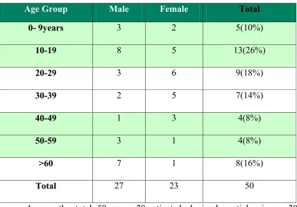
The incidence of focal seizures in the different age groups and the sexes were as follows:
[image:74.612.90.518.311.610.2]AGE & SEX INCIDENCE
Table-1
Age Group Male Female Total
0- 9years 3 2 5(10%)
10-19 8 5 13(26%)
20-29 3 6 9(18%)
30-39 2 5 7(14%)
40-49 1 3 4(8%)
50-59 3 1 4(8%)
>60 7 1 8(16%)
Total 27 23 50
Among the total 50 cases, 20 patients had simple partial seizures, 28 patients had complex partial seizure and 2 patients had 2⁰ Generalization.
SEX DISTRIBUTION
TYPE OF SEIZURE
28
2
TYPES OF SEIZURES
20
TYPES OF SEIZURES
SIMPLE PARTIAL SEIZURE
COMPLEX PARTIAL SEIZURE
2 GENERALIZATION SIMPLE PARTIAL SEIZURE
COMPLEX PARTIAL SEIZURE
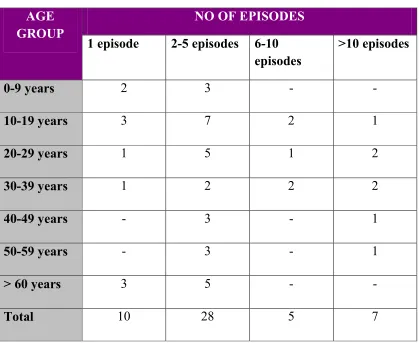
The number of attacks that each patient had was tabulated in relation to the age groups.
Table -2
AGE GROUP
NO OF EPISODES
1 episode 2-5 episodes 6-10 episodes
>10 episodes
0-9 years 2 3 - -
10-19 years 3 7 2 1
20-29 years 1 5 1 2
30-39 years 1 2 2 2
40-49 years - 3 - 1
50-59 years - 3 - 1
> 60 years 3 5 - -
Total 10 28 5 7
[image:76.612.90.509.185.528.2]AURA
[image:77.612.88.510.228.547.2]
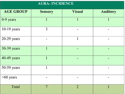
Of the fifty patients, only 8 (16 %) had “Aura”, one patient had 3 forms of aura- sensory, visual and auditory. The distribution of“Aura” was as follows:
Table-3
AURA- INCIDENCE
AGE GROUP Sensory Visual Auditory
0-9 years 1 1 1 10-19 years 3 - - 20-29 years - 1 - 30-39 years 1 - - 40-49 years 1 - - 50-59 years 1 - >60 years - - - Total 7 2 1
Only 6 patients had “precipitating factors” These were as follows:
Emotional Disturbance : 1 (pseudo – seizure) Chronic Alcoholism : 1
Post-Partum dehydration : 1 Anticonvulsant withdrawal : 1 12 Patients had significant illness in the past. They were: Diabetes mellitus Type I : 1
ABNORMAL LAB
Hyperglycemia : 2 Raised ESR : 4 Hyponatraemia : 1
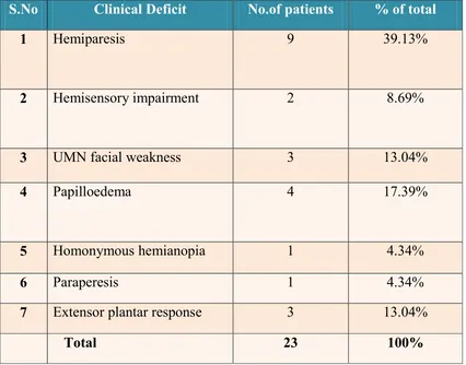
CLINICAL SIGNS
Table-4
S.No Clinical Deficit No.of patients % of total
1 Hemiparesis 9 39.13%
2 Hemisensory impairment 2 8.69%
3 UMN facial weakness 3 13.04%
4 Papilloedema 4 17.39%
5 Homonymous hemianopia 1 4.34%
6 Paraperesis 1 4.34%
7 Extensor plantar response 3 13.04%
[image:80.612.98.524.112.446.2]
In toto among the 50 patients who were examined deficit which amounts to 46%..
In this study, CT brain was abnormal in 37 patients (74%). 20 patients among these 37 patients had deficits on clinical examinations (54.05%). 17 patients with CT brain abno
Among the CT brain lesion
0 5 10 15 20 25 30
CLINICAL DEFICITS
In toto among the 50 patients who were examined - 23 Patients had deficit which amounts to 46%..
CTBRAIN
In this study, CT brain was abnormal in 37 patients (74%). 20 patients among these 37 patients had deficits on clinical examinations (54.05%). 17 patients with CT brain abnormality didn’t show any deficit.
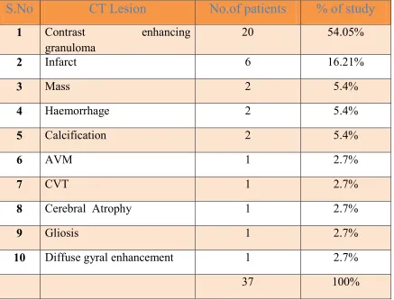
Among the CT brain lesion – Ring enhancing granuloma was the
CLINICAL DEFICITS
23 Patients had
In this study, CT brain was abnormal in 37 patients (74%). 20 patients among these 37 patients had deficits on clinical examinations
most common lesion reported in 20 cases (54.05%). Among these, single contrast enhancing ring lesion was seen in 14 patients, disc enhancing lesion in 3, Double ring enhancing lesion in 1 and multiple ring enhancing lesion in 2. Two patients had shown scolex.
Infarct was seen in 6 cases (16.21%). Mass lesion was reported in 2 cases (5.4%). Calcification in 2 cases, Gliosis Arterio-venous malformation CVT, cerebral atrophy ,diffuse gyral enhancement each in one case.
Granulomas are most commonly reported in the parietal lobe. Among the 20 cases with granuloma, 17 patients had lesions in the parietal lobe. Distribution of granuloma was in the following order, Right parietal in 6 and left parietal in 11. Among the other 3 cases Temporal lobe, frontal lobe each one has one granuloma, other showed multiple lesions.
Among 20 contrast enhancing granulomas
CT LESIONS
Table-5
S.No
CT Lesion
No.of patients
% of study
1 Contrast enhancing granuloma
20 54.05%
2 Infarct 6 16.21%
3 Mass 2 5.4%
4 Haemorrhage 2 5.4%
5 Calcification 2 5.4%
6 AVM 1 2.7%
7 CVT 1 2.7%
8 Cerebral Atrophy 1 2.7%
9 Gliosis 1 2.7%
10 Diffuse gyral enhancement 1 2.7% 37 100% Distribution of infarcts in the CT brain
Distribution of mass lesion Suprasellar -
Parieto occipital -
Distribution of granulomas
Right parietal - 6 Temporal Left parietal - 11 Frontal
0 2 4 6 8 10 12 14 16 18 20
1 1
6 Temporal - 1 11 Frontal - 1 Multiple - 1
Haemorrhage -2 Others
AVM - Parietal 2 Calcification - 1 Gliosis - 1 CVT
1 Cerebral Atrophy
1 Diffuse gyral Enhancement
11
1 1
LOCATION OF GRANULOMA
Parietal
Parietal Occipital
1 Diffuse gyral Enhancement
6 1
LOCATION OF GRANULOMA
RT PARIETAL
LT PARIETAL
TEMPORAL
FRONTAL
MULTIPLE RT PARIETAL
LT PARIETAL
TEMPORAL
FRONTAL
Interestingly among the 37 cases with CT lesions almost in 24 cases lesions were seen in the parietal lobe.
EEG
EEG was abnormal in 27 cases (54%). Among these Generalized changes were present in 10 patients (20%). Lateralizing changes were present in 17 cases (34%)
Following were the EEG abnormalities noted
1. Phase reversal - 7 cases 2. Bilateral spike, sharp waves - 10 cases 3. Focal or unilateral sharp waves - 5 cases 4. Focal slow waves - 5 cases Total cases with EEG abnormalities - 27(54 %) Lateralizing EEG Changes - 17 cases
CLINICAL DEFICITS ,EEG & CT
ABNORMALITIES-CORRELATION
CT CHANGES IN PATIENTS WITH CLINICAL DEFICITS
Among the patients with hemiparesis (9) CT brain was abnormalin 7 patients (77.8%).Among 2 patients with hemisensory deficit CT brain was abnormal in 1 patient (50%) .Among 3 patients with facial weakness of upper motor neuron type CT brain was abnormal in 3 (100%).
Among total of 4 patients with papilloedema CT brain was abnormal in all the 4 patients (100%).One patient with homonymous hemianopia CT brain was abnormal (100%). One patient with Paraparesis had CT abnormality showing a suprasellar mass lesion . CT was abnormal in all the three patients with extensor plantar (100%).
In total 50 patients who were examined, 23 Patients had deficit.CT brain was abnormal in 37 patients. 20 patients among these 37 patients had deficits on clinical examinations. 17 patients with CT brain abnormality didn’t show any deficit.
STATISTICAL SIGNIFICANCE OF POSTICTAL NEUROLOGICAL
CT Brain Lesion
C
li
n
ic
a
l
si
g
n
s
o
f
D
ef
ic
it Present Absent Total
Present 20 3 23 Absent 17 10 27 Total 37 13 50
Χ2 = 2.57
P = <0.01
P SIGNIFICANT
SENSITIVITY- 54 %
SPECIFICITY- 77 %
EEG CHANGES IN PATIENT WITH CT ABNORMARLITY
had shown lateralizing EEG abnormalities. (90.9%). Predominantly granulomas were seen in younger population.
Infarct was seen in 6 cases. Parietal lobe was the commonest site. 4/6 cases showed the infarct in the parietal lobe. EEG was abnormal in 3 cases (50%) of which 2 had lateralized EEG changes and one showed generalized changes.
Among the 2 cases with mass lesion, EEG was abnormal in 1 cases, both of them showed generalized changes.
Table-6
LESION IN CT NO OF PATIENTS
WITH CT LESION
NO OF PATIENTS SHOWING EEG
CHANGES
PERCENTAGE
Granuloma 20 11 55%
Infarct 6 3 50%
Mass lesion 2 1 50%
Hemorrhage 2 0 0%
CVT 1 1 100%
Gyral
enhancement
1 0 0%
Calcification 2 0 0%
AVM 1 1 100%
Gliosis 1 0 0%
Cerebral atrophy
1 1 100%
TOTAL 37 18 48.64%
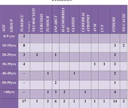
Table-7 ETIOLOGY A G E G R O U P T U B E R C U L O M A N E U R O C Y S T I C E R C O S IS T U M O U R IN F A R C T H A E M O R R H A G E G L IO S IS C E R E B R A L A T R O P H Y A V M C V T O T H E R S N O C A U S E
0-9 yrs 2
10-19yrs 8 3 2
20-29yrs 3 2 1 2
30-39yrs 4 1 1 2
40-49yrs - 1 1 2
50-59yrs - 2 1
>60yrs - 1 3 2 1 4
17 3 2 6 2 1 1 1 1 14 2
Others
0 2 4 6 8 10 12 14 16 18