THYROID PROFILE IN ABNORMAL UTERINE BLEEDING
Dissertation submitted to
THE TAMILNADU DR. M.G.R MEDICAL UNIVERSITY, CHENNAI
With fulfillment of the regulations
For the award of the Degree of
M.D (Obstetrics and Gynaecology)
Branch – II
GOVERNMENT STANLEY MEDICAL COLLEGE
CHENNAI
CERTIFICATE
This is to certify that this dissertation entitled “THYROID PROFILE
IN ABNORMAL UTERINE BLEEDING” submitted by
DR.JAYALAKSHMI.G.SHENOY appearing for part II M.D. branch II
Obstetrics and Gynaecology degree examination in March 2010, is a bonafide
record of work done by her under my direct guidance and supervision as per the
rules and regulations of the Tamil Nadu Dr. M.G.R. Medical University
Chennai, Tamil Nadu. I forward this to The Tamil Nadu Dr. M.G.R Medical
University, Chennai, Tamil Nadu, India.
Professor and Head
Department of Obstetric and Gynaecology Stanley Medical College
Chennai -600 001
THE DEAN
ACKNOWLEDGEMENT
I am greatly indebted to Dr. A.PRIYA M.S., D.O., Dean, Stanley Medical College and Hospital, Chennai for permitting me to utilize the hospital facilities for conducting this study.
I am extremely grateful to Dr. C.VENI M.D., D.G.O., Dip.N.B., professor, HOD and superintendent, Govt, RSRM Lying in Hospital, Chennai for all her support.
I express my deep gratitude to Prof. Dr. A.P. NALINI M.D. D.G.O., Govt, RSRM Lying in Hospital, Stanley Medical College who suggested this topic to me and for her valuable guidance throughout my study.
I am thankful to Prof. Dr. A. KALAICHELVI, M.D. D.G.O., Dip.N.B., Prof. Dr. T. RUCKMANI M.D. D.G.O., Prof. Dr. HEPHZIBAH KIRUBAMANI M.D. D.G.O., Ph.D., for their invaluable help in every step during this study.
CONTENTS
SL.NO Index Page No.
1. INTRODUCTION 1
2 AIM OF THE STUDY 2
3 REVIEW OF LITERATURE 3
4 PHYSIOLOGY OF MENSTRUATION 7
5 PHYSIOLOGY OF THYROID GLAND 11
6 MATERIALS AND METHODS 26
7 ANALYSIS OF THE STUDY 30
8 DISCUSSION 45
9 SUMMARY 48
10 CONCLUSION 50
PROFORMA
BIBLIOGRAPHY
MASTER CHART
INTRODUCTION
Thyroid dysfunction is a common cause of AUB and accounts for
25-35% of cases (Koutras DA, 1997). Thyroid disorders are 10 times more
common in women than men (Sisan et al, 1987). Approximately 1% of the
female population will develop overt hypothyroidism (Turnbridge WMG,
1977,)
Abnormal menstrual cycles are occasionally the first sign of
hypothyroidism and hyperthyroidism. (Wilansky DL, Griesman B, 1992)
The clinical objective is to detect and treat thyroid disease before the
symptoms and signs are significant and intense. Therefore the key to early
diagnosis is to maintain a high index of suspicion and to readily screen for the
presence of abnormal thyroid function. Moreover, thyroid dysfunction is an
easily correctable cause of AUB. Appropriate treatment is rewarded by the
AIM OF THE STUDY
The study is aimed at a cross- section of population presenting to the
department of Obstetrics and Gynecology at the Government RSRM Lying- in
Hospital, Stanley Medical College with complaints of abnormal uterine
bleeding. The study aims to ascertain the following.
1) The association between thyroid dysfunction and AUB in the
reproductive age group (18-45 years).
2) To study the thyroid abnormalities in different types of AUB in the
reproductive age group.
3) To establish if screening for thyroid abnormalities is justified using T3 ,
REVIEW OF LITERATURE
1. Gardner and Hill in 1927 showed an association between
hypothyroidism and menorrhagia
2. Goldsmith et al in 1952 found that 8 out of 10 patients with
hypothyroidism had anovulation with only 2 experiencing normal
ovulation and menses.
3. Rogers et al 1958 stated that the most common abnormality observed by
hypothyroid women is a change in the character of uterine bleeding and
length of the cycle.
4. Scott in 1964 found that 56% of woman with hypothyroidism had
abnormal menstrual patterns with menorrhagia being the most common.
5. Blum and Blum in 1972 have studied the relationship between
subclinical hypothyroidism and menorrhagia.
6. Geenspan et al in 1975 advocated the empirical use of the thyroxine. But
in 1999, prentice et al have stated that the empirical use of thyroxine is
controversial. They have condemned it and advocated the use of TRH in
women with AUB and normal T3, T4, and TSH.
7. Akande in 1975 stated that changes in FSH/LH ratios caused
anovulatory cycles in hypothyroidism.
8. Andrew weeks in 1987 in his study of 650 women with menstrual
disturbances at the Jessop hospital has stated that hypothyroidism is a
9. Keye WR, Yuen B Knopff in 1976 have stated that hyperprolactinemia
causing luteal phase defect is associated with less severe forms of
hypothyroidism
10. Robuschi et al in 1987 have stated that hypothyroidism increases with
age and is more common in older women. Upto 45% of thyroid glands
from women over 60 show evidence of thyroiditis. The incidence of
anti-thyroglobulin antibodies is 7.4% in women over age 75 while
16.9% of woman aged 60 and 17.4% of woman over age 75 have
elevated TSH levels. In women admitted to geriatric wards, 2-4% have
clinically apparent hypothyroidism
11. Klee et al in 1987 have shown the significance and positive predictive
value of TSH assay in thyroid functions tests. They are of opinion that
TSH based testing strategies minimize the problems of abnormal T4
study and significantly reduce the number of TRH stimulation tests
performed.
12. Smith et al in 1987 showed an association between hypothyroidism and
menorrhagia with development of an advanced form of von
willebrand’s disease in untreated hypothyroidism . The hemostatic
defects returned to normal with thyroxine supplementation.
13. Bleney et al in 1990 confirmed the findings of Smith et al. Hingham in
measured. An initial loss of 480 ml decreased to 58 ml following a 3
months treatment with thyroxine.
14. wilansky et al in 1992 performed thyrotropin releasing hormone (TRH)
test in 67 women who complained of excessive menstrual loss. All had
normal levels of thyroxine and thyroid stimulating hormone (TSH).
They found that 22% had abnormal TRH tests and they treated these
women with thyroxine. At follow up between 12 and 36 months later, all
considered their menstrual loss to be normal. In the 16 women with
normal TRH tests, 56% still complained of menorrhagia.
15. Blum and Blum in 1992 studied the possible relationship between
menorrhagia and occult hypothyroidism in IUD- wearing women. They
studied 40 women with menorrhagia secondary to an intrauterine
contraceptive device. They all had normal free thyroxine and TSH
levels. The 10 patients who had the highest TSH levels were given a
TRH test and all proved to have early hypothyroidism. All patients
reported a significant improvement with thyroxine treatment. This recent
development deserves further study.
16. Danese MD et al in 1996 have stated that hypothyroidism is frequent
enough to warrant consideration in most older women. They
recommend screening with highly sensitive TSH assay every 5 years
beginning at age 35 and then every two years beginning at age 60 or
17. Chameron and Fraser in 1998 in their study on the clinical disorders of
the endometrium and the menstrual cycle have stated that thyroid
disorders are the most common endocrine abnormality associated with
menstrual disturbances. Hypothyroidism is a potent cause of
menorrhagia which is amenable to treatment.
18. Shaw RW in 1999 conducted a large comparative analysis to study the
effect of thyroxine replacement on menstrual blood loss in hypothyroid
patients. There was a relative improvement in hemoglobin concentration
and general condition of the patients.
19. Prentice et al – Medical Management of Menorrhagia – 1999 have
stated that all women with unexplained menorrhagia should be tested for
thyroid dysfunction.
The review of literature suggests that there is a strong correlation between
AUB and thyroid dysfunction. It stands as an easily correctable cause of
PHYSIOLOGY OF MENSTRUATION
Menstruation is a very recent phenomenon in the evolutionary time line. It
occurs in very few species even among viviparous animals.The diagnosis and
management of abnormal menstrual function must be based on an
understanding of the physiologic mechanisms involved in the regulation of the
normal cycles. Although the activity of the endometrium is directly controlled
by the ovarian function and by the two hormones secreted by the ovary, the
ovary itself is activated by the pituitary gland, the secretion of which is under
the nervous control of the hypothalamus.
The normal human menstrual cycle can be divided into two segments: the
ovarian cycle and the uterine cycles based on the organ under examination. The
ovarian cycle may be further divided into follicular and luteal phases, whereas
the uterine cycle is divided into the corresponding proliferative and secretory
phases.
(1) At the beginning of each monthly menstrual cycle, levels of gonadal
steroids are low and have been decreasing since the end of the luteal
phase of the previous cycle.
(2) With the demise of the corpus luteum, FSH levels begin to rise and a
cohort of growing follicles is recruited. These follicles each secrete
increasing levels of oestrogen as they grow in the follicular phase. This
(3) Rising oestrogen levels provide a negative feed back on pituitary FSH
secretion which begins to wane by the midpoint of the follicular phase.
Conversely, LH initially decreases in response to rising estradiol levels
but late in the follicular phase the LH level is increased dramatically
(biphasic response).
(4) At the end of the follicular phase (just prior to ovulation), FSH induced
LH receptors are present on granulosa cells and with LH stimulation,
modulate the release of Progesterone.
(5) After a sufficient degree of oestrogenic stimulation, the pituitary LH
surge is triggered, which is the proximate cause of ovulation which
occurs 24-36 hours latter. Ovulation heralds the transition to luteal
secretory phase.
(6) The oestrogen level decreases through the early luteal phase from just
before ovulation until the midluteal phase when it begins to rise again as
a result of corpus luteum secretion.
(7) Progesterone levels rise precipitously after ovulation and can be used as
a presumptive sign that ovulation has occurred.
(8) Both oestrogen and progesterone levels remain elevated throughout the
life of the corpus luteum and then wane with its demise, thereby setting
(9) In the absence of implantation, glandular secretion ceases and an
irregular breakdown of the decidua functionalis occurs. The result is a
shedding of this layer of the endometrium , a process termed menses.
THE NORMAL MENSTRUAL CYCLE
A normal menstrual cycle lasts from 21 to 35 days with 2 to 6 days of flow and
an average blood loss of 20-60ml. (Vollman RF, 1977 and Treloar AE, 1967).
However studies of large numbers of women with normal menstrual cycles have
shown that only approximately two- thirds of adult women have cycles lasting
21-35 days (Friedman E, 1977). The extremes of reproductive life are
characterized by a higher percentage of anovulatory or irregularly timed cycles
(Collett ME et al, 1954).
DEFINITION OF MENSTRUAL CYCLE IRREGULARITIES
(1) oligomenorrhea : Infrequent, irregularly timed episodes
Of bleeding usually occurring at
interval of more than 35 days
(2) Polymenorrhea : Frequent but regularly timed episodes
of bleeding usually occurring at
interval of 21 days or less.
(3) Menorrhagia : Regularly timed episodes of bleeding
That are excessive in amount (>80 ml)
(4) Metrorrhagia : Inter menstrual bleeding
(5) Menometrorrhagia : Excessive, prolonged bleeding that
Occurs at irregularly timed, frequent
Intervals
(6) Hypomenorrhea : Regularly timed bleeding that is
decreased in amount.
(7) Amenorrhea : Absence of menstruation for three
THE THYROID GLAND
Thomas Wharton in 1656, gave the thyroid gland its modern name. For
unknown reasons, thyroid disease is more common in women than in men
(Medvei VC, 1993)
Normal thyroid Physiology
Thyroid hormone synthesis depends on an adequate supply of iodine in the diet.
It is absorbed as iodide and enters the thyroid under the influence of TSH.
Within the gland iodide is oxidized to elemental iodine which is then bound to
tyrosine. Mono and di-iodotyrosines combine to form thyroxine (T4) and
triiodothyronine(T3) (Norman AW, Litwack G, 1987). These compounds are
part of the thyroglobulin molecule which serves as a storage depot for the
thyroid hormone. TSH induces a proteolytic enzyme that result in the release of
iodothyronines into the bloodstream as thyroid hormone. Removal of one iodine
from the phenolic ring of T4 yields T3
One – third of T4 Secreted each day is converted in the peripheral tissues,
largely in the liver and kidney to T3 and about 40% is converted into inactive
Reverse T3. Although T4 in secreted at 20 times the rate of T3 ,T3 is responsible
for most of the thyroid action in the body (Czarnocka B et al, 1985).
Mechanism of Thyroid Hormone Action
Thyroid hormone acts by binding to a specific nuclear DNA bound thyroid
hormone receptor (TR) usually as a hetero dimer with the retinoid X receptor
the RXR-TR complex. T3 has a 15 fold higher binding affinity for TR than
does T4 which explains its function as the active thyroid hormone (Brent GA,
1994)
Bound and free fractions of Thyroid Hormones
Thyroid hormones present in circulation are mainly bound to proteins.
Approximately 70% of thyroid hormones are bound to thyroxine binding
globulin (TBG).The remaining 30% is bound to thyroxin binding prealbumin
and albumin. The binding proteins have greater affinity for T4 and thus allow T3
to have a greater entry into the cells. TBG is synthesized in the liver and the
synthesized in the liver and the synthesis is increased by oestrogens.
Regulation of thyroid hormone secretion – Role of estrogens
Thyroid hormones regulate TSH secretion by suppressing TRH secretion, but
primarily affect the pituitary sensitivity to TRH, by reducing the number of
TRH receptors. Pituitary secretion of TSH is very sensitive to changes in the
circulating level of thyroid hormone. A slight change in the circulating level of
T4 will produce a many fold greater response in TSH. TSH secreting cells are
regulated by T4 but only after the T4 is converted to T3 in the pituitary cells.
Although some tissues depend mainly on the blood T3 for their intracellular T3,
the brain and pituitary depend on their own intracellular conversion of T4
The measurement of T3,T4 and TSH therefore provides the most accurate
The TSH response to TRH is influenced mainly by the thyroid hormone
concentration in the circulation. Estrogen increases the TRH receptor content in
the pituitary. Hence the TSH response to TRH is greater in women than in men
and greater in women taking oral contraceptives.
The smallest doses of the TRH that are capable of producing an increase in
TSH also increase the prolactin levels indicating a physiologic role for TRH in
the control of prolactin secretion.
Thyroid Function Tests
Serum thyroid hormones are measured by radio- immunoassay. Conditions that
elevate the TBG (pregnancy, oestrogen replacement, use of oral contraceptive
pills, hepatitis) necessitate measurement of T3 resin uptake for clarification
(1) Free Thyroxine (FT4): Assays that measure free T4 are usually
displacement assays using an autoantibody to T4. The result is not
affected by changes in TBG and binding.
(2) Total thyroxine (T T4): The total thyroxine, both the portion bound to
TBG and the free unbound portion is measured by displacement assays
and in the absence of hormone therapy and other illnesses estimates the
thyroxine concentration in the blood.
(3) TSH: TSH is measured by highly sensitive assays that can detect
concentrations as low as 0.01μu/L. This is a very sensitive indicator of
thyroid hormone action at tissue level because it is dependent on the
disease, the sensitive TSH assays will provide the best indication of
excess or deficient thyroxine, slight changes in T4 are reflected in a
many fold greater response in TSH. Transient changes in TSH are seen
in severe systemic illness, psychiatric illness, adrenal insufficiency,
corticosteroid therapy, elevated HCG (Since HCG can stimulate the
TSH receptor) and in any acute illness.
(4) Total T3 and Reverse T3: These are rarely required for the accurate
evaluation of a patient with an abnormal TSH level and are of little
value in clinical circumstances. Serum T3 is almost always an indirect
reflection of the serum T4 supply (Berghout A, 1994)
Other tests include the free thyroxine index and radioactive iodine uptake.
THE LABORATORY EVALUATION
For screening purposes or when there is a relatively low clinical suspicion of
thyroid disease, the initial step is to measure the TSH by a sensitive assay. A
normal TSH essential excludes hypo/ Hyperthyroidism. A high TSH requires
the measurement of free T4 to confirm the diagnosis of hypothyroidism. If
the initial TSH is low, especially less than 0.08 μu/ml, then measurement of
high T4 will confirm the diagnosis of hyperthyroidism. If T4 is normal, the
T3 level is measured since some patients will have predominately T3
If T3 is normal it implies that thyroxine secretion is autonomous from TSH
and this is called subclinical hyperthyroidism. Some of these patients will
eventually have increased T4 or T3 levels with true hyperthyroidism.
The algorithm represents a cost-effective and accurate clinical strategy.
(Surks MI, chopra H, Mariesh CN, Nicoloff JT, solomon, TH American
Thyroid Association guidelines for use of laboratory tests in thyroid disorders,
JAMA 263:1529, 1990)
Free Sensitive TSH
High Normal Low
Normal High
Free T3 Free T4
Norma l
Low Normal High
Role of Thyroid in Reproductive physiology/ Pathology
The following facts emphasis the role of thyroid hormone in the female
reproductive physiology.
(1) TSH receptors have been found on granulosa cells.
(2) T3 and T4 have been found in follicular fluids.
(3) T4 has been found to enhance the action of gonadotrophins in
luteinisation and progesterone secretion
The female hormonal milieu and its potential effects on immune surveillance
undoubtedly play a role in the increased risk (10 fold) of women to develop
autoimmune thyroid disease (Gaitan E et al, 1985 and Wenzel BE et al,
1987). The immunoglobulins produced against the thyroid are polyclonal
and the multiple combinations of various antibodies present combine to
create the clinical spectrum of autoimmune thyroid diseases that affect
successful reproductive function.
Foetal and neonatal period.
Very few data exist regarding the role of the thyroid hormones in the
reproductive system of the foetus. No effective human studies are available.
Thyroid excess in mice is shown to cause early maturation of the
reproductive tract and early opening of the vagina. Hypothyroidism in mice
causes small ovaries deficient in cholesterol. No change has been observed
Hypothyroidism
(i) Prepubertal /Pubertal
In both sexes, thyroid hormones influence sexual development and
reproductive function.
Infantile hypothyroidism, if untreated, leads to sexual immaturity. Juvenile
hypothyroidism causes a delay in the onset of puberty followed by
anovulatory cycles.
Paradoxically, primary hypothyroidism may also cause precocious sexual
development and galactorrhea. (Kleinberg DL, New England J.Med. 1977).
The McCune Albright syndrome is characterized by hyperfunctioning
endocrinopathies including hyper/hypothyroidism and sexual precocity, but
the association may be coincidental (Albright F, Maine MJ, 1938).
Precocious puberty with delayed bone age suggests primary hypothyroidism.
Serum TSH is increased, T4 is low and galactorrhea may be present with
increased serum prolactin (Honbo KS et al, 1978). (Kindle et al) have
described a syndrome of precocious menstruation, galactorrhea and sella
enlargement in girls with juvenile hypothyroidism.
2. In adult women
Severe hypothyroidism is associated with diminished libido, amenorrhea or
anovualtion (Grodstein F et al, 1993). Secretion of progesterone is
inadequate and endometrial proliferation persists resulting in excessive and
deficient secretion of luteinizing hormone. Rarely in primary
hypothyroidism, secondary depression of pituitary function may lead to
ovarian atrophy and amenorrhea. Hypothyroidism appears to be associated
with decreased fertility resulting form ovulatory difficulties, and
spontaneous abortions may result, although many pregnancies are
successful. (Lao TTH et al, 1988 and Morimotoc et al, 1990)
The values for plasma gonadotrophins are usually in the normal range in
primary hypothyroidism. In postmenopausal women, levels are usually lower
than euthyroid women of the same age but are nevertheless within the
menopausal range. This provides a valuable means of differentiating primary
from secondary hypothyroidism.(Melmed S,Hershman J,1982)
Myxedematous infiltration can produce enlarged , cystic ovaries (Kansen
KA et al, 1997)
There may be a high incidence of early or potential hypothyroidism in
women presenting with menorrhagia. Hypothyroidism can cause
menorrhagia/ polymenoorrhea.-these symptoms being present in 30-40% of
the cases. (Koutras DA, 1997)
Metabolism of oestrogens in hypothyroidism
The metabolism of oestrogens is also altered. With respect to oestradiol and
oestrone, hypothyroidism favours the metabolism of these steroids via 16 α –
hydroxylation over that via 2 –oxygenation with the result that the formation
2-methoxyoestrone is decreased. The sex hormone binding globulin (SHBG)
concentrations in plasma is decreased with the result that the plasma
concentrations of both testosterone and oestradiol are decreased, but the
unbound fractions are increased. The alterations in steroid metabolism are
corrected by restoration of the euthyroid state (Brenta GA,et al, variation of
SHBG in thyroid dysfunction, 1999).
Effects of hypothyroidsm on the GnRH pulsatality.
TSH is secreted by the pituitary and its excess production causes menstrual
abnormality due to its adverse effects on the GnRH pulse generator and not
by directly affecting the ovary. When there is decrease in GnRH pulsatality,
anovulation can occur. Even slight changes in pulsatality may result in luteal
phase defect (Del pozo, 1979 and warfel W, 1992).
Primary hypothyrodism and hyperprolactinemia
Hyperprolactinemia and anovulation may be associated with primary
hypothyroidism Remarkable enlargement of the pituitary with thyrotroph
hyperplasia and hyperprolactinemia is frequently seen in long standing
primary hypothyroidism (Franks et al, 1975)
A number of mechanisms may be involved.
1) The clearance of prolactin tends to be decreased in hypothyroidism
2) Patients with severe hypothyroidism may have elevated total and free
estradiol levels giving rise to increased prolactin production
stimulated by excess free oestrogen (Tolis G et al, 1979).
3) The third and the most significant mechanism involves the inhibitory
effects of T3 on TRH production and on TRH receptor expression. A
decrease in the T3 feedback in hypothyroidism may induce and
increase in the hypothalamic TRH production and in the number of
TRH receptor in the lactotroph. Increased TRH action on the
lactotroph in turn may stimulate prolactin secretion (Collu R, 1986.)
The duration of hypothyroidism in important with regard to the
mechanism of amenorrhea- the longer the duration, the higher the
incidence of amenorrhea and higher the prolactin levels. This may be
associated with decreasing hypothalamic content of dopamine with
ongoing hypothyroidism. This would lead to an unopposed TRH
stimulatory effect on the pituitary cells that secrete prolactin. Constant
stimulation by the hypothalamic releasing hormones can result in
hypertrophy or hyperplasia of the pituitary (Keye WR, 1976).
Subclinical Hypothyroidism
This term designates a situation in which an asymptomatic patient has a
low normal FT4I (Free thyroxine index) but a slightly elevated serum
TSH level. The TSH elevation in these patients is modest with values
Effects of Hyperthyroidism on Reproductive Function
The two primary causes of hyperthyroidism are Grave’s disease (toxic
diffuse goiter) and plummer’s disease (toxic nodular goiter) (Lazarus JH
in Lancet 349,1997). Thyrotoxicosis in early life may cause delayed
sexual maturation although physical development is normal and skeletal
growth maybe accelerated .Menstrual changes associated with
hyperthyroidism are unpredictable, ranging from amenorrhea to
oligomenorrhea and normal cycles. The intermenstrual interval may be
prolonged or shortened, menstrual flow is initially diminished and
ultimately ceases (McKenzie JM, 1979). Fertility maybe reduced and
the risk of miscarriage is increased. In some patients menstrual cycles
are predominantly anovulatory with oligomenorrhea. In most patients,
however, ovulation occurs as is indicated by the secretory endometrium
(Reid Rl, 1987). Hyperthyroidism seldom causes amenorrhea unless
exophthalmos is present.
The mechanisms involved may be the following:
(i) Increased SHBG levels decrease the clearance of testosterone and
estradiol. Increased peripheral aromatisation of androgens gives
rise to oestrogens due to the increase in peripheral blood flow
(ii) Another more likely mechanism is the disruption in the amplitude
and frequency of LH /FSH pulses due to thyroid hormone
influences on GnRH signaling (DeGroot N, 1979),
Subclincal Hyperthyroidism
The presence of a chronically suppressed serum TSH level with
peripheral free thyroid hormones in the normal range is called subclinical
hyperthyroidism. The incidence is 0.9%.progression to overt
hyperthyroidism is uncommon. The incidence increases in older women
(Felicetta JU, 1987)
Screening for primary hypothyroidism
Only a few patients with amenorrhea/ galactorrhea will have
hypothyroidism that is not clinically apparent. Although it seems
extravagant to measure the TSH in such a large number of patients for
such a small return, because the treatment of hypothyroidism is so simple
and is rewarded by a prompt return of menstrual cycles,TSH
measurement is warranted (Caldwell G, 1985).
The high incidence of hypothyroidism in women, particularly if the
7-10% prevalence of subclinical hypothyroidism is included raises the issue
of whether the cost of systematic periodic screening of an asymptomatic
population is justified (Westman AP, BMJ, 1997). The conclusions
depend to a great extent on the assumptions regarding the effectiveness
elevation alone. An assessment of TSH levels at 5 years intervals in older
women (greater than 50 years ) seems justified, but further analysis of
more extensive screening programs are in order (P.Reid Larsen and Terry
F. Davies, 2003).
Prediction of disease onset
Patients with increased TSH and normal T4 levels progress to overt
thyroid failure at a rate of about 5% per year if thyroid auto antibody
levels are elevated. If the serum TSH alone is elevated without positive
thyroid antibody titres, the annual risk for hypothyroidism drops to
approximately 3% per year. Most clinicians therefore treat women who
have elevated serum TSH concentrations and positive thyroid antibody
tests even in the absence of symptoms. (Vanderpump MPJ, Turnbridge
WMG, 1996)
Thyroid dysfunction and menstrual disturbances in specific
conditions
1) Anorexia nervosa
The various problems associated with anorexia represent a dysfunction of
the body mechanisms regulated by the hypothalamus. Anorexics are
usually amenorrhic. Many symptoms can be explained by the state of
relative hypothyroidism. There appears to be a compensation for the state
inactive metabolite reverse T3 -a state of chemical hypothyroidism
(Herzog DB Copeland PM, 1985)
Exercise and stress induced amenorrhea.
2) In patients with exercise induced amenorrhea, there is decrease in the
frequency of GnRH pulses which is assessed by measuring a decrease
in the frequency of LH pulses (Olson BR, 1989). Athelets have
relatively low T4 levels but amenorrhic atheletes have an overall
suppression of all circulating thyroid hormones including reverse T3.
These patients are usually hypoestrogenic, but less severe alterations
may cause minimal menstrual dysfunction (Anovualtion /luteal phase
defect) (Gennazani AR et al, 1991)
3) Turners syndrome
Patients with Turners, characterized by 45XO karyotype have a
short stature, primary amenorrhea and other abnormalities. A high
prevalence of autoimmune thyroid disorders is noted. Approximately
50% of adult patients with Turners have anti-thyroid peroxidase
(anti-TPO) and antithyroglobulin (anti TG) antibody. Approximately 30% will
develop subclinical / clinical hypothyroidism (Barbesino G et al, 1998).
4) Postpartum thyroiditis
Transient thyrotoxicosis may develop within 3-6 months after delivery and is
often followed by a period of hypothyroidism of several months duration with
hypothyroid phase is apparent. Data suggest that 8-10% of women experience
thyroiditis in the postpartum period (Hayslip et al, 1988). Pregnancy is therefore
an important risk factor with transient thyroiditis developing in some patients
and thyroid failure developing permanently or in the early years after
MATERIALS AND METHODS
The present study of “thyroid profile in AUB” was conducted in Govt.
R.S.R.M. Lying in hospital, attached to Govt. Stanley medical college, Chennai.
This is a cross-sectional study of 250 women with AUB, based on data collected
from women with AUB attending the outpatient department and in-patients
during a period of 10 months from January 2009 to October 2009 at this
hospital. The study group included women with the following complaints.
1. Oligomenorrhea : cycle length greater than 35 days.
2. Hypomenorrhea : Bleeding lasting less than 2 days.
3. Menorrhagia : Blood loss more than 80 ml or more.
4. Polymenorrhea : Cycle length less than 22 days.
5. Amenorrhea : Absence of menstruation for 6 months or 3
consecutive menstrual cycles.
Inclusion criteria
1. Women in the age group of 18-45 years.
2. Women with any of the menstrual disturbances mentioned above.
3. Women who do not have signs of demonstrable pelvic pathology
4. Women with increased BMI.
5. Women who are not on any hormonal preparation.
6. Women who were not using any IUCD in the past two years.
7. Women with signs and symptoms of hypo/hyperthyroidism.
Exclusion criteria
1. Teenage AUB
2. Age more than 45 years.
3. Presence of palpable pelvic pathology like fibroids, polyps or cervical
growths.
4. Presence of general disorders like tuberculosis.
5. Presence of diabetes, hypertension or clotting abnormalities.
6. Patients with history of bleeding diathesis.
7. Patients on drugs like aspirin, heparin, sulpha drugs, antithyroid
medication, eltroxin, glucocorticoids, and amiodarone.
Symptoms and signs of hyperthyroidism
1. Weight loss more than 10 kg in 3 months or subjective weight loss.
2. Diarrhoea
3. Heat intolerance
4. Tremors
5. Tachycardia
Symptoms and signs of hypothyroidism
(i) Weight gain of more than 10 kg in 3 months or subjective weight
gain
(ii) Constipation
(iii) Slow mentation/ lethargy/increased sleepiness.
(iv) Elevated cholesterol
(v) Coarse skin.
PROCEDURE
Patients were selected based on the above criteria and history was taken as
per the proforma including a detailed menstrual history and questions
regarding the signs and symptoms of hypothyroidism and hyperthyroidism
were asked.The following examination was done.
A detailed general examination focusing specifically on the presence/
absence of anemia, thyroid swelling , cardiovascular abnormality, gross
nervous system dysfunction, galactorrhea and abnormal hair distribution.
The height in centimeters and weight in kilograms was measured and the
BMI calculated. An abdominal, speculum examination and pelvic
examination were done to rule out other causes of abnormal bleeding.5 ml of
venous blood was taken in a dry plain glass container without any
anticoagulant for TSH assay and T3,T4 estimation. Morning sample in the
phase –two site immune radiometric assay (IRMA). IRMA- K9 kit supplied
by Board of Radiation and isotope Technology (BRIT), Bombay was used.
The physiological range was 0.3-6.18µIU/ml with due consideration given to
diurnal /pulsatile variation.
T3 and T4 was analysed using RIA K-5/5A kit supplied by BRIT, Bombay .
The physiological range for T4 was 4.8 to 11.5μg/dl. The physiological
ANALYSIS OF THE STUDY
After ruling out those patients with palpable organic pelvic pathology by
gynaec examination and ultrasonogram, A total of 250 patients were included
in the study group.
TABLE – 1
T3 VALUES
Frequency Percentage
< 0.5 ng/ml 34 13.6%
0.5 – 1.5 ng/ ml 206 83.6%
>1.5 ng/ml 10 3.8%
TABLE – 2
T4 VALUES (N 4.8 – 11.5 µg/dl)
T4 (µg/dl) Number Percentage
< 4.8 34 13.6%
4.8 – 11.5 206 83.6%
T3 T4 TSH 0.00% 10.00% 20.00% 30.00% 40.00% 50.00% 60.00% 70.00% 80.00% 90.00%
THYROID PROFILE IN STUDY POPULATION
<NORMAL NORMAL >NORMAL P E R C E N T A G E
18-23 Yrs 24-31 Yrs 32-40 Yrs >40 Yrs 0.00% 10.00% 20.00% 30.00% 40.00% 50.00% 60.00% 70.00% 80.00% 90.00%
AGE DISTRIBUTION IN STUDY GROUP
HYPOTHYROIDISM EUTHYROIDISM HYPERTHYROIDISM AGE GROUP P E R C E N T A G E
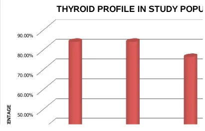
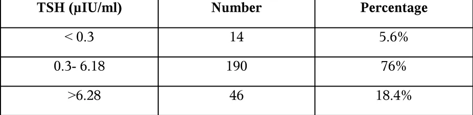
[image:35.595.99.510.75.338.2]TSH VALUES (N 0.3- 6.18 µIU/ml)
TSH (µIU/ml) Number Percentage
< 0.3 14 5.6%
0.3- 6.18 190 76%
>6.28 46 18.4%
Among 250 patients, 190 patients had normal TSH values. Incidence of Clinical hypothyroidism was 13.6%,
Incidence of subclinical hypothyroidism was 4.8% Incidence of clinical hyperthyroidism was 4%
[image:36.595.66.529.106.219.2]Incidence of subclinical hyperthyroidism was 1.6%.
TABLE -4
AGE DISTRIBUTION IN STUDY GROUP
AGE GROUP
Hypothyroidism Euthyroidism Hyperthyroidism
Out of 250 patients,198 patients were in the age group of 24-40 years. Among 198 patients,42 patients were hypothyroid and 11 patients were hyperthyroid.
No.of pts. Percentage No.of pts.
Percentage No.of pts.
Percentage
18-23 Yrs 2 6.06% 29 87.80% 2 6.06%
24-31 Yrs 26 18.05% 110 76.38% 8 5.55%
32-40 Yrs 16 29.62% 35 64.81% 3 5.55%
TABLE 5
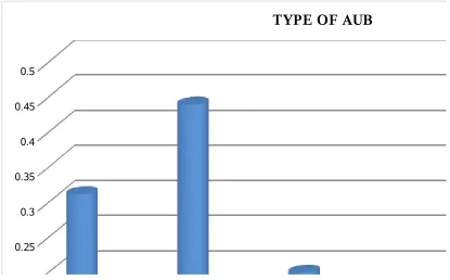
TYPE OF AUB IN STUDY GROUP
Type of AUB No.of patients Percentage
Oligomenorrhea 77 30.80%
Menorrhagia 109 43.60%
Amenorrhea 49 19.60%
Hypomenorrhea 7 2.80%
Polymenorrhea 8 3.20%
Majority of patients in the study group had menorrhagia(43.60%) and
oligomenorrhea(30.80%)
oligo men
orrhea
men orrhag
ia
amen orrhea
Hypo men orrh ea polym enor rhea 0 0.05 0.1 0.15 0.2 0.25 0.3 0.35 0.4 0.45 0.5
TABLE – 6
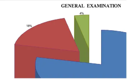
GENERAL EXAMINATION
General Examination Number Percentage
Normal 196 78.40%
Anaemia 45 18.0%
Thyromegaly 9 3.60%
21.6% of the patients had abnormal findings on examination. 78.4% were normal.
78% 18%
4%
GENERAL EXAMINATION
TABLE – 7 BODY MASS INDEX
BMI Range Hypothyroidism Euthyroidism Hyperthyroidism
Increased BMI was found in hypothyroid patients. Decreased BMI was found in hyperthyroid patients.
No.of pts.
Percentage No.of pts.
Percentage No.of pts.
Percentage
<18(lean) - - 2 16.66% 10 83.33%
18-24(Normal) 9 5.52% 150 92.02% 4 2.45%
25-29(overweight)
22 40.74% 32 59.25% -
-30-34(obese )
13 81.25% 3 18.75% -
->35 (morbid obese)
-<18 (lean ) 18-2 4(No rmal) 25-2 9(ov
er w eight ) 30-3 4(ob ese) >35( morbid
obe se) 0.00% 10.00% 20.00% 30.00% 40.00% 50.00% 60.00% 70.00% 80.00% 90.00% 100.00%
BODY MASS INDEX
Hypothyroidism Euthyroidism Hyperthyroidism WEIGHT/M2 P E R C E N T A G E
TABLE – 8
FRACTIONAL CURETTAGE
Endometrial curettage
Hypothyroidism Euthyroidism Hyperthyroidism
Majority of hypothyroid patients(87%) had proliferative pattern due to anovulation. In hyperthyroid patients both proliferative and secretory pattern were seen.
No.of pts. Percentage No.of pts.
Percentage No.of pts.
Percentage
Secretory 6 13.0% 140 73.68% 8 57.14%
[image:40.595.79.524.87.602.2]SECR ETOR Y PROL IFER ATIV E 0.00% 10.00% 20.00% 30.00% 40.00% 50.00% 60.00% 70.00% 80.00% 90.00%
FRACTIONAL CURETTAGE
HYPOTHYROIDISM EUTHYROIDISM HYPERTHYROIDISMTYPE OF ENDOMETRIUM
P E R C E N T A G E TABLE- 9 HEMOGLOBIN ESTIMATION Hemoglobi n
Hypothyroidism Euthyroidism Hyperthyroidism
Majority of hypothyroid patients(65.2%) were anemic due to menorrhagia,whereas hemoglobin was normal in majority of hyperthyroid patients(71.43%)
No.of pts. Percentage No.of pts.
Percentage No.of pts.
Percentage
<7 gms% 30 65.2% 20 10.52% -
-7-9 gms% 5 10.95 30 15.795 4 28.57%
Hb <7 gms% Hb 7-9 gms% Hb >9 gms% 0.00% 10.00% 20.00% 30.00% 40.00% 50.00% 60.00% 70.00% 80.00%
HEMOGLOBIN ESTIMATION
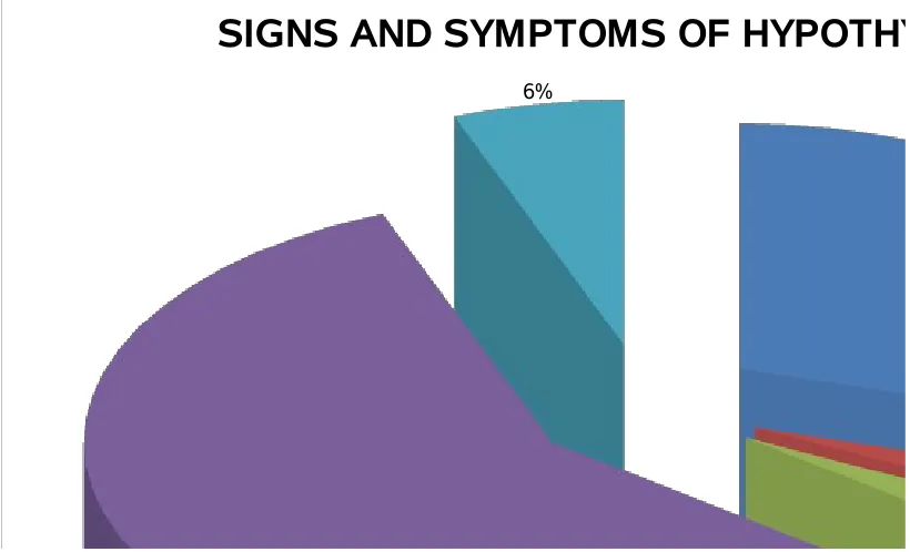
HYPOTHYROIDISM EUTHYROIDISM HYPERTHYROIDISM HEMOGLOBIN P E R C E N T A G E TABLE 10SIGNS AND SYMPTOMS OF HYPOTHYROIDISM
Signs/Symptoms Number of patients percentage
Constipation 10 29.4%
Cold intolerance 1 2.9%
Voice change 1 2.9%
Weight gain 20 58.8%
Lethargy 2 5.9%
29%
3%
3% 59%
6%
SIGNS AND SYMPTOMS OF HYPOTHYROIDISM
[image:43.595.99.509.78.327.2]CONSTIPATION COLD INTOLERANCE VOICE CHANGE WEIGHT GAIN LETHARGY
TABLE – 11
SIGNS AND SYMPTOMS OF HYPERTHYROIDISM
Signs/Symptoms Number of patients Percentage
Heat intolerance 1 10%
Anxiety 1 10%
Weight loss 1 10%
Fatigue 5 50%
Tremors 1 10%
Diarrhea 1 10%
10%
10%
10%
50% 10%
10%
SIGNS AND SYMPTOMS OF HYPERTHYROIDISM
HEAT INTOLERANCE ANXIETY
WEIGHT LOSS FATIGUE TREMORS DIARRHEA
TABLE-12
AMENORRHEA-T3,T4 AND TSH VALUES
Values T3
(N=0.5-1.5 ng/ml)
T4
(N=4.8-11.5µg/dl)
TSH
Out of 49 patients with Amenorrhea,
2 patients (0.8%*) had clinical hypothyroidism
6 patients (2.4%*) had clinical hyperthyroidism
1 patient (0.4%*) had subclinical hypothyroidism
2 patients (0.8%*) had subclinical hyperthyroidism
*→ of the total sample studied
No.of pts. Percentage No.of pts.
Percentage No.of pts.
Percentage
< Normal 2 4.1% 2 4.08% 8 16.32%
Normal 41 83.7% 41 83.7% 38 77.56%
BASED ON T3 BASED ON T4 BASED ON TSH 0.00%
10.00% 20.00% 30.00% 40.00% 50.00% 60.00% 70.00% 80.00% 90.00%
AMENORRHEA
<NORMAL NORMAL >NORMAL
THYROID PROFILE
P
E
R
C
E
N
T
A
G
E
MENORRHAGIA-T3,T4 AND TSH VALUES
Values T3
(N=0.5-1.5 ng/ml)
T4
(N=4.8-11.5µg/dl)
TSH
N=0.3-6.18µIU/ml)
Among 109 patients with menorrhagia, 26 patients(10.4%*) had clinical hypothyroidism
1 patient (0.4%*) had clinical hyperthyroidism
8 patients(3.2%*) had subclinical hypothyroidism
No patient had subclinical hyperthyroidism in this group.
*→ of the total sample studied No.of pts. Percentage No.of
pts.
Percentage No.of pts.
Percentage
< Normal 26 23.85% 26 23.85% 1 2.03%
Normal 82 75.22% 82 75.22% 74 67.89%
BASED ON T3 BASED ON T4 BASED ON TSH 0.00%
10.00% 20.00% 30.00% 40.00% 50.00% 60.00% 70.00% 80.00%
MENORRHAGIA
<NORMAL NORMAL >NORMAL
THYROID PROFILE
P
E
R
C
E
N
T
A
G
E
OLIGOMENORRHEA -T3,T4 AND TSH VALUES
Values T3
(N=0.5-1.5 ng/ml)
T4
(N=4.8-11.5µg/dl)
TSH
N=0.3-6.18µIU/ml)
Out of 77 Oligomenorrheic patients,
4 patients(1.6%*) had clinical hypothyroidism
3 patients(1.2%*) had had clinical hyperthyroidism
1 patient(0.4%*) had subclinical hypothyroidism
2 patients(0.8%*) had subclinical hyperthyroidism in this
group.
*→ of the total sample studied No.of pts. Percentage No.of
pts.
Percentage No.of pts.
Percentage
< Normal 4 5.19% 4 5.19% 5 6.49%
Normal 70 90.90% 70 90.90% 67 87.01%
BASED ON T3 BASED ON T4 BASED ON TSH 0.00%
10.00% 20.00% 30.00% 40.00% 50.00% 60.00% 70.00% 80.00% 90.00% 100.00%
OLIGOMENORRHEA
<NORMAL NORMAL >NORMAL
THYROID PROFILE
P
E
R
C
E
N
T
A
G
TABLE-15
POLYMENORRHEA-T3,T4 AND TSH VALUES
Values T3
(N=0.5-1.5 ng/ml)
T4
(N=4.8-11.5µg/dl)
TSH
N=0.3-6.18µIU/ml)
Among 8 patients with polymenorrhea,
2patients(0.8%*) had clinical hypothyroidism
2patients(0.8%*) had subclinical hypothyroidism
No clinical and subclinical hyperthyroidism was noted in polymenorrhea patients.
No.of pts. Percentage No.of pts.
Percentage No.of pts.
Percentage
< Normal 2 25% 2 25% -
-Normal 6 75% 6 75% 4 50%
*→ of the total sample studied
BASED ON T3 BASED ON T4 BASED ON TSH
0% 10% 20% 30% 40% 50% 60% 70% 80%
POLYMENORRHEA
<NORMAL NORMAL >NORMAL
THYROID PROFILE
P
E
R
C
E
N
T
A
G
The Findings are summarized as follows:
TABLE-16
Incidence of Based on T3,T4 and TSH
Hypothyroidism 13.6%
Hyperthyroidism 4%
Subclinical Hypothyroidism 4.8%
Subclinical Hyperthyroidism 1.6%
In this study,Thyroid dysfunction account for 24 % in AUB patients.
The incidence of thyroid Dysfunction in different types of AUB is as follows:
TABLE-17
AUB Hypo
thyroidism
Hyper thyroidism
Subclinical hypothyroidism
Subclinical hyperthyroidism
Amenorrhea 0.8% 2.4% 0.4% 0.8%
Menorrhagia 10.4% 0.4% 3.2%
-Oligomenorrhea 1.6% 1.2% 0.4% 0.8%
-DISCUSSION
AUB is a benign yet debilitating disease with a strong association with
thyroid disorders. Our study highlights the association between AUB and
thyroid dysfunction by measurement of T4 and TSH in the fasting state in
women with AUB.
Hypothyroidism is observed in 10.4% of women with menorrhagia, and
0.8% of women with amenorrhea. Hyperthyroidism is seen in 1.2% of women
with oligomenorrhea. The overall incidence of thyroid dysfunction is 24%. This
correlates with the study by Wilansky et al, 1992.
Study Incidence of thyroid disorders
Prentice at al, 1999 36%
Wilansky et al, 1992 22%
Present study 24%
Menon et al, 1995 26%
Some patients have a normal serum TSH despite low T3 and T4. This is
explained by a downward resetting of the threshold for TSH inhibition TSH
setpoint for a particular serum T3, T4 increases with age and is also altered by
personal and familial character. TSH values tend to change more rapidly
because the half life of TSH is much shorter than T3 and T4. This should be
considered with regard to abnormal relationships between T3, T4 and TSH.
There are some variations which should be given due consideration before
1) T4- Methodology, pregnancy
2) TSH – diurnal variation, pulsatile secretion
Alteration in the relationship between T4and TSH can be caused by alternate
thyroid stimulating hormones like TSH isoforms, chorionic gonadotrophins, and
TSH receptor stimulating antibody. It can be caused by hormones and drugs like
glucocorticoids, severe non-thyroidal illness, recent thyrotoxicosis and long
standing hypothyroidism. The incidence of thyroid dysfunction in the
population with AUB is 24% according to our study and hence selective
screening of this population would result in a higher yield.
A major benefit of routine testing is the earlier detection of unsuspected overt
thyrotoxicosis or subclinical hypothyroidism or hyperthyroidism. Most
clinicians advocate treatment of women with elevated TSH levels in view of
risk of hypothyroidism subsequently.
T3 - Binding protein ,its values are altered in the following conditions
like illness ,surgery, drugs and age related changes,
A T3 T4 TSH assay can be used as a management tool besides its use in
diagnosis and screening A T3 : T4 eatio of greater than 0.024 during drug
therapy suggests that remission is unlikely . it can be used to identify patients
who have persistent T3 excess despite normal a low serum T4 levels during anti
thyroid therapy. It can be used to distinguish between T3 thyrotoxicosis and
SUMMARY
The present study is a cross-sectional study of 250 women with abnormal
uterine bleeding in the reproductive age group undertaken in a tertiary referral
hospital over a period of 10 months. It was done to ascertain the correlation
between thyroid dysfunction and AUB.
The history was elicited according to the proforma. Anthroprometric
measurement were taken and a detailed examination was done. T3, T4 and TSH
levels were evaluated in the fasting rate and the results interpreted.
1. The study showed a significant correlation (p = 0.004, significant)
between increasing age and thyroid dysfunction.
2. The general examination showed a significant correlation (p=0.003)
with thyroid dysfunction.
3. The Body Mass Index showed a significant correlation (p=0.006) with
thyroid dysfunction.
4. The study showed a significant correlation (p=0.006) between
Hemoglobin Estimation and thyroid dysfunction.
5. The Study showed a significant correlation between thyroid profile
6. The study showed a significant correlation (p=0.002, Significant )
between fractional curettage and hypothyroidism whereas in
hyperthyroidism as the endometrial pattern is unpredictable, it was not
CONCLUSION
It may be concluded from the present study that there is a significant
association between thyroid disorders and AUB. The high incidence (24%)of
thyroid disorders in women with AUB, particularly if the 5-10% of subclinical
hypothyroidism is included, justifies the cost of screening in AUB population.
The risk of progression to overt hypothyroidism (about 5% per year) in patients
with subclinical disease also emphasize the need for screening in AUB
PROFORMA
Name Age
Address Parity
Sterilisation
H/O O/E
Menorrhagia General Condition
Oligomennorrhe Anemia
Amenorrhea Thyromegaly
Polymenorrhea BMI
Others
H/O S/S Hyperthyroidism Thyroid function tests
T3
--- T4
--- TSH
--- Inference
H/O S/S Hypothyroidism other Investigation
--- Hb in gm/dl
BIBLIOGRAPHY
1. Anasti JN, Flack MR, Froehlich, J, Nelson IM, Nisula BC. A Potential
novel mechanism for precocious puberty in juvenile hypothyroidism, J.
Clin. Endocrinol. Metab.80:276, 1995.
2. Balen AH, Shoham Z and Jacobs HS (1933a). Amenorrhea- causes and
consequences In-Asch RH and studd JJW (eds) Annual progress in
reproductive medicine. Carnforth, Lancashire: Pantheon press, pp-
205-34.
3. Ballabio, M.Poshyachinda M, Ekins RP, Pregnancy induced changes in
thyroid function : role of human chorionic gonadotropin as putative
regulator of maternal thyroid, J.Clin.Endocrinol. Metab. 73:824,1991.
4. Blum M. Blum G. The possible relationship between menorrhagia and
occult hypothyroidism in IUD wearing women. Advance contracep.
1992:8:313-317.
5. Bolmet HG, Fieldler K Leindenberger FA. Subclinical hypothyroidism
and infertility. Lancet. 1981;2:1278.
6. Bottazo GF. Dean BM. Autoimmune thyroid disease. Anuu Rev Med.
1986:37 353-354.
7. Boukis MA, Koutrar DA, Souvatzoglou A, et al. Thyroid hormone and
immunological studies in an endemic goiter area. Arch klin med 1968,
8. Brent GA, the molecular basis of thyroid hormone action, new Eng. J.
Med 331-947.1994
9. Brenta G, Schnitman M, Gurfinkiel M, et al variations of sex hormone
binding globulin in thyroid dysfunction. Thyroid 1999;9:273-277.
10.Burrow GN, Fisher DA, Larsen PR, Maternal and fetal thyroid function,
New Engl. J. Med . 331:1072, 1994.
11.Caldwell G, Kellet KA, Gow SM et al . A new strategy for thyroid
function testing. Lancet 1985:1:1117-1119.
12.Cameron IT (1992). Medical Management of menorrhagia .Curr obstet,
Gynaecol 2,136-40
13.Contreras P, Generini G, Michelson H ,Pumarino H, Compmo,
Hyperprolactinemia and galactorrhea: Spontaneous versus iatrogencic
hypothyroidism, J.Clin Endocrinol. Metab 53:1036,1981
14.Cooper D, Ridgway E, Kilman B, et al Metabolic Clearance and
production rates of prolactin in man, J. Clin Invest. 1979; 64:1669-1680.
15.Cooper DS, thyroid hormone treatment: New insights into an old
therapy,JAMA 261:2694,1989.
16.Coulter A , Bradlow J, Agass M, Martin Bates C, Tulloch A. outcomes of
referrals to gynaecology out patient clinics for menstrual problems: an
audit of general practice records. Br. J. obstel Gynacecal
17.Danese Md, powe NR, Sarvin CT, Landenson PW, screening for mild
thyroid failure at the periodic health examination. A decision and cost
effective analysis JAMA 276:285, 1996.
18.Davey DA, Dysfunctional uterine bleeding In: Whitfield CR, ed
Dewhursts textbook of obstetrics and gynaecology for postgraduates, 5th
edn. London, Blackwell, scientific 1995, p 599.
19.Del . Pozo E, wyss H. Tolis G, et al , prolactin and deficient luteal
function, obstet, Gynaecol. 1979; 53:282-286.
20.Doody KM and carr BR (1990). Amenorrhea: in : Chihal HJ, London SN
(eds) Menstrual cycle disorders, obstet. Gynecol Clinical N.Am
Philadelphia : Saunders, 17:361 87.
21.Drexhage HA, Bottazzo GF, Bitenusky L, et al. Thyroid growth blocking
antibodies in primary myxedema. Nature 1981;239:594-595.
22.Edlend M,Magmusson C, Ven shoultz, B. Quality of life a swedish
survey of 220 women. In smith SK, ed. Dysfunctional uterine bleeding
London :Royal society of Medicine press, 1994; pp.36-37.
23. Ericson, GF. An analysis of follicle development and ovum maturation,
seminars Reprod Endocrinol. 4:233,1986.
24.Falsetti L,Pasinetti E, Mazzani MD, Gastaldi A, weight loss and
menstrual cycle clinical and endocrinology evaluation, gynecol
25.Fraser IS. Treatment of menorrhagia In: Drife JO, Ed. Dysfunctional
uterine bleeding and menorrhagia, bailliere’s clinical obstertrics and
gynaecology. London. Bailliere Tindal. 1989:pp.391-402
26.Gennazani Ar, petragtia F, De Ramundo BM, et al, Neuroendocrine
correlates of stress related amenorrhea: Ann N.Y. Acad. Sci
1991;626:125-129.
27.Glinoer D, The regulation of Thyroid function in pregnancy: Pathways of
endocrine adaptation from physiology to pathology, Endocr
Rev.18:404,1997.
28.Glinoer, D, De Nayer P.Bourdoux P, Lemone M.Robyn C, Van
Stirteghem A, Kinthaert, J, Clin . Endocrinol. Metab. 71:276, 1990.
29.Hague WM, Tan SL, Adams , J and Jacobs HS (1987)
Hypergonadotrophic amenorrhea- etiology and outcome in 93 young
women .Int.j.Gynacol. obstet, 25,121-5.
30.Haisen KA, Tho SPT, Hamly M, Moretizzo RW, Mc donough PG,
Massive Ovarian Enlargement in primary Hypothyroidism , fertil steril
67:169,1997.
31.Herzog, DB, Copeland PM. Eating disorders N.Engl J. Med,
1985,313:295-303.
33.Hingham JM, Shaw RW. The effect of thyroxine replacement on
menstrual blood loss in a hypothyroid patient, Br.J.Obstet, Gynaecol,
1992:99:695-696.
34.Hirvonen E, Etiology, clinical features and prognosis in secondary
amenorrhea, Int. J. Fertil. 22:69, 1977.
35.Hung W, August GP, Glasgow AM, Pediatric Endocrinology, medical
Examination publishing Co., Garden City, 1978.
36. Kamilaris TC, De Bold LR, Pavlov SN, et al, Effect of altered thyroid
hormone levels on Hypothalamic pituitary adrenal function . J. Cin
Endocrinal Metab. 1987, 65:994-999.
37.Kellet KA, Van Herle AJ, Honbok, Serum prolactin untreated primary
hypothyroidism AM. J. Med. 1978;64;782-787.
38.Keye WR, Ho Yuen B, Knopff R, et al Amenorrhea, hyperprolactinemia
and pituitary enlargement secondary to primary hypothyroidism. Obset.
Gynecol. 1976; 48:697-702.
39.Kimura M, Amino N. Tamaki H, Mitsuda N, Miyai K, Tamizawa O,
physiologic Thyroid activation in normal early pregnancy is induced by
circulating. hcG, obstet. Gyneocl . 75; 775,1990.
40.Koutras DA, Disturbances of menstruation in thyroid disease. Ann. N.Y.
41.Kramer, M Kaushansky A. Genel M. Adolescent secondary amenorrhea:
association with hypothalamic hypothyroidism , paediatrics
1979;94:300-303.
42.La Barbera A, Miller MM, Ober C, et al., Auto immune etiology in
premature ovarian failure. Am. J. Reprod. Immuno.1988; 16:114-118.
43.Lazarus JH, Hyperthyroidism , Lancet 349:339,1997.
44. Lee PA, Van Dop C, Migeon CJ, Mc Cune Albright syndrome long term
follow up JAMA 256:290,1986.
45. Leon speroff, Robert H.Glas Nathan, G.Kase Clinical gynaecologic
endocrinology and infertility sixth edition, 1999.
46.Mackenzie JM, Zakariya M. Hyperthyroidism in Degroot LJ, Cahill GF,
Martini L, eds. Endocrinology , New York :Greene and Stratton 1979:647.
47.Medvei, VC. The history of clinical Endocrinology. The pantheon
publishing group, New York, 1993.
48.Mindermann T, wibon CB, Thyrotropin Producing pituitary Adenomas, J
Neurosurg 79:521,1993.
49. Munster K, Schmidt I, Helm P. Length and Variation in the menstrual
cycle- a cross sectional study from a Danish country, Br. J. Obstet,
Gynaccol. 99:422, 1992.
50.Natori, S. Karashima, T, Koga S.et al :Effect of thyroid hormone
replacement on reduction of pituitary enlargement and restoration of
51.Nelson L. Rybo G, Treatment of menorrhagia. Am. J. Obstet. Gynecol.
110:713, 1971
52.Norman AW, Litwack G. Thyroid Hormones. In: Norman AW Litwack
G, eds. Hormones, San Diego, Academic press , 1987;221.
53.Olson BR. Exercise induced amenorrhea. Am. Fam. Physician 1989,
39:213-221.
54.Peterson CM. Thyroid disease and fertility In: Gleichon N, ed.
Autoimmunity in reproduction. Immunol. Allergy clin. NA. 1995;
14:725-738.
55.Poretsky L, Garber J, Kleefield J, Primary amenorrhea and pseudo
prolactinoma in a patient with primary hypothyroidism. Am J Med 81:180,
1986.
56.Rebar RW, Kenisberg D, Hodgen GD. The normal menstrual cycle and
the control of ovulation, In :Becker KL, Ed. Principles and practice of
endocrinology and metabolism. 2nd ed. Philadelphia. JB lippincott
1995:868.880
57.Reindollar RH, Novak M, Tho SPT,Mc Donough PG, Adult onset
amenorrhea, AM J Obstet Gynecol 155:531,1986.
58. Reindollar RH, Tho SPT, Mc Donough PG, Delayed Puberty: an updated
59.Royal college of General Practitioners and the office of population
surveys morbidity statistics from general practice 1981-2 London
HMSO, 1986.
60. Scanlon MF, Chass V, Health M, et al, Dopaminergic content of
thyrotropin α and β subunit, and prolactin in euthyroidism and
hypothyroidism J. Clin. Endocrinol. Metab 1981; 53; 360-365.
61.Scott JC,Mussy E menstrual patterns of
myxedema.Am.J.Obstet.Gynaecol.1964;90:161-165
62.Smyth PPA, Hetherton AMT, Smith DF, Radcliff M, O’ Herlihy C,
Maternal Iodine status and thyroid volume during pregnancy correlation
with neonatal iodine intake, J. Clin Endocrinol. Metab 82: 2840, 1997.
63.Surks MI, Chopra IJ, Mariash CN, NiColoff JT, Solomon DH American
Thyroid Association guidelines for use of laboratory tests in thyroid
disorders JAMA 263:1529.1990
64.Tanaka, T, Tamai H, Kuma K, et al. Gonadotrophins response to LHRH
in hyperthyroid patients with menstrual disturbances. Metabolism 1981;
30:323-325.
65.Thomas R, Reid RL. Thyroid disease and reproductive dysfunction
Obstet. Gynaecol.1987;70:789-798.
66.Thomas R, Reid RL. Thyroid disease and reproductive function Obstet.
67.Treloar A E,Boynton RE, Borghilid GB,Brown BW.Variation of the
human menstrual cycle through reproductive life,Int.J.Fertil.12:77,1967.
68.VanHearle AJ,Uller RP,Mathews ML et al Radioimmunoassay for
measurement of Thyroglobulin in human serum.J.Clin
Invest.1973;52;1320-1327.
69.Vander pump MPJ Turnbridge VMG.The thyroid fundamental and
clinical text,7th edition.1996;474-482.
70.Vollman RP,The menstrual cycle,In:Friedman,ed.Major problems in
obstetrics and gynaecology,W.B.SaundersCo.,Philadelphia,1977.
71.Warfel W.Thyroid regulation pathways and its effect on human function
1992;32:145-150.
72.Wilansky DL,Greisman B,Early hypothyroidism in patients with
menorrhagia. Am.J.Obstet.Gyneacol 160;673:1989.
73.Wilkins L. The diagnosis and treatment of Endocrine disorders in
childhood and adolescence,3rd ed.,Charles C,Thomas,Spring Field,1965
Thomas R, Reid RL. Thyroid disease and reproductive dysfunction
obster. Gynecol 1987;70:789-798.
74. Thomas R, Reid RL. Thyroid disease and reproductive function. Obstet
ABBREVIATIONS
AUB - Abnormal Uterine Bleeding
T4 - Thyroxine
T3 - Tri-iodothyronine
TSH - Thyroid Stimulating Hormone TRH - Thyrotropin Releasing Hormone
LH - Luteinising Hormone
FSH - Follicular Stimulating Hormone
HCG - Human Chorionic Hormone
TR - Thyroid hormone Receptor
RXR - Retinoid X Receptor
RT3 - Reverse T3
TBG - Thyroid Binding Globulin SHBG - Sex Hormone Binding Globulin
FT4 - Free Thyroxine
TT4 - Total Thyroxine
GnRH - Gonadotrophin Releasing Hormone Anti TPO - Anti Thyroid Peroxidase
Anti TG - Anti Thyroglobulin
BMI - Body Mass Index
PID - Pelvic Inflammatory Disease
IUCD - Intra Uterine Contraceptive Device IRMA - Immuno Radio Metric Assay