0022-538X/95/$04.0010
Copyrightq1995, American Society for Microbiology
Human Papillomavirus Type 31b Late Gene Expression Is Regulated
through Protein Kinase C-Mediated Changes in RNA Processing
MARY HUMMEL,1HOCK B. LIM,1
ANDLAIMONIS A. LAIMINS1,2*
Department of Microbiology-Immunology1and Department of Biochemistry, Molecular Biology
and Cell Biology,2Northwestern University, Chicago, Illinois 60611
Received 17 January 1995/Accepted 23 February 1995
Expression of the human papillomavirus (HPV) capsid genes, L1 and L2, as well as amplification of viral DNA and virion assembly occur in the terminally differentiated layers of infected stratified squamous epithe-lium in vivo. These processes can be duplicated in the laboratory through the use of organotypic or raft cultures. When CIN612 cells, which contain episomal copies of the high-risk HPV type 31b, are allowed to differentiate in raft cultures, the expression of transcripts encoding the early genes E1∧E4 and E5 is induced. These transcripts are initiated at the differentiation-dependent P742 promoter located in the middle of the E7 open reading frame. Exposure of raft cultures to activators of protein kinase C, such as phorbol esters, results in the further induction of late gene expression as well as virion assembly. In this study, we have investigated the mechanism by which activators of protein kinase C induce late gene expression. The major L1 transcript was found to be encoded by a bicistronic E1∧E4, L1 RNA which initiated at the differentiation-dependent promoter P742. Additional low-level expression of L1-containing RNAs was also observed from the early-region promoter, P97. The major L2 transcripts were found to be encoded by E1∧E4, E5, L2, L1 RNAs which were also initiated in the early region, probably at the differentiation-specific promoter P742. While early and late RNAs were found to be expressed from the same promoter, they differed in utilization of splicing and polyadenylation sites. Raft cultures treated with activators of protein kinase C induced expression of late genes, but no change in the abundance of early RNAs initiated at the P742 promoter was observed. Thus, the increase in late gene expression was likely due to changes in RNA processing or stabilization rather than an increase in the rate of transcription from P742. Regulation of HPV late gene expression therefore occurs at two levels: differentiation-dependent induction of the P742 promoter, which can be mimicked in vitro by growth in raft cultures, and posttranscriptional changes that can be induced by activation of protein kinase C. These posttranscriptional changes may occur through inactivation or down-regulation of splicing factors which inhibit use of the late region polyadenylation site, resulting in increased stability of late region transcripts.
Human papillomaviruses (HPVs) are small DNA viruses which induce proliferative lesions of epithelial tissue (11, 31, 37, 63). Over 60 different types of papillomaviruses have been identified (17), and each exhibits tropism for a particular kind of mucosal or cutaneous epithelium. Over one-third of the known HPV types infect the mucosal epithelia of the anogeni-tal tract, and the high-risk types such as HPV type 16 (HPV-16), HPV-18, HPV-31, and HPV-33 induce lesions which can eventually progress to invasive carcinomas. Genital lesions in-duced by the low-risk types such as HPV-6 and HPV-11 rarely progress to malignancy. DNA sequence analysis indicates a general conservation of genomic organization among HPV types (11, 31, 63). The early regions of all types contain clusters of six to eight open reading frames (ORFs). The E6 and E7 ORFs encode the viral transforming genes (1, 4, 7, 32, 46, 51), while the products of the E1 and E2 genes act to regulate viral DNA replication (12, 23, 24, 60). In addition, the E2 protein may function in the modulation of early gene expression (13, 58). The E4 protein is expressed as a fusion protein with the N-terminal portion of E1 (47) and associates with cytokeratin structures to facilitate viral egress (19). The membrane-asso-ciated E5 protein is believed to contribute to the hyperprolif-eration capability of infected cells. The late region is also
conserved among viral types and encodes the L1 and L2 capsid proteins.
Transcripts encoding the early region ORFs are expressed from a common promoter located at the beginning of the E6 gene and are terminated at a polyadenylation site at the end of the early region (15, 33, 55). Differential splicing of early RNAs generates a variety of polycistronic messages which vary considerably in relative abundance (33). While the initiation sites of the late transcripts of the high-risk viral types have not yet been defined, these transcripts are believed to terminate at a second polyadenylation site at the end of L1. In situ hybrid-ization analysis of biopsies of infected tissue has revealed that expression of HPV genes is closely linked to epithelial cell differentiation (16, 56). The majority of the early genes are expressed in both differentiated and undifferentiated epithelial cells, but expression of the late genes occurs only in the ter-minally differentiated layers of the upper portion of the epi-thelium. One exception to this pattern is the E1∧E4, E5 tran-script, whose expression is also induced upon epithelial cell differentiation. The molecular basis for the induction of differ-entiation-dependent viral expression is not understood but it is likely to be the consequence of cellular factors acting in con-junction with HPV proteins.
Recent advances in tissue culture techniques have allowed for the study of these differentiation-specific viral processes in the laboratory. Dollard et al. (18) demonstrated the synthesis of HPV-11 virus when explants from xenografts were expanded in raft cultures (2). Using an established cell line (CIN612 [6]) which maintains episomal copies of the high-risk HPV-31b
* Corresponding author. Mailing address: Department of Microbi-ology-Immunology, Northwestern University, 303 E. Chicago Ave., Chicago, IL 60611. Phone: (312) 503-0648. Fax: (312) 503-1339.
3381
on November 9, 2019 by guest
http://jvi.asm.org/
(38), Meyers et al. (45) were able to duplicate the differenti-ation-dependent amplification of viral DNA and virus produc-tion in organotypic raft cultures. In previous studies, we have demonstrated the induction of a transcript which encodes the E1∧E4 and E5 ORFs following stratification of CIN612 cells in raft cultures (33). This transcript was initiated at a differenti-ation-dependent promoter at nucleotide (nt) 742 in the middle of the E7 ORF which was designated P742. In situ hybridiza-tion studies of HPV-16-positive biopsy material have also sug-gested the presence of a differentiation-dependent promoter in this region of E7 (30), and similar transcripts have been ob-served in HPV-11 xenografts in nude mice (15, 47, 55). While initial studies using stratified raft cultures of the CIN612 cell line demonstrated the induction of E1∧E4, E5 transcripts, no significant levels of expression of the late genes were observed. When raft cultures of CIN612 cells were treated with activators of protein kinase C (PKC), such as the phorbol ester 12-O-tetradecanoylphorbol-13-acetate (TPA) or synthetic diacyl-glycerol compounds, capsid protein synthesis was observed (45). In the present study, we have used the raft culture system to examine the structure of the late region transcripts and the mechanisms regulating their expression. We find that the late genes, L1 and L2, are expressed from the differentiation-de-pendent promoter, P742, which also directs the expression of the E1∧E4, E5 transcript. Furthermore, late gene expression was observed to be regulated by PKC-dependent changes in posttranscriptional RNA processing.
MATERIALS AND METHODS
Cell culture.CIN612 cells were maintained in E medium with mitomycin-treated fibroblast feeder cells as previously described (6). Collagen raft cultures for in vitro differentiation were prepared as previously described (43). For acti-vation of PKC, raft cultures were treated every 4 days with E medium containing 16 nM TPA for 16 to 24 h as previously described or every 2 days with E medium containing 10mM 1,2-dioctanoyl-sn-glycerol (C8) (45). For activation of PKC in monolayer cultures, cells were seeded and maintained in E medium containing 10mM C8 until they reached confluence (;6 days).
RNA isolation and Northern (RNA) blot analysis.RNA was isolated from monolayer or raft cultures by the guanidinium isothiocynate-CsCl centrifugation method (14) as previously described (33) or with TriZOL reagent (Bethesda Research Laboratories) as directed by the manufacturer. Polyadenylated RNA was selected with PolyATract (Promega). For Northern blot analysis of L1, 3.6 mg of polyadenylated RNA was electrophoretically separated on agarose-form-aldehyde gels, transferred to GeneScreen (DuPont), and hybridized to probes specific for the L1 ORF as previously described (33). One microgram of poly-adenylated RNA was used to analyze L2 transcripts.
RT-PCR analysis of HPV transcripts.Reverse transcriptase-mediated PCR (RT-PCR) was performed as described previously (34). cDNA was made from 2 mg of total cell RNA with 2.5mM random hexanucleotide primers and Super-script II (Bethesda Research Laboratories) as directed by the manufacturer. One-tenth of the cDNA reaction mixture was amplified in 10 mM Tris (pH 8.3)–1.5 mM MgCl2–50 mM KCl–200mM deoxynucleoside triphosphates–1mM
upstream and downstream primer–5 U of Taq polymerase (Boehringer Mann-heim Biochemicals) for 50 cycles of 948C for 1 min, 558C for 1 min, and 728C for 1 min after denaturation at 948C for 2 min. The upstream primer for amplifica-tion of L1 and L2 (primer 763-779) contained 59nt 763 to 779; the downstream primers for L1 and L2 contained 59nt 5760 to 5737 and 4880 to 4860, respec-tively. PCR products were gel purified by electroelution onto NA45 paper (Schleicher & Schuell) and cloned into the SrfI site of PCR-Script SK(1) (Strat-agene). Clones were sequenced with a Sequenase 2.0 DNA sequencing kit (U.S. Biochemicals). Nucleotide positions were assigned according to the HPV-31 DNA sequence (29).
RNase protection assays.An E1∧E4, L1 cDNA clone containing sequences upstream of P742 was constructed to map the 59end of the E1∧E4, L1 transcript. This construct, L1 cDNA 9-2, contains a PvuII-EcoRI fragment consisting of HPV positions 684 (PvuII site) through 877 (E1 splice donor) joined to a segment containing nt 3295 (E4 splice acceptor) through 3590 (E4 splice donor). This segment is joined to a segment containing nt 5552 (L1 splice acceptor) through 5760. The HPV sequences are joined to PCR-Script SK(1) polylinker sequences from the SrfI site to the EcoRI site. This PvuII-EcoRI fragment is cloned into the SmaI and EcoRI sites of Bluescript. The DNA was digested at the
XbaI site in the Bluescript polylinker for use in in vitro transcription reactions.
P46,P39 clone 1, which extends from L2 into L1 and therefore spans the L1 splice junction, was constructed by cloning PCR-amplified HPV-31b positions 4666 to
5760 into the SrfI site of PCR-Script SK(1). This clone was digested with DdeI for use in RNase protection analysis of L2, L1 transcripts. P67,P70 clone 3 was used for RNase protection assays of L2 transcripts initiated in the early region. This clone, which contains 56 bases of sequences upstream of the early region polyadenylation site and extends through the early polyadenylation site into the L2 ORF, was constructed by cloning PCR-amplified positions 4081 to 4320 into the SrfI site of PCR-Script SK(1). The DNA was digested at the NotI site in the PCR-Script polylinker for in vitro transcription. A previously described clone containing early region HPV positions 200 to 747 (33) was used as an internal control in RNase protection assays of the L2 transcript. This clone was digested at the DdeI site at nt 656 for use in in vitro transcription reactions, to generate a probe that protects 91 bases (nt 747 to 656) of early region transcripts con-taining E7. Clones were sequenced to verify their content and to determine orientation. An antisense RNA probe was synthesized in vitro, using a Stratagene in vitro transcription kit and the appropriate polymerase. For RNase protection assays, probes were gel purified, hybridized to 10mg of total cell RNA, digested with RNases, and analyzed as described previously (28, 33). End-labeled HpaII fragments of Bluescript were used as size markers.
RESULTS
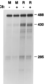
Activation of PKC induces HPV late gene expression in vitro. CIN612 cells can be induced to synthesize HPV-31b virions when they are grown in raft cultures treated with acti-vators of PKC. This treatment has also been shown to induce expression of differentiation-dependent cellular genes such as K10 and filaggrin (45). To investigate the mechanisms by which HPV late genes respond to activators of PKC, CIN612 cells were examined following growth in monolayer or raft cultures or in raft cultures treated with TPA or with C8, a diacylglycerol which activates PKC (10). We have previously used monolayer cultures as models of proliferating basal cells and stratified raft cultures to represent differentiated cells (33). Northern blot analysis with probes specific for the L1 or L2 ORF showed that expression of the late genes was undetectable in monolayer cultures, was slightly induced when the cells were grown in raft cultures, and was strongly induced when raft cultures were treated with C8 (Fig. 1). Identical results were seen with TPA-treated cultures (data not shown). As shown in Fig. 1, the late region of HPV-31b encodes two differentiation-dependent transcripts: a 4.7-kb RNA which hybridized to both L2 and L1, and a 2.3-kb RNA which is unique to L1.
[image:2.612.349.521.66.218.2]Structure of the 2.3-kb L1 transcript.We have shown pre-viously that a transcript encoding the E1∧E4 and E5 proteins is initiated at P742, a differentiation-dependent promoter in the E7 ORF (33). Since expression of the late genes is also differentiation dependent, and many HPV transcripts share
FIG. 1. Activation of PKC induces HPV late gene expression. Polyadeny-lated RNA was isoPolyadeny-lated from CIN612 cells grown in monolayers (M), in raft cultures (R), or in raft cultures treated with C8 to activate PKC. The presence or absence of C8 is indicated1or2. RNAs were electrophoretically separated on agarose-formaldehyde gels, transferred to GeneScreen, and hybridized to probes specific for the L1 (left) or L2 (right) ORF. Sizes are indicated in kilobases.
on November 9, 2019 by guest
http://jvi.asm.org/
common promoters (15, 20, 33, 54, 55), we attempted to iden-tify an L1 transcript initiated at this promoter in RNA from PKC-activated raft cultures. For these experiments, we used RNA from raft cultures treated with TPA. Using an upstream primer from E7 (nt 763 to 779) and a downstream primer in L1 (nt 5760 to 5737), a specific product was amplified by RT-PCR. This product was cloned, sequenced, and found to encode the E1∧E4 ORF in addition to L1 (Fig. 2). The splice junctions used to generate the E1∧E4 fusion protein are the same as those present in other HPV transcripts which we have previ-ously described (nt 877 joined to nt 3295) and are similar to those reported in other HPV strains (15, 20, 33, 47, 54, 55). A splice acceptor at the 59 end of the L1 ORF (nt 5552) was found to be joined to a donor at the end of E4 (nt 3590) to generate a bicistronic RNA with a 15-nt spacer between ORFs. The sequences at the splice junctions conform to consensus donor and acceptor splice site sequences. We were also able to amplify a PCR product consistent in size with an E1∧E4, L1 transcript containing the whole L1 ORF by using a down-stream primer from the 39 end of L1 (data not shown). The structure of this RNA is very similar to that of the L1 transcript expressed in xenograft cultures of the low-risk HPV-11 (15, 55).
The 2.3-kb L1 transcript is initiated at P742.While ampli-fication of the L1 transcript with a primer located in E7 sug-gested that the RNA was likely to be initiated at the differen-tiation-dependent promoter P742, this methodology could not exclude the possibility that the transcript actually initiated fur-ther upstream. Primer extension analysis to identify the 59end of this transcript was not technically feasible because of the low level of L1 transcripts and because the size of an L1-specific product, more than 460 bases, is much larger than can be reliably achieved by primer extension. To provide further evi-dence that the L1 transcript actually initiated at P742, an RNase protection assay was performed with an E1∧E4, L1 cDNA probe containing 59 sequences up to and beyond the promoter at P742 (Fig. 3C). An E1∧E4, L1 transcript initiated at P742 would protect 639 bases of this probe, while a tran-script initiated upstream would protect a larger region. A band
[image:3.612.61.302.67.217.2]of 639 bases was found to be protected by RNA from CIN612 raft cultures treated with TPA (Fig. 3A, lane 5). This 639-base protected fragment was present at very low levels in untreated raft cultures (lane 4) and was absent in monolayers (lane 3). An additional fragment of 697 bases was detected at low levels in RNA isolated from both monolayer and raft cultures. This fragment was derived from a transcript which protects all of the HPV sequences in the probe. It did not result from incom-plete digestion of the probe since additional plasmid sequences which were present in the probe were not protected. Thus, in addition to the E1∧E4, L1 transcript initiated at P742, there is
FIG. 2. Structure of the HPV genome and the 2.3-kb L1 transcript. HPV ORFs are indicated by black boxes. The early and late region polyadenylation sites are indicated as AEand AL, respectively. L1 RNA was amplified by
[image:3.612.320.557.211.618.2]RT-PCR from RNA isolated from CIN612 cells grown in raft cultures treated with TPA. The PCR products were cloned and sequenced. The positions of the primers are indicated by arrowheads. The 59end of the upstream primer is at nt 763; the 59end of the downstream primer is nt 5760. The RNA has the potential to encode an E1∧E4 fusion protein in addition to L1. The E1∧E4 splice, which is present in other HPV transcripts, joins nt 877 to nt 3295. A splice donor at nt 3590 is joined to a splice acceptor at the start of the L1 ORF at nt 5552. The E1∧E4, E5 RNA initiated at P742 is shown for comparison.
FIG. 3. The 2.3-kb late RNA is initiated at P742. (A) RNase protection assay of RNA isolated from untreated monolayers (M), untreated raft cultures (R), or raft cultures treated with TPA to induce PKC. PKC induction is indicated by1 or2. S, standards (HpaII digest of Bluescript); P, probe; C, control showing probe fragments protected by tRNA. The regions of the probe protected by different transcripts are depicted schematically. The promoters used by different transcripts are shown in parentheses. The figure shows only the upper portion of the gel. (B) Shorter exposure of the entire gel in panel A showing probe frag-ments protected by early region transcripts. (C) Schematic of the E1∧E4, L1 transcript and of the E1∧E4, L1 cDNA probe used in the RNase protection assay. The probe contains 58 bases of sequences from E7 upstream of P742 in addition to the entire E1∧E4 ORF, 209 bases of L1, and 43 bases of PCR-Script sequences. Sizes are indicated in bases.
on November 9, 2019 by guest
http://jvi.asm.org/
low-level expression of an E1∧E4, L1 transcript which is initi-ated upstream of P742. This transcript is expressed in both monolayer and raft cultures. Since P97 is not induced by dif-ferentiation or by activation of PKC (33) (see Fig. 3B and 4), we believe that the constitutively expressed E1∧E4, L1 tran-script is initiated at P97, the early region promoter. The abun-dance of this transcript was also slightly increased by treatment of raft cultures with TPA (Fig. 3A, lane 5), and, in some experiments, by PKC activation in monolayer cells (data not shown).
P742 is not induced by activation of PKC.Because of the overlapping nature of HPV transcripts, portions of the E1∧E4, L1 cDNA probe used to map the 59end of the L1 transcript also protected several early region transcripts. In raft cultures, these early transcripts were much more abundant than late region RNAs. This finding is consistent with immunohisto-chemical analyses that demonstrated L1 synthesis in only a small number of E1∧E4-positive cells (52). This differential expression is seen in Fig. 3B, which provides a shorter exposure of the gel shown in Fig. 3A. The E6, E7, E1∧E4, E5 and the E6*, E7, E1∧E4, E5 RNAs initiated at P97 protected a frag-ment of 488 bases and were unchanged in abundance by growth in raft cultures or by PKC activation in raft cultures (Fig. 3B). Additional fragments of 295 and 193 bases were also protected by transcripts which are capable of encoding E2 and E2-C, a truncated form of E2 expressed as a fusion protein with a portion of E1 (32b, 54). These E2 RNAs, however, were not induced by treatment with TPA (Fig. 3) or C8 (Fig. 4). The E2 transcript is also likely to be initiated at P97 (15, 32b). Thus, the abundance of transcripts initiated at P97 is not affected either by differentiation in untreated raft cultures or by PKC activation in raft cultures. In contrast, the E1∧E4, E5 RNA initiated at P742, which protected a fragment 430 bases in length, was induced by growth in raft cultures (Fig. 3B, lane 4) but was not further induced in raft cultures by activation of
protein kinase C (lane 5). The low-level expression of P742 observed in monolayer cells is due to a small number (less than 1%) of cells which spontaneously activate E1∧E4 expression (52).
It was next important to determine if activation of PKC alone was sufficient to increase expression of transcripts initi-ated at the P742 promoter. Using RNase protection assays, we examined the relative levels of expression of early region tran-scripts initiated at the P742 promoter in monolayers and raft cultures with or without treatment with C8 to activate PKC. As shown in Fig. 4, no increase in the level of expression of the E1∧E4, E5 transcript from P742, which protects a fragment of 430 bases, was observed following treatment of monolayer cells with C8. Similarly, no expression of the E1∧E4, L1 transcript initiated at P742 was observed in C8-treated monolayer cells (data not shown). Growth of CIN612 cells in raft cultures, however, substantially increased the abundance of the E1∧E4, E5 RNA initiated at P742, as previously reported (33). As shown in Fig. 3 and 4, expression of this early region transcript was not further augmented in raft cultures by activation of PKC. Thus, activation of PKC in raft cultures increases the abundance of late region transcripts initiated at P742 (Fig. 3) but has no effect on the abundance of early region transcripts initiated at this promoter (Fig. 3 and 4). This finding suggests that rather than increasing the rate of transcription from P742, activation of PKC induces posttranscriptional changes in late viral RNA stability or processing which result in accumulation of transcripts spliced at nt 3590. Polyadenylation of these tran-scripts occurs through sequences located at the end of the late region. These posttranscriptional changes result in an increase in the abundance of the L1 transcript expressed from P742.
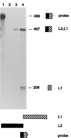
Structure of the L2 transcript.Northern blot analysis indi-cated that a 4.7-kb RNA hybridized to probes specific for both L1 and L2, and we sought to determine the structure of this transcript. Expression of this transcript was dependent upon both growth in raft cultures and treatment with activators of PKC. The 4.7-kb transcript was the only L2 transcript observed by Northern analysis and was large enough to encode the entire ORFs of both L1 and L2. To confirm that this L2 transcript extended into the L1 ORF, an RNase protection assay was performed with a probe spanning the L1/L2 over-lapping region. This region also contained the L1 splice junc-tion for the 2.3-kb message. A fragment of 208 bases, consis-tent in size with an RNA spliced at the L1 splice junction, was observed in RNA from TPA-treated raft cultures (Fig. 5) and in RNA from C8-treated raft cultures (data not shown). An additional protected fragment of 407 bases derived from a transcript which protects all the HPV sequences in the probe was also observed. This transcript was present in both mono-layer and TPA-treated raft cultures but was significantly more abundant in the latter. We conclude that the 4.7-kb transcript containing L2 extends into the L1 ORF.
[image:4.612.133.227.70.244.2]Since the expression of the 4.7-kb L2, L1 transcript in-creased upon differentiation, we reasoned that this transcript might also be initiated at the differentiation-dependent P742 promoter. Using an upstream primer located in the E7 ORF (59 nt 763) and a downstream primer in the L2 ORF, we successfully amplified an L2 transcript initiated in the early region by RT-PCR. Cloning and sequence analysis revealed that the 59end of this L2 transcript contained the same E1∧E4 splice junctions present in early region transcripts as well as in the L1 RNA (Fig. 6). However, the splice junction at nt 3590 which is used to generate the 2.3-kb E1∧E4, L1 transcript was not utilized in the E1∧E4, E5, L2 mRNA. Expression of L2 therefore requires read-through from E5 through the early region polyadenylation sequence and into the L2 ORF. This
FIG. 4. Early region transcripts initiated at P742 are not induced by activa-tion of PKC. RNA from monolayers (M), monolayers treated with C8, raft cultures (R), and raft cultures treated with C8 was analyzed by RNase protection assay using the E1∧E4, L1 cDNA probe used for Fig. 3. The presence or absence of C8 is indicated by1or2. Protected products are the same as those shown in Fig. 3. Only the products protected by the early region transcripts are detectable in this exposure. The E6, E7, E1∧E4, E5 transcripts initiated at P97 protected a fragment of 488 bases. As shown here and in Fig. 3, the abundance of this RNA was not changed by differentiation or by activation of PKC. The E1∧E4, E5 transcript initiated at P742, which protected a fragment of 430 bases, was induced by differentiation in raft cultures but not by treatment of monolayer cells with C8. Addition of C8 to raft cultures did not further increase the abundance of the E1∧E4, E5 RNA. Sizes are indicated in bases.
on November 9, 2019 by guest
http://jvi.asm.org/
L2 transcript also encodes the L1 ORF and is most likely polyadenylated by sequences located at the end of the late region. Since the L2 and L1 ORFs are slightly overlapping, however, it is unlikely that L1 would be efficiently translated from this RNA. Transcripts encoding both L2 and L1 have also been identified in xenograft HPV-11 cultures (15).
The major L2 transcript is initiated in the early region. While the foregoing studies demonstrated that L2 transcripts were initiated in the early region, they did not identify the initiation site. Identification of the L2 initiation site by RNase
protection assays was not feasible since an L2-specific probe extending beyond P742 would be in excess of 1,200 bases in length, which is larger than is achievable for RNase protection assays. To examine potential initiation sites, RT-PCR was per-formed with a 39 primer in L2 and a 59 primer located up-stream of P742 (nt 608 to 628) or downup-stream of P742 (nt 763 to 779). As described above, L2 transcripts could be amplified with 59 primer 763-779 but could not be amplified with a 59 primer located upstream of P742 at nt 628 (primer 608-628) (data not shown). As a positive control, we performed an RT-PCR with primer 608-628 and a downstream primer in the E4 gene. We successfully amplified early region transcripts with these primers, indicating that primer 608-628 is functional in these assays (data not shown). We were also unable to amplify L2 transcripts by using an upstream primer located near P97 (nt 108 to 129), although this primer was functional in control PCRs. Thus, the 59end of the major L2, L1 tran-script, like the E1∧E4, L1 transcript, is likely to be located between nt 628 and 763. We were unable to amplify the E1∧E4, L1 or the E1∧E4, E5, L2, L1 transcripts initiated at P97 by using either primer 608-628 or primer 108-129 as the upstream primer, perhaps because of their low levels of ex-pression.
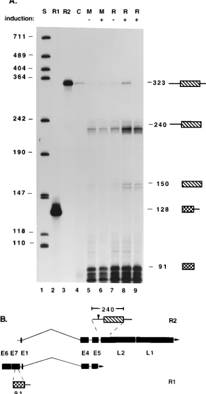
Heteroduplex analysis of RNA from xenograft cultures of HPV-11 suggested that L2, L1 transcripts might be initiated at the beginning of the L2 ORF (15). To determine whether additional L2 transcripts were initiated in the late region, an RNase protection assay was performed with a probe extending from the end of the early region into the L2 ORF (R2; Fig. 7B). L2 transcripts initiated further upstream in the early re-gion would protect all 240 bases of the HPV-specific sequences in the probe, while transcripts initiated at the beginning of L2 would protect only the 150-nt L2-specific portion of the probe. As an internal control, a probe which would be protected by transcripts containing E7 sequences (R1; Fig. 7B) was also included in the assays. As shown previously, these E7 tran-scripts are initiated at P97, and their abundance is not affected by differentiation in raft cultures or by activation of PKC (33) (Fig. 3B and 4). As seen in Fig. 7A, the majority of L2 tran-scripts protected all viral sequences in the probe and were present in both monolayer and raft cultures. However, like the L1 transcripts, the L2 transcripts were much more abundant in raft cultures treated with either TPA (lane 8) or C8 (lane 9). Differentiation in raft cultures alone was not sufficient to in-duce expression of this L2 transcript (lane 7). Similarly, addi-tion of activators of PKC to monolayer cultures failed to in-duce significant L2 expression (lane 6). The major protected species were heterogeneous and slightly smaller than expected, most likely because of artifactual digestion at the ends of the RNA-RNA hybrids. In addition, low-level expression of an RNA consistent in size with an L2 transcript initiated or spliced at the beginning of the L2 ORF was detected in both monolayer and raft cultures. Taken together, our studies indi-cate that the primary L2 transcript also contains L1 and, like the E1∧E4, L1 transcript, is likely to be initiated at P742. Since these transcripts contain the early region polyadenylation site, PKC-mediated induction of L2 expression occurs through changes in utilization of polyadenylation sites and/or changes in stability of late region transcripts.
DISCUSSION
In HPV-infected basal cells, viral gene expression is limited to the early region, and viral DNA is maintained as an episome at low copy number. Amplification of viral DNA and expres-sion of the late genes occurs only in the terminally
[image:5.612.118.241.67.306.2]differenti-FIG. 5. RNase protection analysis of the L1 splice junction. RNAs from monolayers (lane 3) or TPA-treated raft cultures (lane 4) were hybridized to an antisense RNA probe spanning the L1 splice junction. The probe is depicted schematically at the bottom. E1∧E4, L1 RNA protects only the 208-base L1-specific part of the probe. RNAs extending from L2 into L1 protect all of the 407-base HPV-specific sequences in the probe. Both transcripts were present at low levels in undifferentiated monolayer cultures but were induced when cells were allowed to differentiate in raft cultures. Lane 1, undigested probe. The probe contains 83 bases of plasmid sequences in addition to HPV positions 5760 to 5353. Lane 2, products protected by tRNA. Sizes are indicated in bases.
FIG. 6. Structure of the L2 RNA. L2 transcripts were amplified by RT-PCR from RNA isolated from TPA-treated raft cultures. PCR products were cloned and sequenced. Arrowheads indicate the positions of the primers. The probable structure of the 4.7-kb L2 RNA detected by Northern blot hybridization is shown with that of the E1∧E4, E5 and E1∧E4, L1 transcripts. The positions of the early and late region polyadenylation sites are indicated as AEand AL, respectively.
on November 9, 2019 by guest
http://jvi.asm.org/
[image:5.612.58.299.539.675.2]ated layers of the epithelium, where viral particles are assem-bled and shed. In this study, we have demonstrated that the majority of the HPV-31b L1 transcripts are expressed as a bicistronic 2.3-kb E1∧E4, L1 RNA initiated at P742, a
differ-entiation-dependent promoter located in the middle of the E7 ORF. We have previously shown that the E1∧E4, E5 transcript is also expressed from this differentiation-dependent promoter (33). While differentiation-dependent activation of the P742 promoter is required for high-level E1∧E4, L1 expression, it is not sufficient. Posttranscriptional changes are also required for late gene expression, and in the raft system these are depen-dent on activation of PKC. Expression of L1 from P742 re-quires changes either in utilization of both splicing and poly-adenylation sites or in the stability of RNAs.
We have also identified differentiation-dependent and con-stitutively expressed 4.7-kb L2, L1 transcripts. While we have not conclusively identified the L2, L1 initiation site, we have shown that the majority of L2, L1 transcripts are initiated in the early region. We have shown that, like the 2.3-kb E1∧E4, L1 RNA, induction of L2, L1 RNA requires both growth in raft cultures and activation of PKC. It is therefore likely that, like the E1∧E4, L1 RNAs, the differentiation-dependent tran-scripts encoding L2 and L1 are initiated at P742, while the constitutively expressed transcripts are initiated at P97. Since our RNase protection assays were done with total cell RNA, some of the constitutively expressed transcripts containing L2 sequences could be derived from nuclear precursor RNAs. Transcripts containing L1 and L2 which are predominantly nuclear have been detected in the less differentiated cells be-low the granular layer by in situ hybridization. Analysis of HPV-11 xenograft cultures also identified L2 transcripts initi-ated at P742 but suggested that there might be an L2-specific promoter at the beginning of the L2 ORF (15). The L2 pro-moter could not be positively identified in those studies be-cause of the low level of L2 expression. Our data are consistent with initiation and/or splicing of some transcripts at the begin-ning of L2, but these transcripts are significantly less abundant than those initiated at P97 or P742. Although a polypyrimidine tract precedes the L2 ORF, there is no AG dinucleotide at the putative splice junction (29, 32a). If spliced L2 products exist, they would have to utilize a nonconsensus splice junction. The majority of the L2 transcripts do not arise from use of L2-specific splicing or initiation sites, however. The major L2 transcripts are initiated in the early region and extend through the early region polyadenylation site. Induction of L2 expres-sion therefore appears to be regulated primarily through changes in polyadenylation site usage. Thus, expression of both L1 and L2 is regulated at the level of initiation of transcription as well as through posttranscriptional mechanisms.
Among the papillomaviruses, the transcription pattern of HPV-31b L1 RNAs appears to be most similar to that of HPV-6 and HPV-11, which are low-risk genital-specific types. In these types, L1 is expressed from a bicistronic E1∧E4, L1 RNA (15, 55) whose initiation site has been mapped to the E7 ORF by heteroduplex analysis (15). In contrast, the L1 tran-scripts of HPV-8, which infects cutaneous epithelium, are ini-tiated in the upstream regulatory region (57). Two alterna-tively spliced L1 RNAs which contain sequences from E4 but apparently do not encode genes other than L1 were identified in HPV-8-infected cells. Similar L1 transcripts are present in bovine papillomavirus type 1, which induces fibropapillomas in cattle. The bovine papillomavirus type 1 L1 and L2 RNAs are expressed from a differentiation-dependent promoter in the upstream regulatory region, and changes in RNA processing may also be important in regulating expression of these tran-scripts (5, 26).
[image:6.612.74.279.76.468.2]Studies in HPV-16 have identified a negative regulatory element at the 39 end of the late region that affects RNA stability (35, 36). While these studies indicated that the ele-ment affects the stability of polyadenylated RNA, subsequent
FIG. 7. The major L2 transcript is initiated in the early region. (A) RNase protection assay of RNA isolated from CIN612 cells grown in monolayers (M; lanes 5 and 6) or raft cultures (R) with (lanes 8 and 9) or without (lane 7) induction of PKC. Addition of activators of PKC is indicated by1or2. PKC was induced in monolayer cells with C8 and in raft cultures with either TPA (lane 8) or C8 (lane 9). No differences in the transcripts induced by TPA and C8 were observed. Lane 1, standards (S; HpaII digest of Bluescript), lane 2, R1 probe used as internal control for the amount of RNA present; lane 3, R2 probe spanning the early region polyadenylation site and extending into L2; lane 4, negative control (C) showing probe fragments protected by tRNA. Some undi-gested R2 probe is present in lanes 4 to 8. Numbers at the left indicate sizes (in bases) of Bluescript molecular weight markers; numbers at the right indicate the sizes (in bases) of probe fragments protected by HPV transcripts. Protected regions are depicted at the right. (B) Schematic of the probes used in the RNase protection assay. The 128-nt R1 probe contains 91 bases of E7 sequences which are protected by E6, E7, E1∧E4, E5 transcripts initiated at P97 and 37 bases of plasmid sequences. The abundance of the transcript does not change upon differentiation or activation of PKC. The 323-nt R2 probe contains 83 bases of plasmid sequences in addition to 240 bases of HPV sequences extending from the 39untranslated portion of the early region, through the early region polyad-enylation site (arrowhead), and into the L2 ORF (striped box). L2 transcripts initiated in the early region protect all of the 240 HPV-specific sequences in the probe, while transcripts initiated at the beginning of L2 protect only the 150-nt L2-specific portion of the probe.
on November 9, 2019 by guest
http://jvi.asm.org/
analyses of late gene expression of bovine papillomavirus type 1 and HPV-16 have suggested that this element acts as a 59 splice site which prevents use of the late region polyadenyla-tion site (27), perhaps by interfering with exon definipolyadenyla-tion (53). Placement of this putative splice site downstream of the chlor-amphenicol acetyltransferase gene inhibited chlorchlor-amphenicol acetyltransferase expression in transient assays, while deletion of this site resulted in an increase in reporter expression (27), suggesting that this site binds a negative regulatory factor. The sequence CAGGTAAACG, which is very similar to the 59 splice site consensus sequence (C/A)AGGT(A/G)AGT, is present in HPV-31 at nt 6997 to 7008, approximately 225 bases upstream of the late polyadenylation site (29). A negative regulatory factor bound to this site might prevent use of the late region polyadenylation site. This putative splicing factor could be either a virally encoded factor or a differentiation-dependent cellular factor. In support of this model, there is increasing evidence that splicing factors may also be involved in polyadenylation. Mutation of the AAUAAA polyadenyla-tion site specifically depresses splicing of the final intron of chimeric minigenes in vitro (48). In addition, the presence of a 39splice site can stimulate polyadenylation in vitro (50), while insertion of a 59splice site can depress use of a polyadenylation site in vitro and in vivo (49). Furthermore, splicing factors such as the U1 small nuclear ribonucleoprotein A protein have been shown to interact directly with sequences required for efficient polyadenylation in simian virus 40 (39). This interaction is functionally significant, since abrogation of binding inhibits polyadenylation in vitro. It is possible that activation of PKC can cause inactivation or down-regulation of this factor and an increase in the efficiency of polyadenylation of late region mRNAs. Since polyadenylation is an important determinant in mRNA stability (8, 9), this would result in increased abun-dance of the late region transcripts. According to this model, late transcripts initiated at both P97 and P742 should increase in abundance upon activation of PKC. Some increase in the amount of E1∧E4, L1 transcripts initiated at P97 was observed in PKC-activated raft cultures (Fig. 3) and, in some experi-ments, in PKC-activated monolayer cultures as well.
Several examples of developmental or cell-type-specific reg-ulation of RNA processing have been previously described (reviewed in references 40 and 44). RNA processing of
Dro-sophila sex determination genes is controlled by gene-specific
positive or negative regulators of splice site selection (3, 40, 59). Similar mammalian factors have not yet been identified, but there is evidence for tissue-specific expression of factors that regulate exon usage (21), indicating that a common mech-anism may exist. Activation of PKC could therefore induce HPV-31b late gene expression by directly or indirectly altering the levels of gene-specific factors which regulate RNA process-ing. Alternatively, PKC could alter the relative abundance of general splicing factors. A family of evolutionarily conserved pre-mRNA splicing factors known as SR proteins have been identified (61, 62). Changes in the relative amounts of some SR proteins are known to alter splice site selection in vitro (22, 25, 41, 42), and tissue-specific differences in the relative concen-trations of splicing factors have been observed (62), suggesting that regulation of the relative concentrations of splicing factors could control differentiation-specific changes in splice site uti-lization. Rather than changing the abundance of factors in-volved in splicing, a third possibility is that PKC alters the activity of these factors through changes in their phosphoryla-tion state. Whatever the mode of acphosphoryla-tion of PKC, HPV late gene expression requires that these effects occur in conjunction with changes in the differentiation program of epithelial cells,
since little or no induction of late message was observed in monolayer cultures treated with PKC activators.
In summary, our studies demonstrate that HPV late gene expression is regulated both at the initiation of transcription as well as by posttranscriptional mechanisms. Differentiation-spe-cific transcription factors activate expression of the P742 pro-moter and, in conjunction with PKC-mediated changes in RNA processing or stabilization, result in the accumulation of late messages. Since papillomaviruses are likely to have evolved to exploit cellular factors to regulate expression of their genes, the characterization of the differentiation-depen-dent transcription factors regulating expression of P742 and of the PKC-dependent factors controlling posttranscriptional events is likely to provide insight into factors controlling dif-ferentiation-dependent expression of keratinocyte genes as well.
ACKNOWLEDGMENTS
We thank Mark Frattini for advice on RNA purification and anal-ysis, David Klumpp for assistance with computer graphics, and Pat Spear, Kathy Rundell, Elliot Androphy, and Dennis McCance for critical reading of the manuscript.
This work was supported by grants from the NCI (CA 59655) and NIAID (AI 34637).
REFERENCES
1. Androphy, E. J., N. L. Hubbert, J. T. Schiller, and D. R. Lowy. 1987. Identification of the HPV-16 E6 protein from transformed mouse cells and human cervical carcinoma cell lines. EMBO J. 6:989–992.
2. Asselineau, D., and M. Prunieras. 1984. Reconstruction of simplified skin— control of fabrication. Br. J. Dermatol. Suppl. 111:219–221.
3. Baker, B. S. 1989. Sex in flies: the splice of life. Nature (London) 340:521– 524.
4. Barbosa, M. S., and R. Schlegel. 1989. The E6 and E7 genes of HPV-18 are sufficient for inducing two-stage in vitro transformation of human keratino-cytes. Oncogene 4:1529–1532.
5. Barksdale, S. K., and C. C. Baker. 1993. Differentiation-specific expression from the bovine papillomavirus type 1 P2443and late promoters. J. Virol. 67:5605–5616.
6. Bedell, M. A., J. B. Hudson, T. R. Golub, M. E. Turyk, M. Hosken, G. D.
Wilbanks, and L. A. Laimins.1991. Amplification of human papillomavirus genomes in vitro is dependent on epithelial differentiation. J. Virol. 65:2254– 2260.
7. Bedell, M. A., K. H. Jones, S. R. Grossman, and L. A. Laimins. 1989. Identification of human papillomavirus type 18 transforming genes in im-mortalized and primary cells. J. Virol. 63:1247–1255.
8. Bernstein, P., S. W. Peltz, and J. Ross. 1989. The poly(A)-poly(A)-binding protein complex is a major determinant of mRNA stability in vitro. Mol. Cell. Biol. 9:659–670.
9. Bernstein, P., and J. Ross. 1989. Poly (A), poly(A) binding protein and the regulation of mRNA stability. Trends. Biochem. Sci. 14:373–377. 10. Boynton, A. L., J. F. Whitfield, and L. P. Kleine. 1983. Ca21
/phospholipid-dependent protein kinase activity correlates to the ability of transformed liver cells to proliferate in Ca21-deficient medium. Biochem. Biophys. Res. Commun. 115:383–390.
11. Broker, T. R., and M. Botchan. 1986. Papillomaviruses: retrospectives and perspectives. Cancer Cells 4:17–36.
12. Chaing, C. M., M. Ustav, A. Stenlund, T. Ho, T. R. Broker, and L. T. Chow. 1992. Viral E1 and E2 proteins support replication of homologous and heterologous papillomavirus origins. Proc. Natl. Acad. Sci. USA 89:5799– 5803.
13. Chin, M. T., R. Hirochika, H. Hirochika, T. R. Broker, and L. T. Chow. 1988. Regulation of human papillomavirus type 11 enhancer and E6 promoter by activating and repressing proteins from the E2 open reading frame: func-tional and biochemical studies. J. Virol. 62:2994–3002.
14. Chirgwin, J. M., A. E. Przybyla, R. J. MacDonald, and W. J. Rutter. 1979. Isolation of biologically active ribonucleic acid from sources enriched in ribonuclease. Biochemistry 18:5294–5299.
15. Chow, L. T., M. Nasseri, S. M. Wolinsky, and T. R. Broker. 1987. Human papillomavirus types 6 and 11 mRNAs from genital condylomata acuminata. J. Virol. 61:2581–2588.
16. Crum, C. P., G. Nuovo, D. Friedman, and S. J. Silverstein. 1988. Accumu-lation of RNA homologous to human papillomavirus type 16 open reading frames in genital precancers. J. Virol. 62:84–90.
17. deVilliers, E.-M. 1989. Heterogeneity of the human papillomavirus group. J. Virol. 63:4898–4903.
on November 9, 2019 by guest
http://jvi.asm.org/
18. Dollard, S. C., J. L. Wilson, L. M. Demeter, W. Bonnez, R. C. Reichman,
T. R. Broker, and L. T. Chow.1992. Production of human papillomavirus and modulation of the infectious program in epithelial raft cultures. Genes Dev.
6:1131–1142.
19. Doorbar, J., S. Ely, J. Sterling, C. McLean, and L. Crawford. 1991. Specific interaction between HPV-16 E1-E4 and cytokeratins results in collapse of the epithelial cell intermediate filament network. Nature (London) 352:824– 827.
20. Doorbar, J., A. Parton, K. Hartley, L. Banks, T. Crook, M. Stanley, and L.
Crawford.1990. Detection of novel splicing patterns in a HPV16-containing keratinocyte cell line. Virology 178:254–262.
21. Emeson, R. B., F. Hedjran, J. M. Yeakley, J. W. Guise, and M. G. Rosenfeld. 1989. Alternative production of calcitonin and CGRP mRNA is regulated at the calcitonin-specific splice acceptor. Nature (London) 341:76–80. 22. Eperon, I. C., D. C. Ireland, R. A. Smith, A. Mayeda, and A. R. Krainer.
1993. Pathways for selection of 59splice sites by U1 snRNPs and SF2/ASF. EMBO J. 12:3607–3617.
23. Frattini, M. G., and L. A. Laimins. 1994. Binding of the human papilloma-virus E1 origin-recognition protein is regulated through complex formation with the E2 enhancer-binding protein. Proc. Natl. Acad. Sci. USA 91:12398– 12402.
24. Frattini, M. G., and L. A. Laimins. 1994. The role of the E1 and E2 proteins in the replication of human papillomavirus type 31b. Virology 204:799–804. 25. Fu, X.-D., A. Mayeda, T. Maniatis, and A. R. Krainer. 1992. General splicing factors SF2 and SC35 have equivalent activities in vitro and both affect 59and 39splice site selection. Proc. Natl. Acad. Sci. USA 89:11224–11228. 26. Furth, P. A., and C. C. Baker. 1991. An element in the bovine papillomavirus
late 39untranslated region reduces polyadenylated cytoplasmic RNA levels. J. Virol. 65:5806–5812.
27. Furth, P. A., W.-T. Choe, J. H. Rex, J. C. Byrne, and C. C. Baker. 1994. Sequences homologous to 59splice sites are required for the inhibitory activity of papillomavirus late 39untranslated regions. Mol. Cell. Biol. 14: 5278–5289.
28. Gilman, M. 1989. Ribonuclease protection assay, p. 4.7.1–4.7.8. In F. M. Ausubel, R. Brent, R. E. Kingston, D. D. Moore, J. G. Seidman, J. A. Smith, and K. Struhl (ed.), Current protocols in molecular biology. John Wiley & Sons, New York.
29. Goldsborough, M. D., D. DiSilvestre, G. F. Temple, and A. T. Lorincz. 1989. Nucleotide sequence of human papillomavirus type 31: a cervical neoplasia-associated virus. Virology 171:306–311.
30. Higgins, G. D., D. M. Uzelin, G. E. Phillips, P. McEvoy, R. Marin, and C. J.
Burrell.1992. Transcription patterns of human papillomavirus type 16 in genital intraepithelial neoplasia: evidence for promoter usage within the E7 open reading frame during epithelial differentiation. J. Gen. Virol. 73:2047– 2057.
31. Howley, P. M. 1991. Papillomavirinae and their replication, p. 743–768. In B. N. Fields and D. M. Knipe (ed.), Fundamental virology, vol. 2. Raven Press, New York.
32. Hudson, J. B., M. A. Bedell, D. J. McCance, and L. A. Laimins. 1990. Immortalization and altered differentiation of human keratinocytes in vitro by the E6 and E7 open reading frames of human papillomavirus type 18. J. Virol. 64:519–526.
32a.Hummel, M. Unpublished data. 32b.Hummel, M., et al. Unpublished data.
33. Hummel, M., J. B. Hudson, and L. A. Laimins. 1992. Differentiation-induced and constitutive transcription of human papillomavirus type 31b in cell lines containing viral episomes. J. Virol. 66:6070–6080.
34. Kawasaki, E. S. 1990. Amplification of RNA, p. 21–27. In M. A. Innis, D. H. Gelfand, J. J. Sninsky, and T. J. White (ed.), PCR protocols: a guide to methods and materials. Academic Press, Inc., New York.
35. Kennedy, I. M., J. K. Haddow, and J. B. Clements. 1990. Analysis of human papillomavirus type 16 late mRNA 39processing signals in vitro and in vivo. J. Virol. 64:1825–1829.
36. Kennedy, I. M., J. K. Haddow, and J. B. Clements. 1991. A negative regu-latory element in the human papillomavirus type 16 genome acts at the level of late mRNA stability. J. Virol. 65:2093–2097.
37. Laimins, L. A. 1993. The biology of human papillomaviruses: from warts to cancer. Infect Agents Dis. 2:74–86.
38. Lorincz, A. T., W. D. Lancaster, and G. F. Temple. 1986. Cloning and characterization of the DNA of a new human papillomavirus from a woman with dysplasia of the uterine cervix. J. Virol. 58:225–229.
39. Lutz, C. S., and J. C. Alwine. 1994. Direct interaction of the U1 snRNP-A protein with the upstream efficiency element of the SV40 late polyadenyla-tion signal. Genes Dev. 8:576–586.
40. Maniatis, T. 1991. Mechanisms of alternative pre-mRNA splicing. Science
251:33–34.
41. Mayeda, A., D. M. Helfman, and A. R. Krainer. 1993. Modulation of exon skipping and inclusion by heterogeneous nuclear ribonucleoprotein A1 and pre-mRNA splicing factor SF2/ASF. Mol. Cell. Biol. 13:2993–3001. 42. Mayeda, A., and A. R. Krainer. 1992. Regulation of alternative pre-mRNA
splicing by hnRNP A1 and splicing factor SF2. Cell 68:365–375. 43. McCance, D. J., R. Kopan, E. Fuchs, and L. A. Laimins. 1988. Human
papillomavirus type 16 alters human epithelial cell differentiation in vitro. Proc. Natl. Acad. Sci. USA 85:7169–7173.
44. McKeown, M. 1992. Alternative mRNA splicing. Annu. Rev. Cell Biol.
8:133–155.
45. Meyers, C., M. G. Frattini, J. B. Hudson, and L. A. Laimins. 1992. Biosyn-thesis of human papillomavirus from a continuous cell line upon epithelial differentiation. Science 257:971–973.
46. Munger, K., W. C. Phelps, V. Bubb, P. M. Howley, and R. Schlegel. 1989. The E6 and E7 genes of the human papillomavirus type 16 together are necessary and sufficient for transformation of primary human keratinocytes. J. Virol.
63:4417–4421.
47. Nasseri, M., R. Hirochika, T. R. Broker, and L. T. Chow. 1987. A human papillomavirus type 11 transcript encoding an E1∧E4 protein. Virology 159: 433–439.
48. Niwa, M., and S. M. Berget. 1991. Mutation of the AAUAAA polyadenyl-ation signal depresses in vitro splicing of proximal but not distal introns. Genes Dev. 5:2086–2095.
49. Niwa, M., C. C. MacDonald, and S. M. Berget. 1992. Are vertebrate exons scanned during splice site selection? Nature (London) 360:277–280. 50. Niwa, M., S. D. Rose, and S. M. Berget. 1990. In vitro polyadenylation is
stimulated by the presence of an upstream intron. Genes Dev. 4:1552–1559. 51. Phelps, W. C., C. L. Yee, K. Munger, and P. M. Howley. 1988. The human papillomavirus type 16 E7 gene encodes transactivation and transformation functions similar to those of adenovirus E1A. Cell 53:539–547.
52. Pray, T., and L. A. Laimins. 1995. Differentiation-dependent expression of E∧E4 proteins in cell lines maintaining episomes of human papillomavirus type 31b. Virology 206:679–685.
53. Robberson, B. L., G. J. Cote, and S. M. Berget. 1990. Exon definition may facilitate splice site selection in RNAs with multiple exons. Mol. Cell. Biol.
10:84–94.
54. Rohlfs, M., S. Winkenbach, S. Meyer, T. Rupp, and M. Durst. 1991. Viral transcription in human keratinocyte cell lines immortalized by human pap-illomavirus type-16. Virology 183:331–342.
55. Rotenberg, M. O., L. T. Chow, and T. R. Broker. 1989. Characterization of rare human papillomavirus type 11 mRNAs coding for regulatory and struc-tural proteins, using the polymerase chain reaction. Virology 172:489–497. 56. Stoler, M. H., S. M. Wolinsky, A. Whitbeck, T. R. Broker, and L. T. Chow. 1989. Differentiation-linked human papillomavirus types 6 and 11 transcrip-tion in genital condylomata revealed by in situ hybridizatranscrip-tion with message-specific RNA probes. Virology 172:331–340.
57. Stubenrauch, F., J. Malejczyk, P. G. Fuchs, and H. Pfister. 1992. Late promoter of human papillomavirus type 8 and its regulation. J. Virol. 66: 3485–3493.
58. Thierry, F., and M. Yaniv. 1987. The BPV-1 E2 trans-acting protein can be either an activator or a repressor of the HPV18 regulatory region. EMBO J.
6:3391–3397.
59. Tian, M., and T. Maniatis. 1992. Positive control of pre-mRNA splicing in vitro. Science 256:237–240.
60. Ustav, M., and A. Stenlund. 1991. Transient replication of BPV-1 requires two viral polypeptides encoded by the E1 and E2 open reading frames. EMBO J. 10:449–457.
61. Zahler, A. M., W. S. Lane, J. A. Stolk, and M. B. Roth. 1992. SR proteins: a conserved family of pre-mRNA splicing factors. Genes Dev. 6:837–847. 62. Zahler, A. M., K. M. Neugebauer, W. S. Lane, and M. B. Roth. 1993. Distinct
functions of SR proteins in alternative pre-mRNA splicing. Science 260:219– 222.
63. zur Hausen, H., and A. Schneider. 1987. The role of papillomaviruses in human anogenital cancer, p. 245–263. In N. D. Salzman and P. M. Howley (ed.), The Papovaviridae. Plenum Press, New York.