JOURNAL OF VIROLOGY, Sept. 1994, p. 5556-5567 Vol.68, No.9 0022-538X/94/$04.00+0
Copyright © 1994,American Society for Microbiology
Ectopic
Expression of Gamma Interferon in the Eye Protects
Transgenic Mice
from
Intraocular
Herpes
Simplex Virus
Type
1
Infectionst
KATHRIN
GEIGER,'
EDWARD L.HOWES,2ANDNORASARVETNICKI*DepartmentofNeuropharmacology, The ScrippsResearch Institute, LaJolla, Califomia 92037,1 and
Department of
AnatomicPathology, University of
Califomia,
SanFrancisco,
Califomia
941102Received 31 March 1994/Accepted 8 June 1994
Transgenic (rhoy) miceprovide a model for studying the influence ofgammainterferon (IFN--y) produced in the eye on ocular and cerebral viral infection. To establish this model, we injected BALB/c- and C57BL/6-derived transgenic and nontransgenic miceofdifferentages intravitreallywithherpes simplexvirus type 1 (HSV-1) strain F. Eye andbrain tissuesofthese micewere assessed forpathological and immunocy-tochemicalchanges. HSV-1 infection induced severe retinitis of the injected eyes and infection of thebrain in all mice. In transgenic mice inoculated withHSV-1, the left, nontreated eyes were protectedfrom retinitis, whereas nontransgenic mice developed bilateral retinitis.Additional intravitreal injection of IFN-,ywith the virus protected the noninoculated eyes of nontransgenic mice. Three-week-old nontransgenic mice diedfrom HSV-1 infection, whereas transgenic mice of the same age andnontransgenic mice intravitreally treated with IFN-y survived. Ocular IFN--y production increased the extent ofinflammation intransgenic micebutdid not havea significant influence on the growth ofHSV-1 until day 3 after inoculation and didnot influence the neuroinvasionof thisvirus. Thus, the
etfects
ofIFN--y were not caused byanearlyblock of viralreplication. Possiblemechanisms ofIFN-yaction include activation of the immune response, alteration oftheproperties ofthevirus, and directprotection of neurons.Herpessimplex virus type 1 (HSV-1) infection of the human eye causes recurrentkeratoconjunctivitis, frequently resulting in blindness (33a). HSV-1 can also induce acute necrotizing retinitis and encephalitis in immunocompetent and immuno-suppressed patients (13, 29). However, immunodeficient indi-viduals with HIV infection have a higher risk of developing
herpeticinfection of the retina and the brain. Severe forms of
the AIDS-dementia complex might be associated with second-ary infection of the brains with several viruses of the herpes family, mainly cytomegalovirus (12, 31, 41, 42).Itisnotknown how these virusesenterthe brain and cause chronic infection. The eye, including theretina, and the brain shareanimmune privilege featuring an efficient blood-tissue barrier and the absence ofmajor histocompatibility complex (MHC) expres-sion under normal conditions (21,40, 48,52,53).However, it hasbeen shown thatmostallografts are ultimately rejected in the brain(48),asopposedto thesituation in the eye(55-57). Thus,theimmuneprivilege might be more extendedinthe eye, since there isastrongactivesuppression of cellular infiltration and ofdelayed-type hypersensitivity to alloantigens presented inthe eye, which is mediated by high amounts of
immunosup-pressive factors produced in the eye, such as transforming
growth factor beta and ot-melanocyte-stimulating hormone
(54). Incontrast to the brain (11), even peripherally induced
delayed-type hypersensitivity is abrogated after intraocular exposure with the sameantigen (51). Possibly, this impairment of the cellular immune response to viral infection influences the outcome of intraocular viral infection and facilitates the
*Corresponding author. Mailing address: Department of
Neuro-pharmacology, CVN 10, The Scripps Research Institute, 10666 N. Torrey Pines Rd., La Jolla, CA 92037. Phone: (619) 554 7066. Fax:
(619)
554 6477.tManuscript8557-NP fromTheScripps Research Institute.
entrance of virusestothe brainvia the eye asopposedtothe peripheralroute.
Gamma interferon
(IFN-y)
is likely to be an importantfactor influencingthe intraocular milieu. Thiscytokine
proba-bly reverses immunosuppression of the eye by antagonizing transforming growth factor beta, amajor immunosuppressant present in the intraocular fluids (8), and thus is capable of influencing the intraocular microenvironment (14). Further-more, IFN-,y hasits own antiviral properties, among which is its impact on viral replication (18, 23, 37). Some studies suggested that the effects of IFN-,y might not be entirely favorable during corneal HSV-1 infection, since thecytokine seems toincrease tissue damage(18).
We have developed a transgenic murine model in which IFN-^yexpression overwhelms the immunosuppressive proper-ties of the intraocular compartment. Withapparently normal development of eyes and brain, these rhoy transgenic mice expressIFN--y in the retinalphotoreceptorsfrom thefirst week oflifetothe age of8 to 9weeks. Rho-y mice show alterations of the retinaby2 to3 weeks of age,beginningwithshortening
of the photoreceptor outer segments, and subsequently lose theirphotoreceptorsatthe age of8 to 9weeks.Additionally, these animals develop cataracts and exhibit upregulation of both MHC class I and class II and increased infiltration of macrophages. Infiltration ofTcellswasnotsignificantinthese mice.Notransgeneexpressionwasdetected inthe brain (14). We infected 4- to 6-week-old transgenic mice, which still have photoreceptors producing IFN-y, as well as
nontrans-genicmice of differentgeneticbackgroundswith HSV-1 strain
F (44). We used intravitreal injection because this route of infection facilitates thedirect contact betweenvirus and retina and promotes the spread of infection to the brain (3). To eliminate the effects of systemic IFN-y production, we also infected 3-week-old mice whicharereportedtohave aspecific
developmental lack of IFN-y production (1). We found that
5556
on November 9, 2019 by guest
http://jvi.asm.org/
IFN-y
PROTECTS MICE FROM INTRAOCULAR HSV-1 INFECTION 5557A
*,
-wo
70to
tA4
r%> - @
C
r *' *.a
*rc t
-*;'44 W n > < ''U' 0 z { s; }
W
>
,
+*;
viX +
^.sssn-^b~~~~~~~~~~~~.
<
3 .LW*~~~~~~~~~~~~~~~~~~~~~~~~~~~~1a -. AAo
'to
* i
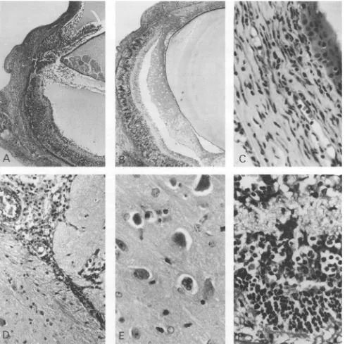
FIG. 1. Virus-inducedpathologyin eyesand brains of 5-to6-week-old miceby day6after inoculation of HSV-1.
(A)
Rhoymouse,C57BL/6 derived,infected with HSV-1 strain F. Therighteyedisplaysanincrease of cells in thecornea,the iris,theciliary
body,
and the retina. Note free mononuclear cells in the anterior chamber andvitreous and exudate in both compartments.Remnants of the inoculum arerecognizable
in thesubretinal space. PAS; magnification, x48.(B)Nontransgenic mouse,BALB/c derived,infected withHSV-1. Inthe
right
eye, infiltration of all tissues and massive retinal destructionareclear.Notetherelativelyfewinfiltrating
inflammatory
cellscompared
withpanel
A.PAS;magnification,
x48.(C) Detail of thecorneafrom thenontransgenicmouseinpanelBshowingcellularinfiltration of the wholecornea.PAS;magnification,
x480.(D)Detail of the brain fromrho-ymouseinpanelAwithmononuclear infiltration around the choroidplexus.PAS;
magnification,
x240.(E)
Detail of the brain from thesame mouseshowingswollenlargeneuronsandcellghosts.PAS;magnification,x480.(F)
Detail of the retinafrom therhoymouseinpanelA. Massive destruction of the retinalarchitecture is apparent,asisinfiltration of the whole retina and
underlying
choroid withinflammatorycells. PAS;magnification, x480.
VOL.68, 1994
on November 9, 2019 by guest
http://jvi.asm.org/
[image:2.612.67.557.111.600.2]5558 GEIGER ET AL.
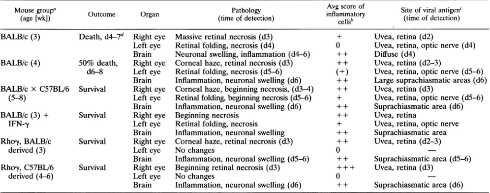
TABLE 1. Pathology and course of disease inyoungmice infected with HSV-1strainF
Mousegroup' Pathology Avg score of Siteof viralantigenC
(age [wk]) Outcome Organ (time of detection)
inflammatory
(time ofdetection)cellSb tm fdtcin
BALB/c(3) Death,d4-7d Right eye Massive retinal necrosis(d3) + Uvea,retina(d2)
Left eye Retinal folding, necrosis (d4) 0 Uvea, retina, opticnerve (d4) Brain Neuronalswelling, inflammation(d4-6) ++ Diffuse (d4)
BALB/c(4) 50% death, Right eye Corneal haze, retinalnecrosis (d3) ++ Uvea, retina(d2-3)
d6-8 Left eye Retinalfolding, necrosis(d5-6) (+) Uvea, retina, opticnerve (d5-6) Brain Inflammation,neuronalswelling (d6) + + Largesuprachiasmaticareas(d6) BALB/cx C57BL/6 Survival Right eye Corneal haze, beginningnecrosis,(d3-4) + + Uvea,retina(d3)
(5-8) Left eye Retinal folding, beginning necrosis(d5-6) + Uvea,retina, opticnerve(d5-6) Brain Inflammation, neuronal swelling (d6) ++ Suprachiasmatic area(d6)
BALB/c(3) + Survival Right eye Beginning necrosis + + Uvea, retina
IFN-,y Left eye Retinal folding, necrosis + Uvea,retina, opticnerve
Brain Inflammation,neuronalswelling + + Suprachiasmatic area
Rhoy,BALB/c Survival Righteye Cornealhaze,retinal necrosis(d3) + + Uvea,retina(d2-3)
derived (3) Lefteye Nochanges 0
Brain Inflammation, neuronal swelling(d5-6) + + Suprachiasmatic area(d5-6) Rho-y, C57BL/6 Survival Right eye Beginning retinalnecrosis(d3) + + + Uvea,retina(d3)
derived (4-6) Left eye Nochanges 0
Brain Inflammation, neuronal swelling (d6) + + Suprachiasmatic area(d6)
aEach group consisted of three mice of the same age,usuallylittermates.Experimentswereperformedtwice,except for thoseinvolving injectionwith antiIFN--y
(three mice)andinjection ofIFN-yin 3-week-old mice(five mice).Inall other groups,six animalsweretested.
bBased on individual scores of all mice in a group. 0, noinflammation; +, fewinflammatory cells;++, moderateinflammation;+ ++, strong inflammation.
c
Determined
byimmunostainingusingthe indirect immunoperoxidase method with DAB as achromogen.-,antigennotdetected.ddn,day afterinfection.
IFN--yaffordedprotection fromviral infection.Thus, therho-y
model provided a means to analyze the effects of ocular IFN--y production on the progress of HSV-1 infection tothe brain.
MATERUILSAND METHODS
Animals. Initially,toinduce ectopicexpression ofIFN-y in photoreceptors of the retina, we used the rhodopsin promoter to introduce the rho-y transgene into C57BL76 mice, which werethen crossed for four tosix generations with BALB/c and
C57BL/6mice(14).These animalswereroutinely screened for
the rho-y transgene by PCR amplification of tail DNA and Southern blothybridization, classedastransgenicor
nontrans-genic (littermates),and used forthisstudy at3to 8weeks of
age. All animals were maintained and handled under
veteri-nary supervision in accordance with National Institutes of
Health guidelines.
Virus andinjection protocols.Apreparation(1
pI)
contain-ing 2 x 10⁵PFUof HSV-1strainF(27, 44), passagedoncebyG. Lewandowski, was injected into the vitreous of each
ani-mal's right eye under visual control. Asharp puncture 2 mm behind the corneal limbus permitted access to theposterior chamber, where injection of virus through 10-pI Hamilton syringes with sterile, replaceable needles (26 gauge) was followed by injection of 2 p.l of air to prevent reflux. We injected the virus into the right eyes of C57BL/6- and BALB/ c-derived transgenic and nontransgenic animals, using three to
four age-matched animals per group and time point, and performed the experiments at least twice. Therefore, the groupscontained in total six animals unless stated otherwise. Mice which had received eitheraloweramountof virusor an accidentalinjuryof the lensduring injection, asconfirmedby histology and immunocytochemistry, were removed fromthe study. Micewereinoculated with virus eitherat3 and 4 weeks of age or at 5 to 6 weeks of age. Immunostaining for viral antigen on histological sections and the development of pa-thology confirmed the presence of infection. Either 1 p.1 of
IFN-y (1 mg/ml/107 IU; Genentech)or
anti-IFN-y
(1 mg/ml;Pharmingen) was coinjected with thevirus,and theprocedure wasrepeated onday4 after the initialinjection. All animals' left eyesremained untreated. Animals mock infectedby phos-phate-buffered saline (PBS) inoculation served as controls. Infected miceweremonitoreddailyforsignsofdisease,suchas inflammatory changes of the skin and eyelids, rough coat, abnormal stretching, lethargy, ataxia, limb paralysis, and sei-zures.Animalssufferingfrom seizuresorothersignsofsevere encephalitis were euthanized. The other mice were killed by cervicaldislocation or halothane overdose 3 to 10 days after viral injection. Both eyes and the brains were either fixed in 10% zinc formalin orimmediatelyfrozen in OCTcompound. Torecoverinfectiousvirus, we homogenized(i)wholeright eyes and (ii)left eyes and brains ofatleastthree animals per group in 400 and 1,000 p.1 of PBS, respectively, with a mechanical homogenizer (Ultra-Turrax; IKA Laboratories).
FIG. 2. Pathology of the retina in HSV-infected mice. PAS; magnification, x560.(A)Five-week-old rho-y transgenic mouse, right eye, 8 days afterintravitrealinfection with HSV-1. Note beginning destruction of the retinal architecture and inflammatory cells in all layers of the retina, especially along aretinal vessel(arrowhead),and in the choroid. (B) Retina of anuninfectedrhoy mouse at 5 weeks of age, with folding of the photoreceptor layer, loss of the outer photoreceptor segments, and inflammatory cells in the photoreceptor layer between retinal folds and in the ganglioncelllayer (arrowheads). (C) Normal retina of a 5-week-old BALB/c mouse without infection for comparison. s, photoreceptor segments consistingof inner and outersegments(darkerarea); on, outer nuclear layer containing the nuclei of the photoreceptors and the radial glia(Muller cells); in,inner nuclear layer containing the nuclei of amacrine and bipolar cells; gl, ganglion cell layer containing the nuclei of the ganglion cells andmostofthe retinal vessels. Note(i) the broad photoreceptor layer without folding or inflammatory cells and(ii)the intact segments of the photoreceptors.
J. VIROL.
on November 9, 2019 by guest
http://jvi.asm.org/
IFN-y PROTECTS MICE FROM INTRAOCULAR HSV-1 INFECTION 5559
*3S e ..
.: > .: :. J@R} si xSs:e.
*:e =:v _ __St .. i,!
St |tU <
n 4-i:¢<a>f: i.
sh
gw
},
.r
_.
...
a|
<o w t
z irsw
_.~5i= Y
VOL.68, 1994
on November 9, 2019 by guest
http://jvi.asm.org/
5560 GEIGER ET AL.
:.s
_...
..
~~$
A
-.
_,AV
6s ...4. 1:
.4 * .4 w i ?¢
*bw *P
.... ...
r
#'
*: 4*:1
..4:.
s 4
'S*-,, * ¾
*:. : - 4:zs'
4....
V
NRWI"
'st .. -,.-0 v
..W. .:,
-AM.. r
.r: t:: :: :< :.
..
..._.
i, ..:}
r..
P* ,8.
..J;:-N...:
.': .: :t":... 'x' A
_.W..:
*', '$t.*..
as:s.# ..
sir ; '
..::'
:f.:'::.
Si w X e
^ :'
:.b.S Si' .. .2.
;:s w 2 i sf: n}
#S .^, i,<**
Y:
*s e *
*44~~~~~~~~~~~~~~~~~~~,i
q4.B :-:..s.
E
s >J. VIROL.
-AL-lw-11
's..
-t' .17, - ;.".,:
&. 1,11.:
,i
:":.:
-t- g"...'; .:'
.:.-., AW
.1
ss
.l.
on November 9, 2019 by guest
http://jvi.asm.org/
IFN-y PROTECTS MICE FROM INTRAOCULAR HSV-1 INFECTION 5561 All right eyes of a group were pooled together. The same
procedure was used for left eyes and brains. After removing
the cell debris by centrifugation at 2,000 rpm (Sorvall RT 6000B), we performed plaque assays on Vero cells with an incubation time of 60 min at 37°C, subsequent Dulbecco modified Eagle medium-agarose overlay, and development for 2days. Viralyield was calculated from serial dilutions on plates yielding 10 to 100 plaques(7).
Histology and immunocytochemistry. Paraffin-embedded sectionsof brain and eye tissueswere stained with hematoxy-lin-eosin orperiodic acid-Schiff(PAS). Frozen serial sections, 6 ,umthick, were cut and stained with hematoxylin or analyzed by immunocytochemistry. Immunostainingwas performed by the indirectavidin-biotin-peroxidase complex method(Vector Laboratories) on frozen sections afterfixationincold acetone for 5 min. Primaryantibodies (polyclonalanti-HSV-1 [Dako],
monoclonal MAC-1 [BoehringerMannheim],monoclonal Ly2, L3T4,and NK[Pharmingen],andmonoclonalF4/80[Serotec]) were applied at a concentration of 5
p.g/ml;
0.05% diamino-benzidine (DAB)-0.04% nickel sulfate-0.02% hydrogen per-oxide servedas achromogen. Counterstaining was performed either in hematoxylin (2 g/ml) or in 1.4% methyl green-PBS. Ly2(CD8)- and L3T4(CD4)-positive cells were counted in a light microscope at X20 magnification on 6-,um-thick sec-tions of fresh-frozen whole eyes and counterstained with methyl green, using an underlying 1-mm grid as a reference. The counted areas included the corneal limbus but excluded thesclera, extraocular tissue, and optic nerve outside the eye.RESULTS
Manifestation of disease. All mice inoculated with HSV-1 strain F developed ocular, neuronal, and systemic signs of infection (Fig. 1). Histological changes of the eyes included necrotizing retinitis similar to the acute retinal necrosis syn-dromeassociated with HSV-1 infection of humans(13). How-ever, thecourseof diseasevaried depending on both the age of the infected mice and the presenceofIFN-yinthe inoculated eye.
Survival and protection from neurologic disease inyoung mice. The eyes and brains of all HSV-1-inoculated animals becameinfected(Fig. 1 to3).5All 3-week-old and about50%
of the 4-week-old nontransgenic animals developed signs of
encephalitis, such as hunched posture, rough coat, lethargy,
anorexia,andataxia,and diedwithin 4to8days,whereas adult
BALB/cmice (5to 6weeksold)andrho-y mice of allstudied
ages survived without clinical signs of encephalitis. Three-week-old nontransgenic mice which had received additional intraocularinjectionofIFN-ytogetherwith the virus survived
aswell(Table 1).
Protection fromHSV-1-induced infection of the
contralat-eraleye.Inrho-y transgenic mice,the nontreated left eyewas
protected from retinitis.Incontrast, allnontransgenic animals developed viral infection and retinitis in both eyes. Intravitreal
coinjection of exogenous IFN--y with the virus prevented the
development of retinitis in the left, nontreated eye (Tables 1
and 2) in adult, but not 3-week-old, nontransgenic mice.
Anti-IFN-y
coinjected with the virus did not alter the outcomeof infection (Table 2).
C57BL/6
and Balb/c mice proved equally susceptible to infection and toretinaldamage.Morphology and viral antigen in infected tissues. Viral antigen wasfound in all tissues of theinoculatedeye(Fig.3A),
the optic nerve (Fig. 3B), the brain, and in most cases the
trigeminal nerve, as determined by immunostaining. C57BL/6 mice hadsignificantly less viralantigeninthe cornea than did BALB/cmice, although they showedequal involvement of the uvea andthe retina (not shown). Additionally, the noninocu-lated eyes ofnontransgenicmiceyielded HSV antigen by day 6, whereasnoantigencould be foundbyimmunostainingin the nontreated eyes oftransgenic mice.
Viral antigen and pathological changes of the brain were located mostly in theopticandtrigeminal projection,including
the geniculate and the colliculus superior. The infection was mostpronounced in the suprachiasmatic area. Aggregatesof virus-infected cells and mononuclearcells, mostly small
lym-phocytes, were often located in the vicinity of blood vessels
(Fig. 1D). Infected neuronal areas, especially the inner retina and the suprachiasmatic area of the brain (Fig. 1E and F),
containedrounded, swollen cells. No multinucleated giant cells werevisible. Viralantigen seemedtodisappearfaster from the brains of transgenic micecompared withnontransgenic mice,
which by immunocytochemistry still had vast quantities of antigen in their brains atday 10 (not shown).
Strongerinflammatory responsetoviral infectionin trans-genic mice. The inflammatory response to HSV-1 comprised
natural killercells,macrophages(Fig. 3C),andTcells(Fig.3D andE)in the eyes andbrain,asconfirmed byimmunostaining
for the antibodies MAC-1, NK, LFA-1, Ly2, and L3T4. Viral antigen and polymorphonuclear infiltration appeared simulta-neously in the injectedeyes.Both theextentand thenumbers
of infiltrating cells were greater in the oculated eyes of
transgenic mice than in those ofnontransgenic animals (Fig.
IF). In nontransgenic animals, inflammation was confined
mainly to the uvea and corneaatearly stages of infection(days 3 to 4); later, the retina was infiltrated. These infiltrates contained arelatively high number of natural killer cells and macrophages (Fig. 3C), whereas the inoculated eyes of
trans-genic mice contained morelymphocytes than did the eyes of
nontransgenicanimals(Fig. 3D and E).
Subsequently,thenoninjectedeyesofnontransgenicanimals
became infected, and inflammatory cells appeared around
virallyinfected cells in the retina and theunderlyingchoroid.
However, the number of inflammatory cells in these eyes remained extremelylow, in contrastwiththe high amountof viral antigen and ocular destruction seenin thesemice, while
transgenicmicewereprotected and showedno inflammation
of the noninoculated eye above the background usually ob-served inourtransgenicmice (14).
At the same time, viral antigen and inflammatory cells appeared in the brains of all animals. However, in thebrain,
FIG. 3. Viral antigen and inflammation in infected mice at day 6 after intravitreal injection. Immunostaining was done by the indirect avidin-biotin-peroxidase complexmethod with DABasthechromogen.(A) Rho-y transgenicmouseinfected with HSV-1showingviralantigenin thechoroidand alllayersof the retina.Counterstained withhematoxylin;magnification, x340. (B)Viralantigenintheopticnerveofthesame
mouse.Counterstained withhematoxylin;magnification, X480. (C) Nontransgenicmouseinfected with HSV-1.Stainingwiththeantibody MAC-1
indicatesmacrophagesand naturalkiller cells in theperipheralretina. Notepositivecells in alllayersof theretina,thechoroid,andthe sclera. Counterstained withhematoxylin; magnification, X340.(D) Nontransgenicmouseinfected with HSV-1.Stainingwaswithantibody L3T4(CD4). Positivecellsarelocated mainlyintheganglioncell layer.Counterstained withmethylgreen;magnification, X340. (E) Rhoytransgenicmouse
infected with HSV-1.StainingwaswithantibodyL3T4(CD4).Positive cells appear in alllayersof the retina.Counterstainedwithmethylgreen; magnification, x340.
VOL. 68, 1994
on November 9, 2019 by guest
http://jvi.asm.org/
5562 GEIGER ET AL.
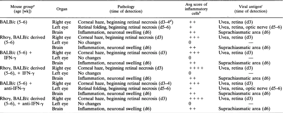
TABLE 2. Pathologyand course of diseasein adult mice treated with IFN--y oranti-IFN--yand infected with HSV-1strainF
Mousegroup Organ Pathology Avg scoreof Viralantigen'
(age [wk])
Ogn(time
ofdetection)inflammatory(tmofdecin
cells' tm fdtcin
BALB/c(5-6) Right eye Corneal haze,beginning retinal necrosis(d34d) ++ Uvea,retina(d3)
Left eye Retinal folding,beginning retinal necrosis (d5-6) + Uvea, retina, optic nerve(d5-6) Brain Inflammation, neuronal swelling (d6) +±+ Suprachiasmaticarea(d6) Rhoy,BALB/c derived Right eye Cornealhaze,beginning retinal necrosis(d3) + + + Uvea, retina(d3)
(5-6) Left eye Nochanges 0
Brain Inflammation,neuronalswelling(d6) + + Suprachiasmatic area(d6) BALB/c(5-6) + Right eye Corneal haze,beginning retinal necrosis(d3) + + + Uvea, retina (d3)
IFN--y Lefteye Nochanges 0
Brain Inflammation,neuronalswelling(d6) + + Suprachiasmaticarea(d6) Rhoy,BALB/c derived Right eye Cornealhaze,beginning retinalnecrosis(d3) + + + + Uvea, retina(d3)
(5-6), + IFN-y Lefteye Nochanges 0
Brain Inflammation,neuronalswelling(d6) + + Suprachiasmaticarea(d6) BALB/c (5-6)+ Right eye Corneal haze,beginning retinal necrosis(d3-4) + + + Uvea, retina(d3)
anti-IFN--y Left eye Retinal folding,beginning retinal necrosis(d5-6) + Uvea, retina,opticnerve(d5-6) Brain Inflammation,neuronalswelling(d6) + + Suprachiasmatic area(d6) Rhoy,BALB/c derived Right eye Corneal haze,beginning retinal necrosis(d3) + + + + Uvea, retina (d3)
(5-6), +anti-IFN-y Left eye Nochanges 0
Brain Inflammation,neuronalswelling (d6) + + Suprachiasmaticarea(d6)
Forcompositionsof groups, seeTable1,footnotea. +IFN-y,additional intraocularinjectionofIFN--y;+Anti-IFN--y,additional intravitrealinjectionofanti-IFN--y.
All mice survived.
bBased onindividualscoresofallmice in a group. 0, noinflammationor noinflammation abovebackgroundintransgenic mice; +, fewinflammatorycells;++, moderateinflammation; + ++,stronginflammation; + + ++, wholeorganinfiltratedwithinflammatorycells.
C4See Table 1, footnotes cand d.
these differences were less drastic; both transgenic and non-transgenic mice developed nearly the same amount of inflam-mation.
The effect of addingIFN-ytothe viral inoculum wasnotable in that injected eyes of nontransgenic animals given the mixture showed nearly the same morphology and immune response asdid their transgenic littermates.
Increase of CD4 and CD8 cells in infected tissues of transgenic mice. To ascertain the type of inflammatory cells
observed,wequantified CD4(L3T4)and CD8(Ly2)cells. The
difference between transgenic mice, which showed three- to
five-times-higher total cell counts and relatively more CD8
cells from the beginning of infection (Fig. 4), and nontrans-genic animals was quite striking. In the noninjected eyes of nontransgenic mice, these T-cell subsets were still enhanced
(Fig. 4), but their numbers did not match the amount of
herpetic pathology. The cell counts and the CD8/CD4 ratio were scarcely influenced by administration ofanti-IFN-y and were notsignificantly different in the brains of transgenic and nontransgenic mice at 5 weeks of age(not shown). Additional intraocular treatment with IFN-y produced an increase of L3T4- and Ly2-positive cells in the inoculated eyes of non-transgenic mice.
EnhancedMHC expressionininfected tissuesoftransgenic mice. In both transgenic and nontransgenic animals, all af-fected tissuesexpressedanoverall increase of MHC classIand classII antigensonactivated glial cells and infiltrating inflam-matory cells. Both MHC classes were expressed more
abun-dantlyintransgenic animals, whichalready express MHC class
I andclassIIin the retinawithoutviralinfection(14), than in thenontransgenic group (Fig. 5).
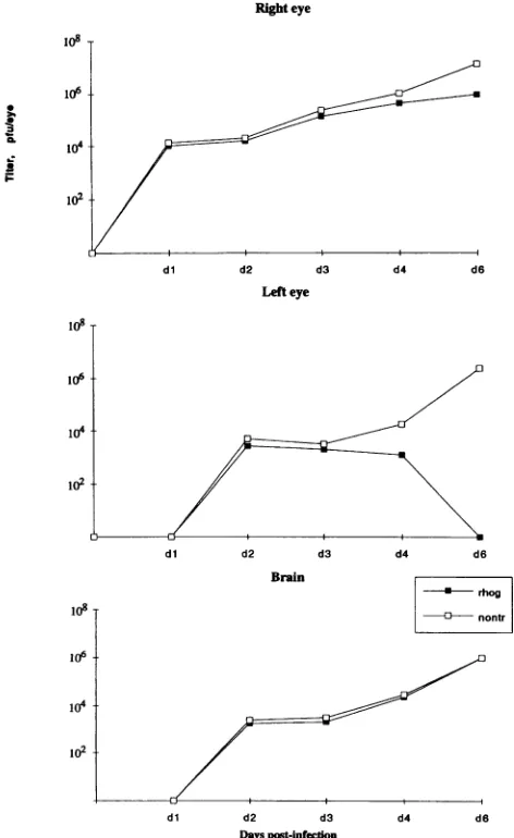
No early viral replication block in transgenic mice. To
determinewhether the survival oftransgenic mice after HSV-1
infection resulted from ablock of viral replication induced by IFN--y,weattempted to recover infectious virus frominfected tissue homogenates. Virus wasrecoverableby day 1from the injected (right) eyes of all animals studied and from the brains and the noninjected (left) eyes on day 2, a time when viral
antigenwas not yetdetectableby immunostaining,which isa considerably less sensitive method for the detection ofvirus. Novirus wasrecovered from noninfected control animals or from animalsinfected for more than 3 weeks. At days 1 to 2, tissues of transgenic and nontransgenic mice infected with HSV-1 contained similar amounts of virus (Fig. 6). Subse-quently, the relative amount of viral yield decreased in the
transgenicmice.Byday4,novirus could be recovered from the
noninoculated left eyes of these mice.
DISCUSSION
Intraocularproduction ofIFN-y rendered miceresistantto intraocular infection with HSV-1. After transgenic mice re-ceived HSV-1injectionsinoneeye,theirectopic expressionof IFN-,yprotected the other eye from virus-inducedpathology,
although itbrieflycontained infectiousvirus. The same effect was achieved in adult nontransgenic mice by coinjection of IFN--y with the virus. However, thecytokine didnot prevent the virus fromspreadingtothebrain.
The lethal susceptibility to HSV-1 strain F of 3-week-old BALB/c mice could be relatedtothe selective defect ofIFN-y
production in mice of this age (1). This defect is correlated
with the lack ofIFN-yproductioninresponsetoviral infection inmacrophages(28)and theunresponsivenessof naturalkiller cellstostimulation withIFN-,y(43).Adult-levelresponsiveness to stimulation with IFN--y is reached at 4 to 5 weeks of age
(43),whereas the fullcapacitytoproducethiscytokinefollows
much later (1). Protection afforded in 3-week-oldtransgenic
micecould be due to the local productionof IFN-y. Consis-tently, 50% of the 4-week-oldnontransgenicanimals whichare
reportedtohavesystemicIFN-,yproduction, although
consid-erably below adult levels(1),survived.However, theamountof
additionally givenIFN-,ywasapparentlyinsufficienttoprotect
the second eyes of youngnontransgenicmice.Thus,wedonot know whether otherfactors, suchasinduction of resistanceto virus-induced damage on a cellular level or an age-related
J. VIROL.
on November 9, 2019 by guest
http://jvi.asm.org/
IFN-y PROTECTS MICE FROM INTRAOCULAR HSV-1 INFECTION 5563
200
rA
c
0
100
u
IU 100
A.
right
eye0
TU CD8(Ly2)
CD4
(L3T4)
l0
0
d3 d6 dlO
d3
d6
dlO dlOrho rho rho Balb Balb
Balb
Balb/IFNB.
left
eye0
* CD8
(Ly2)
*CD4
(L3T4)
0-OA
d3 d6 dlO d3 d6 dlO dlO
rho rho rho Balb Balb Balb Balb/IFN
FIG. 4. Counts of CD4 (L3T4)and CD8(Ly2)cellsineyesof5-to
6-week-old mice infectedwith HSV-1. Sections of6 p.mwereanalyzed;
immunostainingwaswith DABasthe chromogen, and counterstaining
waswithmethylgreen.Ly2- and L3T4-positive cellswerecounted in
thelight microscopeat X20 magnification of wholeeyes.Thecounted
areasincludedthe corneal limbus but excluded the sclera, extraocular
tissue, and opticnerveoutside theeye.The plotusesmeanvalues of groupsof three animals(5-to6-week oldBALB/c-derivedmice).The
variance ofrecountsthesameeyestayedbelow5%; however,
individ-ual mice variedasmuchas10% withina group.Errorbarsrepresent
thestandarddeviation of eachgroup(xcn -1). dn, day after infection.
increased susceptibility of neurons to HSV-1 (32), play an
additional role.
The properties ofIFN-y that could be responsible for the observedeffects includeblock of viral replication, activation of macrophages and natural killer cells, and enhancement of cytotoxicity. Alternatively, IFN-y could act indirectly via the increasedMHC expression in transgenic mice (5, 6,10, 17, 19, 36, 39, 46, 54). Because we found nearly equal amounts of infectiousvirus in transgenic and nontransgenic mice until day 2after infection, atimewhen the viruswasalready presentin
thesecondeye, anyblockof viral replication wouldoccurtoo
latetoaffect its invasionof the brain. This resultfitswell with
earlierreports describingan 18-h delay between the presence
of IFN--y and the beginning of effects on viral replication in
culture (2a, 23). Although we cannot exclude the possibility thatasmallpool of replicatingviruscaninfluence theoutcome
ofinfectionatalatestage,ourresults doexplain the failure of
IFN-y
to prevent the neuroinvasion of HSV-1. Since weobserved an influence of IFN--y in our model, which was similar to that observed on cultured cells, it is not likely that retinal changes present in uninfected transgenic mice have an influ-ence on the behavior of the virus. Thus, it is possible that a disturbance of the blood-retina barrier in transgenic mice has an influence on the intraocular immune privilege (35, 52). Exactly why the second eyes of transgenic mice do not develop pathology, despite the small amounts of virus recovered at day 2, why these mice show no clinical signs of encephalitis, and why they survive an otherwise lethal infection when infected at 3 weeks of age remain conjectural. Once in the brain, the virus should replicate with equal speed in transgenic and nontrans-genic mice, unless the cytokine has either directly influenced the propagation characteristics of the virus during its passage through the eye or sent a systemic signal protecting infected neurons from death. IFN-y might similarly send a systemic signal to boost an immune response in the brain, although increased inflammation in the brain would not necessarily have a positive effect. The amounts of inflammation in the brains of transgenic and nontransgenic mice did not differ greatly. Instead, the lack of neurologic symptoms in young transgenic mice could indicate its protective effect. Since HSV-1 strain F has been shown to develop latency (33, 34), it is not likely that the virus is ever cleared from surviving infected cells. There-fore, the more rapid loss of virus detection from the brains of transgenic mice could mean that
IFN-y
favors the pathway toward latency. Additional mechanisms, including synergistic action with antibodies (49, 50) and other cytokines such as tumor necrosis factor alpha (45) or protection of infected neurons from being killed by cytotoxic T cells (30), should be considered. This might involve a prevention of apoptosis as demonstrated for antibody action in Sindbis virus (16, 26). Furthermore, we do not know the role of IFN--y-induced production of nitric oxide in the development of disease (9, 15, 22).We detected a greater infiltration of leukocytes in the eyes of the transgenic mice than in those of nontransgenic animals, possibly reflecting a faster and more adequate immune re-sponse in the
rhoy
animals. Since the infiltrates contained naturalkiller cells, macrophages, and cytotoxic T cells, multi-ple mechanisms could be responsible. The already present MHC class I expression on retinal cells in transgenic mice (14) could play an important role in this process by presenting HSV antigen to CD8 cells.The finding that
C57BU/6
andBALB/c
mice were equally susceptible to infection with HSV-1 was unexpected.C57BL/6
mice, whose MHC class I haplotype has been recognized as a major factor for geneticallydeterminedprotection (38, 47), are regarded as comparatively resistant to HSV-1 infection. How-ever, the intraocular mode of infection and the viral strains that we used apparently overrode possible genetic protection, although this does not necessarily support the conclusion that MHC class I might be less important than other factors in accounting for the activities of this virus, since MHC class I is not likely to be expressed on neurons under normal conditions. Apart from infiltrating cells, the MHC class I expression that we found in the retinas and brains of nontransgenic mice is probably located on activated glial cells, consistent with earlier studies of HSV-1 strain F infection in rats (60).The substantial number of CD4 and CD8 cells present in this model confirms the importance of cytotoxic T cells, which are not present in uninfected rho-y mice (14), in intraocular viral infection (4, 20, 24, 25, 61, 62) and suggests that CD8 cells, the amounts of which are low in the left untreated eyes of nontransgenic mice, might have a crucial role in the outcome VOL.68, 1994
on November 9, 2019 by guest
http://jvi.asm.org/
[image:8.612.53.299.77.427.2]J.VIROL. 5564 GEIGER ET AL.
J, gL:wl_t; eis '.J; 6NEe =&iSls-:s
ib i EX 'MG ;- iS
*:giS;Ss*,L?*e
.,$9F.EJt
illl L 4.S S r .C
.l|lg-t!WiF...?;S
s | .l.: :::
gi _ ::7
M.s
.s .. . .s
:. p. ss
-;e: :3'
?i
:t: ':
's
.&v. .*. .L.
.:
11....C..,
.. S%'~. ( ,. *
C, ,at
4'*C,}**.;SI, S
%4$
s w
£'9rs
* X
^*
& .s-J
tJ
I .I
-
i,
xuz<-;>s ar
-e.s
.V
Tt: §
*'t;t.:.v M 9s. a slr4,. v; ggs.>G
tsB
,.PE,aL.s@w1S.
>, .s- S;£et ot e;4Pw
*,kJ
}::i:-||,.
.,.,w..
veFQ^#|
)...
f?4*-\#(
liL-sr?Zflsb*t*
gB+...^.
SN'.._7
P.
4.
*,
.1 :,
UL-r, MZ4'.
-rl-";t
:,.: p !% --l"'.."'. ...
'k 4--;s -lk-v .. All,
t .1 "
i It
.-.!C-
-1-., 'A
~-S'
on November 9, 2019 by guest
http://jvi.asm.org/
IFN--y PROTECTS MICE FROM INTRAOCULAR HSV-1 INFECTION 5565
FIG. 5. Immunostaining for MHC class I in right eyes of transgenic and nontransgenic mice infected with HSV-1 at day 6. Indirect immunoperoxidase technique withDAB asthe chromogen, counterstainingwith hematoxylin; magnification, X400. (A)Rhoytransgenic mouse. Staining of the retina with MHC class Iantibody (H-2)shows positive cells through all layers of the retina.(B) Nontransgenic mouse with only afew positive cells in the inner retina andin the choroid. (C)Rhoytransgenic mouse. Staining of the retinawith MHC class I antibody (H-2) shows positive cells in all layers of the retina. (D) Nontransgenicmousewith few positive cells in the inner retina and in the choroid.
ofdisease. If so, the
IFN-y
could be cajference with antige Our data showti infection of the eye this occurs withoul suggests either inv( regulation by the
108
106
.I* 1-1
CL
2
r-102
io6t
v MHChyperexpression induced by ectopic changes in the immunosuppressive properties of the
intraocu-pable
ofoverriding a possibly subtle inter- lar microenvironment might be responsible for the protection nexpression (58, 59) by the virus. of the second eyes in transgenic mice (2). The intraocular hat IFN--y canprotect animals from HSV-1 immune privilege could beinfluenced either by IFN--y directly withoutinfluencing its neuroinvasion. That or by a cytokine-induced disturbance of the blood-retina t a significant increase in retinal damage barrier. Although development of rhoy mice is apparently )lvement ofsynergistic factors or counter- normal (14), IFN--y couldhave a subtle effect on the neuronal intraocular microenvironment. Possible development which could influence the susceptibility of these neurons to virus infection. However, it is not likely that the observed alterations of theneuronal morphology in rho-y mice are to influence the neuronal spread of HSV-1. These alter-Right eye ations are mainly present in the photoreceptors of the retina, and thespread of HSV-1 to the brain is notdependent on the function of thephotoreceptors. Infact,the earliestappearance of viralantigen by stainingoccurred in theganglion celllayer of rho-y and nontransgenic mice without a significant differ-ence.Therefore, the virus could traveldirectly to the brain via the ganglion cells. A further pathway, using the iris and the ciliary ganglion, is also notlikelytobeanatomically influenced by the presence of IFN--y in the eye.The mechanisms involved in protective effects of IFN-y
remain speculative. Thus, they are likely to induce a more chronic course of disease, including the possibility of local
dl d2 d3 d4 d6 reactivation and slowly developing dysfunction of infected
Lefteye cells, parallel to the phenomena observed in AIDS dementia
(31). The rho-y mouse has proved to be a suitable tool for
manipulatingIFN-yfunction in theeye,particularly intermsof viral infection.
ACKNOWLEDGMENTS
This work was supported by NIH grant MH 47680. K.Geigerwas
supported byafellowshipfrom the DeutscheForschungsgemeinschaft;
N.Sarvetnickwassupported bya careerdevelopmentaward from the Juvenile Diabetes Foundation.
WethankFloydBloom and GailLewandowski, Scripps,forhelping
to initiate thesestudies,forprovidingthevirus,andforcommentson dl d2 d3 d4 d6 the manuscript. We thankTerry Nash, Scripps, for help in setting up Brain the virus recovery assay. We
especially
thankJ.Stevens, UCLA, for* rhog helpful suggestions concerningthis work.
nontr
REFERENCES
dl d2 d3
Days post-infection
FIG. 6. Recoveryof infectious virusfromeyesan
6-week-old mice infected with HSV-1 strain F.:
experimentconsisted of three mice. Wholerighteyes
and left eyes and brainswere pooled separately, hI
fixed volumeasdescribedin Materials andMethods,E
plaqueassayonVero cells. Thevalues shownarebas
oftwoidenticalexperiments,the results ofwhich had
afactorof 3.rhog, rho-y;nontr, nontransformed.
1. Adkins, B., A. Ghanei, and K. Hamilton. 1993. Developmental regulation of IL-4, IL-2 andIFN-gamma production by murine
peripheralTlymphocytes.J.Immunol. 151:6617-6626.
2. Atherton, S. S., and J.W. Streilein. 1987. Virus-specific DTH
preventscontralateral retinitisfollowingintracameral inoculation
of HSV-2. Curr.EyeRes. 6:133-139.
2a.Balish, M.J.,M. E.Abrams,A. M.Pumfery,and C. R. Brandt.
d4 d6 1992.Enhanced inhibition of herpes simplex virus type 1 growthin
human corneal fibroblastsbycombinations of interferon-a and--.
J. Infect.Dis.166:1401-1403.
idbrains of 5-to 3. Boerman,R.H.,A. C. B.Peters,B. R.Bloem,A. K.Raap,and M.
Each group per van der Ploeg. 1992. Spread of herpes simplex virus to the
(threepergroup) cerebrospinal fluid and the meninges in experimental mouse
omogenized in a encephalitis.ActaNeuropathol. 83:300-307.
andprocessedfor 4. Bonneau, R. H., and S. R. Jennings. 1990. Herpes simplex
sedonthemeans virus-specific cytolyticTlymphocytesrestrictedtoanormallylow
avariance below responderH-2 alleleareprotectivein vivo.Virology174:599-604. 5. Brandt,C.R.,andC. A. Salkowski. 1992. Activation ofNKcellsin
0o2
106
106
VOL.68, 1994
108T
I
le
on November 9, 2019 by guest
http://jvi.asm.org/
[image:10.612.59.295.262.647.2]5566 GEIGER ET AL.
mice following corneal infection with herpes simplex virus type-1. Invest. Ophthalmol. Visual Sci. 33:113-120.
6. Charteris, D. G., and S. L. Lightman. 1992. Interferon-gamma (IFN-gamma) production in vivo in experimental autoimmune uveoretinitis. Immunology 75:463-467.
7. Cooper,P. D.1967. The plaqueassayof animal viruses. Methods
Virol. 3:243-311.
8. Cousins, S. W.,M.M.McCabe, R.Danielpour,andJ. W. Streilein. 1991. Identification of transforming growth factor-beta as an
immunosuppressive factor inaqueoushumor.Invest.Ophthalmol.
Visual Sci. 32:2201-2211.
9. Croen, K. D. 1993. Evidence for antiviral effect of nitric oxide. Inhibition of herpes simplex virustype1replication. J. Clin. Invest. 91:2446-2452.
10. Dayton, E. T., M. Matsumoto-Kobayashi, P. Perussia, and G.
Trinchieri. 1985. Role of immune interferon in the monocytic differentiation of human promyelocytic cell lines by leukocyte conditioned medium. Blood 66:583-594.
11. Denkins, Y. M., and M. L. Kripke. 1993. Effect ofUVirradiation
on lethal infection of mice with Candida albicans. Photochem.
Photobiol. 57:266-271.
12. Fiala,M., E. J. Singer, M. C. Graves, W. W. Tourtellotte, J. A.
Stewart, C. A. Schable, R. H.Rhodes,etal.1993.AIDS dementia complexcomplicated by cytomegalovirus encephalopathy. J. Neu-rol. 240:223-231.
13. Gartry, D. S.,D. J. Spalton, A. Tilzey, and P. G. Hykin. 1991. Acuteretinalnecrosis syndrome.Br. J. Ophthalmol. 75:292-297. 14. Geiger,K., E. Howes, M.Gallina,X. J. Huang, G. H. Travis, and
N. Sarvetnick. 1994. Transgenic mice expressing IFN-,y in the retina develop inflammation and photoreceptor loss. Invest. Oph-thalmol. Visual Sci.35:2667-2681.
15. Goureau, O.,M. Lepoivre, and Y.Courtois.1992. Lipopolysaccha-ride and cytokines induce a macrophage-type of nitric oxide
synthase in bovine retinal pigmented epithelial cells. Biochem. Biophys. Res. Commun. 186:854-859.
16. Griffin, D. E., B. Levine,W. R. Tyor, and D. N. Irani. 1992. The immune response in viral encephalitis. Semin. Immunol.
4:111-119.
17. Hamel, C.P., B.Detrick,and J. J.Hooks.1990.Evaluation ofIa
expression in rat ocular tissues following inoculation with inter-feron-gamma. Exp. Eye Res. 50:173-182.
18. Hendricks, R.L., T. M. Tumpey, and A. Finnegan. 1992. IFN--y
andIL-2areprotective in the skin but pathologic in thecorneasof
HSV-1-infected mice.J.Immunol. 149:3023-3028.
19. Hughes, C. C.,D. K. Male, and P. L. Lantos. 1988.Adhesion of lymphocytestocerebral microvascular cells: effects of
interferon-gamma, tumor necrosis factor and interleukin-1. Immunology
64:677-681.
20. Inatsuki, A.,M.Yasukawa, and Y. Kobayashi. 1989. Functional alterations of herpes simplexvirus-specific CD4+ multifunctional
Tcell clones followinginfection with humanTlymphotropic virus
typeI.J.Immunol. 143:1327-1333.
21. Jiang, L. Q., M. Jorquera, and J. W. Streilein. 1993. Subretinal
space and vitreous cavity as immunologicallyprivileged sites of
retinalallografts. Invest. Ophthalmol. Visual Sci. 34:3347-3354. 22. Karupiah, G., Q.W.Xie,R. M. Buller,C.Nathan,C. Duarte, and
J. D. MacMicking. 1993. Inhibition of viral replication by inter-feron-gamma-induced nitric oxide synthase. Science
261:1445-1448.
23. Klotzbucher, A., S. Mittnacht, H. Kirchner, and H. Jacobsen.
1990. Differenteffects of IFN-y andIFNoa/I on"immediateearly"
geneexpression of HSV-1. Virology 179:487-491.
24. Kolaitis,G., M. Doymaz, and B. T. Rouse. 1990. Demonstration of
MHC classII restrictedcytotoxicTlymphocytes in mice against
herpessimplex virus. Immunology71:101-106.
25. Kuzushima, K., K.-I. Isobe, T. Morishima, A. Takatsuki, and I.
Nakashima. 1990.Inhibitory effect of herpes simplex virus
infec-tion to target cells on recognition of minor histocompatibility
antigens bycytotoxicTlymphocytes. J. Immunol. 144:4536-4540.
26. Levine, B., J. M. Hardwick, B. D. Trapp, T. 0. Crawford, R. C.
Bollinger,and D.E. Griffin.1991. Antibody-mediatedclearance of
alphavirusinfection fromneurons. Science254:856-560. 27. Lewandowski, G.A.,D.Lo, andF. E. Bloom. 1993. Interference
withmajorhistocompatibilitycomplex class1I-restrictedantigen presentation inthe brainby herpessimplexvirustype1:apossible mechanismofevasionof the immuneresponse.Proc.Natl.Acad. Sci. USA 90:2005-2009.
28. Lucchiari, M. A., and C. A. Pereira. 1990. A major role of macrophageactivationby interferon-gammaduringmouse hepa-titis virustype3 infection. II.Age-dependentresistance. Immuno-biology181:31-39.
29. Marsh, R.J. 1989. Ocularmanifestations of AIDS. Br. J. Hosp. Med. 42:224-230.
30. Martz, E., and S. R. Gamble. 1992. How do CTL control virus infections? Evidence for prelytic halt of herpes simplex. Viral Immunol.5:81-91.
31. Masliah, E.,C. L.Achim,N.Ge,R.DeTeresa,R. D.Terry,and C. A.Wiley. 1992. Spectrum of human immunodeficiency virus-associated neocorticaldamage.Ann.Neurol.32:321-329. 32. McKendall, R.R., and W. Woo. 1987. Possible neural basis for
age-dependentresistancetoneurologicdisease from herpes sim-plexvirus. J.Neurol.Sci. 81:227-237.
33. Meignier, B., B. Norrild,and B. Roizman. 1983. Colonizationof murineganglia byasuperinfectingstrain ofherpessimplexvirus. Infect. Immun. 41:702-708.
33a.NationalAdvisoryEye Council. 1987. Vision research. A national plan. 1987. Evaluation andupdate.National Institutes ofHealth, Bethesda,Md.
34. Nesburn, A. B., R.Dickinson,M.Radnoti,andM.J.Green.1976. Experimental reactivation of ocular herpes simplex in rabbits. Surv.Ophthalmol.21:185-190.
35. Niederkorn,J.Y. 1990. Immuneprivilegeand immuneregulation in theeye.Adv. Immunol. 48:191-226.
36. Palliard, X., R. De Waal Malefijt, H. Yssel, D. Blanchard, I.
Chretien, J. Abrams, J. De Vries, et al. 1988. Simultaneous production of IL-2, IL-4,and IFN--yby activated human CD4+ and CD 8+ Tcell clones. J. Immunol. 141:849-855.
37. Park, C. H., and M. A. Latina. 1993. Effects ofgamma-interferon
onhumantrabecularmeshwork cellphagocytosis.Invest. Ophthal-mol. Visual Sci. 34:2228-2236.
38. Pepose, J. S.,andJ. A.Whittum-Hudson. 1987. An immunoge-netic analysis of resistance to herpes simple retinitis in inbred strains of mice. Invest. Ophthalmol.Visual Sci. 28:1549-1552. 39. Percopo, C. M., J. J. Hooks, T. Shinohara, R. Caspi, and B.
Detrick. 1990.Cytokinemediated activation ofaneuronal retinal resident cell provokes antigen presentation. J. Immunol. 145: 4101-4107.
40. Pollak, I. F., and R. D. Lund. 1990. The blood-brain barrier
protectsforeign antigensin thebrainfrom immune attack. Exp.
Neurol. 108:114-121.
41. Price,R.W., B.Brew, J.Sidtis,M.Rosenblum, and A. C. Scheck. 1988.The brain in AIDS: centralnervous systemHIV-1infection and AIDS dementiacomplex. Science239:586-592.
42. Price, R.W., B. J. Brew, and M. Rosenblum. 1990. The AIDS dementia complex and HIV-1 brain infection: a pathogenetic model of virus-immune interaction. Res. Publ. Assoc. Res. Nerv. Ment.Dis. 68:269-290.
43. Provinciali, M., M. Muxxioli, and N. Fabris. 1989. Timing of
appearanceand disappearance of IFN and IL-2 induced natural
immunityduring ontogenetic development andaging. Exp. Ger-ontol.24:227-236.
44. Roizman, B.,P. G. Spear,andE. D.Kieff. 1972.Herpes simplex viruses I and II: abiochemical definition.Perspect.Virol. 8:129-169.
45. Rossol-Voth, R., S. Rossol,K. H.Schutt,S.Corridori,W. deCian, and D. Falke. 1991. In vitro protective effect oftumor necrosis factor alpha against experimental infection withherpes simplex
type1. J. Gen.Virol. 72:143-147.
46. Shuai, K.,C. Schindler,V. R. Prezioso,andJ.E. Darnell. 1992. Activation oftranscription by IFN--y: tyosinephosphorylationofa
91-kd DNAbindingprotein.Science 258:1808-1812.
47. Simmons, A. 1989. H-2-linked genes influence the severity of herpes simplexvirusinfection oftheperipheralnervoussystem.J. Exp.Med. 169:1503-1507.
48. Sloan,D.J.,M.J. Wood,and H. M.Charlton. 1991.The immune
responsetointracerebral neuralgrafts.Trends Neurosci. 14:341-346.
J. VIROL.
on November 9, 2019 by guest
http://jvi.asm.org/
IFN-,y PROTECTS MICE FROM INTRAOCULAR HSV-1 INFECTION 5567
49. Spiezia, K. V., B. J. Dille, I. K. Mushahwar, L. Kifle, and G. F. Okasinski. 1990. Prevalence of specific antibodies to herpes simplex virus type 2 asrevealed by an enzyme-linked immunoassay andWestern blot analysis. Adv. Exp. Med. Biol. 278:231-242. 50. Staats, H. F., J. E. Oakes, and R. N. Lausch. 1991.
Anti-glycoproteinD monoclonalantibodyprotectsagainst herpes sim-plexvirus type 1-induced diseases in mice functionally depleted of selected T-cell subsets or asialo GM1+ cells. J. Virol. 65:6008-6014.
51. Streilein, J. W. 1990. Anteriorchamber-associated immune devi-ation: the privilege of immunity in the eye. Surv. Ophthalmol. 35:67-73.
52. Streilein, J. W. 1993. Immune privilege as the result of local tissue barriers immunosuppressive microenvironments. Curr. Opin. Im-munol. 5:428-432.
53. Streilein, J. W., G. A. Wilbanks, and S. W. Cousins. 1992. Immunoregulatory mechanisms in the eye. J. Neuroimmunol. 39:185-200.
54. Streilein, J. W., G. A. Wilbanks, A. Taylor, and S. Cousins. 1992. Eye derived cytokines and the immunosuppressive intraocular microenvironment:areview. Curr. Eye Res.11(Suppl.):41-47. 55. Subba Rao, D. S. V., and J. B.Grogan. 1977. Host response to
tissuesplacedin the anteriorchamber of the eye: demonstration of migration inhibition factor and serum blocking activity. Cell. Immunol. 33:125-133.
56. Subba Rao, D. S. V., and J. B. Grogan. 1979. Suppression of graft-versus-host reactions in rats bearing implants in the anterior chamber of the eye.Transplantation 27:75-78.
57. Vessella, R. L., S. Raju, J. V. Cockrell, and J. B. Grogan. 1978. Host responsetoallogeneic implants in the anterior chamber of the rat eye. Invest. Ophthalmol. Visual Sci. 17:140-148. 58. Walev, I., J. Kunkel, W.Schwaeble, K. Weise, and D. Falke. 1992.
Relationship between HLA I surface expression and different cytopathic effects produced after herpes simplex virus infection in vitro. Arch. Virol. 126:303-311.
59. Walker, C., M. Selby, A.Erickson, D. Cataldo, and J.-P. Valensi. 1992. Cationic lipids direct a viral glycoprotein into the class I major histocompatibility complex antigen-presentation pathway. Proc. Natl.Acad. Sci. USA 89:7915-7918.
60. Weinstein, D. L., D. G.Walker,H.Akiyama, and P. L. McGeer. 1990. Herpes simplexvirus type I infection of the CNS induces majorhistocompatibility complex antigen expression on rat micro-glia. J. Neurosci. Res. 26:55-65.
61. Whittum-Hudson, J. A., and J. S. Pepose. 1987. Immunologic modulation ofvirus-induced pathology in a murine model ofacute
herpetic retinal necrosis. Invest. Ophthalmol. Visual Sci. 28:1541-1548.
62. Yasukawa, M., A. Inatsuki, T.Horiuchi,and Y.Kobayashi. 1991. Functional heterogeneity among herpes simplex virus-specific human CD 4+ Tcells. J. Immunol. 146:1341-1347.
VOL.68, 1994