0022-538X/94/$04.00+0
Copyright© 1994, American Society for Microbiology
Processing
of the Intracellular Form of the West Nile
Virus
Capsid
Protein by the Viral NS2B-NS3 Protease:
an In
Vitro Study
VLADIMIR F. YAMSHCHIKOV AND RICHARD W. COMPANS*
Departmentof Microbiology and Immunology, Emory University Schoolof Medicine, Emory University,
Atlanta,
Georgia
30322Received 5 May 1994/Accepted 20 June 1994
According to the existing model of flavivirus polyprotein processing, one of the cleavages in the amino-terminalpart oftheflavivirus polyprotein by host cell signalases resultsinformation of prM (precursor to one of the structural proteins, M) and the membrane-bound intracellular form of the viral capsid protein
(Ci.t)
retaining the prM signal sequence at its carboxy terminus. This hydrophobic anchor is subsequently removed bythe viralprotease,resultinginformationof the matureviral capsid protein found in virions (Cv.r). We have preparedinvitro expression cassettes coding for both forms of the capsid protein, for the prM protein, for the C-prM precursor, and for the viral protease components of West Nile flavivirus and characterized their translation products. UsingCi.t
andC.,
translation products as molecular markers, we have observed processingof the intracellular form of the West Nile capsid protein by the viral protease in vitro both upon cotranslation oftheC-prM precursor and the viral protease-encoding cassette and by incubation of C-prM translation products with a detergent-solubilized extract of cells infected with a recombinant vaccinia virus expressingtheactive viral protease.Thecleavage ofCi.t
bytheviral proteaseatthepredicted dibasicsite was verifiedbyintroductionof pointmutations into thecleavage siteand anadjacent region.These studiesprovide thefirst direct demonstration of processing of the intracellular form of the flavivirus capsid protein by the viral protease.The flavivirus genome, consisting ofasingle positive-strand RNA molecule, is translated in infected cells into a large
polyproteinofmorethan 350 kDa that isprocessedto
individ-ualflavivirus proteinswhicharearrangedin the genome in the
order 5'-C-prM-E-NS1-NS2A-NS2B-NS3-NS4A-NS4B-NS5-3'
(reviewedin reference6).Several host cell andvirus-specified
enzymaticactivities have been demonstrated tobeinvolved in
processing of this polyprotein. While prM-E-NS1 junctions were shown to be cotranslationally processed by host cell
signalases (2, 10, 27, 32, 33, 38),aviral protease(3, 31) which
wasidentified within the amino-terminal third of the flavivirus NS3protein (la, 12) andwasfoundtofunction asaNS2B-NS3
complex (5,9, 11, 29,37)has been demonstratedtobe involved
in processing of the NS2A-NS2B-NS3-NS4A and NS4B-NS5
junctions. Cleavages effected by the viral protease occur after
the dibasic sequence KR or RR, followed by small non-branched amino acids such as Gly or Ser. Processing at the NS4A-NS4Bjunction wasshown to be mediated by host cell signalases, with internal cleavage of NS4A by the NS2B-NS3 protease appearing to be a prerequisite (14, 28). Alternate
cleavages within NS2A were also reported (7, 10, 24), and
recently, the NS2B-NS3 protease was shown to effect this
cleavage for yellow fever (YF)virus (26). Propercleavage at
thecarboxyendof NS1dependsonthe presence ofNS2A and
the carboxy-terminal octapeptide of NS1 (10, 14), but the
enzymatic activityinvolved is stilltobe identified.
Two other cleavages after basic amino acids occur late in flavivirus infection and involve furtherprocessingofprM and
*Correspondingauthor.Mailingaddress: Departmentof
Microbi-ologyandImmunology, EmoryUniversitySchoolofMedicine, Emory University, 1510Clifton Rd., Atlanta, GA 30322. Phone: (404) 727-5947. Fax:(404)727-8250.
an intracellular form of the Cprotein (reviewedin reference
6). The late cleavage within prM, found mostly in immature intracellular virions, constitutes one of the latest events in flavivirus maturation andyields the M protein found in infec-tious extracellular virions (reviewed in reference 6). This cleavage depends on an acidic environment (13, 30), and a
cleavage site motif suggests the involvement of some host
protease (reviewed in reference 6). The second cleavage mentioned generates the virion form ofprotein C(Cvir), which lacks ahydrophobic sequence present at the carboxy terminus of its intracellular form
(Cint)
(27, 34). Originally, it was hypothesized (31) that C-prM cleavage by host signalases results in the formation of membrane-associatedCint
whichis, assuggestedonthe basisof thesimilarityof the cleavagesites, furtherprocessed by the NS2B-NS3 protease with formation ofCvir (6). Recently, experimental evidence (19, 39) which, in
general, supports the above hypothesis was presented.
How-ever,thesequentialorder of thesetwocleavageeventsremains uncertain. We havesuggested (39) that whilesignalase cleav-ageofC-prM occurspriortoNS2B-NS3-mediatedcleavageof
Cint,
the formeroneis facilitatedbyinteraction of the protease complex either with the prM signal peptideor withadjacentC-terminalregionsof
Cint.
Inaddition,wehavesuggested thatthe latter isalsofacilitatedby capsid-RNAinteractionandmay be concomitant with RNA encapsidation. The opposite se-quential order ofthese twocleavageevents, with
NS2B-NS3-mediated cleavage of the C-prM precursor at the upstream dibasic siteas aprerequisite forsubsequent processingof the
prMsignal peptideatthesignalase cleavagesite,has alsobeen
suggested (1, 19).
Sinceprocessingof the
Cint
protein bythe viralproteasehasbeen postulated (6) but has not been demonstrated yet, we
haveattemptedtoreproduce directlytheNS2B-NS3-mediated
5765
on November 9, 2019 by guest
http://jvi.asm.org/
cleavage of
Cj,
In this study, the protease activity of the NS2B-NS3 complex towardCin,
wasinvestigatedinvitro,and its specificity wasassessed by introduction of point mutations in the cleavage site.MATERUILS AND METHODS
Cells and viruses. Vaccinia virus recombinant vW{5'-C->NS3243} carryingacDNAgenome fragmentof WestNile virus (WN) has been described previously (39). Along with the WN structuralproteins, proteins NS1 and NS2A, this recom-binant expresses the active WN protease consistingofproteins NS2B and 243 N-terminal amino acids of protein NS3. He-LaT4 cells (20) were used throughout thisstudy asdescribed previously (39).
Construction of gene cassettes. Standard procedures for DNAmanipulationsandcloningwerefollowed(22).ForPCR amplification, VentR thermophilic DNA polymerase (New England Biolabs, Beverly,Mass.)waschosen because ofhigher
fidelity (25) and was used according to the manufacturer's
recommendations. All recombinant plasmids were
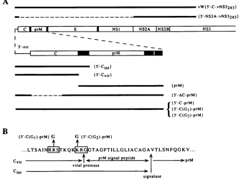
character-izedby restriction analysis and sequencing. Ifnot mentioned otherwise, enzymes were purchased from New England Bio-labs, except for Asp-718, which wasobtained from Boehringer Mannheim (Indianapolis, Ind.). Gene cassettes used in this studyare presentedinFig. 1.
Construction of the W{5'-C-prM} and W{prM} cassettes has been describedpreviously (39). Briefly, the former includes the entire5'-nontranslatedregion and genes of theWNcapsid
and prMproteins. FollowingATG, the latter codes for the last
two positively charged amino acids of
Cvir
(see Fig. 1 fordetails), the prM signal peptide, and the prMprotein. Both
cassettes end with the prM carboxy-terminal anchoring se-quence uptothe last amino acidof theEsignal peptide, and threenonrelated amino acidsresulted froma5'-CTTAATTA ATTAAG-3' universal stop codon-HindIlI linker addition.
The {C-prM} cassette was placed under control of the SP6
promoter,and the {prM} cassettewasplacedunder control of the T7 promoter.
A W{5'-Cvir} cassette was obtained by PCR amplification
from the original pGEM-K plasmid (G. Wengler, Giessen, Germany; see also reference 39) with primers to the SP6 promoter(as direct) andtothe 388-to411-nucleotide (nt) (4, 27) region of the WN genome (as reverse). The last primer introduced the TAA stop codon immediately after the last amino acid of
Cvir,
as a part of the HindIll recognition sequence. TheresultingfragmentwasdigestedwithEcoRI and HindIll and cloned intopGEM3Zvector(Promega, Madison,Wis.)under control of theT7promoter.
A
W{5'-Cjnt}
cassetteunder control of theT7 promoterwas created by extension of the above Cvir cassette in order to includeafragmentencoding the prMsignalpeptide whichwas found (4) to be a part of the intracellular form of the WNcapsid protein.A391-bp AluI (nt 71)-AluI ((nt462) fragment
from pGEM-K was ligated with the universal stop codon-HindlIl linker, which restored the codon of the last
Cin,
amino acid(4)andintroduced theTAAstopcodon immediately after it. Theligated fragment wassubsequently digested with SstIIandHindIll, and acarboxy terminus-encoding fragment was exchanged with the SstII-HindIII carboxy terminus-encoding
fragmentin the W{5'-Cvir} cassette.
A W{5'-AC-prM} cassette was constructed bydigestion of
theW{5'-C-prM}-containing plasmid withBglII (located9 nt upstream from the C initiation codon), filling in with Klenow
polymerase,digestion with Hindll (nt 310[4]), ligation with an
Nco FO linker, 5'-AGCCATGGCT-3', which supplied an
in-A
vW15'-C->NS3243)
- - - 5'-NS2A->NS3243)
lFIC|DrM | E I NSI NS2A INS2B NS3
5'-ntr
=~
15S-Cint1
(5,-Cvir
tprM)
(5'-AC-prM)
f(5'-C-prM)
115'-C(G2 )-prM)
B
( 5'-C(GI)-prMJG(, ({5'-C(G2)-prM
...LTSAINE6KQKICIRGTAGFTILLGLIACAGAVTLSNFQGKV...
Cv_r _t_X prMsignalpeptide - )PrM
virTalpotease
Cint 30
signalase
FIG. 1. Aschematic representationofproteinsencodedbythe WN in vitro expression plasmids. (A) The 5'-terminal half of the WN genome, with the amino-terminal half of the encoded polyprotein drawn as anopen bar, is shown in the middle. Individual flavivirus proteinsaremarked, with the vertical bars denoting their boundaries. Thethick vertical bars representhydrophobicregions serving assignal andstop-transfer sequences. In the upper part of thefigure,the thick horizontal bar represents the expression cassette carried by the recombinantvaccinia virus whichwasused forpreparationofthe WN protease-containingextract;theprotease-encoding gene cassette used forsynthesisof the WNNS2B-NS3243protease invitro is drawn below. The second open bar represents a scaled part ofthe WN genome encodingthe5'-nontranslated regionand the C-prMprecursor.The Cint membrane anchor/prM signal peptide and the prM membrane anchorconsistingofaprM stop-transfersequenceand the E signal peptide divided by a single Arg residue are indicated bythe filled boxes.Below, the thick horizontal lines show gene cassettes encoded by in vitro transcription plasmids designated as in Materials and Methods. The dashed lines denote the genome regions which were deleted. (B) Thesingle-letter amino acid representation of the WN polyprotein sequenceatthe C-prMjunction. Thecarboxy termini of Cvir and
Cint
and the amino terminus ofprM are denoted by the horizontal arrows; theviralprotease andsignalasecleavage sites aremarkedbythe vertical arrows below the sequence. Two dibasicsites, RRS and KRG(provisionallymarked as no. 1 and no.2),whichwere
subjected tomutagenesis,aredenoted byopenboxes. Themutations which were introduced are shown above the sequence, with
corre-spondingcassettedesignationsbeside eachmutation.
frame ATG(underlined),digestion withNcoI,andreligation. Thisprocedure createdaMet(amino acid 1)-Val(aminoacid 72) deletion in the 105-amino acid sequence of Cvir with replacement of Val(amino acid 72) for Ala.
AW{5'-NS2A---NS3243} cassettewhich, along with NS2A,
encodes components of the activeWNproteasewascreatedby ligation of an EcoRI-BglII fragment coding for the 5'-non-translated region of WN, aBglII-PstI adapter (5'-GATCTC
GAIRTCTGCA-3',
upper strand; 5'-GACATCGA-3', lower strand) which supplied anin-frame ATG (underlined),and aNsiI (nt 3509)-Asp-718 (nt 5330) fragment encoding a WN
NS2A--NS3243 precursor, and cloning between the
EcoRI-Asp-718 sitesinpGEM3Zunder control of theT7promoter. Thevectoradds4amino acids ofanonrelated sequencetothe carboxy end of the encoded precursor. The WN 5'-nontrans-lated region was added to facilitate expression of encoded proteins(29).
PCR-mediated point mutagenesiswas used to obtain vari-ants of the {C-prM} cassette with point mutations in the
on November 9, 2019 by guest
http://jvi.asm.org/
[image:2.612.313.554.80.261.2]C
*,-Cvir
cleavage site and adjacent region (see Fig. 1 for details). In general, mutated cassettes were synthesized by two-step PCR amplification of pGEM-K segments as de-scribed previously (15). The 5'-C-coding half was amplified withprimers to theSP6 promoter (as direct) and to the 381- to 405-nt region (as reverse). The reverse primer introduced a C->Gpoint mutation at nt 388, causing replacement of a CGG triplet (Arg) for a GGG triplet (Gly). The prM half was separately amplified from pGEM-K with primers to the 394- to 422-nt (as direct) and to the 952- to 971-nt (as reverse) regions. The direct primer in the last case introduced two point mutations at nt 406 (A-->G) and 407 (A->G), causing replace-ment of an AAA triplet (Lys) for a GGA triplet (Gly). The first reverse and second direct primers were designed in such a way that mutations were located outside of their overlapping region (whichconsisted of 12 nt). Thus, the second round of PCR with the amplified halves, with the first direct and second reverse primers, resulted in a mixture of fragments bearing either or both mutations. After digestion withEcoRl (12 nt upstream from the beginning of the WN 5' region) andMluI
(nt 920; at the end of the prM gene), the resulting fragments were exchanged with the EcoRI-MluI fragment in the pW{5'-C-prM} plasmid by ligation and cloning, and individual mutated cassettes {5'-C(G1)-prM} and {5'-C(G2)-prM}, as well as{5'-C(GIG2)-prM},
were identified by sequencing. Thisap-proach enabled us to use two mutating oligonucleotides in-stead of four.
RNA synthesis. All cassettes were transcribed with T7
polymerase, except for the {5'-C-prM} cassette, which was
transcribed with SP6 polymerase (Pharmacia, Piscataway, N.J.). The plasmids containing the {Cvir},
{Cin,},
{prM},
{C-prM}, and {AC-prM} cassettes were linearized by
HindIll
locateddownstream from the termination codon. The
pW{5'-NS2A->NS3243} plasmid was linearized by Asp-718, which is
found in the WN cDNA sequence. Two micrograms of linear-ized plasmids was transcribed in a 50-pI reaction mixture as described previously (22), except that ATP, UTP, and CTP were at 0.5mM, GTP was at 0.25 mM, andm7GpppG(all from NewEngland Biolabs) was at a 1 mM concentration. A total of 35 Uofplacental RNase inhibitor (Pharmacia) and 50 to 55 U of RNA polymerases were used per reaction. After 1 h of incubation at 37°C (T7) or40°C(SP6), 1
plI
of theenzymeswas again added, and mixtures were incubated for an additional hour. RNA was deproteinized by phenol-chloroform extrac-tion (22) and ethanol precipitated from 2 M ammonium acetate in order to remove unincorporated triphosphates. TheRNAconcentration was estimated from the assumption that
40 p.g/ml corresponds to one A260 unit. The optical density of
DNA was subtracted on the basis of the assumption that 50 ,ug/ml corresponds to one A260 unit.
Translation invitro. RNA transcripts were translated with a
Translation Kit, Reticulocyte, Type II (Boehringer Mann-heim), as recommended by the manufacturer. Where men-tioned,
25-pul
translation reaction mixtures were supplemented with 1RA
of canine microsomal membranes (Boehringer Mannheim). For translation, 0.5 pug of individual RNAs was used.Atotal of 20 ,uCi ofL-[35S]methionine(>1,000 Ci/mmol,10
puCi/RI;
Amersham, Arlington Heights, Ill.) was used tolabeltranslation products, and mixtures were incubated for 30 to 60minat30°C. After 20min of centrifugation at 14,000 rpm on an Eppendorf model 5415C benchtop centrifuge at
4°C,
membrane pellets were rinsed with 50 plAof 20 mM HEPES
(N-2-hydroxyethylpiperazine-N'-2-ethanesulfonic
acid) (pH 7.5)-100 mM potassium acetate-1 mM EDTA-1 mM dithio-threitol-100 ,ug of bovine serum albumin per ml, centrifuged for 10 min, andresuspended in 100 plA of the Laemmli samplebuffer (16). When mixtures were analyzed directly, 5 pA of translation reaction mixtures was diluted to 100 pA with the sample buffer. For immunoprecipitation, translation mixtures were diluted to 50 pA with the radioimmunoprecipitation assay buffer (22), 1 plA of
anti-WN
(strain Eg 101) hyperimmune mouse ascites fluid (obtained from C. Calisher, Centers for Disease Control and Prevention, Fort Collins,Colo.)
was added, and samples were incubated overnight at4°C.
Proteins were collected by shaking with 10 pA of a 50% suspension of protein A-agarose conjugate (Boehringer Mannheim) for 1 hr at4°C.
Following brief centrifugation, pellets were washed twice with 100 pA of the radioimmunoprecipitation assay buffer and once with 10 mMTris-HCl
(pH 7.5) and resuspended in 50 pA of the sample buffer.For deglycosylation with peptide N-glycosidase F (New England Biolabs), sample aliquots were diluted five times with 10 mM sodium phosphate (pH
7.5)-1%
octylglycoside and incubated for 1 h with 500 U of peptide N-glycosidase F at37°C.
Proteins were precipitated with 10 volumes of acetone and resuspended in the sample buffer.Preparation of protease-containing extract and trans-cleav-age assay. HeLaT4 (approximately 3x
107)
cells were infected withvW{5'-C->NS3243}
(39) at a multiplicity of infection of 10 for 1 h and incubated in growth medium for 5 h at37°C.
Cells were harvested by scraping, washed once with phosphate-buffered saline, and resuspended in 1 ml of cold 50 mMTris-HCl
(pH 7.5)-75 mMNaCl-5
mM EDTA. After three cycles of freeze-thawing, the suspension was centrifuged on an Eppendorf model 5415C benchtop centrifuge (14,000 rpm; 5min), and the supernatant was discarded. The pellet was
resuspended in 300 pA of the same buffer, and 30
[lI
of 10% Triton X-100 was added. After vigorous vortexing, the suspen-sion was incubated at ambient temperature for 10min,
and the insoluble material was removed by 5min
of centrifugation on the benchtop centrifuge. The supernatant was collected and stored at-20°C
in aliquots. The same protocol was used to prepare the Triton X-100 extract from noninfected cells.For trans-cleavage assay, membrane pellets obtained from translation reactions as described above were resuspended in 10
[lI
of the Triton X-100 extracts, incubated at30°C
for 30 min, and diluted to 100RI
with the sample buffer.Electrophoretic
analysis.
RNA integrity was verified by polyacrylamide gel electrophoresis (PAGE) on5%gels in the presence of urea with subsequent silver staining (Silver Stain Plus; Bio-Rad Laboratories, Hercules, Calif.). Protein samples, after heat denaturation for 5min
at95°C,
were analyzed on 12% polyacrylamide gels in the Laemmli system (16, 22). After electrophoresis, gels were processed for fluorography as de-scribed earlier (39).RESULTS
Characterization of translation products. Experimental demonstration of flavivirus capsid processing is complicated by the low immunogenicity of the capsid protein, which makes preparation of specific anticapsid antibodies difficult (8, 32, 39). For this reason, we have prepared
Cvir
andCint
expression cassettes. Since these cassettes contain the authentic transla-tion start codons, and stop codons were introduced immedi-ately afterCvir
andCi.t
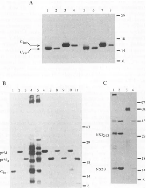
carboxy-terminal amino acids, we expected that translation in vitro would produce authentic molecular markers for the capsid processing products. Single major products were observed upon translation of the{Cvir}
(Fig. 2A, lanes 1 and 5) and
{Ci.t}
(lanes 3 and 7) transcripts. The presence of microsomal membranes during translation (lanes 5 to 8) had no detectable effect on electrophoreticon November 9, 2019 by guest
http://jvi.asm.org/
A
2
Cin _.._
rt~Mu
./=v
v)l
rB
2 3 4 -- 6} 7 8 9 10 11
SPI .. ..
-43
&V:
pr\1 4 ..40 w m
pir%l,l ow~~~~~~~~~~~~~~~S: .mso
\~f1 -*_d-.
Cint *0
to....
29
-14
-6
FIG. 2. Characterization ofinvitro transl
ofprestainedmolecularweight markers(Gil N.Y.)areindicatedontheright, and identific on the left. (A) Analysis of products syr
translation of the Cvir and
Cint
marker-er transcripts ofthe{5'-Cvir} (lanes 1, 2, 5,and4, 7,and8)cassettesweretranslatedinthera
the absence (lanes 1 to 4) or in the pres
microsomal membranes, and either complete
membrane pellets (lanes 5 and 7),or immi
(lanes 2, 4, 6, and 8)wereanalyzed by PAGE
and Methods.(B) Analysis of C-prM transla
the {5'-C-prM} (lanes 2 to 5), {5'-AC-prN. {prM} (lanes 8to11)weretranslatedinvitro
presenceof microsomal membranes; thepro
noprecipitated (lanes 2, 3, and 6 to 9), or
pellets (lanes 4, 5, 10, and 11), subjectedto treatment(lanes 3, 5, 7, 9, and 11), and analyzi
inMaterials and Methods.The
Cint
moleculmembranepelletis shown inlane1.prMd den (C) Analysis ofproducts synthesizedupon i]
{5'-NS2A-*NS3243} transcript. Translation
presence(lanes 1 and 2)or absence (lanes membranes.Themembrane pellet (lane 1),ci
ortheirimmunoprecipitates (lanes2 and4)w
describedinMaterials andMethods.
mobility or relative amounts of either phoretic mobility of both proteinswasfi
expected from their calculated moleculz Da;
Cint,
13,289 Da). Thecapsid protein Arg and presumably retains a relativcharge under the conditions of electrc affects itsmobility toward the anode. Bo'
werefoundinthemembrane fractionup
2A, lanes 5 and 7). For
Cinl,
this observation is in agreement 7 X with previous reports (4, 6, 27, 32, 38). For Cvir, such associa--239 tion was suggested earlier (32, 39) but has not been directly demonstrated yet. Approximately one-third of the total incor-porated radioactivity was recovered in each case, and we suggest that incomplete membrane sedimentation upon cen--IX trifugation may account for the incomplete recoveryof Cvir andd
-Cint.
At lowdilution (1:50), the antiserum specificallyprecipi--14 tates Cvir (Fig. 2A, lanes 2 and 6) and
Cint
(lanes 4 and 8), although the amounts ofimmunoprecipitated proteins repre-- 6 sented only 1 to 2% of the total synthesized proteins, as estimated from radioactivity counts. The low recovery of the C proteins presumably indicates that the hyperimmuneanti-c serumraised
against
WN-infectedmousetissues containsalowl2^R34 concentration of WN C-specific antibodies. However, misfold-- ing of the Cproteins synthesized in vitro also cannot beruled
{97
out. At higher dilutions (1:250 and higher), no C-specific immunoprecipitation was detected (results not shown). Such a Ill-68
steep decrease in the efficiency of immunoprecipitationsug-43 gests a low affinity of the C-specific antibodies present in the
antiserum.
Immunoprecipitation of the products obtained upon trans-NS32443
_-9
lation of the{C-prM}
transcript in the presence of membranes yielded a single band with an apparent molecular mass of approximately 22 to 25 kDa (Fig.2B, lane 2), which apparently represented a glycosylated protein, as demonstrated bydegly-18 cosylation (lane 3). No such product was detected by immu-NS2B - i4
noprecipitation
when microsomal membranes were not in-*2A
B _14 cluded during translation (results not shown). Apparently, the-6 sameproductwas obtainedupontranslation ofthe
{IAC-prM}
(lanes 6 and 7) and {prM} (lanes 8 and 9) transcripts, thus suggesting that this band represents the glycosylated prM ationproducts.Positions protein. Neither
Cint
(Fig.
2B, lane 1,marker)
northeC-prM
bco BRL, Grand Island, precursor was
readily
detectableby
immunoprecipitation.
edproteinsareindicated However,upon
longer
exposure, afaint bandcomigrating
withithesized in vitro upon the
Ci.t
marker could be observed(results
notshown).
nacoding
cassettes. RNA Aproduct
resembling
Cin,
wasfoundinthe membranepellet
bb r
eticulocyte
it lysatein3
obtained aftercentrifugation
of theC-prM
translation mixturesence(lanes
5 to 8) Ofi(Fig.
2B,lane4).
Incontrasttotranslation oftheC-containing
mixtures(lanes1and3), cassettes, the membrane
pellet
fromC-prM
translationre-unoprecipitated proteins vealedarather
complex
mixture ofproducts
possibly resulting
asdescribed inMaterials from internal initiationand/orprematuretermination of trans-ition.RNAtranscriptsof lation.
Deglycosylation
ofthis mixture enabledus toidentify
4} (lanes 6 and 7), and the
prM
protein (Fig.
2B,
comparelanes 2and 4and3 and5)
asdescribed above in the and the
possible C-prM
precursor(appearing
as adistinctbandducts
were
eather
mmmu-
of about 32 kDa in lane5)
and also revealed that the bandpeptide
N-glycosidase F around 14 kDa represents a mixtureof
Cin,with some otherpedpbyPAGE asdescribed
product. This otherproduct
was also detected in themem-larmarker obtained as a brane
pellet
of the translated{prM}
transcript
(lane 10),
iotesdeglycosylated prM.
appeared
tobeglycosylated
(compare
lane 11, afterdeglyco-nvitro translation of the
sylation),
andpossibly
resulted from premature translationwas carried out in the termination within the
prM
coding
sequence. This minor 3 and 4) of microsomal productwasnotdetected inimmunoprecipitates (Fig.
2B,laneompletemixture(lane 3),
8).
Thus,despite
thecomplexity
of theC-prM
translation,ereanalyzed byPAGEas
mixture,
wewere able toidentify
Ci.t
in
themembrane pellet,andwe used this membrane fraction in subsequent
Cin,
pro-cessing assays.Translation of the {5'-NS2A--NS3243} in the presence of
protein.
The electro- microsomal membranesyielded
threemajor
products
with oundtobe lower than apparent molecular masses ofapproximately
14, 29, and 40armasses
(Cvir,
11,703 kDa which wereclearly
seen inimmunoprecipitates (Fig.
2C,is enriched in
Lys
and lane2)
and,lessclearly,
inmembranepellets (lane
1).
The14-rely high
netpositive
and29-kDabandswereearlieridentifiedasNS2B andNS3243
)phoresis
whichlikely
(37,
39).
Aminorband between themmight
represent NS2A thtranslationproducts
(37,
39).
Wecouldnotreadily
identify
the 40-kDaband,
whichon
centrifugation
(Fig.
may represent either NS2A-NS2B(approximately
40kDa),
on November 9, 2019 by guest
http://jvi.asm.org/
[image:4.612.59.297.78.384.2]NS2B-NS3243 (approximately42kDa) precursor, orboth.The major band found upon translation in the absence of mem-branes was approximately 65 kDa (Fig. 2C, lane 3) and apparently represented the uncleaved NS2A-NS2B-NS3243 precursor (approximately66 kDa). It is interesting that minor cleavage products of this precursor in the absence of added membranes were also detected by immunoprecipitation (lane 4). A similar observationwas reported for YF virus by Cham-bers et al. (9). Thus, formation of individual NS2B andNS3243 indicated thattranslation of the {5'-NS2A-*NS3243} transcript in vitro yields the active NS2B-NS3243 protease, and it was subsequently used in
Cin,
processing assays.Processing of
Ci.,
by theNS2B-NS3243 protease. First, we investigatedprotease-mediated processing ofCint
by cotrans-lation of the{C-prM} and {NS2A->NS3243} transcripts with subsequent PAGE analysis of the membrane pellets. Upon cotranslation of the two transcripts, a band comigrating with the Cvir marker (Fig. 3A, lane 2) was readily observed in a position betweenCint
and NS2B (lane 4). This band was not observed when the {C-prM} transcript was translated alone (lane 5), and onlyCin,
(lane 3, marker) couldbe detected in the membrane pellets. Since this band comigrated with the Cvir molecularmarker, we have concluded thatthis band is likely to represent Cvir which resulted from processing ofCint
at its KR/G site by the NS2B-NS3243 protease.However, several amino acids upstream from this site, the WN capsid protein contains another similar sequence, RRS (Fig. 1), which might be also utilized in vitro. Although the KR/G configuration is found at almost all WN cleavage sites mediated by the viral protease (3, 37), a KR/Scleavage site is present between WN NS3 and NS4A proteins. It was shown thatconservative R-*K mutations in the dibasic consensus are tolerated to some extent by the YF protease (26),but replace-mentofRfor a noncharged amino acid abolished cleavage. On thebasis of these findings, we prepared two mutated {C-prM} cassettes, bearing the R-->G and K-*G mutations attheRRS and KRGsites(Fig. 1), respectively, and studied their process-ing in the same system as for the wild-type cassette. We have chosenaglycine residue in order to minimize a possible effect ofanamino acid side chain on the flexibility and/or conforma-tionof the region adjacent to the prM signal peptide. Cell-free translation of both mutated cassette transcripts resulted in similar mixtures of translation products, as observed with the wild-type cassette. Both
C(Gl)int
andC(G2)Ant
exhibited a similar and slightly faster mobility (compared with wild-typeCint),
which presumably resulted from reduction in the netpositivecharge of theseproteins as a consequence of the R->G
and K->G replacements. When a transcript of the mutated cassette
{5'-C(G1)-prM}
was cotranslated with {NS2A->NS3243}, Cvir (Fig. 3B, lane 2, marker) was again observed
betweenthe
Cint
and NS2B bands (Fig. 3B, lane 5) and was not detected when the mutated transcript was translated alone(Fig. 3B, lane4). In contrast, the appearance of Cvir was not
detected in either case for another cassette, {5'-C(G2)-prM}
(Fig.3B, lanes 6and 7),indicating that in the presence of the
NS2B-NS3243protease
C(G2)jnt>C(G2)vjr
processing did notoccur. These results demonstrated that we indeed observed specific processing of
Cint
atthepredicted KR/G site (27), and the nonconservative K->G mutation in the cleavage site was nottolerated by the viral protease.Formation of the active WN protease composed of NS2B and theNS3243domain was also observed upon expression of
theWN {5-C->NS3243} cassette in vivo (39). We prepared a
Triton X-100-solubilized extract from HeLaT4 cells which were infected with recombinant vaccinia virus vW{5-C->
NS3243} andattempted to demonstrate its processing activity
[image:5.612.318.560.81.230.2]-
w3m3:
FIG. 3. Processing of
Ci.,
by theNS2B-NS3243protease.Forclarity, only the bottom parts of the gels are shown.Positions oftheprestained molecular weight markers and the identified proteins are denoted on the right and left sides,respectively. (A) ProcessingofCintbytheviral protease supplied in trans by cotranslation of the {5'-C-prM} and {5'-NS2A--->NS3243} transcripts in the rabbitreticulocyte lysate in the presence of membranes. Translation products of the {5'-NS2A-.NS3243} (lane 1), {5'-Cvir} (lane 2),{5'-Cint}
(lane 3), {5'-NS2A- NS3243} plus {5'-C-prM} (lane 4), and {5'-C-prM} alone (lane 5)transcripts were recovered as membrane pellets and analyzed by PAGE asdescribed in Materials and Methods. (B) Processing ofCint
bearingmutations at the viral protease cleavage site by the viral protease supplied in trans bycotranslation of the mutated C-prM and the viralprotease-encoding transcripts in the rabbit reticulocyte lysate in thepresence ofmicrosomal membranes. Translation products of the {5'-NS2A->NS3243} (lane 1), {5'-Cvir} (lane 2),{5'-Cinl}
(lane 3), {5'-C(G1)-prM} (lane 4), {5'-C(G1)-prM} plus {5'-NS2A- >NS3243} (lane 5), {5'-C(G2)-prM} (lane 6), and {5'-C(G2)-prM} plus{5'-NS2A-*NS3243} (lane 7) transcripts were recovered as membrane
pellets andanalyzed by PAGE as described in Materials and Methods. (C) Processing of wild-type and mutated
Cint
by the viral protease obtained as a Triton X-100-solubilized extract from HeLaT4 cells infected with thevW{5'-C--+NS3243} vaccinia virus recombinant. The membrane pellets recovered after translation of the{5'-C-prM}(lanes 3and 4),{5'-C(G1)-prM} (lanes 5 and 6), and{5'-C(G2)-prM} (lanes 7and 8)transcripts were solubilized in the Triton X-100 extract of the infected (lanes 4, 6, and 8) or uninfected (lanes 3, 5, and 7) cells, incubated for 30minat30°C, and analyzed by PAGE as described in Materials and Methods. TheCin,
andCvir
molecularmarkers obtained as membrane pellets are present in lanes 1 and 2, respectively. As explained inResults, the electrophoretic mobilities of both mutated variantsofCint
aswell as that ofC(GI)vir
differ slightly from that of the wild-typeprotein.toward
Cint
prepared in vitro. The band comigratingwith the Cvir marker (Fig. 3C, lane 2) was again observed when the membrane pellet obtained after translation of the {C-prM} transcript was solubilized and incubated in an extract of cells infected with the above recombinant (Fig. 3C, lane 4). This bandwas notobserved when an extract of uninfected cells was used(Fig. 3C, lane 3), and only adoublet comigrating with theCint
marker (Fig. 3C, lane 1) wasdetected. Similarly, formation ofC(Gi)vir
was seen upon incubation of the membrane pellet obtained after translation of the mutated {C(G1)-prM} cas-settetranscript with the extract from the infected cells (Fig. 3C, lane 6), butnot with the extract from thenoninfected (Fig. 3C, lane 5) cells. In contrast, formation of C(G2)vir was not observed in either case, when membrane pellets obtained after translation of the mutated{C(G2)-prM} cassette were used in theassay (Fig. 3C, lanes 7 and 8). Theseresults are in complete agreement with the evidence obtained from the above in vitro cotranslation assays.&
0LI
a r-,4 %,_, is -W
#
aw . -i-1, w = I-M
\,'i,
46
*
on November 9, 2019 by guest
http://jvi.asm.org/
DISCUSSION
In the general scheme of flavivirus polyprotein processing suggested byRiceet al. (31), signalase cleavageoftheC-prM
precursorwould result in formation of Cretainingastretch of
hydrophobic amino acids which served as an internal signal
sequence for theprM protein. C-terminal sequencesof the C proteins of WN(27) and Kunjin (34)viruses reveal that this hydrophobicsequenceis absent in thematurecapsid proteins, butaC variant with lowerelectrophoretic mobilityisobserved
ininfected cells. Thesefindingsledtothesuggestion (27)that the membrane-associated form of C which resulted from signalase cleavage is further processed by some protease of viral origin to its mature form found in virions, and this cleavage event is connected with virion formation. After identification of the flavivirusproteaseas amembrane-bound NS2B-NS3complex (la, 3, 5, 9, 11, 12, 29, 31, 37),theevidence obtainedwas summarized in a hypothesis that processing of the intracellular form of C protein is performed by the NS2B-NS3 protease (6). Until recently, there was no direct
experimentalverification of thishypothesis. Amberget al. (1) recently reportedinvitroprocessingofacleavagesite
contain-ing peptide substrate by NS2B-NS3 protease which was
ob-tained as a detergent-solubilized extract from cells infected eitherbyYFvirusorbyrecombinant vaccinia viruses
express-ingYFNS2B and the NS3 protease domain. However,in vitro processingof the whole
Cint
proteinhasnotbeenreported.To develop an assaywhich would enable a direct study ofthese processing events, we have prepared Cvir andCjnt
expressioncassettesin ordertousetheir translationproductsasmolecular
markers for both forms of the Cprotein. Cell-free translation of the Cvir and
Cint
transcripts in both the presence andthe absence of microsomal membranes led toformation ofmajor products with expected differences in molecular masses (27).In agreement with previous reports (4, 6, 27, 32, 38),the
Cint
protein, which was expected to be membrane bound by the hydrophobic sequence retained at its carboxy terminus, wasfoundinthe membranepelletobtained aftercentrifugationof the translation mixture. Interestingly, Cvir, which lacks this hydrophobic anchor,also sediments with themembranes,with
an efficiencyof itsrecoveryfrom the membranepellet
compa-rabletothatof
Cint.
Such association ofCvirwith membraneswas notreported before, but is in agreementwith the obser-vation (32) that only the amino-terminal part of C in the C-prM precursorappearedtobe accessibletoprotease diges-tion in vitro. On the basis of thisreport,wehavesuggested (39)
that, along with the carboxy-terminal anchor in
Cinl,
acon-servedhydrophobic region around residue 50 ofCvir (21)may
be responsible for interaction of an internal part of C with
membranes.
Amorecomplexmixture ofproducts, possibly resultingfrom internal initiation and/or premature translation termination,
wasobserveduponcell-free translation of the {C-prM}
tran-script.Sinceourgoalwastostudy processingof
Cint,
wedidnotidentify all the observed by-products. A band which comi-grated with the
Cint
molecular marker was found in the membrane pellet obtained upon centrifugation of the above translationmixture. When the {C-prM}and{NS2A->NS3243} transcriptswere cotranslated in the presence of microsomal membranes, processingofCint
resultinginCvir
formationwasreadily detected. While R-*G replacement in the upstream RRSsequence hadno effectonprocessingof
Cint
bythe viralprotease, processing was completely abolished by the K->G
mutation inthe KR/G site. These resultssupport the conclu-sion thatformation ofCvirresults fromprocessingof
Cin,
atthe KR/G site. Since no Cvirwas detected in the absence of the{NS2A-NS3243} transcript, we conclude that
Cin,
is processed by the NS2B-NS3243 protease. We also have demonstrated a similar proteaseactivity in lysates of cells infected with recom-binant vaccinia virus vW{5-C->NS3243}. In accordance with the above cotranslation assays,Cint
>Cvir
processingwas ob-served both for the wild-type cassette and for the mutant cassettewith the R->G replacement inthe RRSsite, but not for the cassette bearing the K->G mutation in the KR/G cleavage site. These experiments constitute the first demon-stration ofCint_>Cvir
processing in vitrobythe NS2B-NS3243 proteasesupplied in trans.We havesuggested (39) that
Cint>Cvir
cleavage bythe viral protease depends on or is facilitated by interaction of the capsid with viralRNA. Little is known about thespecificity of flavivirus capsid-RNA interaction or the presence of encapsi-dationsignal(s)onthe flavivirus genomic RNA. Foranumber ofRNAviruses, sequences whichwerefound tobe critical for RNA-nucleocapsid interaction were identified near the 5' ends of these RNAs. Examples include Sindbis virus (36), flock house virus(40),andmurine(23), avian (35), and human(17) retroviruses. Although the 5' segment of flavivirus RNA was always present during translation experiments, more studies are required toinvestigate its role in encapsidation.ACKNOWLEDGMENT
This study was supported by Research Award V22/181/33 and V22/181/72from the World HealthOrganization.
REFERENCES
1. Amberg, S. M., A. Nestorowicz, D.W.McCourt,andC. M. Rice. 1994. NS2B-3proteinase-mediated processingin theyellowfever virus structural region: in vitro and in vivo studies. J. Virol. 68:3794-3802.
la.Bazan, J. F., and R. J.Fletterick. 1989.Detectionofatrypsin-like serineprotease domain inflaviviruses andpestiviruses. Virology 171:637-639.
2. Biedrzycka, A., M. R.Cauchi, A. Bartholomeusz, J. J. Gorman, and P.J. Wright. 1987. Characterization of proteasecleavagesites involved in the formation of envelope glycoprotein and three nonstructural proteins of dengue virus type 2, New Guinea C strain. J. Gen. Virol. 68:1317-1326.
3. Castle, E., U.Leidner, T. Nowak, G. Wengler, and G. Wengler. 1986.Primarystructureof the West Nileflavivirusgenomeregion codingfor all nonstructuralproteins. Virology149:10-26. 4. Castle, E.,T. Nowak, U. Leidner, G. Wengler, and G. Wengler.
1985. Sequence analysis of the viralcore proteinand the
mem-brane-associatedproteinsVl and NV2of theflavivirusWestNile virus and of the genome sequence of these proteins. Virology 145:227-236.
5. Chambers,T.J., A. Grakoui,andC.M.Rice. 1991.Processingof yellowfever virus nonstructuralpolyprotein: acatalyticallyactive NS3 proteinasedomain and NS2Barerequired forcleavages at
dibasic sites. J. Virol.65:6042-6050.
6. Chambers,T. J., C. S. Hahn, R. Galler, and C. M. Rice. 1990. Flavivirus genomeorganization, expression and replication. Annu. Rev. Microbiol. 44:649-688.
7. Chambers,T.J.,D.W. McCourt, and C.M. Rice. 1989.Yellow fever virusproteins NS2A, NS2B, and NS4B: identification and partial N-terminal amino acid sequence analysis. Virology 169: 100-109.
8. Chambers,T.J.,D. W.McCourt,andC.M. Rice.1990.Production ofyellow fevervirus proteins in infected cells: identification of discretepolyprotein speciesandanalysis of cleavage kineticsusing region-specificpolyclonal antisera. Virology 177:159-174. 9. Chambers,T.J., R. C. Weir, A. Grakoui, D.W.McCourt, J. F.
Bazan, R. J. Fletterick,andC. M. Rice. 1990. Evidence that the N-terminal domain of nonstructuralproteinNS3 fromyellowfever virus isaserine proteaseresponsible forsite-specificcleavages in the viralpolyprotein. Proc.Natl. Acad. Sci.USA87:8898-8902. 10. Falgout,B.,R.Chanock, and C.-J. Lai. 1989. Properprocessing of
on November 9, 2019 by guest
http://jvi.asm.org/
dengue virus nonstructural glycoprotein NS1 requires the N-terminalhydrophobicsequenceand the downstreamnonstructural protein NS2A. J. Virol. 63:1852-1860.
11. Falgout, B., M. Pethel, Y.-M. Zhang, and C.-J. Lai. 1991. Both nonstructural proteins NS2B and NS3 are required for the pro-teolytic processing of dengue virus nonstructural proteins. J. Virol. 65:2467-2475.
12. Gorbalenya, A. E., A. P. Donchenko, V. M. Blinov, and E. V. Koonin. 1989. Cysteine proteasesof positive strand RNA viruses andchymotrypsin-like serine proteases: adistinctprotein super-familywith a commonstructuralfold.FEBS Lett. 243:103-114. 13. Heinz, F. X., K. Stiasny, G.Piischner-Auer, H. Holzmann, S. L.
Allison, C. W. Mandl, and C. Kunz. 1994. Structural changes and functional control of thetick-borne encephalitis virus glycoprotein E by the heterodimeric association with protein prM. Virology 198:109-117.
14. Hori,H., and C.-J. Lai. 1990.Cleavage ofdengue virusNS1-NS2A requires an octapeptide sequence at the C terminus of NS1. J. Virol.64:4573-4577.
15. Horton, R. M., H. D. Hunt, S. N. Ho, J. K. Pullen, and L. R. Pease. 1989. Engineering hybrid genes without the use of restriction enzymes: genesplicing byoverlap extension. Gene 77:61-68. 16. Laemmli, U. K. 1970. Cleavage of structuralproteins during the
assembly of the head of bacteriophage T4. Nature (London) 227:680-685.
17. Lever, A., H. Gottlinger, W. Haseltine, and J. Sodroski. 1989. Identification of a sequence required for efficient packaging of humanimmunodeficiencyvirus type 1 RNAintovirions. J.Virol. 63:4085-4087.
18. Lin, C., S. M. Amberg, T. J. Chambers, and C. M. Rice. 1993. Cleavageat anovelsite in the NS4Aregionbytheyellow fever virus NS2B-3 proteinase is a prerequisite forprocessing at the downstream4A/4Bsignalasesite.J.Virol.67:2327-2335. 19. Lobigs, M. 1993. Flavivirus premembrane protein cleavage and
spike heterodimer secretion require the function of the viral proteinase NS3.Proc.Natl.Acad. Sci. USA 90:6218-6222. 20. Maddon, P. J., A. G.Dalgleish,J.S. McDougal, P. R.Clapham,
R.A.Weiss, and R. Axel. 1986. The T4 gene encodestheAIDS virus receptor and is expressed in the immune system and the brain. Cell47:333-348.
21. Mandl, C. W., F. X. Heinz, and C. Kunz. 1988. Sequenceof the structuralproteins of tick-borne encephalitis virus(Western sub-type)andcomparative analysis with other flaviviruses. Virology 166:197-205.
22. Maniatis, T., E. F. Fritsch, and J. Sambroolk 1982. Molecular cloning: a laboratory manual. Cold Spring Harbor Laboratory, ColdSpring Harbor,N.Y.
23. Mann, R., R. C. Mulligan, and D. Baltimore. 1983. Construction of retrovirus packaging mutant and its use to produce helper-free defectiveretrovirus.Cell 33:153-159.
24. Mason, P. W. 1989. Maturation of Japanese encephalitis virus glycoproteins produced by infected mammalian and mosquito cells. Virology169:354-364.
25. Mattila, P., J. Korpela, T. Tenkanen, and K. Pitkanen. 1991. Fidelity of DNA synthesis by the Thermococcus litoralis DNA polymerase-anextremely heatstable enzyme withproofreading
activity.Nucleic Acids Res. 19:4967-4973.
26. Nestorowicz, A., T. J. Chambers, and C. M. Rice. 1994. Mutagen-esis of theyellow fevervirus NS2A/2B cleavage site: effects on proteolytic processing,viral replicationandevidence for alterna-tiveprocessingof the NS2Aprotein.Virology 199:114-123. 27. Nowak, T., P. M. Farber, G. Wengler, and G. Wengler. 1989.
Analyses of the terminal sequences of West Nile virus structural proteins and of the in vitro translation of theseproteins allow the proposal of a complete scheme of the proteolytic cleavages involved in theirsynthesis. Virology 169:365-376.
28. Preugschat, F., and J. H. Strauss.1991. Processingof nonstruc-tural proteins NS4AandNS4B ofdengue2virus in vitroand in vivo.Virology 185:689-697.
29. Preugschat, F., C.-W. Yao, and J. H. Strauss. 1990. In vitro processing ofdengue virus type 2 nonstructural proteins NS2A, NS2B, and NS3. J.Virol.64:4364-4374.
30. Randolph, V. B., G. Winkler, and V. Stollar. 1990. Acidotropic amines inhibit proteolytic processing of flavivirus prM protein. Virology174:450-458.
31. Rice,C. M., E. M. Lenches, S. R. Eddy, S. J. Shin, R. L. Sheets, andJ. H. Strauss.1985. Nucleotide sequenceofyellow fever virus: implicationforflavivirusgene expression andevolution. Science 229:726-733.
32. Ruiz-Linares, A., A. Cahour, P. Despres, M. Girard, and M. Bouloy. 1989.Processing of yellow feverviruspolyprotein:role of cellular proteases in maturation of the structuralproteins.J. Virol. 63:4199-4209.
33. Speight, G.,G.Coia,M.D.Parker, and E. G.Westaway. 1988. Gene mapping and positive identification of the nonstructural proteins NS2A, NS2B, NS3, NS4B, and NS5 of the flavivirus Kunjinandtheircleavage sites. J. Gen. Virol. 69:23-34. 34. Speight, G., and E. G.Westaway. 1989.Carboxy-terminal analysis
of nineproteinsspecified bytheflavivirusKunjin: evidence that only the intracellular core protein is truncated. J. Gen. Virol. 70:2209-2214.
35. Watanabe, S.,and H. M. Temin. 1983. Encapsidationsequences forspleen necrosisvirus,and avianretrovirus,arebetween the5' longterminal repeat and the start of the gag gene. Proc. Natl. Acad. Sci. USA 79:5986-5990.
36. Weiss, B., N.Nitschko,I.Ghattas,R.Wright, and S.Schlesinger. 1989.Evidence forspecificityintheencapsidation of Sindbis virus RNAs.J. Virol.63:5310-5318.
37. Wengler,G.,G.Czaya,P. M.Farber,andJ.H.Hegemann. 1991. In vitro synthesis ofWest Nile virus proteinsindicates that the amino-terminal segment of theNS3 proteincontains the active
center of the protease whichcleaves the viral polyprotein after multiplebasic amino acids. J. Gen.Virol.72:851-858.
38. Wright,P.S.,M. R.Cauchi,and M. L.Ng. 1989. Definition of the carboxy termini of the three glycoproteins specified by dengue virus type 2.Virology171:61-67.
39. Yamshchikov, V. F., and R. W.Compans.1993.Regulation oflate
eventsinflaviviruspolyproteinprocessingandmaturation. Virol-ogy 192:38-51.
40. Zhong, W.,R.Dasgupta,and R.Rueckert.1992. Evidence that the packagingsignalfor nodaviral RNA2 isabulgedstem-loop.Proc. Natl. Acad. Sci. USA 89:11146-11150.