RESEARCH NOTE
Changes in cardiac function
and hemodynamics during robot-assisted
laparoscopic prostatectomy with steep
head-down tilt: a prospective observational
study
Naomi Ono, Junko Nakahira
*, Shoko Nakano, Toshiyuki Sawai and Toshiaki Minami
Abstract
Objective: Robot-assisted laparoscopic prostatectomy requires the patient to be placed in a steep head-down tilt. The aim of our study was to investigate changes in cardiac index and left ventricular end-diastolic volume in a steep had-down tilt. This is a prospective observational study. We investigated the influence of steep head-down tilt on car-diac function and hemodynamics without fluid restriction in 12 men of American Society of Anesthesiologists physi-cal status I–II undergoing robot-assisted laparoscopic prostatectomy. We measured left ventricular ejection fraction, left ventricular end-diastolic volume and cardiac index by transesophageal echocardiography, cardiac index using a FloTrac® sensor, heart rate and arterial blood pressure, before and 5 min after tilting the operating table.
Results: The following variables changed significantly after tilting and establishment of the pneumoperitoneum: left ventricular ejection fraction (before 62.5%, after 55.5%; P = 0.040), systolic blood pressure (before 116 mmHg, after 128 mmHg; P = 0.001) and diastolic blood pressure (before 59 mmHg, after 70 mmHg; P = 0.002). There were no significant changes in cardiac index or left ventricular end-diastolic volume measured by transesophageal echocardi-ography, or cardiac index by FloTrac® sensor. Left ventricular ejection fraction decreased, whereas cardiac index and left ventricular end-diastolic volume did not change, indicating that steep head-down tilt and pneumoperitoneum during robot-assisted laparoscopic prostatectomy did not greatly influence cardiac function.
This study was registered as a clinical study with the Japanese Official Clinical Trial Registry (Trial Registration Number JMA-IIA00158 on 7th January, 2014)
Keywords: Robot-assisted laparoscopic prostatectomy, Steep head-down tilt, Transesophageal echocardiography
© The Author(s) 2017. This article is distributed under the terms of the Creative Commons Attribution 4.0 International License (http://creativecommons.org/licenses/by/4.0/), which permits unrestricted use, distribution, and reproduction in any medium, provided you give appropriate credit to the original author(s) and the source, provide a link to the Creative Commons license, and indicate if changes were made. The Creative Commons Public Domain Dedication waiver (http://creativecommons.org/ publicdomain/zero/1.0/) applies to the data made available in this article, unless otherwise stated.
Introduction
Robot-assisted techniques have gained popularity because they overcome several of the shortcomings of conventional laparoscopic techniques. The problems associated with robotic prostatectomy are a consequence of four main factors: the steep Trendelenburg position of the patient, the insufflation of carbon dioxide to produce
a pneumoperitoneum, spatial restrictions because of the bulk of the equipment set over the patient, and the pos-sibility of unexpected visceral injury or blood loss [1].
A few cases of cardiopulmonary deterioration, evi-denced by pulmonary edema, pulmonary embolism and worsening of mitral regurgitation, have been reported during this procedure [2–4]. We investigated the changes in hemodynamic parameters and cardiac function during robot-assisted laparoscopic prostatectomy (RALP) with-out fluid restriction.
Open Access
Main text
Methods
The study protocol was approved by the Ethics Commit-tee of Osaka Medical College (Reference Number 1339), and all participants provided written informed consent. This study was registered as a clinical study with the Japanese Official Clinical Trial Registry (Trial Registra-tion Number JMA-IIA00158). Twelve men scheduled for RALP under general anesthesia were assessed in this prospective observational study. All subjects were selected after routine screening and all met the criteria for American Society of Anesthesiologists (ASA) physical status classification I–II. Routine preoperative evaluation included electrocardiography, chest radiographs, hema-tocrit and a screening chemistry panel to aid in identify-ing underlyidentify-ing myocardial ischemia, chronic obstructive pulmonary disease, anemia and hyperglycemia. Patients with cardiac disease, including dysrhythmia and coro-nary artery disease, and those with moderate or severe obstructive pulmonary disease, did not meet the criteria for this study. All RALP procedures were performed on
the same ALPHAMAXX® operating table, (MAQUET
Holding B.V. and Co. KG, Rastatt, Germany) and with the da Vinci® Surgical System (Intuitive Surgical, Sunnyvale,
CA, USA).
No premedication was given. Anesthesia was induced with propofol 1.5–2.0 mg/kg, inhaled sevoflurane 3.0% and remifentanil 0.3–0.5 µg/kg/min, and maintained with remifentanil 0.1–0.3 μg/kg/min and inhaled sevoflurane 1.5%. Rocuronium 0.5 mg/kg was used to facilitate oro-tracheal intubation and was repeated as needed for mus-cle relaxation. The patients were intubated with a cuffed endotracheal tube and their lungs mechanically venti-lated in volume-controlled mode with a mixture of oxy-gen and air: the fraction of inspired oxyoxy-gen was 0.4, the tidal volume was 7 mL/kg predicted ideal body weight and positive end-expiratory pressure (PEEP) of 5 cm H2O
was provided. After induction of anesthesia, ventilation was adjusted to achieve an end-tidal carbon dioxide ten-sion of 32–36 mmHg. If oxygen saturation measured by pulse oximetry was less than 97%, the tidal volume was increased to 8.5 mL/kg, and the PEEP increased. No changes were made in ventilator settings between before- and post-tilting measurements. At least 1000 mL of intravenous crystalloid or colloid was infused before the patient was placed in the steep head-down tilt.
Patients’ clinical records were reviewed and relevant patient background information, including age, body mass index and intraoperative variables, were collected. Pre-tilting measurements were performed under stable anesthesia and steady-state conditions with the patients in the horizontal position before surgery began. Post-tilt-ing variables were measured 5 min after the patients had
been positioned in a 28° head-down tilt and a pneumop-eritoneum of 12–15 mmHg had been achieved. Measured variables comprised left ventricular end-diastolic volume (LVEDV), left ventricular ejection fraction (LVEF) and cardiac index (CI) by transesophageal echocardiography (TEE), blood pressure (BP) in the radial artery, CI using a FloTrac® sensor and Vigileo® monitor (Edwards Lifes-ciences, Irvine, CA, USA), central venous blood oxygen saturation (ScvO2) using a PreSep® central venous
oxi-metry catheter (Edwards Lifesciences), and heart rate and regional hemoglobin oxygen saturation (rSO2) using an
INVOS™ 5100C cerebral/somatic oximeter (Medtronic Minimally Invasive Therapies, Minneapolis, MN, USA). A multiplane TEE probe, iE33® X7-2t transducer (Philips N.V., Amsterdam, the Netherlands) was introduced into the esophagus after induction of anesthesia. The LVEF was calculated in the mid-esophageal four-chamber view, whereas the left ventricular diastolic and end-systolic areas were measuring using an iE33 ultrasound system using the modified Simpson’s method (Philips). Urine output was determined by insertion of a urinary catheter during the surgical procedure and a proportion of the urine was included in the total blood loss.
Preliminary data for LVEDV recorded in a pilot study of nine patients undergoing RALP were 125.8 ± 25.2 mL (mean ± standard deviation [SD]) before tilting and 116.6 ± 27.4 mL after tilting. The SD was therefore con-sidered to be 25.2 and the expected difference in LVEDV 10 mL. Sample size calculation informed by these data, with an α error level of 5% and an expected pre-and post-tilting difference in LVEDV of 10 mL, found that 60 sub-jects would be required to obtain an 80% power goal for comparing pre- and post-head down tilt. However, the study was stopped prematurely after 12 subjects had been enrolled as the researchers moved to the different insti-tutes. The pre- and post-tilting measurements obtained were compared using Wilcoxon signed-rank tests, with P < 0.05 considered statistically significant. Data are pre-sented as medians with interquartile ranges. All statisti-cal analyses were performed using GraphPad Prism 6 software (GraphPad Software, La Jolla, CA, USA).
Results
We obtained data from 12 patients. There were no epi-sodes of severe bradycardia. A crystalloid was infused in all cases and an additional 500 mL of colloid was infused in three cases. There were no instances of uncon-trollable hemorrhage or respiratory disorder intra- or post-operatively.
median LVEF (before 62.5%, after 55.5%; P = 0.040), sys-tolic BP (before 116 mmHg, after 128 mmHg; P = 0.001), diastolic BP (before 59 mmHg, after 70 mmHg; P = 0.002) and rSO2 (left: before 64%, after 67%, P = 0.034; right:
before 65%, after 70%, P = 0.003) (Table 2). There were no significant changes in median LVEDV measured by TEE (before 124.0 mL, after 119.5 mL; P = 0.955), CI measured by TEE (before 2.5 L/min/m2, after 2.2 L/min/
m2; P = 0.143), CI by FloTrac® sensor (before 2.3 L/min/
m2, after 2.3 L/min/m2, P = 0.350) or SvcO
2 (before 83%,
after 85%, P = 0.200).
Discussion
We examined changes in circulatory status by measuring hemodynamic and cardiac function brought about by 28° head-down tilting and establishment of the pneumoperi-toneum in men undergoing RALP. We found that head-down tilt and pneumoperitoneum significantly decreased LVEF but that LVEDV and CI measured by TEE, and CI measured with a FloTrac® sensor, did not change. These findings indicate that a steep head-down tilt and pneu-moperitoneum during RALP did not greatly influence cardiac function in our study patients.
Augmented venous return may result in an increase in right ventricular filling. The high inspiratory pressures required for mechanical ventilation following position-ing of a patient in a steep head-down tilt and creation of a pneumoperitoneum reportedly increase right ven-tricular afterload, reducing right venven-tricular ejection and consequently increasing right ventricular volumes [2]. A steep Trendelenburg position and pneumoperitoneum may also increase left ventricular afterload because blood flow to the left ventricle is augmented by the increase in
right ventricular output. Pulmonary artery wedge pres-sure is reportedly more than twofold that of initial val-ues [5]. Although left ventricular end-diastolic pressure (LVEDP) provides the most useful information concern-ing left ventricular afterload, we did not measure this in our study. We found no significant changes in car-diac output (CO) or CI measured by TEE and FloTrac®
[image:3.595.305.538.102.414.2]sensing; both these variables reflect stroke volume and heart rate, neither of which changed. Ejection fraction is determined by LVEDV and left ventricular end-sys-tolic volume. Although stroke volume and LVEDV did not change, LVEF decreased in our cohort, suggesting that the decrease in LVEF was not clinically significant. Rosendal et al. reportedly identified no changes in car-diac contractility throughout RALP procedures, although the afterload increased more than twofold [6, 7]. Several studies have reported values for CO and CI after steep head-down tilting for RALP, CI having been reported differently in different studies. Haas et al. reported an increase in CO, Lester et al. and Rosendal et al. reported no changes in CI, whereas Darlong et al., Danic et al.
Table 1 Relevant patient characteristics and intraopera-tive variables
Data are expressed as medians with interquartile ranges
Variable Data, n = 12 (interquartile range) Patient background
Age (years) 67 (65–68) Height (cm) 165.3 (160.3–168.8) Body weight (kg) 71.6 (64.3–76.7) Body surface area (m2) 1.78 (1.70–1.85) Body mass index (kg/m2) 26.2 (23.6–28.5) Intraoperative variables
Anesthetic time (min) 304.0 (290.8–311.5) Operation time (min) 204.5 (195.5–234.0) Pneumoperitoneum time (min) 173.0 (152.0–185.0) Volume infused (mL) 2750 (2175–3375) Volume of urine (mL) 105 (40–298) Volume of blood loss (mL) 200 (100–587.5)
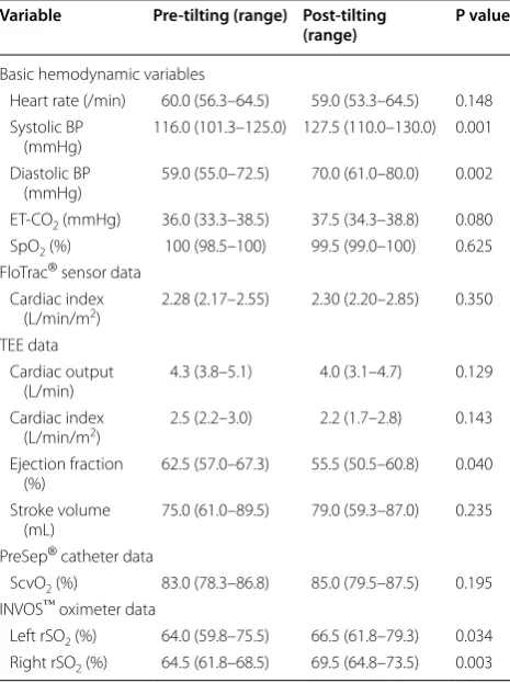
Table 2 Comparison of circulatory variables
Data are expressed as medians with interquartile ranges
ET-CO2 end-tidal carbon dioxide, LVEDV left ventricular end-diastolic volume,
rSO2 regional saturation of oxygen, SpO2 percutaneous oxygen saturation, ScvO2
central venous blood oxygen saturation
Variable Pre-tilting (range) Post-tilting
(range) P value
Basic hemodynamic variables
Heart rate (/min) 60.0 (56.3–64.5) 59.0 (53.3–64.5) 0.148 Systolic BP
(mmHg) 116.0 (101.3–125.0) 127.5 (110.0–130.0) 0.001 Diastolic BP
(mmHg) 59.0 (55.0–72.5) 70.0 (61.0–80.0) 0.002 ET-CO2 (mmHg) 36.0 (33.3–38.5) 37.5 (34.3–38.8) 0.080 SpO2 (%) 100 (98.5–100) 99.5 (99.0–100) 0.625 FloTrac® sensor data
Cardiac index
(L/min/m2) 2.28 (2.17–2.55) 2.30 (2.20–2.85) 0.350 TEE data
Cardiac output
(L/min) 4.3 (3.8–5.1) 4.0 (3.1–4.7) 0.129 Cardiac index
(L/min/m2) 2.5 (2.2–3.0) 2.2 (1.7–2.8) 0.143 Ejection fraction
(%) 62.5 (57.0–67.3) 55.5 (50.5–60.8) 0.040 Stroke volume
(mL) 75.0 (61.0–89.5) 79.0 (59.3–87.0) 0.235 PreSep® catheter data
ScvO2 (%) 83.0 (78.3–86.8) 85.0 (79.5–87.5) 0.195 INVOS™ oximeter data
[image:3.595.57.289.112.291.2]and Felabella et al. all reported decreases in CI [2, 6, 8,
9]. Age, cardiovascular status, obesity and volume sta-tus may influence these variables [1]. According to a study in which a fluid challenge was administered, arte-rial elastance is an important influence on mean artearte-rial pressure (MAP) response to head-down tilting and crea-tion of a pneumoperitoneum [10]. We did not restrict fluids in our study, nor did we measure LVEDP, which is an important variable. Given that we did not restrict vol-ume of infused fluids, a high LVEDP may have helped us to identify fluid overloading. We were unable to measure LVEDP or left atrial pressure in our patients as we did not catheterize the pulmonary artery, a substantially more invasive means of measuring CO and CI. We also did not measure pulmonary venous flow velocity pattern to make assessments of changes in LVEDP.
Several authors have recommended fluid restriction during radical prostatectomy to facilitate adequate visu-alization of the surgical field and minimize blood loss, because this procedure may induce adverse respiratory, cardiovascular, and neurophysiological changes such as severe laryngeal edema, respiratory complications and brachial plexus injury [10, 11]. In contrast, some surgeons have insisted that restricted fluid management (defined as <2000 mL for each case) causes postoperative oliguria [12], and the surgeons in our institute judge that intra- or postoperative hypovolemia may result in more serious adverse events than an obscured operative field during bladder neck transection. The policy of our institution is therefore not to restrict intravenous fluids during RALP.
A patient with undiagnosed mild mitral valve insuf-ficiency reportedly experienced transitory exacerbation of that insufficiency during RALP [2]. A possible expla-nation for this is increased left ventricular after- and pre-loads; however, the degree of mitral insufficiency reverted to the initial status after the patient was reposi-tioned [2].
In our study, the percutaneous rSO2 increased with
head-down tilt, consistent with other studies [10, 13]. We also measured ScvO2, which did not change. These
findings support the conclusion that the effects of steep head-down tilt and pneumoperitoneum on the cerebral circulation are relatively small. Using second-genera-tion near infrared spectroscopy, Kalmar et al. recently reported that cerebral perfusion pressure was maintained above the lower threshold of cerebral autoregulation dur-ing RACP, likely because of simultaneous increases in MAP and central venous pressure [10].
Conclusions
We found that LVEF decreased after steep head-down tilting and establishment of pneumoperitoneum for
RALP, whereas CI and LVEDV measured by TEE, and CI measured by FloTrac®, did not change. These find-ings indicate that the steep head-down tilt and pneumop-eritoneum during RALP did not greatly influence cardiac function in our cohort. Additionally, assessment of CI with TEE did not provide more useful information con-cerning cardiac function than was provided by FloTrac® in these patients.
Limitations
The limitations of our trial include its small size, the cri-teria for patient selection and the use of volume-con-trolled ventilation during RALP. Different settings for tidal volume and PEEP, or pressure-controlled ventila-tion, may have produced different results. In our insti-tute, only patients who meet the criteria for ASA I–II undergo RALP. Because a pneumoperitoneum and steep Trendelenburg position are well tolerated by individuals with normal cardiac function, further clinical trials are required to evaluate those with cardiac or valve dysfunc-tion or high pulmonary vascular pressure undergoing this procedure.
Abbreviations
BP: blood pressure; CI: cardiac index; CO: cardiac output; CO2: carbon dioxide;
LVEDP: left ventricular end-diastolic pressure; LVEDV: left ventricle end-diastolic volume; LVEF: left ventricular ejection fraction; MAP: mean arterial pressure; PEEP: positive end-expiratory pressure; RALP: robot-assisted laparoscopic pros-tatectomy; rSO2: regional saturation of oxygen; ScvO2: central venous blood
oxygen saturation; TEE: transesophageal echocardiography.
Authors’ contributions
NO and JN designed the study, interpreted the data and drafted the manu-script. SN corrected the data and reviewed the manumanu-script. TS confirmed the analysis of the data and reviewed the manuscript. TM designed the study and reviewed the manuscript. All authors read and approved the final manuscript.
Acknowledgements None.
Competing interests
The authors declare that they have no competing interests.
Availability of data and materials
The datasets during and/or analysed during the current study available from the corresponding author on reasonable request.
Consent for publication Not applicable.
Ethics approval and consent to participate
The study protocol was approved by the Ethics Committee of Osaka Medical College (Reference Number 1339); all participants provided written informed consent.
Funding None.
Publisher’s Note
• We accept pre-submission inquiries
• Our selector tool helps you to find the most relevant journal • We provide round the clock customer support
• Convenient online submission • Thorough peer review
• Inclusion in PubMed and all major indexing services • Maximum visibility for your research
Submit your manuscript at www.biomedcentral.com/submit
Submit your next manuscript to BioMed Central
and we will help you at every step:
Received: 23 August 2016 Accepted: 22 July 2017
References
1. Baltayian S. A brief review: anesthesia for robotic surgery. J Robot Surg. 2008;2:59–66.
2. Haas S, Haese A, Goetz AE, Kubitz JC. Haemodynamics and cardiac function during robotic-assisted laparoscopic prostatectomy in steep Trendelenburg position. Int J Med Robot. 2011;7:408–13.
3. Hong JY, Oh YJ, Rha KH, Park WS, Kim YS, Kil HK. Pulmonary edema after da Vinci-assisted laparoscopic radical prostatectomy: a case report. J Clin Anesth. 2010;22:370–2.
4. Secin FP, Jiborn T, Bjartell AS, Fournier G, Salomon L, Abbou CC, et al. Multi-institutional study of symptomatic deep venous thrombosis and pulmonary embolism in prostate cancer patients undergoing laparo-scopic or robot-assisted laparolaparo-scopic radical prostatectomy. Eur Urol. 2008;53:134–45.
5. Gainsburg DM. Anesthetic concerns for robotic-assisted laparoscopic radical prostatectomy. Minerva Anestesiol. 2012;78:596–604. 6. Rosendal C, Markin S, Hien MD, Motsch J, Roggenbach J. Cardiac and
hemodynamic consequences during capnoperitoneum and steep Tren-delenburg positioning: lessons learned from robot-assisted laparoscopic prostatectomy. J Clin Anesth. 2014;26:383–9.
7. Lestar M, Gunnarsson L, Lagerstrand L, Wiklund P, Odeberg-Wernerman S. Hemodynamic perturbations during robot-assisted laparoscopic radical prostatectomy in 45° Trendelenburg position. Anesth Analg. 2011;113:1069–75.
8. Darlong V, Kunhabdulla NP, Pandey R, Chandralekha Punj J, Garg R, et al. Hemodynamic changes during robotic radical prostatectomy. Saudi J Anaesth. 2012;6:213–8.
9. Falabella A, Moore-Jeffries E, Sullivan MJ, Nelson R, Lew M. Cardiac func-tion during steep Trendelenburg posifunc-tion and CO2 pneumoperitoneum
for robotic-assisted prostatectomy: a trans-oesophageal Doppler probe study. Int J Med Robot. 2007;3:312–5.
10. Kalmar AF, Foubert L, Hendrickx JF, Mottrie A, Absalom A, Mortier EP, et al. Influence of steep Trendelenburg position and CO2 pneumoperitoneum
on cardiovascular, cerebrovascular, and respiratory homeostasis during robotic prostatectomy. Br J Anaesth. 2010;104:433–9.
11. Casati A, Spreafico E, Putzu M, Fanelli G. New technology for noninvasive brain monitoring: continuous cerebral oximetry. Minerva Anestesiol. 2006;72:605–25.
12. Baltayian S. A brief review: anesthesia for robotic prostatectomy. J Robot Surg. 2008;2:59–66.