T E C H N I C A L N O T E
Open Access
Highly efficient method for isolation of total RNA
from adipose tissue
Susanna Cirera
Abstract
Background:RNA extraction is a crucial step for monitoring gene expression. Poor RNA quality (including degradation and remaining impurities) can result in misleading results. Isolation of RNA from animal tissues with high lipid content can be challenging. Especially, it is not trivial to isolate high quality RNA with a reasonable yield from adipose tissue. The aim of this study was to provide an optimized protocol for isolating total RNA from
adipose tissue. This was achieved by combining the advantages of the two routinely used methods, TRI ReagentW
and miRNeasy.
Findings:The miRNeasy method results in cleaner samples but more prone to degradation while the TRI ReagentW method results in samples contaminated with salts and solvents but more intact. The new protocol combines the best of both methods resulting in RNA of high quality and suitable for downstream experiments like RT-qPCR, microarrays and high-throughput sequencing.
Conclusions:The current protocol for total RNA isolation from adipose tissue yields sufficient amount of high quality total RNA free of contaminants.
Keywords:Adipose tissue, Total RNA isolation, RNA purity, RNA integrity
Background
Adipose tissue has traditionally been viewed as a passive storage of energy in the body but has recently been re-cognized as a complex and highly active metabolic and endocrine organ [1]. As such, adipose tissues are of key importance for research in diabetes, metabolic syndrome and obesity which as a result of its epidemic proportions worldwide has become the focus of interest for many scientists.
Due to its high content in fatty acids and in some in-stances its low cell number, adipose tissue poses some challenges when high quality RNA needs to be isolated for downstream gene expression applications (i.e. arrays, high-throughput sequencing or RT-qPCR). Many rou-tinely used protocols for isolation of RNA from adipose tissue result in RNA that is partially degraded and/or poor yield of RNA or the small RNAs are lost in the process. All these issues can cause misleading results and they are a big challenge specially when working with
limited amount of tissue (i.e. human biopsies). Therefore there is a need for a simple, reliable and effective proto-col to isolate high quality total RNA with sufficient yield from adipose tissues. Here an optimized and simple protocol which combines two widely used methods and allows rapid purification of high quality total RNA from adipose tissue is presented. Performance of the new protocol has been evaluated and compared with the standard methods for RNA purification.
Methods
Porcine retroperitoneal adipose tissue, snap frozen in liquid nitrogen and stored at −80°C was used for the present study. Animal care, maintenance and slaughter have been conducted according to the Danish “Animal Welfare Act”(LBK 1343 of 04/12/2007).
Four samples (2 lean animals and 2 fat animals) were used for the TRI ReagentW (MRC Inc., US) and miR-Neasy (Qiagen, Germany) methods. The 4 samples were the same for these 2 methods. For the combined protocol the number of samples was increased to 10 (5 lean ani-mals and 5 fat aniani-mals). The phenotypic characterization Correspondence:[email protected]
Department of Veterinary Clinical and Animal Sciences, Section for Animal Genetics, Bioinformatics and Breeding, Faculty of Health and Medical Sciences, University of Copenhagen, Frederiksberg, Denmark
of the animals as “lean” or “fat” is based on the body weight and on metabolic parameters [2].
RNA isolation from adipose tissue was performed using TRI ReagentW method (MRC Inc., US) and miR-Neasy method (Qiagen, Germany) and a combination of both (combined protocol). Around 100 mg of tissue were used to isolate total RNA with the three mentioned methods. Using TRI ReagentW and miRNeasy the exact protocols from the manufacturers were followed. For the
combined protocol, the homogenization was done in 2 ml of TRI ReagentWbuffer using a gentleMACS™Octo Dissociator system (Milteny Biotec, GmbH, Germany) with M tubes (Milteny Biotec, GmbH, Germany) and the RNA_02 program recommended by the manufac-turer. After homogenization the samples were incubated at room temperature for 5 minutes. Subsequently a cen-trifugation step at 12000 g at 4°C for 10 min was per-formed and the resulting fat monolayer (see Figure 1) was carefully avoided when pipetting the rest of the sam-ple into a clean 1.5 ml tube. 400 μl of chloroform was then added to the sample and mixed by vortexing (tho-roughly mixing is important for subsequent phase sepa-ration). After 3 minutes at room temperature the sample was centrifuged at 12000 g at 4°C for 30 min. After cen-trifugation, the sample separates into three phases with the RNA in the upper phase. The RNA phase was trans-ferred to a new tube without disturbing the interphase. The volume of the sample was precisely measured and 1.5 volumes of 100% ethanol were added and the sample was mixed thoroughly by inverting the tube several times. The sample was then loaded on the miRNeasy spin column (Qiagen) and from this step the manufac-turer’s protocol was followed. At the end the RNA was eluted in 30μl of RNAse-free water.
[image:2.595.57.291.89.257.2]Quantity and quality were assessed by Nanodrop ND-1000 spectrophotometer using OD260for calculation of the concentration and the ratios 260/280 and 260/230 Figure 1Fat monolayer.This fat monolayer appears after the first
centrifugation step in all 3 protocols and it should be carefully avoided.
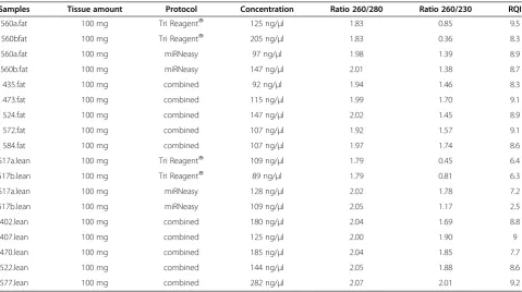
Table 1 Quantitative and qualitative measurements of the total RNA isolated with the 3 protocols
Samples Tissue amount Protocol Concentration Ratio 260/280 Ratio 260/230 RQI
560a.fat 100 mg Tri ReagentW 125 ng/μl 1.83 0.85 9.5
560bfat 100 mg Tri ReagentW 205 ng/μl 1.83 0.36 8.3
560a.fat 100 mg miRNeasy 97 ng/μl 1.98 1.39 8.9
560b.fat 100 mg miRNeasy 147 ng/μl 2.01 1.38 8.7
435.fat 100 mg combined 92 ng/μl 1.94 1.46 8.3
473.fat 100 mg combined 115 ng/μl 1.99 1.70 9.1
524.fat 100 mg combined 147 ng/μl 2.02 1.45 8.9
572.fat 100 mg combined 107 ng/μl 1.92 1.57 9.1
584.fat 100 mg combined 107 ng/μl 1.97 1.74 8.6
517a.lean 100 mg Tri ReagentW 109 ng/μl 1.79 0.45 6.4
517b.lean 100 mg Tri ReagentW 89 ng/μl 1.79 0.81 6.3
517a.lean 100 mg miRNeasy 128 ng/μl 2.02 1.78 7.2
517b.lean 100 mg miRNeasy 109 ng/μl 2.05 1.17 2.5
402.lean 100 mg combined 180 ng/μl 2.04 1.69 8.8
407.lean 100 mg combined 125 ng/μl 2.00 1.90 9
470.lean 100 mg combined 185 ng/μl 2.04 1.85 7.7
522.lean 100 mg combined 144 ng/μl 2.05 1.88 8.6
577.lean 100 mg combined 282 ng/μl 2.07 2.01 9.2
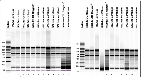
[image:2.595.59.539.449.717.2]for assessing the purity of the samples. Integrity of the isolated RNA was inspected by electrophoresis in a 1.4% agarose gel and by measuring the RQI value on an Experion™ system (BioRad) using Eukaryote Total RNA StdSens kit (BioRad) (see Table 1 and Figure 2). RNA isolation from the three methods was evaluated and compared (see Table 1, Figure 2 and Figure 3). Student’s T-test was performed to compare the ratios 260/280 and 260/230 and the RQI between the 3 methods.
Findings and discussion
Due to the high lipid content and relatively low cell num-ber of the adipose tissue, many methods for isolation of RNA from this tissue have been tested ([3-8], among others). Some of these methods are very time consuming and/or require high amount of starting tissue. Others do not report the purity as measured by microfluidics: I.e. Guan & Yang [4] and Duckett et al. [5] implemented a protocol similar to the combined method described in the present study. However, the authors did not test the integ-rity of the RNA with microfluidics. Verification of the RNA integrity by microfluidics (Experion or Bioanalyzer) is crucial in order to evaluate if the isolated RNA is of suf-ficient quality for downstream applications [9]. This short-coming makes direct comparison of the methods difficult. Penaet al.[6] used a simplified version of the TRI ReagentW method but their RNA integrity results were considerably lower than the ones reported with our combined protocol. Recently, Pratt et al. [8] have developed a protocol
(modified from Duckett et al. [5]) using the miRVana miRNA Isolation kit and they report comparable yield and integrity results to our method. Nevertheless, the authors start the protocol with 1 gr of adipose tissue and the RNA integrity results are reported from only 2 samples.
In the present study a protocol for total RNA isolation from adipose tissue was developed based on widely used methods. The purity of the samples isolated with the new protocol was excellent: A 260/280 ratio of 2.00 ± 0.049 (1.81 ± 0.023 for TRI Reagent and 2.01 ± 0.03 for miRNeasy) and a 260/230 ratio of 1.73 ± 0.189 which was significantly higher (P< 0.05) than the other two (0.61 ± 0.25 for TRI Reagent and 1.43 ± 0.25 for miR-Neasy). The RNA quality index (RQI) assessed by the ladder 473.fat.combined 524.fat.combined 572.fat.combined 560a.fat.TRI Reagent 560a.fat.miRNeasy
®
584.fat.combined 407.lean.combined 470.lean.combined 522.lean.combined 577.lean.combined 517a.lean.TRI Reagent
®
517a.lean.miRNeasy ladder 560b.fat.TRI Reagent
®
560b.fat.miRNeasy 435.fat.combined 517b.lean.miRNeasy 517b.lean.TRI Reagent
®
[image:3.595.57.540.90.355.2]402.lean.combined 402.lean.combined 407.lean.combined 470.lean.combined 522.lean.combined 577.lean.combined
Figure 2ExperionTMgel image.The RQI corresponding values can be found in Table 1. Lane identity is given at the top of the gel image.
[image:3.595.307.538.572.706.2]Experion™ system resulted also in excellent values for the samples isolated with the new protocol, RQI = 8.7 ± 0.36 (7.62 ± 1.55 for TRI Reagent and 6.82 ± 2.98 for miRNeasy) which is quite unusual for adipose samples (see Table 1, Figure 2 and Figure 3).
The RNA yield was also higher for the combined protocol, 148.4 ± 56.25 (132 ± 50.84 for TRI Reagent and 120.25 ± 21.93 for miRNeasy).
The TRI ReagentW method showed problems with the purity of the isolated RNA. The ratio of 260/280 was lower than 1.8-2 indicating the presence of proteins. Fur-thermore, the 260/230 ratio, which should ideally be 1.8 or greater, was consistently very low (under 1) revealing remains of solvents (i.e. phenol/chloroform) or chaotropic salts (guanidine isothiocyanate) in the RNA solution (see Table 1). It is well known that all phenol-based methods have almost always little remaining of the organic solvents in the final isolated sample and that all these impurities remaining in the sample can compromise downstream applications.
The miRNeasy method resulted in RNA samples with 260/280 ratios around 2 and 260/230 ratios over 1. Nevertheless, the RNA yield for the samples originating from fat animals was 22-28% lower than with the TRI ReagentW method (see Table 1), even though we started both methods with the same amount of tissue and with exactly the same sample. Furthermore the integrity of the RNA, especially for the samples originated from lean animals was lower than with the TRI ReagentWmethod, indicating that the lipids carried over after the phase sep-aration had more drastic effects during the process of the miRNeasy RNA isolation than with the TRI ReagentW method.
The homogenization step is very crucial for the quality of the final isolated RNA. Independently of the homo-genization buffer used (TRI ReagentWor QIAzol) we ex-perienced that if the disruption was performed using a mortar and pestle, the fatty acids were abundant in the sample after the homogenization and the fat monolayer was very evident after the initial centrifugation step (see Figure 1). In contrast, if the homogenization was per-formed using the gentleMACS™machine most of the fat tended to stick to the walls of the M tubes used for homogenization and the monolayer, after the initial centri-fugation step, was very small and easy to avoid and there-fore the problem of fat carried over to the next steps was minimized. In the samples from lean animals, the fat did not stick so much to the plastic walls of the M tubes and the fat monolayer was more evident and difficult to avoid after the first centrifugation step. Therefore, the samples from lean animals performed worse than the samples orig-inated from fat animals (lower 260/230 and 260/280 ra-tios, more degradation in the agarose gel and lower RQI, see Table 1 and Figure 2) in all the three protocols used in
the present study but the effects were much less dramatic when using the new protocol.
In the step of phase separation, after addition of chloroform and 15 min. centrifugation at 12000 g and 4°C, the interphase was usually very diffuse and difficult to avoid. In order to make it more compact to avoid car-rying it over when transferring the RNA upper phase, the centrifugation was extended from 15 minutes to 30 minutes.
When running the RNA samples in agarose gels or in the Experion™ system the samples from the miRNeasy protocol were slightly more degraded than the ones from the TRI ReagentW and the new protocols (see Table 1 and Figure 2).
Conclusion
For total RNA isolation from adipose tissue, the miR-Neasy method results in cleaner samples but more prone to degradation and the TRI ReagentW method results in samples contaminated with salts and solvents but more intact. The new protocol combines the best of both methods yielding sufficient amount of high quality total RNA from adipose tissue free of contaminants and sui-table for downstream experiments like RT-qPCR, micro-arrays and high-throughput sequencing. At present, we use this protocol routinely in our laboratory starting from frozen adipose tissue and from isolated mature adi-pocytes with excellent results.
Competing interests
The author declares not to have any competing interests.
Acknowledgements
I thank Peter K. Busk and Peter Karlskov-Mortensen for critical comments to the manuscript.
Received: 3 January 2013 Accepted: 1 November 2013 Published: 18 November 2013
References
1. Kershaw EE, Flier JS:Adipose tissue as an endocrine organ.
J Clin Endocrinol Metab2004,89:2548–2556.
2. Kogelman LJ, Kadarmideen HN, Mark T, Karlskov-Mortensen P, Bruun CS, Cirera S, Jacobsen MJ, Jørgensen CB, Fredholm M:An f2 pig resource population as a model for genetic studies of obesity and obesity-related diseases in humans: design and genetic parameters.Front Genet.
2013,4:29.
3. Tavangar K, Hoffman AR, Kraemer FB:A Micromethod for the isolation of total RNA from adipose tissue.Analitical Biochem1990,186:60–63. 4. Guan H, Yang K:RNA isolation and real-time quantitative RT-PCR.In
Adipose Tissue Protocols. Methods in Molecular Biology, vol 456.Edited by Yang K. Totowa, NJ: Humana press; 2008:259–270.
5. Duckett SK, Pratt SL, Pavan E:Corn oil or corn grain supplementation to steers grazing endophyte-free tall fescue. II. Effects on subcutaneous fatty acid content and lipogenic gene expression.J Anim Sci2009, 87:1120–1128.
6. Pena RN, Cánovas A, Estany J:Technical note: Efficient protocol for isolation of total ribonucleic acid form lyophilized fat and muscle pig samples.J Anim Sci2010,88:442–445.
8. Pratt SL, Burns TA, Owens MD, Duckett SK:Isolation of total RNA and detection procedures for miRNA present in bovine-cultured adipocytes and adipose tissues.Methods Mol Biol2013,936:181–194.
9. Bustin SA, Benes V, Garson JA, Hellemans J, Huggett J, Kubista M, Mueller R, Nolan T, Pfaffl MW, Shipley GL, Vandesompele J, Wittwer CT:The MIQE guidelines: minimum information for publication of quantitative real-time PCR experiments.Clin Chem2009,55:611–622.
doi:10.1186/1756-0500-6-472
Cite this article as:Cirera:Highly efficient method for isolation of total RNA from adipose tissue.BMC Research Notes20136:472.
Submit your next manuscript to BioMed Central and take full advantage of:
• Convenient online submission
• Thorough peer review
• No space constraints or color figure charges
• Immediate publication on acceptance
• Inclusion in PubMed, CAS, Scopus and Google Scholar
• Research which is freely available for redistribution