R E S E A R C H A R T I C L E
Open Access
Elevated serum leptin levels in patients with acute
myocardial infarction; correlation with coronary
angiographic and echocardiographic findings
Hadi AR Hadi Khafaji
1, Abdul Bari Bener
2, Nasser M Rizk
3and Jassim Al Suwaidi
1,4*Abstract
Background:To assess the relationship between serial serum leptin levels in patients with acute myocardial infarction (AMI) who received thrombolysis and the degree of coronary atherosclerosis, coronary reperfusion, echocardiographic findings, and clinical outcome. 51 consecutive patients presenting with AMI were studied. Clinical characteristics including age, sex, body mass index (BMI) and cardiovascular risk factors were recorded. Serial serum leptin levels at the time of admission and subsequently at 0, 6, 12, 24, 36, 60 hours afterwards were obtained. Coronary angiography was performed in 34 patients; the relation between serum leptin levels and evidence of coronary reperfusion as well as the extent of coronary atherosclerosis according to the coronary artery surgery study classification (CASS) were evaluated. Echocardiographic evaluation was performed in all patients. 36 matched patients were enrolled as control group who had serum leptin level 9.4 ± 6.5 ng/ml.
Results:The patients mean age was 50.5 ± 10.6 years. There were 47 males and 3 females. 37.1% were diabetics, 23.5% were hypertensive, 21.6% were dyslipidemic and 22.7% were obese (BMI≥30). Leptin concentrations (ng/ml) increased and peaked at the 4th sample (36 hrs) after admission (mean ± SD) sample (1) =9.55 ± 7.4, sample (2) =12.9 ± 8.4, sample (3) =13.8 ± 10.4, sample (4) =18.9 ± 18.1, sample (5) =11.4 ± 6.5, sample (6) =10.8 ± 8.9 ng/ml. There was a significant correlation between serum leptin and BMI (r = 0.342;p= 0.03). Leptin levels correlated significantly to creatine kinase level on the second day (r = 0.43,p≤0.01). Significant correlation of mean serum leptin with the ejection fraction (P<0.05) was found. No difference in timing of peak serum leptin between patients who achieved coronary reperfusion vs. those who did not (p= 0.8). There was a trend for an increase in the mean serum leptin levels with increasing number of diseased vessels. There was no correlation between serum leptin levels and outcome neither during the hospitalization nor at 9 months follow up.
Conclusion:Serum leptin levels increase after myocardial infarction. Serum leptin level may be a predictor of the left ventricular ejection fraction and the degree of atherosclerosis but not of coronary reperfusion.
Keywords:Serum leptin, Acute myocardial infarction, Angiographic findings, Echocardiography
* Correspondence:jha01@hmc.org.qa;jalsuwaidi@hotmail.com 1Heart Hospital, Hamad Medical Corporation, Doha, Qatar 4
Department of Adult Cardiology Hamad Medical Corporation - Heart Hospital, P.O. Box 3050, Doha, Qatar
Full list of author information is available at the end of the article
Background
Leptin is the 16,000 Dalton protein product of the obes-ity gene (ob) [1]. Leptin has a role in the body weight maintenance in humans. It is released into the blood stream, where it binds to leptin-binding protein and is transported into the cerebrospinal fluid [2-5] and exerts its major effect on the hypothalamus. The role of leptin in coronary artery vasoreactivity has been raised in a study by Sundell J et al. [6] in obese and non-obese patients, suggesting that leptin might have a role in the regulation of the myocardial blood flow. A preliminary study in a small number of patients suggested that leptin levels might increase with acute myocardial infarction [7]. Leptin is found to have multiple roles in the cardio-vascular system. A number of investigators suggested its role as a vasoactive substance [7]. There is also some evidence that leptin may have a role in obesity-related hypertension [8]. In a human study, a correlation was seen between the serum concentration of leptin and blood pressure among patients with essential hyperten-sion [9]. Leptin also may have a prothrombotic effect [10]. This effect appears to be mediated through the platelet leptin receptor. In a previous study we suggested that TNF alpha may represent a modulator of leptin ac-tion in the hypothalamus, such finding may have impli-cation in the setting of acute myocardial infarction [11].
The aims of the current study were to evaluate serial levels of serum leptin among patients presenting with acute myocardial infarction (AMI) and whether there is a correlation between leptin and coronary reperfusion as well as with angiographic and echocardiographic data.
Methods Study design
This study was conducted at Hamad General Hospital. 51 patients who were admitted to the Coronary Care Unit with a diagnosis of acute coronary syndrome (ACS) were studied; after written informed consent for partici-pation in the study was obtained from participants. The definition of ACS in this study is according to the defin-ition for acute myocardial infarction by the joint com-mittee of American College of Cardiology/European Society of Cardiology [12]. Patients with major co-mor-bid conditions including renal failure were excluded from the study. Hamad Medical Corporation institu-tional review board approved the study.
Baseline clinical characteristics including age, sex, car-diovascular risk factors, and complete carcar-diovascular physical examination findings were recorded. The mode of administered therapy was also recorded. 49 patients with ST elevation myocardial infarction (STEMI) were given thrombolytic therapy with either metalyse, or streptokinase [13]. Tow patients suffered non-ST eleva-tion myocardial infarceleva-tion (NSTEMI). None of these
patients developed acute complication such as cardio-genic shock or acute homodynamic disturbance. Height, weight, body mass index, smoking habit, alcohol intake, diabetes mellitus, hypertension, lipid profile including serum cholesterol, high density and low density lipopro-tein, and serum creatinine of patients were analyzed. Measurements of serum creatinine kinase, creatinine kinase-MB portion, troponin T levels were taken at the time of admission (0 time) and subsequently at 6, 12, 24, 36, 60 hours afterwards. Full echocardiographic studies were performed for all patients on the 2ndday of admis-sion. Variables including left ventricular ejection fraction (LV EF%), left ventricular end-systolic dimension (LVESD), left ventricular end-diastolic dimension (LVEDD), left atrial size, right ventricular dimension (RVD) and right ventricular systolic pressure (RVSP) were all recorded. Coronary angiography was performed in 36 patients as a routine clinical check up. Extent of diseased vessels involved; whether single vessel, two -vessel, or three-vessel diseases, and whether re-canalization achieved (defined non-invasively as rapid resolution of ST segment eleva-tion reperfusion arrhythmias and chest pain resolueleva-tion after thrombolytic therapy or angiographically by the evidence of non-occluded culprit coronary artery) were recorded.
36 matched (age and BMI) stable cardiac patients with the same age and sex were taken as a control group and their data were collected from the cardiology out patient department.
Biochemical analyses
by Elecys 2010, Roche, according to the manufacturer pro-tocols. Imprecision, between run (CVs), for troponin & CKMB were 2% & 3% respectively [17].
Normal fasting level of serum leptin
Normal fasting ranges for serum leptin are directly cor-related with the degree of adiposity, with BMI range 18– 25; serum leptin level for men (3.8 ± 1.8 ng/ml), and for women (7.4 ± 3.7 ng/ml). Serum leptin level rises ap-proximately 2.5 times faster in women per unit BMI as compared to man. Keeping in mind that a diurnal rhythm of serum leptin concentrations, the values being 20 to 40 percent higher in the middle of the night as compared with daytime [18,19]. The peak shifts are in parallel with shifts in the timing of meals [20].
Coronary angiography and echocardiographic analysis
Coronary angiography was performed according to the standard Judkin technique femoral approach either on admission or during the recovery period. Several views
of each coronary artery were analyzed. The severity of arterial stenosis (defined as maximal percent reduction in luminal diameter) was determined according to visual estimation using the coronary artery surgery study clas-sification (CASS) study analysis. Significant coronary stenosis was defined as a 70% lumen narrowing or> than 50% lumen narrowing of the left main coronary ar-tery. The extent of coronary artery disease was classified as 1, 2 or 3 vessels according to number of major coron-ary arteries with significant stenosis [21].
[image:3.595.63.539.101.449.2]Transthoracic 2 dimensional echocardiography 3 megahertz, 5500 Hewlett Packard Sonos machine was used for the study. Images were captured in the 5 stand-ard views, parasternal long axis views, short axis view and 4, 2 &3 chamber views according to criteria of American society of echocardiography. The measure-ment of left ventricular dimensions was performed from 2 dimensional targeted M-mode at end diastolic and end systolic dimensions and subsequently LV ejection frac-tion was calculated. Left atrial size at end systole was Table 1 Clinical baseline characteristics compared with mean and peak leptin sample
Variables
Mean Leptin Peak Leptin
Normoleptinemia hyperleptinemia Normoleptinemia hyperleptinemia
N = 13 (29.5%) N = 31 (70.4%) N = 5 (13.1%) N = 33 (86.8%)
Sex Male 11(84.6%) 31(100.0%) 4(80.0%) 32 (97.0%)
Female 2(15.4%) 0(0.0) 1(20.0%) 1(3.0%)
Age group ≤50 years 7(53.8%) 17(54.8%) 2(40.0%) 17 (51.5%)
>50 years 6(46.2%) 14(45.2%) 3(60.0%) 16 (48.5%)
Diabetic Yes 8(61.5%) 9(29.0%) 5(100.0%) 10 (30.3%)
No 5(38.5%) 22(71.0%) 0(0.0) 23 (69.7%)
Hypertensive Yes 4(30.8%) 5(16.1%) 1(20.0%) 6(18.2%)
No 9(69.2%) 26(83.9%) 4(80.0%) 27(81.8 %)
Body mass index (Kg/m2) <25 5(45.5%) 2(6.9%) 2(40.0%) 3(10.0%)
25-30 4(36.4%) 19(65.5%) 3(60.0%) 18(60.0%)
>30 2(18.2%) 8(27.6%) 0(0.0) 9(30.0%)
High Cholesterol>5.2 mmol/l Yes 5(38.5%) 4(12.9%) 1(20.0%) 7(21.2%)
No 8(61.5%) 27(87.1%) 4(80.0%) 26(78.8%)
HDL* Abnormal (<1)mmolL 10(90.9%) 10(37.0%) 5(100.0%) 12(41.4%)
Normal(>1) mmol/L 1(9.1%) 17(63.0%) 0(0.0) 17(58.6%)
LDL ≤3.3 mmol/L 4(40.0%) 20(69.0%) 3(75.0%) 19(61.3%)
>3.3 mmol/L 6(60.0%) 9(31.0%) 1(25.0%) 12(38.7%)
Triglyceride ≤1.7 mmol/L 6(66.7%) 7(24.1%) 1(25.0%) 11(35.5%)
>1.7 mmol/L 3(33.3%) 22(75.9%) 3(75.0%) 20(64.5%)
MI type Anterior 8(66.7%) 19(61.3%) 4(80.0%) 19(59.4%)
Inferior 4(33.3%) 10(32.3%) 1(20.0%) 11(34.4%)
Non ST Elevation 0(0.0) 2(6.5%) 0(0.0) 2(6.3%)
measured from the parasternal long axis view. Right ven-tricular dimensions were recorded from the parasternal long axis view. Conventional Doppler measurements were obtained using tricuspid inflow, mitral early and late diastolic inflow velocities, volumetric relaxation times were also recorded. Wall motion abnormalities were recorded using 16 segment models [22].
Statistical analysis
The data were analyzed by using the Statistical Packages for Social Sciences [SPSS] version 19 [23]. Data were
expressed as mean and standard deviation (SD) unless otherwise stated; studentttest was used to ascertain the significance of difference between mean values of two continuous variables and confirmed by non-parametric Mann Whitney test. Fisher exact and Chi square test were performed to test for difference in proportions of categorical variable between two and more groups. The Pearson’s correlation coefficient was used to evaluate the strength association between two variables. The level of
P<0.05 was considered as the cut off value for signifi-cance. Repeated measures ANOVA utilized to test differ-ences in serial leptin measures.
Results
[image:4.595.54.545.101.379.2]Baseline clinical characteristics of the studied patients shown in Table 1. The mean age of patients was 50.5 ± 10.6 years. There were 47 males and 4 females. 37.1% had diabetes mellitus, 23.5% had hypertension, 21.6% had high cholesterol, and 22.7% were obese (BMI≥30). Thirty six matched (with age and BMI) stable cardiac patients were taken as a control group. The mean serum leptin level of the control group was 9.43 ± 6.5 ng/ml.
Table 2 shows echocardiographic and coronary angio-graphic characteristics compared with mean and peak serum leptin sample.
Table 2 Echocardiographic & coronary angiographic characteristics compared with mean and peak leptin sample
Variables Mean Leptin Peak Leptin
Normoleptinemia Hyperleptinemia Normoleptinemia Hyperleptinemia
N = 13 N = 31 N = 5 N = 33
L. Atrium 1.9-4.0 cm 10(76.9%) 19(63.3%) 3(60.0%) 22(68.8%)
>4.0 cm 3(23.1%) 11(36.7%) 2(40.0%) 10(31.3%)
LVEDD ≤5.6 cm 11(84.6%) 23(76.7%) 4(80.0%) 26(81.3%)
>5.6 cm 2(15.4%) 7(23.3%) 1(20.0%) 6(18.8 %)
RVD ≤3.0 cm 12(92.3%) 30(100%) 5(100.0%) 31(96.9%)
>3.0 cm 1(7.7%) 0(0.0) 0(0.0) 1(3.1%)
RVSP ≤30 mmHg 4(30.8%) 13(44.8%) 2(40.0%) 14(45.2%)
>30 mmHg 9(69.25%) 16(55.2%) 3(60.0%) 17(54.8%)
FS% ≤25% 6(46.2%) 12(40.0%) 3(60.0%) 17(53.1%)
>25% 7(53.8%) 18(60.0%) 2(40.0%) 15(46.9%)
EF% ≤45% 9(69.2) 16(53.3%) 1(20.0%) 13(40.6%)
>45% 4(30.8%) 14(46.7%) 4(80.0%) 19(59.4%)
No of vessels 1VD 4(57.1%) 6(37.5%) 2(50.0%) 8(50.0%)
2VD 3(42.9%) 6(37.5%) 2(50.0%) 4(25.0%)
3VD 0(0.0) 6(37.5%) 0(0.0) 4(25.0%)
Recanalisation Yes 7(87.5%) 15(71.4%) 4(80.0%) 15(75.0%)
(noninvasive assessment) No 1(12.5%) 6(28.6%) 1(20.0%) 5(25.0%)
Normoleptinemia: serum leptin level for male = (3.8 ± 1.8 ng/ml), and for female (7.4 ± 3.7 ng/ml) with BMI range 18–25. Hyperleptinemia: serum leptin level>(3.8 ± 1.87 ng/ml) for males, for females>(7.4 ± 3.77 ng/ml) with BMI range 18–25.
[image:4.595.56.292.565.696.2]Figure 1 shows the mean values of serum leptin level during the course of AMI at different time points. Lep-tin concentrations (ng/ml) increased and peaked at fourth reading (36 hrs) after admission and gradually decreased thereafter (mean ± SD sample (1) =9.55 ± 7.4, sample (2) = 12.9 ± 8.4, sample (3) =13.8 ± 10.4, sample (4) =18.9 ± 18.1, sample (5) =11.4 ± 6.5, sample (6) =10.8 ± 8.9) ng/ml,P value 0.04 based on repeated mea-sures ANOVA.
There was a significant correlation between serum lep-tin and BMI (r = 0.34; p= 0.03). Leptin levels also corre-lated significantly with serum creatine kinase level on the second day (r = 0.43, p≤0.01). There was no differ-ence in timing of peak serum levels between patients who had evidence of coronary reperfusion compared to those who did not achieve coronary reperfusion (p= 0.8),
also there was no significant difference in mean serum leptin level in patients with anterior MI vs. inferior MI (mean ± SD = 11.71 ± 7, 9 vs.11.86 ± 6.6 ng/ml,p= 0.9) re-spectively. However, there was significant correlation with left ventricular ejection fraction, LVEF (r =−0.25,
p= 0.01). No significant correlation between mean serum leptin and left atrial size (r =−0.09;p= 0.5), left ventricu-lar systolic (r = 0.017, p= 0.9) or diastolic dimension (r = 0.1;p= 0.47) were found.
Figure 2 shows that mean serum leptin, which appeared to increase with the increase in the number of diseased vessels (mean, SVD = 9.2, 2VD = 12.0, 3VD = 12.9,p= 0.6), although this was not statistically significant.
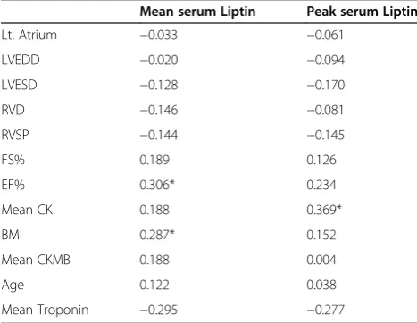
[image:5.595.58.290.89.244.2]Correlation between mean and peak leptin level and variables of interest showed significant correlation of mean serum leptin with the ejection fraction and body mass index (P<0.05), and significant correlation of cre-atinine kinase with mean leptin level (P<0.05) (Table 3).
Discussion
Studies on the role of serum leptin in coronary artery disease are scarce [24-34] (Table 4). To the best of our knowledge, this study is among few studies that analyzed the serial serum leptin levels in the setting of acute myo-cardial infarction and the first that evaluated its correl-ation with angiographic and echocardiographic findings in this high-risk group.
In the United States population, increased leptin con-centrations was significantly associated with increased risk of myocardial infarction and stroke in men and women, independent of traditional cardiovascular risk factors and obesity status [36]. We have shown previously a significant correlation between serum leptin and hs-CRP in stable cardiac patients [37]. Study by Stangl et al. [38] concluded that patients with coronary artery disease exhibited higher serum leptin concentrations than controls matched for age, gender & BMI, suggesting that leptin could contribute to the development of cardiovascular disease, possibly via activation of the sympathetic nervous system. The Trp64Arg variant of theβ-adrenoceptor did not influence serum leptin levels [38]. Leptin might be a marker of risk of coronary artery disease, at least in men, and contributes to the risk profile in subjects with insulin resistance. Lep-tin concentrations were significantly higher in diabetic and coronary artery disease patients than in controls. Body weight, serum triglyceride concentration and systolic blood pressure were all significantly related to the logarithm of the serum leptin concentration in stable cor-onary artery disease patients [39]. Wallace et al. [40] docu-mented that leptin is an independent risk factor for coronary artery disease using data from the west Scotland coronary prevention study. It has been suggests that leptin might participate in the catabolic state leading to
[image:5.595.56.290.518.699.2]Figure 2Serum leptin level by number of diseased vessels (VD). (SVD = 9.2, 2VD = 12.0, 3VD = 12.9 ng/ml,p= 0.6).
Table 3 Correlation between mean Leptin level and variables of interest
Mean serum Liptin Peak serum Liptin
Lt. Atrium −0.033 −0.061
LVEDD −0.020 −0.094
LVESD −0.128 −0.170
RVD −0.146 −0.081
RVSP −0.144 −0.145
FS% 0.189 0.126
EF% 0.306* 0.234
Mean CK 0.188 0.369*
BMI 0.287* 0.152
Mean CKMB 0.188 0.004
Age 0.122 0.038
Mean Troponin −0.295 −0.277
*Two sided p value less than 0.05.
development of cardiac cachexia in the course of congest-ive heart failure [41].
The current study demonstrates elevation of serum leptin levels in the acute phase of myocardial infarction and it peaks at 36 hours after admission (doubled). Whether leptin is released from the myocytes or it acts only as an acute phase reactant protein after its release from adipose tissue, was the main issue of this project. The fact that there was no earlier peak in serum leptin in patients who had coronary reperfusion after thrombo-lytic therapy compared to those who don’t achieve reperfusion suggests that leptin is not released from the myocytes and is mainly an acute phase reactant similar to high sensitivity C- reactive protein. This finding is concordant to the study in Poland, which included 35 patients with AMI and showed that plasma leptin levels in diabetic patients were significantly higher in AMI than in the period of convalescence. These findings sug-gest that leptin may play an important role in the
metabolic changes taking place during the first days of AMI [24].
[image:6.595.55.541.102.464.2]Correlating mean and peak serum leptin levels; serum leptin is positively correlated with the extent of diseased coronary vessels (1-, 2-, and 3 -vessel disease) (Figure 2), although statistically not significant, expanding the study sample may be confirmatory of this finding. Furthermore no significant correlation with either evidence of coron-ary re-canalization or with the type of MI (whether anter-ior or inferanter-ior) was found. We observed a significant correlation between serum leptin and LVEF with no sig-nificant correlation with other echocardiographic find-ings. Significant correlations were found between high serum leptin level and BMI as was demonstrated by pre-vious studies (Table 3), with no significant correlation to other cardiovascular risk factor; diabetes, hypertension, total cholesterol LDL, HDL or triglyceride in this patients’ population. In contrast to our study which has followed the leptin up to 60 hours after the onset of Table 4 Studies on leptin in acute myocardial infarction
study patients conclusion
Hadi Khafaji et al. 2011 Qatarcurrent study
87 pts: 51 AMI pts + 36 matched control
Leptin level is significantly correlated with BMI, CK, and LVEF. But neither with the type of MI (anterior vs. inferior) nor with the severity of angiographic findings and other echocardiographic parameters.
Yan GT,et al. * 2005 China [28]
AMI and CS pts Leptin levels of both AMI & coronary atherosclerosis pts are significantly without a significant difference between each other, no correlation for leptin with CRP, TnT & endothelin
Krasnodebski et al. 2010 Poland [24]
58 patients with AMI Plasma leptin levels in diabetic pts are significantly in acute stage of AMI than in the period of convalescence.
Wallander M,et al. 2008Sweden [29]
181 AMI Circulating levels of leptin on the first morning after an AMI are associated with the presence of abnormal Glucose tolerance at discharge & with poor long-term prognosis.
Piestrzeniewicz et al. 2007 Poland [31]
40 obese and 40 non-obese men.
Leptinemia is associated with fasting glucose, triglyceride levels, CRP & uric acid & negatively with HDL-C; leptin may be a pathogenetic factor in cardiovascular disease
Taneli et al. 2006 Turkey [30]
35 pts stable angina + 40 STEMI, + 30 control
S. leptin is significantly in both stable angina &STEMI groups. No significant correlation between leptin levels & selected risk factors
Amasyali et al. 2006Turkey [25]
41 pts with AMI have thrombolysis
Failure of reperfusion therapy with streptokinase is significantly in patients with admission plasma leptin≥14 ng/mL vs. pts with admission plasma leptin <14 ng/mL.
Selvakumar et al. 2005 India [32]
94 pts with acute ST EMI + 46 controls
leptin level is in patients with acute STEMI
Meisel et al. 2001 Isreal [26]
30 pts, AMI Leptin peaked on the 2nd day of hospitalization with a 2-fold from its baseline level on admission (p<0.02). On day 3, leptin levels, & were 46%, 9%, &6% above baseline on days 3, 4 and 5, respectively.
Fujimaki et al. 2001 Japan [35]
21 AMI pts 15 control
Hypoleptinaemic AMI pts had significantly plasma LDH vs. normoleptinaemic pts. No differences in other serum markers between normo vs. hyperleptinaemic AMI
Söderberg et al. 1999 Sweeden [33]
62 men with first-ever AMI
High leptin (OR 8.97; 95% CI: 1.73-46.5) & cholesterol (OR 5.18; 95% CI: 1.34-20.0) levels significant risk factors for AMI in a multivariate model
Stejskal et al. 1988 Czech** [34]
16 AMI, 22 unstable angina
Early after the acute coronary event, leptinemia in persons with AMI statistically positively correlated with concentration of interleukine-6 & subsequently of markers of coronary lesion severity (cTnI).
*Article in Chinese, information taken from the abstract, ** Article in Czech information taken from the abstract.
AMI, investigators from Turkey studied the influence of plasma leptin concentrations obtained at the time of ad-mission and 6 hours afterwards in 41 AMI patients who were treated with thrombolytic therapy. The investigators found that failure of reperfusion therapy with streptokin-ase was significantly higher in patients with admission plasma leptin concentrations≥14 ng/mL as compared to patients with admission plasma leptin concentrations
<14 ng/m, i.e. hyperleptinemia decreased the chance of successful reperfusion . Left ventricular ejection fraction was slightly but significantly higher in patients with ad-mission plasma leptin concentrations≥14 ng/mL than in patients with admission plasma leptin concentrations
<14 ng/m (p= 0.031) [25]. Another study involving 30 consecutive AMI patients with a similar profile to the current one, showed that leptin levels reached its peak on the second day of hospitalization, with a 2-fold in-crease from baseline level on admission (p<0.02). On day 3, leptin levels declined, and were 46%, 9%, and 6% above baseline on days 3, 4 and 5, respectively, suggesting that leptin may have a role in the metabolic changes tak-ing place durtak-ing the first days after an AMI. [26].
The predictive power of leptin on cardiovascular dis-ease was addressed in a report from the Quebec cardio-vascular study[27], eighty-six patients who developed ischemic heart disease were compared with referent matched for a number of traditional cardiovascular risk factor including body mass index. Leptin did not emerge as a predictor in coronary artery disease. However, fun-damental differences between the two studies, first and most importantly, patients in our present study were all proven first ever AMI cases according to newly defined criteria [12], whereas in Quebec, the study group consti-tuted a mixture of stable and unstable angina [27]. Fur-thermore Fujimaki et al. [35] correlated serum leptin with other myocardial infarction markers and interleukin level in 15 aged-matched controls and found a signifi-cant negative correlation between these two markers. These studies again suggest that leptin may play an im-portant role in the metabolic changes taking place dur-ing the first days of myocardial infarction.
Limitations of the study
Since this study included a small number of patients, the result should be interpreted cautiously.
Conclusion
Serum leptin acts as an acute phase reactant in AMI patients. Significant correlation was found in mean serum leptin level with BMI, CK, and LVEF. There was no significant difference in mean serum leptin level in patients with anterior vs. inferior infarction and statisti-cally no significant correlations of serum leptin with
severity of angiographic findings and other echocardio-graphic parameters.
Abbreviations
BMI: body mass index; SVD: single vessel disease; 2VD: two vessel disease; 3VD: three vessel disease; LVEF: left ventricular ejection fraction; RVD: right ventricular dimension; RVSP: right ventricular systolic pressure; STEMI: ST elevation myocardial infarction.
Competing interests
The authors declare that they have no competing interests.
Authors’contributions
HARHK- participated in the design of the study, patients’recruitment, writing, analyzing and reviewing the paper. AB- performed the statistical analysis. NR- participated in the design of the study and the performance of laboratory investigations. JA- participated in the design of the study and patients’enrollment. All authors read and approved the final manuscript.
Acknowledgements
This study was funded by the Medical Research Center of Hamad Medical Corporation, Doha, Qatar [Grant No 263]. The study was conducted in collaboration with biomedical department of the Qatar University, Doha, Qatar. We sincerely acknowledge: Ms. Nora Basem and Mrs. Wafa Khalil for their laboratory work and their technical support in this study.
Author details
1
Heart Hospital, Hamad Medical Corporation, Doha, Qatar.2Dept. of Adult Cardiology, Dept. of Medical Statistics and Epidemiology, Doha, Qatar. 3
Biomedical Department, Qatar University, Doha, Qatar, Heart Hospital, Hamad Medical Corporation, Qatar, Biomedical Department, Qatar University, Doha, Qatar.4Department of Adult Cardiology Hamad Medical Corporation -Heart Hospital, P.O. Box 3050, Doha, Qatar.
Received: 8 December 2011 Accepted: 24 April 2012 Published: 29 May 2012
References
1. Zhang Y, Proenca R, Maffei M, Barone M, Leopold L, Friedman JM: Positional cloning of the mouse obese gene and its human homologue.
Nature1994,372:425.
2. Halaas JL, Gajiwala KS, Maffei M, Cohen SL, Chait BT, Rabinowitz D, Lallone RL, Burley SK, Friedman JM:Weight-reducing effects of the plasma protein encoded by the obese gene.Science1995,269:543.
3. Pelleymounter MA, Cullen MJ, Baker MB, Hecht R, Winters D, Boone T, Collins F:Effects of the obese gene product on body weight regulation in ob/ob mice.Science1995,269:540.
4. Campfield LA, Smith FJ, Guisez Y, Devos R, Burn P:Recombinant mouse ob protein: evidence for a peripheral signal linking adiposity and central neural networks.Science1995,269:546.
5. Sharma K, Considine RV:The Ob protein (leptin) and the kidney.Kidney Int
1998,53:1483.
6. Sundell J, Huuppon R, Raitakari OT, Nuutila P, Knuuti J:High serum leptin is associated with attenuated coronary vasoreactivity.Obes Res
2003,11(6):776–782.
7. Matsuda K, Teragawa H, Fukuda Y, Nakagawa K, Higashi Y, Chayama K: Leptin causes nitric-oxide independent coronary artery vasodilation in humans.Hypertens Res2003 Feb,26(2):147–152.
8. Aizawa-Abe M, Ogawa Y, Masuzaki H, Ebihara K, Satoh N, Iwai H, Matsuoka N, Hayashi T, Hosoda K, Inoue G, Yoshimasa Y,et al: Pathophysiological role of leptin in obesity-related hypertension.J Clin Invest2000,105:1243.
9. Agata J, Masuda A, Takada M, Higashiura K, Murakami H, Miyazaki Y, Shimamoto K:High plasma immunoreactive leptin level in essential hypertension.Am J Hypertens1997,10:1171.
10. Bodary PF, Westrick RJ, Wickenheiser KJ, Shen Y, Eitzman DT:Effect of leptin on arterial thrombosis following vascular injury in mice.JAMA2002, 287:1706.
and activator of transcription proteins in the hypothalamus of normal rats in vivo.Endocrinology2001 Jul,142(7):3027–3032.
12. Alpert JS, Thygesen K, Antman E, Bassand JP:Myocardial infarction redefined–a consensus document of the Joint European Society of Cardiology/American College of cardiology Committee for redefinition of of myocardial infarction.J Am Coll Cardiol2000,36(3):959–973.
13. Hadi HAR:Al Suwaidi J, Bener A, Al Binali H: Thrombolytic therapy use in acute myocardial infaction and outcome in Qatar,int J.Cardiol2005, 102:249–254.
14. Ma Z, Gingerich RL, Santiago JV, Klein S, Smith CH, Landt M:
Radioimmunoassay of Leptin in Human Plasma.Clin ChemJune 1996, 42:942–946.
15. Fridewald WF, Levy RI, Frederickson DS:Estimation of LDLcholesterol concentration without use of the Preparative Ultra-centrifuge.Clin Chem
1972,18:499–502.
16. Nauck M, Warnick GR, Rifai N:Methods for measurement of LDL-cholesterol: a critical assessment of direct measurement by homogeneous assays versus calculation.Clin Chem2002,48:236–254. 17. Panteghini M, Pagani F:Yeo KTJ, Apple FS, Christenson RH, Dati F, Mair J,
Ravkilde J.Wu AH: Committee on Standardization of Markers of Cardiac Damage of the IFCC. Evaluation of imprecision for cardiac troponin assays at low-range concentrations. Clin Chem2004,50:327–332.
18. Boden G, Chen X, Kolaczynski JW, Polansky M:Effects of prolonged hyperinsulinemia on serum leptin in normal human subjects.J Clin Invest
1997,100:1107.
19. Licinio J, Negrao AB, Mantzoros C, Kaklamani V, Wong ML, Bongiorno PB, Mulla A, Cearnal L, Veldhuis JD, Flier JS,et al:Synchronicity of frequently sampled, 24-h concentrations of circulating leptin, luteinizing hormone, and estradiol in healthy women.Proc Natl Acad Sci U S A1998,95:2541. 20. Schoeller DA, Cella LK, Sinha MK, Caro JF:Entrainment of the diurnal
rhythm of plasma leptin to meal timing.J Clin Invest1882,1997:100. 21. Rogers WJ, Coggin CJ, Gersh BJ, Fisher LD, Myers WO, Oberman A, Sheffield
LT:Ten-year follow-up of quality of life in patients randomized to receive medical therapy or coronary artery bypass graft surgery: The Coronary Artery Surgical Study (CASS).Circulation1990,82:1647.
22. Cerqueira MD, Weissman NJ, Dilsizian V, Jacobs AK, Kaul S, Laskey WK, Pennell DJ, Rumberger JA, Ryan T, Verani MS:American Heart Association Writing Group on Myocardial Segmentation and Registration for Cardiac Imaging. Standardized myocardial segmentation and nomenclature for tomographic imaging of the heart: a statement for healthcare professionals from the Cardiac Imaging Committee of the Council on Clinical Cardiology of the American Heart Association.Circulation2002, 105:539.
23. Norusis MJ:SPSS/PC + for windows. Base System and Advanced Statistical User’s Guide, Window Version.Chicago, Illinois1998,12.
24. Krasnodebski P, Bak MI, Opolski G, Karnafel W:Leptin in acute myocardial infarction and period of convalescence in patients with type 2 diabetes mellitus.Kardiol Pol.2010 Jun,68(6):648–653.
25. Amasyali B, Aytemir K, Kose S, Kilic A, Abali G, Iyisoy A, Kursaklioglu H, Turan M, Bingol N, Isik E,et al:Admission plasma leptin level strongly correlates with the success of thrombolytic therapy in patients with acute myocardial infarction.Angiology2006,57(6):671–680.
26. Meisel SR, Ellis M, Pariente C, Pauzner H, Liebowitz M, David D, Shimon I: Serum leptin levels increase following acute myocardial infarction.
Cardiology2001,95(4):206–211.
27. Després JP, Lupien PJ, Moorjani S, Dagenais GR, Cantin B, Mauriege P, Lamarche B, Couillard C:Leptenemia is not a risk factor for ischemic heart disease in men. Prospective results from Quebec Cardiovascular study.
Diabetic Care1998,21:782–786.
28. Yan GT, Xue H, Lin J, Hao XH, Zhang K, Wang LH:Correlation analysis of increase in serum level of leptin with that of C reactive protein, troponin T and endothelin in patients with acute myocardial infarction.Zhongguo Wei Zhong Bing Ji Jiu Yi Xue.2005 Sep,17(9):530–532.
29. Wallander M, Söderberg S, Norhammar A:Leptin: a predictor of abnormal glucose tolerance and prognosis in patients with myocardial infarction and without previously known Type 2 diabetes.Diabet Med2008 Aug,25 (8):949–955.
30. Taneli F, Yegane S, Ulman C, Tikiz H, Bilge AR, Ari Z, Uyanik BS:Increased serum leptin concentrations in patients with chronic stable angina pectoris and ST-elevated myocardial infarction.Angiology2006, 57(3):267–272.
31. Piestrzeniewicz K, Luczak K, Komorowski J, Maciejewski M, Goch JH:The relationship between leptin and obesity and cardiovascular risk factors in men with acute myocardial infarction.Cardiol J.2007,14(3):252–259. 32. Selvakumar D, Selvakumar PV, George P, Jose VJ Mariappan P:Serum leptin
levels in acute myocardial infarction.Indian Heart J2005,57(1):39–43. 33. Söderberg S, Ahrén B, Jansson JH, Johnson O, Hallmans G, Asplund K,
Olsson T:Leptin is associated with increased risk of myocardial infarction.J Intern Med.1999 Oct,246(4):409–418.
34. Stejskal D, Růzicka V, Bartek J, Horalík D:Leptinemia in persons with acute myocardial infarct.Vnitr Lek.1998 Oct,44(10):588–592.
35. Fujimaki S, Kanda T, Fujita K, Tamura J, Kobayashi I:The significance of measuring plasma leptin in acute myocardial infarction.J Int Med Res
2001,29(2):108–113.
36. Sierra-Johnson J, Romero-Corral A, Lopez-Jimenez F, Gami AS, Sert Kuniyoshi FH, Wolk R, Somers VK:Relation of increased leptin concentrations to history of myocardial infarction and stroke in the United States population.Am J Cardiol2007,100(2):234–239.
37. Khafaji HAR Hadi, Bener A, Osman M, Al-marri A, Al Suwaidi J:The impact of diurnal fasting during Ramadan on the lipid profile, hs-CRP, and serum leptin in stable cardiac patients.Vascular Health and Risk Management2012,8:7–14.
38. Stangl K, Cascorbi I, Laule M, Stangl V, Vogt M, Ziemer S, Roots I, Wernecke K, Baumann G, Hauner H:Elevated serum leptin in patients with coronary artery disease: no association with the Trp64Arg polymorphism of the beta3-adrenergic receptor.Int J Obes Relat Metab Disord.2000 Mar, 24(3):369–375.
39. Al-Daghri N, Al-Rubean K, Bartlett WA, Al-Attas O, Jones AF, Kumar S:Serum leptin is elevated in Saudi Arabian patients with metabolic syndrome and coronary artery disease.Diabet Med2003 Oct,20(10):832–837. 40. Wallace AM Mc Mahan AD, Packed CJ, Kelly A, Shepherd J, Gaw A, Sattar N:
Plasma Leptin and the risk of cardiovascular disease in the wast of Scotland coronary prevention study.Circulation2001,104:3052–3056. 41. Schuler G, Moebius-Winkler S, Erbs S, Gielen S, Adams V, Schoene N, Linke
A, Kratzsch J, Schule PC:Elevated serum levels of leptin and soluble leptin receptor in patients with advanced heart failure.EurJ Heart Fail2003, 33(1).
doi:10.1186/1756-0500-5-262
Cite this article as:Khafajiet al.:Elevated serum leptin levels in patients with acute myocardial infarction; correlation with coronary angiographic and echocardiographic findings.BMC Research Notes20125:262.
Submit your next manuscript to BioMed Central and take full advantage of:
• Convenient online submission
• Thorough peer review
• No space constraints or color figure charges
• Immediate publication on acceptance
• Inclusion in PubMed, CAS, Scopus and Google Scholar
• Research which is freely available for redistribution