This is a repository copy of
CXCL4/Platelet Factor 4 is an agonist of CCR1 and drives
human monocyte migration
.
White Rose Research Online URL for this paper:
http://eprints.whiterose.ac.uk/132464/
Version: Published Version
Article:
Fox, James Martin orcid.org/0000-0002-2473-7029, Kausar, Fahima, Day, Amy et al. (7
more authors) (2018) CXCL4/Platelet Factor 4 is an agonist of CCR1 and drives human
monocyte migration. Scientific Reports. 9466. ISSN 2045-2322
[email protected]
https://eprints.whiterose.ac.uk/
Reuse
This article is distributed under the terms of the Creative Commons Attribution (CC BY) licence. This licence
allows you to distribute, remix, tweak, and build upon the work, even commercially, as long as you credit the
authors for the original work. More information and the full terms of the licence here:
https://creativecommons.org/licenses/
Takedown
If you consider content in White Rose Research Online to be in breach of UK law, please notify us by
www nature com scientificreports
CXCL Platelet Factor is an
agonist of CCR and drives human
monocyte migration
James M Fox Fahima Kausar Amy Day Michael Osborne Khansa Hussain
Anja Mueller
Jessica Lin Tomoko Tsuchiya Shiro Kanegasaki James E Pease
Activated platelets release micromolar concentrations of the chemokine CXCL Platelet Factor Deposition of CXCL onto the vascular endothelium is involved in atherosclerosis facilitating monocyte arrest and recruitment by an as yet unidenti ed receptor Here we demonstrate that CXCL drives chemotaxis of the monocytic cell line THP Migration and intracellular calcium responses induced by CXCL were pertussis toxin sensitive implicating a GPCR in signal transduction Cell treatment
with chondroitinase ABC ablated migration suggesting that cis presentation of CXCL by cell surface
glycosaminoglycans to a GPCR is required Although CXCR has been previously described as a CXCL receptor THP cells were unresponsive to CXCR ligands and CXCL induced migration was insensitive to a CXCR antagonist suggesting that an alternative receptor is involved Interrogating CC class chemokine receptor transfectants we unexpectedly found that CXCL could induce the migration of CCR expressing cells and also induce CCR endocytosis Extending our ndings to primary human monocytes we observed that CXCL induced CCR endocytosis and could induce monocyte chemotaxis in a CCR antagonist sensitive manner Collectively our data identify CCR as a previously elusive monocyte CXCL receptor and suggest that CCR may play a role in in ammation where the release of CXCL is implicated
Chemokines represent a large family of small peptides that typically signal via G protein-coupled receptors (GPCRs) and which recruit leukocytes to inlammatory sites and lymphoid microenvironments1. Chemokines
are classiied as belonging to one of four distinct groups based on the conservation of amino-terminal cysteine residues2. he chemokines belonging to two main classes contain a pair of cysteine residues, which are either
adjacent (CC class) or separated by a single amino acid (CXC class). Signaling is considered to be class-restricted, with CC-chemokines activating CC receptors and CXC chemokines activating CXC receptors. Both CC and CXC chemokines have been shown to be highly expressed in the atherosclerotic plaques of humans and rodents, implying that enhanced leukocyte recruitment to the plaque is a driver of disease3. Supportive of this, deletion of
several key chemokine receptors has been shown to protect against the development of atherosclerosis in murine models4–6, suggesting that targeting chemokine receptors may be therapeutically beneicial7.
CXCL4/Platelet Factor-4 was the irst member of the chemokine family to be discovered8 and is found at
signiicant levels in atherosclerotic plaques, where its abundance correlates with lesion severity9. Several lines of
evidence suggest that CXCL4 is an important player in atherogenesis. CXCL4 is released in micromolar concen-trations following platelet activation and its deposition on the endothelium has been shown to exacerbate ather-osclerotic lesion formation in Apolipoprotein E-deicient mice10. Consistent with this role, deletion of CXCL4 on
the same genetic background results in reduced atherosclerotic lesion size11. CXCL4 exerts numerous efects on
monocytes, although its roles appear to be complex. here is some debate as to whether or not CXCL4 alone is able to induce monocyte migration12,13, although CXCL4 has been shown to form functional heterodimers with
CC chemokines such as CCL5/RANTES that promote monocyte arrest on endothelium14. hese CXCL4/CCL5
heterodimers are atherogenic, since inhibition of their formation by small molecule antagonists is protective in a mouse model of atherosclerosis15.
Receptor Biology Group In ammation Repair and Development Section National Heart and Lung Institute Faculty of Medicine Imperial College London London SW AZ UK Research Institute National Center for Global Health and Medicine Toyama Shinjuku ku Tokyo Japan Present address Department of Biology University of York Heslington York YO DD UK Present address School of Pharmacy UEA Norwich NR TJ UK Correspondence and requests for materials should be addressed to J E P email j pease imperial ac uk)
Received: 10 March 2016
Accepted: 5 June 2018
Published: xx xx xxxx
www.nature.com/scientificreports/
Although the chemokine receptors CXCR3-A and CXCR3-B have been shown to mediate endothelial and T-cell signaling in response to CXCL416–18, the receptor by which CXCL4 signals in human monocytes has proved
elusive. his has hampered eforts to further elucidate the biology of this chemokine. In this study we provide several clear lines of evidence to demonstrate that CXCL4 is an agonist of the CC chemokine receptor CCR1 and that this receptor mediates CXCL4-signaling in human monocytes. his has implications for its targeting in the treatment of atherosclerosis and other inlammatory diseases.
Results
Functional responses induced by CXCL treatment of monocytic THP cells
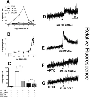
he monocytic line THP-1 was initially examined by chemotaxis assays using a modiied Boyden chamber. Cells were highly responsive to CCL7, as we have previously described19, with a 10 nM concentration of CCL7 inducing a robustresponse (Fig. 1A). Migration to CXCL4 was less potent, with migration only observed at a concentration of 1 µM CXCL4. No migration of THP-1 cells to the CXCR3 ligands CXCL10 and CXCL11 was observed (Fig. 1A), nor could we detect signiicant levels of cell surface CXCR3 expression with a speciic antibody nor speciic binding of radiolabeled CXCL10 (Supplementary Fig. 1). his suggests that in contrast to CXCL4 signaling in T cells17,18,
CXCL4 responses in THP-1 cells are not mediated by either of the CXCR3 variants. his was supported by the lack of efect of a CXCR3 antagonist on CXCL4 induced responses (Supplementary Figure 1) and is in agreement with other studies citing either a lack of CXCR3-B mRNA in monocytes20 or no efect of a CXCR3 antagonist on
CXCL4-induced monocyte responses21. Using an extended concentration range of CXCL4, a bell-shaped
[image:3.595.163.521.47.434.2]migra-tory curve typical of other chemokines was observed (Fig. 1B). In keeping with the indings of several groups
www.nature.com/scientificreports/
studying CXCL4 signaling in a variety of cell types12,22–24, micromolar concentrations of CXCL4 were required
to induce migration, suggesting that the ainity of CXCL4 for its receptor is likely to be low. To assess the relative contributions of chemokinesis and chemotaxis to THP-1 cell migration, optimal concentrations of CCL7 and CXCL4 were placed either in the bottom compartment of a modiied Boyden chamber or in both the top and bottom compartments (Fig. 1C). Whilst the inclusion of CCL7 in the upper compartment signiicantly reduced THP-1 migration (indicative of a largely chemotactic component), the inclusion of CXCL4 in the upper compart-ment had no signiicant efect upon CXCL4-induced migration. his suggests that a gradient of CXCL4 is not required to induce migration of THP-1 cells when assayed with a modiied Boyden chamber.
CXCL4 treatment of THP-1 cells was also observed to give rise to an increase in intracellular calcium (Fig. 1D), as was treatment with the chemokine CCL7 (Fig. 1E). Pre-treatment of THP-1 cells with pertussis toxin ablated both the induction of intracellular calcium release (Fig. 1F and G) and THP-1 cell migration (Supplementary Fig. 1) in response to either CXCL4 or CCL7, suggesting that these signals are mediated via GPCRs that couple to Gαi proteins. Petersen and colleagues have previously reported that CXCL4 binds to one or more chondroitin sulphate-decorated glycoproteins on the surface of neutrophils, which is necessary for the biological activity of CXCL4, since enzymatic removal of cell surface chondroitin ablates functional responses25,26. Similarly, we
observed that pre-treatment of THP-1 cells with the enzyme chondroitinase ABC signiicantly reduced the migra-tion of THP-1 cells to not only CXCL4 but also to CCL7, whilst heparinase treatment had no efect (Fig. 2A and B). hese data suggest that one or more chondroitin sulphate-decorated glycoproteins present CXCL4 in cis to a speciic GPCR.
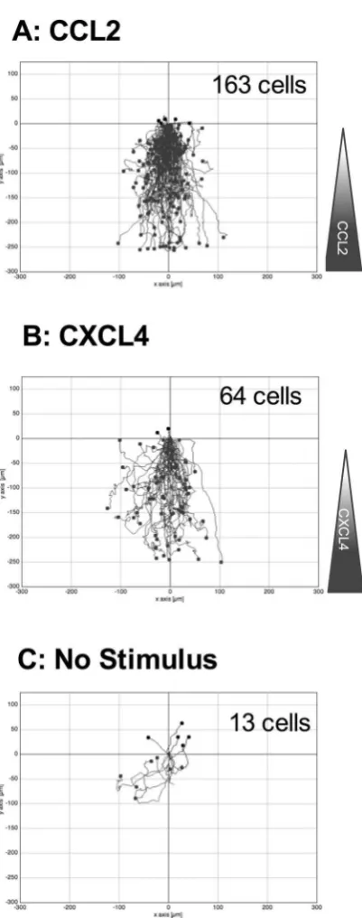
To supplement our Boyden chamber chemotaxis data, we switched to a novel horizontal device for imaging leukocyte migration in real time, namely the TAXIScan27. In this device, cells are introduced at one end of a 5 µm
[image:4.595.157.360.49.395.2]deep microchannel and chemoattractant is introduced at the other. Migration of cells to chemoattractant within the channel is then monitored via a charge coupled device camera, with the data subjected to manual cell track-ing (Supplementary Fig. 2). THP-1 cells were introduced into the chambers and their migration in response to CCL2, to CXCL4 or in the absence of stimulation was recorded over a 2 hr period (Fig. 3). Migration to CCL2 was typically robust, with many cells migrating towards the source of chemokine (Fig. 3A, Supplementary Video 1). Likewise, several cells were seen to migrate along a gradient of CXCL4 (Fig. 3B, Supplementary Video 2). In both cases, cells were seen to generate relatively long protrusions in the direction of the gradient prior to migration
www.nature.com/scientificreports/
[image:5.595.157.358.36.545.2]in the same direction. In contrast, relatively few cells migrated along the microchannel in the absence of stim-ulation (Fig. 3C, Supplementary Video 3). he tracks of individual cells were subjected to further analysis. he mean track length of cells responding to CXCL4 or CCL2 was seen to be signiicantly greater than that of cells not subjected to a stimulus (Fig. 4A). Velocity Directionality Box (VD-Box) plots of migration were subsequently generated, in which each symbol corresponds to an individual cell (Fig. 4B). In a visual system, better designed to assess cellular migration and diferentiate between chemotaxis and chemokinesis, cells exposed to gradients of either CXCL4 or CCL2 had a signiicantly higher directionality value compared to unstimulated cells (plotted to the right most area of the graph) indicating that both CXCL4 and CCL2 induce the directional migration (chem-otaxis) of THP-1 cells. he velocity of cells migrating in response to CXCL4 or CCL2 trended towards an increase over basal cell velocity but this was not signiicant.
www.nature.com/scientificreports/
CXCL is an agonist of CCR
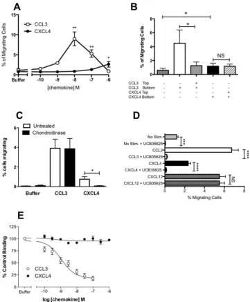
We have previously shown that responses to CCL7 in THP-1 cells are medi-ated predominantly via CCR119. We observed that the most robust responses in the Boyden chamber assay toCXCL4 were obtained with THP-1 cells that were also the most responsive to CCL7. We therefore postulated that contrary to the current dogma that chemokine receptor activation is class-restricted (i.e. CXC chemokines acti-vate only CXC receptors), CXCL4 might be acting by a known CCL7 receptor. To test this hypothesis, we trans-fected the mouse pre-B cell line L1.2 with plasmids encoding the three CC chemokine receptors at which CCL7 is known to be an agonist, namely CCR1, CCR2 and CCR3. All three receptors were readily expressed in a transient manner (Fig. 5A,B,C) and cells correspondingly responded to their cognate ligands CCL3, CCL2 and CCL11 in Boyden chamber assays (Fig. 5D,E,F). Notably, a signiicant response over basal migration to micromolar con-centrations of CXCL4 was singularly observed in CCR1 transfectants (Fig. 5D), albeit a less potent and eicacious response than that observed with CCL3, suggesting that CXCL4 is a partial agonist of CCR1 in this assay.
We next sought to validate whether a well-characterized small molecule antagonist of CCR1 named UCB 3562528,29 was able to block CXCL4 signaling. Using L1.2 cells stably expressing high levels of CCR1, we again
observed robust migration responses in Boyden chamber assays to both CCL3 and CXCL4 (Fig. 6A). As we had observed with THP-1 cells, migration to CXCL4 in the Boyden chamber assay was unafected by the inclusion of CXCL4 both above and below the ilter (Fig. 6B). his was in contrast to CCL3 responses which were signiicantly inhibited by including CCL3 both above and below the ilter. As was the case with THP-1 cells, (Fig. 2) preincuba-tion of L1.2 cells expressing CCR1 with chondroitinase was found to signiicantly inhibit the migrapreincuba-tion to CXCL4 whilst responses to CCL3 remained intact (Fig. 6C). Preincubation of cells with a 10 µM concentration of UCB 35625 was able to ablate migration to optimal concentrations of both CCL3 and CXCL4 but had no signiicant efect upon migration to the CXCR4 ligand CXCL12 (Fig. 6D). Unlike naïve L1.2 cells29, the L1.2 CCR1
transfect-ants displayed a detectable level of migration in the absence of chemokine but this was signiicantly increased by exposure to 1 µM CXCL4 (p = 0.0025). Basal migration was signiicantly inhibited by preincubation with UCB 35625, suggesting that when expressed at high levels, CCR1 shows a degree of constitutive activity, which is able to drive migration, as previously reported by Gilliland and colleagues30. Ligand binding assays were also
[image:6.595.157.359.47.373.2]under-taken in which CXCL4 was observed to be unable to compete with radiolabelled CCL3 for binding to CCR1, unlike unlabeled CCL3 which was able to displace radiolabelled CCL3 with ease (Fig. 6E).
www.nature.com/scientificreports/
In addition to initiating receptor signaling, chemokines also classically induce receptor endocytosis follow-ing bindfollow-ing. We therefore assessed the ability of 50 nM CCL3 or 1 µM CXCL4 to induce receptor endocytosis in THP-1 cells, the L1.2-CCR1 line and freshly isolated human monocytes (Fig. 7). Incubation of THP-1 cells with CCL3 at 37 °C induced a signiicant loss of cell surface CCR1 whilst CXCL4 was without signiicant efect (Fig. 7A). In contrast, when L1.2 CCR1 transfectants were examined, incubation with either CCL3 or CXCL4 was able to induce signiicant CCR1 endocytosis (Fig. 7B). Similarly, when human monocytes were examined, both CCL3 and CXCL4 induced signiicant CCR1 endocytosis at levels similar to those induced in the L1.2 CCR1 cell line. Collectively, these data conirm our indings that CXCL4 is a CCR1 agonist, although the cellular back-ground in which CCR1 is expressed appears to dictate the eicacy of CXCL4-induced endocytosis.
Blockade of CCR ablates monocyte migration in response to CXCL
Having implicated CCR1 in CXCL4 signaling using a transfectant system and a monocytic cell line we sought to translate our migratory indings to assays using freshly isolated human monocytes. he TAXIScan device27 was employed to examine [image:7.595.156.518.44.467.2]realtime migration of monocytes. his system also circumvents the problems of monocytes adhering to the ilters of modiied Boyden chambers, which can make analysis problematic. Exposure of monocytes to a gradient of CCL2 and CXCL4 resulted in robust migration, whilst considerably fewer monocytes were observed to migrate in the absence of chemokine (Fig. 8A,B,C, Supplementary Videos 4, 5 and 6). Unlike the THP-1 cells, no long protrusions were observed to be generated prior to migration. Tracking of individual monocytes revealed that
www.nature.com/scientificreports/
the mean track length of monocytes migrating along a gradient of CCL2 was signiicantly shorter than those of monocytes exposed to a gradient of CXCL4 or not exposed to a stimulus (Fig. 9A). his inding was further clariied when the directionality and velocity parameters were assessed in a VD-B plot (Fig. 9B). Cells migrating in response to both CCL2 and CXCL4 had signiicantly greater directionality than those undergoing basal migra-tion. In contrast, when the velocity of migration was examined, CCL2 stimulation resulted in signiicantly slower migration than observed in either basal migration or migration in response to CXCL4 stimulation. Collectively, these data show that that both CCL2 and CXCL4 induce the chemotaxis of monocytes, with CCL2 inducing a slower, more methodical migration.
[image:8.595.154.516.43.481.2]To conirm that the CXCL4-induced migration was mediated via CCR1, we assessed whether it was sensitive to pre-treatment with the CCR1 antagonists UCB 35625 and BX-471. he accumulated distance migrated by monocytes (i.e. the sum of the individual cell tracks) either along a CXCL4 gradient, along a CCL2 gradient or in the absence of stimulation was examined using monocytes pretreated with vehicle or with 10 µM UCB 35625
www.nature.com/scientificreports/
or BX-471. Migration in response to CXCL4 was signiicantly inhibited by pretreated of cells with 10 µM UCB 35625 when compared to vehicle treated cells (Fig. 10A) whilst basal migration, or migration along a gradient of CCL2 was unafected (Fig. 10B). his is in line with our previous indings regarding the lack of activity of this compound in blocking CCL2 responses in monocytes28. Similarly, pretreatment of cells with 10 µM BX-471, an
[image:9.595.156.339.42.603.2]alternative CCR1 antagonist, was seen to signiicantly inhibit the migratory responses to CXCL4 but not the basal migration (Fig. 10C).
www.nature.com/scientificreports/
Discussion
he release of micromolar concentrations of CXCL4 by activated platelets has been implicated in the process of atherogenesis by virtue of its many efects upon monocytes, notably their endothelial arrest, recruitment to the sub-endothelial space and efects on survival and diferentiation10,14,20,23,31. We present here a body of data
[image:10.595.156.333.42.592.2]that clearly identiies CXCL4 as a CCR1 agonist. Notably, we observed that CCR1 could induce the migration of CCR1-transfctants, albeit with reduced potency and eicacy when compared to the principal CCR1 ligand, CCL3.
www.nature.com/scientificreports/
his is reminiscent of our earlier indings with CXCR3, where CXCL4 was observed to have reduced potency and eicacy when compared with the other CXCR3 ligands17. Since the current dogma suggests that chemokines are
class restricted, i.e. CXC chemokines activate only CXC chemokine receptors, the inding that CXCL4 activates CCR1 was unexpected and serves to underline the apparent plasticity of some chemokine receptors with respect to ligand recognition. For example, β-defensin induces T-cell migration via CCR632, whilst ubiquitin has been
shown to be a ligand for CXCR433. Similarly, the atypical chemokine receptor ACKR1, previously known as Dufy
Antigen Receptor for Chemokines (DARC) is well known to bind chemokines of both CC and CXC classes34.
In keeping with the indings of many other groups studying aspects of CXCL4 signalling in a variety of cell types12,17,18,22–24, micromolar concentrations of CXCL4 were required to observe biological efects, suggesting
that the ainity of CXCL4 for its receptor is likely to be low. Petersen and colleagues previously reported that CXCL4 bound to the surface of neutrophils with a Kd of less than 600 nM which they attributed to the binding of tetrameric CXCL4 to a chondroitin sulphate-containing cell surface glycoprotein of around 250 kDa, considera-bly larger than most GPCRs25,26. Similarly, von Hundelshausen and colleagues have shown that CXCL4-induced
monocyte arrest on ECs requires chondroitin sulphate14. We observed that pre-treatment with the enzyme
chon-droitinase ABC markedly reduced the migration of THP-1 cells and CCR1 transfectants to CXCL4 which sug-gests that one or more glycoproteins decorated with chondroitin sulphate provide cis presentation of CXCL4 to CCR1 on the monocyte surface, which is critical for receptor activation (Fig. 11). A similar mechanism by which glycosaminoglycans on the surface of T-cells present CCL5 to the receptor CCR5 has previously been put forward35, with treatment with a cocktail of glycanases resulting in a loss of T-cell responses to CCL5. Consistent
with this, we were unable to efectively compete CCL3 from the surface of CCR1 transfectants with CXCL4. his is reminiscent of our earlier study examining CXCL4 activation of CXCR3, where we were unable to demonstrate displacement of CXCL11 from CXCR3 by CXCL4, despite clearly showing CXCR3-mediated CXCL4 signalling, with glycosamingoglycans necessary for CXCL4 function17.
[image:11.595.156.360.46.377.2]In addition to activity on CCR1 transfectants, we observed that CXCL4 could induce the migration of the monocytic cell line THP-1 in a modiied Boyden chamber. hese data were ably supported by realtime imag-ing of cell migration usimag-ing a TAXIScan system in which CXCL4 clearly induced the directed migration of both THP-1 cells and monocytes. Although an earlier report suggested that CXCL4 was chemotactic for neutrophils and monocytes13, this was subsequently contested by Petersen and colleagues who failed to observe monocyte
www.nature.com/scientificreports/
migration in response to a highly puriied preparation of CXCL4 in Boyden chamber assays12. heir argument
was that the original migration observed by Deuel and colleagues13 was likely due to contamination with other
chemokines in the platelet-derived CXCL4 preparation. Here, we have used a highly pure recombinant form of CXCL4 (greater than 98 percent purity as determined by Western blotting and HPLC analysis) so can con-idently attribute the observed chemotactic activity to CXCL4 alone. In keeping with our CCR1 transfectant data, CXCL4-induced migration of THP-1 cells and monocytes was CCR1-dependent, as shown by the action of the small molecule antagonists UCB 35625 and BX-471. It is unclear why in the Boyden chamber assays, the inclusion of chemokine above the membrane had no efect on CXCL4 responses, suggesting at face value, that the CXCL4-induced migration was chemokinetic (non-directional). his is clearly not the case, as the TAXIScan data (movies and VD-B plots) shows the migration to be chemotactic. he laws of assessing chemokinesis in Boyden chambers are well known, hence the initial development of Dunn and Zigmond chambers and systems such as TAXIScan. It is debatable in modiied Boyden chamber assays whether or not a chemoattractant gradient exists for the duration of the assay and the validity of the “checkerboard” correction for chemokinesis measurements has been questioned36. Our data highlights the need for direct visualisation to accurately examine the migratory
behavior of cells in a chemotactic gradient, as suggested by others37.
In addition to its ability to induce monocyte arrest and migration, CXCL4 can also act as a diferentiation and survival factor for monocytes, generating what has been dubbed the “M4” macrophage23. he transcriptional
[image:12.595.159.357.40.448.2]program induced by CXCL4 appears to be distinct from those induced by classical and non-classical activa-tion, including apparently contradictory pro-atherosclerotic and anti-atherosclerotic components, such as down
www.nature.com/scientificreports/
regulation of the atheroprotective scavenger CD163 and down regulation of the atherogenic scavenger CD3620.
Notably, the M4 macrophage takes up much less modiied low density lipoprotein (LDL) than those diferenti-ated by M-CSF treatment23. If we extrapolate our indings here that CCR1 is a bona ide receptor for CXCL4 on
monocytes, then our indings may explain at a mechanistic level why deletion of CCR1 in mice has been shown to enhance atherosclerosis in two independent studies38,39. his is in stark contrast to several other studies of
chemokine receptor-deicient mice that display various levels of atheroprotection4,5. In light of our data here, we
speculate that the accelerated atherosclerosis seen in the CCR1 deicient mice may be due to the selective loss of CCR1:CXCL4 signalling in macrophages within plaques that normally would lead to lower modiied LDL uptake. Although CXCL4 deletion has previously been shown to be atheroprotective on the Apolipoprotein E null back-ground11, such a broad stroke removes a number of potentially pro-atherosclerotic CXCL4 activities outside of
CCR1 signalling, such as promoting oxidised LDL binding to the vasculature and inhibiting oxLDL endocytosis by the LDL receptor40 in addition to T cell recruitment via CXCR3-B17. We would suggest that when
contemplat-ing targetcontemplat-ing CXCL4 in the treatment of atherosclerosis, alongside the pro-atherosclerotic activities of CXCL4, the anti-atherogenic properties of M4 macrophages must also be considered41.
Assuming the in vitro data described here extrapolates to the clinical setting, then it would strongly argue against targeting CCR1 in atherosclerosis and may point to unforeseen side efects following the long-term use of such molecules for the treatment of other inlammatory disorders. Of note, the small molecule CCR1 antag-onist CCX354 has shown eicacy in a phase II clinical trial for the treatment of rheumatoid arthritis and larger, longer-term clinical trials have been planned42. Since accelerated atherosclerosis and increased mortality is oten
observed in arthritic patients (reviewed in43), blockade of CCR1 may be detrimental. Further experimentation to
test such a hypothesis in vivo is highly desirable.
Methods
Materials
Recombinant chemokines were from PeproTech EC Ltd (London, UK). he CCR1 antagonists UCB 35625 and BX-471 were from BioTechne Ltd. (Abingdon, UK) and have previously been described28,44.Bordetella pertussis toxin and the mouse isotype-matched control IgG1 (MOPC 21 clone) were from Sigma-Aldrich (Poole, UK), as were heparinase and chondroitinase ABC. Radioiodinated CXCL10 and CCL3 were from Perkin Elmer Life Sciences (Boston, MA). he anti-human CCR1 and CXCR3 mAbs (MAB145 and MAB160, respectively) were from BioTechne Ltd. he anti-HA mAb was from Covance Biosciences (Crawley, UK). he secondary anti-mouse-FITC conjugated antibody was from DAKO UK Ltd, (Cambridge, UK). All other reagents, unless noted, were from Life Technologies (Paisley, UK).
[image:13.595.155.393.44.338.2]Cell Preparation and Maintenance
Blood was taken from healthy normal subjects with written informed consent. he protocol was approved by the Brompton, Hareield and NHLI ethics committee. All experimentswww.nature.com/scientificreports/
were carried out in accordance with the approved guidelines. PBMCs were isolated as previously described28.
THP-1 and L1.2 cells were maintained as previously described44. Where indicated, cells were treated
over-night with 100 ng/ml pertussis toxin (Sigma-Aldrich) to inhibit Gαi signaling. For studies of monocyte chem-otaxis, monocytes were separated from whole blood using a Rosette-Sep monocyte puriication kit (StemCell Technologies, Grenoble, France) according to the manufacturer’s instructions. Transient transfection of L1.2 cells with HA-tagged chemokine receptor constructs was by electroporation as previously described44.
Flow cytometric analysis of chemokine receptor expression
Staining of HA-tagged CCR1, CCR2 and CCR3 transfectants was by the use of an anti-HA monoclonal as previously described44. Detection of CCR1with MAB145 was as described previously45. Flow cytometry analysis was carried out using a LSR Fortessa or
FACS Calibur (BD Bioscience, Oxford, UK); gating excluded cell debris and cell doublets from analysis.
Cell Migration Assays
Modiied Boyden Chamber assays were carried out as previously described44 using5 µm pore chambers (Neuroprobe Inc, Gaithersburg, MD). In experiments employing pertussis toxin, cells were pretreated with 100 ng/ml for 18 hours at 37 °C. Similarly, in experiments employing the glycosidases hepari-nase and chondroitihepari-nase ABC, cells were incubated with agitation with one unit/ml of enzyme for 30 minutes at 37 °C as previously described25, before being washed extensively in phosphate bufered saline. In experiments
employing the CXCR3 or CCR1 antagonist, chemokine dilutions were made in a solution containing a ixed con-centration of the competitor. In experiments to assess the contribution of chemokinesis, the same concon-centration of chemokine placed in the lower well of the chemotaxis chamber was present in the assay bufer, eliminating the chemoattractant gradient.
For real time analysis of migrating THP-1 cells and freshly isolated monocytes, a 12-channel TAXIScan was employed27 and used according to the manufacturer’s protocol (Efector Cell Institute, Tokyo. Japan). Where
indicated, cells were pretreated with a ixed concentration of antagonist for 30 minutes at 37 °C prior to carrying out the assay. One µl of a suspension containing 500,000 cells/ml was loaded into each chamber and following alignment of the cells at one end of the terrace, 1 µl of chemokine (1 µM CCL2 or 20 µM CXCL4) was added to the other end of the terrace (260 µm away) and cells were allowed to migrate along the ensuing chemokine gradient for 2 hr whilst maintained at 37 °C (Supplementary Figure 2). Sequential image data were captured at every minute as individual jpegs. To calculate the mean track length, data were subsequently processed with ImageJ (National Institutes of Health), equipped with the manual tracking tool plugin (Fabrice Cordelieres, Institut Curie, Orsay (France) and chemotaxis tool plugin (Ibidi, Martinsried, Germany). Individual experiments consisted of duplicate conditions and data illustrated are collated from several experiments as highlighted in the igure legend. he numbers of cells tracked under each condition are shown in the igure. For each individual cell, the mean track length was calculated by the chemotaxis tool plugin as described previously46. he accumulated
distance parameter (Fig. 10) refers to the total distances traveled by all cells for a particular condition in an indi-vidual experiment.
Cell migration was also manually tracked using TAXIScan Analysis sotware in order to generate plots of Velocity versus Directionality. his is an accepted methodology for accurately distinguishing chemotaxis from chemokinesis with the TAXIScan system27. he direction of cell migration is expressed as the angle (rad) towards
the concentration gradient, namely π/2 indicates that the cell is migrating toward the concentration gradient, whereas −π/2 indicates that the cell is migrating against the concentration gradient. Velocity-Directionality-Box (VD-B) plots were subsequently generated47 in which the vertical axis shows the median value of velocity and the
horizontal axis shows the median value of the directionality. For THP-1 cells, tracking was undertaken during the initial 30 minutes of migration, whereas for slower moving monocytes, tracking was carried out for the irst 60 minutes of migration.
Intracellular Calcium Measurements
hese were performed as previously described19 using cells thatwere loaded with the luorescent dye FURA-2 AM (Life Technologies). Real time data were recovered using a luorimeter (LS-50B, Perkin-Elmer, Beaconsield, UK). Data are expressed as the relative ratio of luorescence emitted at 510 nm ater sequential stimulation at 340 and 380 nm.
CCR Endocytosis Assays
THP-1 cells were cultured with 80 nM calcitriol (Sigma-Aldrich) for 48 hr to enhance CCR1 expression48. Similarly, CCR1-L1.2 transfectants were cultured overnight with 10 mM sodiumButyrate to enhance CCR1 expression44. Human monocytes were used within an hour of puriication. Cells were
resuspended at 500,000 cells/ml in RPMI, 0.1 % BSA and kept on ice. Aliquots of these cells (50 µl) were incu-bated with either an identical volume of bufer alone or bufer containing CCL3 or CXCL4, giving a inal con-centration of 50 nM CCL3 or 1 µM CXCL4. Samples were either let at 4 °C for 30 minutes or incubated at 37 °C for 30 minutes ater which they were stained for CCR1 expression by low cytometry as described above. Data is presented as the percentage of CCR1 staining observed in the bufer treated samples that were incubated on ice.
Statistical Analysis
Statistical analyses were carried out using Prism 6 (GraphPad Sotware, La Jolla, CA). Unless otherwise stated, data relect the mean values ± SEM from the number of experiments shown in parenthe-sis. Unless otherwise stated, statistics refer to repeated measures ANOVA with Bonferroni’s post-test. *P ≤ 0.05, **P ≤ 0.01, ***P ≤ 0.001 and ****P ≤ 0.0001. NS denotes not signiicant.www.nature.com/scientificreports/
References
1. Bachelerie, F. et al. International Union of Pharmacology. LXXXIX. Update on the Extended Family of Chemokine Receptors and Introducing a New Nomenclature for Atypical Chemokine Receptors. Pharmacol. Rev. 66, 1–79 (2013).
2. Zlotnik, A. & Yoshie, O. he chemokine superfamily revisited. Immun. 36, 705–716 (2012).
3. Weber, C., Zernecke, A. & Libby, P. he multifaceted contributions of leukocyte subsets to atherosclerosis: lessons from mouse models. Nat. Rev. Immunol. 8, 802–815 (2008).
4. Boring, L., Gosling, J., Cleary, M. & Charo, I. F. Decreased lesion formation in CCR2−/− mice reveals a role for chemokines in the initiation of atherosclerosis. Nat. 394, 894–897 (1998).
5. Combadière, C. et al. Decreased atherosclerotic lesion formation in CX3CR1/apolipoprotein E double knockout mice. Cir. 107, 1009–1016 (2003).
6. Combadière, C. et al. Combined inhibition of CCL2, CX3CR1, and CCR5 abrogates Ly6C(hi) and Ly6C(lo) monocytosis and almost abolishes atherosclerosis in hypercholesterolemic mice. Cir. 117, 1649–1657 (2008).
7. Koenen, R. R. & Weber, C. herapeutic targeting of chemokine interactions in atherosclerosis. Nat. Rev. Drug Discov. 9, 141–153 (2010).
8. Deuel, T. F., Keim, P. S., Farmer, M. & Heinrikson, R. L. Amino acid sequence of human platelet factor 4. Proc. Natl. Acad. Sci. USA
74, 2256–2258 (1977).
9. Pitsilos, S. et al. Platelet factor 4 localization in carotid atherosclerotic plaques: correlation with clinical parameters. hromb. Haemost. 90, 1112–1120 (2003).
10. Huo, Y. et al. Circulating activated platelets exacerbate atherosclerosis in mice deicient in apolipoprotein E. Nat. Med. 9, 61–67 (2002).
11. Sachais, B. S. et al. Elimination of platelet factor 4 (PF4) from platelets reduces atherosclerosis in C57Bl/6 and apoE−/− mice.
hromb. Haemost. 98, 1108–1113 (2007).
12. Pervushina, O. et al. Platelet factor 4/CXCL4 induces phagocytosis and the generation of reactive oxygen metabolites in mononuclear phagocytes independently of Gi protein activation or intracellular calcium transients. J. Immunol. 173, 2060–2067 (2004). 13. Deuel, T. F. et al. Platelet factor 4 is chemotactic for neutrophils and monocytes. Proc. Natl. Acad. Sci. USA 78, 4584–4587 (1981). 14. von Hundelshausen, P. et al. Heterophilic interactions of platelet factor 4 and RANTES promote monocyte arrest on endothelium.
Blood 105, 924–930 (2005).
15. Koenen, R. R. et al. Disrupting functional interactions between platelet chemokines inhibits atherosclerosis in hyperlipidemic mice.
Nat. Med. 15, 97–103 (2009).
16. Lasagni, L. et al. An alternatively spliced variant of CXCR3 mediates the inhibition of endothelial cell growth induced by IP-10, Mig, and I-TAC, and acts as functional receptor for platelet factor 4. J.Exp. Med. 197, 1537–1549 (2003).
17. Mueller, A. et al. CXCL4-induced migration of activated T lymphocytes is mediated by the chemokine receptor CXCR3. J. Leuk. Biol.
83, 875–882 (2008).
18. Korniejewska, A., McKnight, A. J., Johnson, Z., Watson, M. L. & Ward, S. G. Expression and agonist responsiveness of CXCR3 variants in human T lymphocytes. Immuno. 132, 503–515 (2011).
19. Martinelli, R., Sabroe, I., LaRosa, G., Williams, T. J. & Pease, J. E. he CC chemokine eotaxin (CCL11) is a partial agonist of CC chemokine receptor 2b. J. Biol. Chem. 276, 42957–42964 (2001).
20. Gleissner, C. A. et al. CXCL4 Downregulates the Atheroprotective Hemoglobin Receptor CD163 in Human Macrophages. Circ. Res.
106, 203–211 (2010).
21. Gouwy, M. et al. CXCL4 and CXCL4L1 Diferentially Afect Monocyte Survival and Dendritic Cell Diferentiation and Phagocytosis.
PloS one 11, e0166006–24 (2016).
22. Kasper, B. & Petersen, F. Molecular pathways of platelet factor 4/CXCL4 signaling. Eur. J. Cell Biol. 90, 521–526 (2011).
23. Gleissner, C. A., Shaked, I., Little, K. M. & Ley, K. CXC Chemokine Ligand 4 Induces a Unique Transcriptome in Monocyte-Derived Macrophages. J. Immunol. 184, 4810–4818 (2010).
24. Struyf, S. et al. Angiostatic and chemotactic activities of the CXC chemokine CXCL4L1 (platelet factor-4 variant) are mediated by CXCR3. Blood 117, 480–488 (2011).
25. Petersen, F., Bock, L., Flad, H. D. & Brandt, E. A chondroitin sulfate proteoglycan on human neutrophils speciically binds platelet factor 4 and is involved in cell activation. J. Immunol. 161, 4347–4355 (1998).
26. Fleischer, J. et al. Platelet factor 4 inhibits proliferation and cytokine release of activated human T cells. J. Immunol. 169, 770–777 (2002).
27. Nitta, N., Tsuchiya, T., Yamauchi, A., Tamatani, T. & Kanegasaki, S. Quantitative analysis of eosinophil chemotaxis tracked using a novel optical device — TAXIScan. J. Immunol. Meth. 320, 155–163 (2007).
28. Sabroe, I. et al. A small molecule antagonist of chemokine receptors CCR1 and CCR3. Potent inhibition of eosinophil function and CCR3-mediated HIV-1 entry. J. Biol. Chem. 275, 25985–25992 (2000).
29. de Mendonça, F. L. et al. Site-directed mutagenesis of CC chemokine receptor 1 reveals the mechanism of action of UCB 35625, a small molecule chemokine receptor antagonist. J. Biol. Chem. 280, 4808–4816 (2005).
30. Gilliland, C. T., Salanga, C. L., Kawamura, T., Trejo, J. & Handel, T. M. he Chemokine Receptor CCR1 Is Constitutively Active, Which Leads to G Protein-independent, β-Arrestin-mediated Internalization. J. Biol. Chem. 288, 32194–32210 (2013).
31. Scheuerer, B. et al. he CXC-chemokine platelet factor 4 promotes monocyte survival and induces monocyte diferentiation into macrophages. Blood 95, 1158–1166 (2000).
32. Yang, D. et al. Beta-defensins: linking innate and adaptive immunity through dendritic and T cell CCR6. Sci. 286, 525–528 (1999). 33. Saini, V., Marchese, A. & Majetschak, M. CXC chemokine receptor 4 is a cell surface receptor for extracellular ubiquitin. J. Biol.
Chem. 285, 15566–15576 (2010).
34. Graham, G. J., Locati, M., Mantovani, A., Rot, A. & helen, M. he biochemistry and biology of the atypical chemokine receptors.
Immuno. letters 145, 30–38 (2012).
35. Burns, J. M., Gallo, R. C., DeVico, A. L. & Lewis, G. K. A new monoclonal antibody, mAb 4A12, identifies a role for the glycosaminoglycan (GAG) binding domain of RANTES in the antiviral efect against HIV-1 and intracellular Ca2+ signaling. J. Exp. Med. 188, 1917–1927 (1998).
36. Rhodes, J. M. Measurement of chemotaxis in Boyden chamber ilter assays. Is the checkerboard correction valid? J. immunological methods 49, 235–236 (1982).
37. Muinonen-Martin, A. J., Veltman, D. M., Kalna, G. & Insall, R. H. An improved chamber for direct visualisation of chemotaxis. PloS one 5, e15309 (2010).
38. Potteaux, S. et al. Chemokine receptor CCR1 disruption in bone marrow cells enhances atherosclerotic lesion development and inlammation in mice. Mol. Med. 11, 16–20 (2005).
39. Braunersreuther, V. et al. Ccr5 but not Ccr1 deiciency reduces development of diet-induced atherosclerosis in mice. Arter. hromb. Vasc. Biol. 27, 373–379 (2007).
40. Sachais, B. S. et al. Platelet factor 4 binds to low-density lipoprotein receptors and disrupts the endocytic machinery, resulting in retention of low-density lipoprotein on the cell surface. Blood 99, 3613–3622 (2002).
www.nature.com/scientificreports/
42. Tak, P. P. et al. Chemokine receptor CCR1 antagonist CCX354-C treatment for rheumatoid arthritis: CARAT-2, a randomised, placebo controlled clinical trial. Ann. Rheum. Dis. 72, 337–344 (2012).
43. Shoenfeld, Y. et al. Accelerated atherosclerosis in autoimmune rheumatic diseases. Circ. 112, 3337–3347 (2005).
44. Vaidehi, N., Pease, J. E. & Horuk, R. Modeling small molecule-compound binding to G-protein-coupled receptors. Meth. Enzym.
460, 263–288 (2009).
45. Phillips, R. M. et al. Variations in eosinophil chemokine responses: an investigation of CCR1 and CCR3 function, expression in atopy, and identiication of a functional CCR1 promoter. J. Immunol. 170, 6190–6201 (2003).
46. Zengel, P. et al. µ-Slide Chemotaxis: a new chamber for long-term chemotaxis studies. BMC Cell Biol. 12, 21 (2011).
47. Yamauchi, A., Degawa-Yamauchi, M., Kuribayashi, F., Kanegasaki, S. & Tsuchiya, T. Systematic single cell analysis of migration and morphological changes of human neutrophils over stimulus concentration gradients. J. immunological methods 404, 59–70 (2014). 48. Parker, L. C., Whyte, M. K. B., Vogel, S. N., Dower, S. K. & Sabroe, I. Toll-like receptor (TLR)2 and TLR4 agonists regulate CCR
expression in human monocytic cells. J. Immunol. 172, 4977–4986 (2004).
Acknowledgements
his work was supported by grants from the Medical Research Council (PhD Studentship to J.M.F.), the British Heart Foundation (A.M., PG/2000055) and the Royal Embassy Of Saudi Arabia (PhD Studentship to K.H., R1036). We are grateful to members of the section for their helpful discussions.
Author Contributions
J.E.P. conceived the experiments, J.M.F., F.K., A.D., M.O., K.H., A.M., J.L. and J.E.P. conducted the experiments, J.M.F., T.T., S.K. and J.E.P. analysed the results. All authors reviewed the manuscript.
Additional Information
Supplementary information accompanies this paper at https://doi.org/10.1038/s41598-018-27710-9.
Competing Interests: he authors declare no competing interests.
Publisher's note: Springer Nature remains neutral with regard to jurisdictional claims in published maps and institutional ailiations.
Open Access This article is licensed under a Creative Commons Attribution 4.0 International License, which permits use, sharing, adaptation, distribution and reproduction in any medium or format, as long as you give appropriate credit to the original author(s) and the source, provide a link to the Cre-ative Commons license, and indicate if changes were made. he images or other third party material in this article are included in the article’s Creative Commons license, unless indicated otherwise in a credit line to the material. If material is not included in the article’s Creative Commons license and your intended use is not per-mitted by statutory regulation or exceeds the perper-mitted use, you will need to obtain permission directly from the copyright holder. To view a copy of this license, visit http://creativecommons.org/licenses/by/4.0/.