EFFECT OF LOWER EXTREMITY STRENGTH
TRAINING ON GAIT IN CEREBRAL PALSY
CHILDREN
ProjectSubmitted to
TheTamilnaduDr. MGRMedicalUniversity
In partial fulfillment for the degree of
MASTEROFPHYSIOTHERAPY
(PEADIATRICPHYSIOTHERAPY)
271540041
Cherraan’s
College of
Physiotherapy
Cherraan’s Institute of Health Sciences
Coimbatore, Tamilnadu, India
2
CERTIFICATE
The work embodied in the thesis entitled “Effect of Lower Extremity Strength Training on Gait in Cerebral Palsy Children” Submitted to The Tamilnadu Dr. MGR Medical University in partial fulfillment for the degree of Master of physiotherapy was carried out by candidate bearing register number 271540041 at Cherraan’s College of Physiotherapy under my supervision. This is an original work done by her and has not been submitted in part or full for any other degree/diploma at this of any other university/ institute. The thesis is fit to be considered for evaluation for award of the degree of master of physiotherapy
.
Signatureofsupervisor Principal
Mrs. Aroona MPT Mrs . Selvarani MPT
Date: Date:
INTERNAL EXAMINER EXTERNAL EXAMINER
3
DECLARATION BY THE STUDENT
The work embodied in the thesis entitled “EFFECT OF LOWER
EXTREMITY STRENGTH TRAINING ON GAIT IN CEREBRAL PALSY CHILDEREN” Submitted to The Tamilnadu Dr. MGR Medical University in partial fulfillment for the degree of Master of physiotherapy was the original work carried out by me and has not been submitted in part or full for any other degree / diploma at this or any other university / institute. All the ideas and references have been duly acknowledged.
.
Signatureofsupervisor signature ofstudent
Mrs. Aroona MPT Bavapriya .P
(professor)
4
ACKNOWLEDGEMENT
Dissertation of this kind would not have been possible without valuable contribution of many individuals.
First and foremost I would like to thank the Almighty, who showered his blessing1sqw`ws in all walks of my life.
I take this extreme privilege to thank MRS. SELVARANI, MPT, Principal, cherrann’s college of physiotherapy, for her encouragement and inspiration during the course of this study.
I submit my heartfelt thanks to my guide MRS. AROONA, MPT
(Pediatric), cherraan’s college of physiotherapy, for her valuable advices, inspiring guidance, timely support constant encouragement, diligent and sincere review of the work which enable me to complete my study in a very good manner.
I thank all my Assistant professors who with all patience gave me helping hands whenever needed .
I am highly indebted to my friends for their love, support and encouragement in the completion of this work.
5
6
S.NO INDEX PAGE NO
1 INTRODUCTION 7
2 REVIEW OF LITERATURE 11
3 MATERIALS AND METHODOLOGY 3.1 Study design
3.2 Study type 3.3 Study setting 3.4 Sample size
3.5 Sampling technique 3.6 Study duration 3.7 Inclusion criteria 3.8 Exclusion criteria 3.9 Procedure
3.1 Outcome measures 3.11 Materials used 3.12 Testing of tools
17
4 DATA ANALYSIS AND RESULTS
25
5
DISCUSSION 35
6
CONCLUSION 39
7
RECOMMENDATIONS & LIMITATIONS 41 8
7
8
Cerebral palsy is a collection of disorders characterized by an insult to
the developing brain that produces physical disability as the primary or distinguishing feature Dr.Little described cerebral palsy as a persistent disorder of posture and movement appearing early in life and due to a developmental non progressive disorder of the brain.
The spastic form of cerebral palsy is the most common and in those patients, additional clinical signs may include, muscle shortening, diminished selective control and weakness. It is well recognized that muscle weakness is a major impairment in people with cerebral palsy. Motor problems affecting gait includes disruption to both neuromuscular and musculoskeletal system. In patients with neurological pathology musculoskeletal problem are secondary to primary neuromuscular problem.
Central lesions of the CNS results in impaired programming affecting leg muscle activation. Impaired programming can manifest in gait as.
Inability to recruit a muscle appropriately.
Increased activation of muscle that is unrelated to spasticity mediated stretch.
Inability to modulate a muscle activity throughout the gait cycle.
Many children with cerebral palsy quickly became exhausted because of the disease’s impact on the muscle. According to the researchers the average speed of cerebral palsy kids is about 40m/s or 57% of normal walking speed. These children walk slowly they required high energy expenditure often lack endurance and suffer from muscle weakness.
While spasticity was once thought to be the primary contributor to the motor dysfunction noted in cerebral palsy, many have challenged this perspective and now consider ‘negative’ signs such as muscle weakness to be more harmful to function.
9
themselves12, aberrations in the reciprocal inhibition pathways in agonist-antagonist muscle pairs13 and heightened stretch responses or spasticity.
(Patla 1995; Buschner and DeLateur 1991) Walking21 does not normally tax the various lower extremity muscle groups to their full capacity. The only muscle that comes close to their maximum output during gait is the ankle Plantar flexor, which normally provide a major source of propulsive power. In the presence of plantar flexor weakness, alternative power source are used resulting in changes in locomotors characteristic like stride length & velocity. Several researchers have shown a positive association between lower extremity muscle strength an speed7,8 . A person needs at least a minimum level of strength to walk at a given speed. Even children with cerebral palsy who have mild disabilities demonstrate substantial weakness compared with age-related peers4,6 .
Many treatments exist to improve gait and function of person with cerebral palsy. They include tendon lengthening , transfer or release, selective dorsal rhizotomy , Botulinum toxin, Baclofen, stretching . None of these treatments increase muscle strength.
More than 50 years ago, Phelps contended that resisted exercise to develop strength or skill in a weakened muscle or an impaired muscle group was an integral part of treatment in cerebral palsy14 .
Directly loading the muscle through specific exercises, activities , or sufficiently intense electrical stimulation is the only direct way to increase muscle strength in cerebral palsy and may be particularly useful in augmenting or maximizing the functional outcomes of other interventions that address different component of the motor disorder2,3,8.
10
BACKGROUND AND NEED FOR THE STUDY
Children with cerebral palsy walk slowly; they require high energy expenditure, often lack endurance because of muscle weakness. The purpose of this study is to examine whether strengthening lower extremity improves gait speed, cadence, stride length and motor activity in cerebral palsy children.
HYPOTHESIS
Null Hypothesis: There is no significant change in the gait parameters and muscle strength after lower extremity strength training in cerebral palsy children.
Alternate Hypothesis: There is a significant change in the gait parameters muscle strength after lower extremity strength training in cerebral palsy children.
OPERATIONAL DEFINITIONS
CEREBRAL PALSY – Dr. John Little stated that cerebral palsy is a persistent disorder of movement and posture appearing early in life and due to a developmental non progressive disorder of brain.
GAIT is the manner of moving the body from one place to another by alternatively and repetitively changing the location of the feet, with the condition that at least one feet is in contact with the walking surface.
CADENCE – Cadence is the number of the steps taken by a person per unit time. Cadence may be measured the number of steps per second or per minute.
STRIDE LENGTH – Stride length is the linear distance between two successive events that are accomplished by the same lower extremity during gait.
11
LITERATURE
12
1. RossSA, EngsbergJR(2007), no relationship exists between spasticity and strength and that increasing muscle strength does not alter spasticity5.
2. Lee JH, Sung IY, Yoo ZY (2007), strengthening exercise could be a useful method to improve gait function of children with spastic cerebral palsy. Increased gait speed and stride length & decreased double support phase31.
3. Diane L, Damiano (2004), says that children with cerebral palsy suffer biomechanical misalignment on the one hand and are denied proper use of their muscles on the other.
4. Joanne Bundonis (2004), strength training has cardiovascular neuromuscular system benefits as well as psychological, fitness and functional benefits29.
5. Blunell SW, Shepherd RB, DeanCM, Adams RD, CahillBM (2003),
task specific strengthening exercises run as a group circuit class, resulted in improved strength and functional performance30.
6. McBurney et al (2003), found that the children had increased strength, decreased activity limitations improved mobility after performing a lower extremity strengthening home program28.
7. RichSmith Rose (2002), the idea underlying the energetics of walking is that the amount of energy required to walk, the shorter time and distance an individual will be able to endure ambulation without tiring32.
13
9. Damiano DL, Martellotta TL, Quinlivan J, Abel MF (2001), in a sample of children with a broader range of involvement, those with greater muscle spasticity in the antagonist tended to have greater agonist weakness.
10. Teixeira-Salmela LF, Olney SJ, Nadeau S, Brouwer B (1999),
in chronic stroke direct muscle strengthening improved functional performance in persons whose recovery had plateau before this intervention and was not shown to increase spasticity40.
11. Damiano DL, Abel MF (1998), spastic cerebral palsy children reported higher gait velocity primarily as a result of increased cadence, with a greater capacity to walk faster7.
12. Damiano DL, Kelly LE, Vaughan CL. (1995), hamstring strength was measured before and after a quadriceps-strengthening program in children with cerebral palsy to determine whether the program caused an inadvertent increase in strength in the spastic muscle due to abnormal co contraction or stretch responses elicited in the antagonist during agonist strengthening. The quadriceps showed a mean strength increase of more than 50% with no significant change in the hamstring values.
13. Damiano DL, Kelly LE, Vaughan CL. (1995) Kramer JF, MecPhail HEA. (1994), leg strength has shown to be related to freely selected walking velocity and to the Gross Motor Function Measure in children and adolescents with cerebral palsy.
14. Gowland et al (1992), inadequate requirements of agonist motor neurons and not increased activity in the antagonist is the primary basis for disorders of motor control following a CNS lesion34.
14
walking and gross motor ability. They stated that improvements in muscular strength may be associated with improvements in walking efficiency8.
16. While spasticity was once thought to the primary contributor to the motor dysfunction noted in cerebral palsy, many have challenged this perspective and now consider ‘negative’ signs such as muscle weakness to be more harmful to function.
17. Buchner et al (1992), proposed a theory that there is a curvilinear relationship between gait speed and muscle strength. The benefit of a muscle-strengthening program on gait speed depends of the target group22.
18. Mossberg KA, et al (1990), stated that the children with spastic diplegic cerebral palsy ambulate at about half the speed of the children without of cerebral palsy25.
19. Smidt (1990), defined gait as the manner of moving the body from one place to another by alternatively and repetitively changing the location of feet with condition that at least on foot is contact with walking surface.
Strength training program have shown that an increase in overall strength relates to improved functional changes, these improvements have noted with GMFM measure, increases physical activity & increases self-selected walking velocities.
15
21. Damiano DL, Vaughan CL, Able MF (1995), muscle weakness is a major impairment in people with cerebral palsy. They eliminated the concern that heavy resistance exercise would elicit unwanted muscle activity in antagonist muscle2.
22. Damino, DL Kelly LE, Vaughn CL (1995), increase in knee strength can improve gait and function in persons with cerebral palsy3.
23. Children with cerebral palsy not only consume more energy per minute of walking but because they also walk more slowly, their consumption is not very fuel efficient.
24. Pery et al (1992), flexion of the hips and knees is called a crouched gait and is often seen in spastic cerebral palsy as a compensatory gait pattern for inadequate hip extension35, 36.
25. Rymer & Katz (1989), there is no agreement for the role of spasticity a positive sign of lesions to neurons of the motor cortex in the loss of functional performance39.
26. In a single case study, Horvat (1987), found increased range of motion in a spastic muscle after strengthening its antagonist, which countered the suspicion of increasing muscle tight-ness resulting from strengthening27.
27. Holden and Coworkers (1984, 1986), used ink patches placed on patient’s shoes and a digital stopwatch to derive timing and distance parameters of gait38.
16
29. Dr Little club (1959), described cerebral palsy as a persistent disorder of the movement an posture appearing early in life and due to a developmental non-progressive disorder of the brain.
30. Darcy Ann Umphred Resistance is an important clinical treatment. Resistance is often used to facilitate intrafusal and extrafusal muscle contraction1.
31. Resistance can be applied manually, mechanically or by the use of gravity in an activity. Resistance also recruits more motor units.
32. Steindler, (1955) defined gait as human gait is a constant interplay between loss and recovery of equilibrium and therefore a series of narrowly escaped catastrophes.
33. Time and distance parameters are sensitive measures of gait abnormalities and often so asymmetry, prolonged stance phase, decreased velocity and increased cadence in children with pathology.
34. Speed of gait is a scalar quantity24.It is simply the magnitude of the vector velocity. Speed of the gait is product of stride length and cadence. It is measured in m/sec or cm/sec.
35. Stride length is the linear distance between two successive events that are accomplished by the same lower extremity during gait.
36. Cadence is the number of steps taken by a person per unit time. cadence may be measured as the number of steps per second or per minute.
17
MATERIALS AND
18
3.1 STUDY DESIGN
Experimental design.
3.2 STUDY TYPE
Pretest-posttest randomized control trail.
3.3 STUDY SETTING
The study was one in a clinical setting in cherraan’s college of physiotherapy with the consent of parents and children.
3.4 SAMPLE SIZE
No of sample - 30 subjects. Experimental group - 15 subjects. Control group - 15 subjects.
3.5 SAMPLING TECHNIQUE
Simple randomized sampling technique.
3.6 STUDY DURATION
The study is done for the period of 5 weeks, 5 days a week, 20-30 minutes per day.
3.7 INCLUSION CRITERIA Diplegic cerebral palsy.
Age 5-14 years children who could walk for at least 45m.
Children who ambulate independently.
Children who use assistive devices.
19
3.8 EXCLUSION CRITERIA
Subjects who underwent any surgeries within 3 months.
Subjects who took botox injections within 3 months.
Children with athetosis, dystonia and other types of cerebral palsy.
Children with cardio respiratory disease.
Children with poor mentation.
Children with severe contractures.
3.9 PROCEDURE
Intervention: subjects were allocated in 2 groups: control and Experimental group.
4 weeks of strength training exercises like bilateral squats, lateral step ups, bilateral heel raises, dorsiflexors strengthening are given for 5 days a week for 5 weeks for the experimental group.
Control group receives conventional physical therapy.
Additional resistance would be provided by adding weight to a back pack. An initial training weight would be determined to allow the patient to complete 3 sets of 8 to 10 repetition of each exercise.
Theraband is used for additional resistance for dorsiflexors.
I would provide verbal and written instructions in progression of the program.
In order to monitor an assess progress muscles performance would be measure by MMT and 2 min walk test to obtain a base line measure and would be reassessed every week.
3.10 OUTCOME MEASURES
20
21
MATERIALS USED
1.
Tray
2.
Theraband
3.
Trace sheet
4.
Cotton
5.
Ink
6.
Inch tap
7.
Backpack with weight
22
23
3.10 TESTING OF TOOLS
3.10.1.2-MINUTE WALK TEST
PROCEURE
Children were allowed a minimum of 5 minutes rest before starting the
test and were then seated at a starting point inside the outline of a 20m level track. They were told that whenever they were given the instruction to start they were to keep walking around the track as fast possible for 2 minute. They were told that they were not allowed to run. Distance was calculated.
3.10.2 MUSCLE STRENGTH ASSESSMENT
Manual muscle testing is the most common clinical approach to testing strength. Amundsen, 1990 assesses a subject ability to move a body segment through its ROM or against gravity or against externally applied resistance An ordinal scale is used to grade strength from 0(no contraction) to 5 (full movement against gravity and maximal resistance). The muscle power of hip abductors, hip extensors, knee extensors, dorsiflexors and plantar flexor muscle strength are evaluated in both the group.
3.10.3 GAIT ASSESSMENT
Procedure
24
25
26
DATA ANALYSIS
The data are analyzed by descriptive and inferential statistics. Mean and standard deviation are calculated by descriptive statistics. Paired t-test is used for within group and independent t-test is used for between groups.
[image:26.612.70.544.249.536.2]Between group T-Test
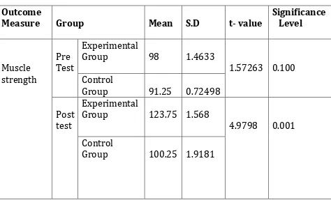
Table 1 group statistics Outcome
Measure Group Mean S.D t- value
Significance Level Muscle strength Pre Test Experimental
Group 98 1.4633
1.57263 0.100 Control
Group 91.25 0.72498 Post
test
Experimental
Group 123.75 1.568
4.9798 0.001 Control
Group 100.25 1.9181
27
Table 2 Group Statistics Outcome
Measure Group Mean S.D t-value Significance Level Distance Walked Pre Test Experimental
Group 63.333 15.69253
0.695 0.497 Control
Group 58.144 15.97209 Post
test
Experimental
Group 78.000 10.01249
2.795 0.013 Control
Group 60.988 15.26618
From Table2, the parameter distance walked is statically significant (0.013) at the t-value (2.795) between the groups.
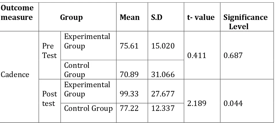
Table 3 Group Statistics Outcome
measure Group Mean S.D t- value Significance Level Cadence Pre Test Experimental
Group 75.61 15.020
0.411 0.687 Control
Group 70.89 31.066 Post
test
Experimental
Group 99.33 27.677
2.189 0.044 Control Group 77.22 12.337
[image:27.612.71.548.417.630.2]
28
Table 4 Group statistics Outcome
measure Group Mean S.D t- value Significance Level Speed Pre Test Experimental
Group 32.444 07.3885
.350 .731 Control
Group 30.850 11.4862 Post
test
Experimental
Group 37.889 06.6086
2.197 .043 Control Group 30.494 07.6331
[image:28.612.71.549.383.588.2]From Table 4, the parameter speed is statistically significant(0.043) for a t-value of (2.189) .
Table 5 Group statistics Outcome
measure Group Mean S.D t- value Significance Level Stride Length Pre Test Experimental
Group 46.678 08.5041
.453 0.657 Control
Group 45.033 06.8189 Post
test
Experimental
Group 54.756 10.0716
2.676 .017 Control Group 44.556 05.4130
From Table 5, the parameter Stride length is statistically significant (0.017) for a t-value of (2.656) between the groups .
29
Outcome
Measure Group Mean S.D t- value Significance Level
Distance walked
Pre Test 60.333 15.69235
-3.498 0.008 Post Test 78.000 10.01249
Cadence Pre Test 75.61 15.020
-2.951 0.18 Post Test 99.33 27.677
Speed Pre Test 32.444 07.3885
-2.231 0.05 Post Test 37.889 06.6086
Stride
Length Pre Test 46.678 08.5041 -2.537 0.035 Post Test 54.756 10.0716
Muscle
strength Pre Test 98 1.7396 2.670411 .025 Post Test 123.75 1.4632
From the above table, the mean of distance walked has increased from 66.33(pre test) to 78.00 (post test) and is statistically significant (.008).
The parameter of gait, cadence has increased from mean value of 75.61 in the pre test to 99.33 in the post test, which is statistically significant(.018).
The mean speed has increased from 32.444(pre test) to 37.889 (post test) which is also statistically significant (.05)
Stride length has increased from its mean value of 46.678 in (pre test) to 54.756, which is also statistically significant (.035)
30
COMPARISON BETWEEN EXPERIMENTAL GROUP AND CONTROL
GROUP IN MUSCLE STRENGTH
98
123.75 91.25
100.25
0 20 40 60 80 100 120 140
PRE TEST POST TEST
31
COMPARISON BETWEEN EXPERIMENTAL GROUP AND CONTROL
GROUP IN CADENCE
75.61
99.33 70.89
77.22
0 20 40 60 80 100 120
PRE TEST POST TEST
32
COMPARISON BETWEEN EXPERIMENTAL GROUP AND CONTROL
GROUP IN DISTANCE WALK
63.33
78 58.14 60.98
0 10 20 30 40 50 60 70 80 90
PRE TEST POST TEST
33
COMPARISON BETWEEN EXPERIMENTAL GROUP AND CONTROL
GROUP IN SPEED
32.44
37.88 30.85 30.9
0 5 10 15 20 25 30 35 40
PRE TEST POST TEST
34
COMPARISON BETWEEN EXPERIMENTAL GROUP AND CONTROL
GROUP IN STRIDE LENGTH
46.67
54.75 45.03 44.55
0 10 20 30 40 50 60
PRE TEST POST TEST
35
DISCUSSION
36
Cerebral palsy is a disorder of posture and movement that results from a non progressive lesion or injury to immature brain. Guralnicketal411995 stated that impaired mobility is a critical determinant of independence and a major contributor to physical disability. The type gait abnormality observed depends on the type and extent of CNS pathology, the constellation of resulting impairments.
Crenna and Inverno24 1994 have suggested a conceptual framework based on four main impairments contributing to disordered gait in patients with supraspinal lesions.
Defective muscle activation (Paretic component)
Abnormal velocity dependent recruitment of muscles during lengthening (spasticity component)
Loss of selectivity in motor output( co contraction component)
Changes in mechanical properties of muscle tendon system(non neural component)
Muscles in gait act both concentrically to generate motion eccentrically to control motion thus weakness can result in both inability to generate force to move the body forward an unrestrained motion resulting from lack of muscle control. Muscle strength is more crucial for individuals with disability than for those without. In this study, lower extremity strengthening exercises was done for a period of 5 days a week for 5 weeks.
From table 6, in the experimental group, after intervention the gait parameters improved significantly. The mean distance walked has improved significantly (17.67) after a period of 5 weeks.
37
from 75.61 to 99.33 ).However stride length has also increased (from 98 in the pre test to 123.75 in the post test)
During the first (2-4) weeks strength can be achieved without structural change in the muscle, but not without neural adaptation. There are more neuro functional changes than structural changes within the muscle.
During the first week of strengthening exercise there is a reduction in the coactivation of other muscles, (muscle synergies, pathological movement) it result in decrease in energy expenditure, movement control improvement . Hence the improved gait performances have occurred because of motor neurological changes.
This improvement is caused by adaptive changes that occur in the nervous system in response to strength training. The EMG findings of high intensity short duration(4 weeks) exercises done in cerebral palsy children have indicated other adaptation mechanisms that may contribute to increased neuronal outflow with training, including increase in maximal firing frequency, increased excitability decreased presynaptic inhibition of spinal motor neurons and down regulation of inhibitory pathways14.
During the first several weeks of resistive training, gains in strength are almost exclusively neural in nature , meaning the body is leaning to recruit the correct muscles in the proper sequence while inhibiting unnecessary muscle recruitments. The physiologic changes, such as an increase in contractile proteins, stored nutrients, and anaerobic enzymes, take several weeks to develop. LeMura15states that once the ”learning” phase begins to diminish, remodeling of the muscle is beginning to take place and strength gains continue”.
Short-term resistance training has been reported also to induce hypertrophy of slow and fast muscle fibers, induce alterations in muscle fiber architecture and fiber type distribution and other morphological changes.
38
Cadence value increased from a mean value of (70.89 to 77.22), it also it was not statically significant. The other 2 parameters stride length & speed declined slightly from their original mean value.
Although the control group received conventional physical therapy, there were no significant improvements in gait performance within a short interval of 5 weeks.
39
40
41
RECOMMENDATIONS
AND LIMITATIONS
42
RECOMMENDATIONS
Joint range of motion and spasticity of the agonist muscle may be measured.
Other groups of cerebral palsy may be trained by strengthening exercises.
Balance training may also be incorporated along with strength training.
Large sample size may be used.
LIMITATION
Small sample size.
Long term effects are not monitored.
43
BIBLIOGRAPHY
&
44
REFERECES
1. Darcy Ann Umphred, neurological rehabilitation.
2. Damiano DL, Vaughan CL, Abel MF. Muscle response to heavy resistance exercise in children with spastic cerebral palsy. Dev Med Child Neurol. 1995; 37:731-739.
3. Damiano DL, Kelly LE, Vaughn CL, Effects of quadriceps femoris muscle strengthening on crouch gait in children with spastic diplegia. Phys Ther 1995;75:658-667.
4. Damiano DL, Wily ME. Lower extremity strength profiles in spastic cerebral palsy. Dev Med Child Neurol. 1998;1998:100-107.
5. Engsberg JR, Ross SA, Bark TS, Changes in ankle spasticity and strength following selective dorsal rhizotomy and physical therapy for spastic cerebral palsy. 1999;91:727-732.
6. Engsberg JR, Ross SA, Olree KS, et al. Ankle spasticity and strength in children with spastic diplegia. Physiother Rev. 1947;96-103.
7. Damiano DL, Abel MF. (1998) Functional outcomes of strength training in spastic cerebral palsy. Archives of physical medicine and Rehabilitation. 79: 119-25
8. Kramer JF, MacPhail HEA. (1994) Relationship among measures of walking efficiency, gross motor ability an isokinetic strength in adolescents with cerebral palsy. Pediatric physical therapy 6: 3-8.
9. Wiley ME, Damiano DL. (1998) Lower-extremity strength profiles in spastic cerebral palsy. Development Medicine & Child Neurology 40:100
45
with spastic cerebral palsy. Developmental Medicine & Child Neurology 38: 1117-25.
11. Leonard CT, Moritani T, Hirschfeld H, Forrsberg H. (1990 ) Deficits in reciprocal inhibition of children with cerebral Palsy as revealed by H reflex testing, Developmental Medicine & Child Neurology 32: 974-84.
12. Dietz V, Berger W. (1995) cerebral palsy and muscle transformation. Developmental Medicine & Child Neurology 37: 180-4. Activity quarterly 2:
13. McCubbin JA, Shasby GB. (1985) Effects of isokinetic exercise On adolescents with cerebral palsy. Adapted physical 56-64.
14. Richard Koscielny American Association of intensive paediatric physical therapy.
15. Clinical Exercise physiology-Application and physiological Principles Linda W.LeMura, Serge P. Von Dulliard, Lippincott Williams&Wilkins 2004.
16. Exercise an sports science Reviews American College of Sport Medicine, Vol 31, No 2, 2003.
17. Thomas R. Baechle, Roger W. Earle, Human Kinetics 2000 Essentials of Strength Training and Conditioning.
18. Jack H. Wilmore, David L. Costill, Human Kinetics 1999 Physiology of Sport and Exercises.
19. Steven J. Fleck, Williams J. Kraemer, Human Kinetics 1997. Designing Resistive Training Programs.
46
21. Anne Shumwaycook PT, Phd, Marorie H. Woollacott, Phd,Marori H. motor control theory an practical applications.
22. Buchner DM, Beres ford SA, Larson EB, effect on physical activity on health status in older adults II intervention studies. Ann rev public health 1992:13:69-88.
23. Holden(1984)-PT, VOL 64(1) 35-40.
24. Gary L. Smidt- Gait in rehabilitation 1-14, 292-307.
25. Mossberg KA, Linton KA, Fricke K. Ankle foot orthoses: effect on energy expenditure of gait in spastic diplegic children. Arch phys medical rehab 1990; 71: 490-494.
26. Dodd KJ, Damino Dl, A systematic review of effectiveness of strength training programs for people with cerebral palsy. Arch phys medical rehab2002 AUG; 83(8);1157-64.
27. Horvat M. (1987) Effects of progressive resistance training program on an individual with spastic cerebral palsy. American corrective Therapy Journal 41: 7-11.
28. Mcburney H, Taylor NF, Dodd KJ, Graham HK.A qualitative analysis of the benefits of strength training for young people with cerebral palsy.
29. Joanne Bundonis, Grimenstein J, Diienno M Fiunctional exercise and strengthening in the neurologically impaired child(clinical notes) Toms River, NJ:Princeton University; November2004.
30. Blunell SW, Shephard RB, Dean CM, Adams RD, Cahill BM. Functional strength training in cerebral palsy; a pilot study of group circuit training classes for children.
47
32. Rich Smith, Maximizing Energy Efficiency, Rehab management Oct 2002.
33. Eagleton M, Iams A, McDowellJ, MorrisonR, Evans CL The effect of strength training on gait in adolescents with cerebral palsy. Paeiatr Phys Ther 2004;16 (1); 22-30.
34. Gowland C.Staging motor impairment after stroke. Stroke 1990;21(supp):11-19-11-21.
35. Perry HJ.Newsam C. Function of the hamstrings in cerebral palsy. In Sussman Med. The diplegic child.
36. Montgomery J. Assessment and treatment of locomotor deficits in stroke. In: Duncan PW, Badke MB. Stroke rehabilitation: the recovery of motor control. Chicago: Year Book, 1987:223-259.
37. Designing Resistive Training Programs, Steven J, Fleck, Williams J. Kraemer, Human Kinetics 1997.
38. Holland MK, Gill KM, MagliozziMR, et al clinical gait assessment in the neurologically impaired:reliability and meaningfulness Phys Ther 1984;64:35-40.
39. Katz, Rymer Z. Spastic hypertonia mechanism and measurement. Arch.Phy.Med.rehab 1989:70:144-155.
40. Texeria-Salmela LF, Olney SJ, NadeauS, BrouwerB, Muscle strengthening and physical conditioning to reduce impairment disability in chronic stroke survivors Arch Phys Med Rehabil 1999:1211-1215.
Web site:
www.pubmed.gov
www.emedicine.com
48
Annexure I
Consent Form
I voluntarily agree to allow my child / ward to participate in the study on “the effect of lower extremity strength training on gait in the study on” the effect of children. This study will be carried out for duration of 40 to 45 min, 5 days a week for 5 weeks. All the information given will be kept strictly confidential and used only for research purpose.
49
Annexure II
Manual muscle testing
MRC grading
Grade 0 - No tension is palpated in the muscle or tendon on maximum voluntary effort
Grade 1 - Tension is palpated in the muscle (or) tendon but no motion occurs. Grade 2 – Movement with gravity eliminated.
Grade 3 - Movement against gravity .
50
Annexure III
GMFCS score sheet
Child’s name
Assessment date
GMFCS level
Level I Children walk indoors and outdoors and climb stairs without limitations.
Level II Children walk indoors and outdoors and climb stairs holding onto a railing but experience limitation walking on uneven surfaces and inclines and walking in crowds or confined spaces.
Level III Children walk indoors or outdoors on a level surface with an assistive mobility device and orthoses on level surfaces and climb stairs with support .
Level IV Children may continue to walk for short distances on a walker or rely more on wheeled mobility at home, school and community .
51
Annexure IV
ASSESSMENT CHART
Name
Age
Sex
Class
Muscle power assessment
Muscle Pre test Post test Hip Flexors R L R L Hip Extensors
Hip Abductors Hip Adductors Knee Extensors Knee Flexors Ankle DF Ankle PF
Gait Function assessment
Name Age Speed
Distance
walked in two minutes