EVALUATION AND COMPARATIVE ANALYSIS
OF NEWER COMPOUND FIXATIVES
DISSERTATION
SUBMITTED TO TAMILNADU DR.M.G.R. MEDICAL UNIVERSITY,
CHENNAI
in partial fulfilment of
the requirements for the degree of
M.D. (PATHOLOGY)
BRANCH - III
TIRUNELVELI MEDICAL COLLEGE HOSPITAL,
TIRUNELVELI- 627011
CERTIFICATE
I hereby certify that this dissertation entitled “EVALUATION AND COMPARATIVE ANALYSIS OF NEWER COMPOUND FIXATIVES” is a
record of work done by Dr.S. MUTHUSELVI, in the Department of Pathology,
Tirunelveli Medical College, Tirunelveli, during her postgraduate degree course
period from 2014- 2017. This work has not formed the basis for previous award of any
degree.
Dr. K. SITHY ATHIYA MUNAVARAH M.D.,
DEAN
CERTIFICATE
I hereby certify that this dissertation entitled “
EVALUATION AND
COMPARATIVE ANALYSIS OF NEWER COMPOUND FIXATIVES”
isa record of work done by
Dr.S. MUTHUSELVI
, in the Department of Pathology,Tirunelveli Medical College, Tirunelveli, during her postgraduate degree course
period from 2014- 2017, under my guidance and supervision, in the Department of
Pathology Tirunelveli Medical College & Hospital, Tirunelveli, in partial fulfilment of
the requirement for M.D., (Branch III) in Pathology examination of the Tamilnadu Dr.
M.G.R Medical University will be held in APRIL 2017. This work has not formed the
basis for previous award of any degree.
Prof. Dr. J. SURESH DURAI,M.D.,
Department of pathology,
Tirunelveli Medical College,
Prof. K. SHANTARAMAN.MD.,
Professor and Head, Department of Pathology, Tirunelveli Medical College Tirunelveli- 627011.
DECLARATION
I solemnly declare that this dissertation titled “EVALUATION AND COMPARATIVE ANALYSIS OF NEWER COMPOUND FIXATIVES”
submitted by me for the degree of M.D, is the record work carried out by me during
the period of 2014-2016 under the guidance of Prof.Dr. J. SURESH DURAI, M.D,
Professor of Pathology, Department of Pathology, Tirunelveli Medical College,
Tirunelveli. The dissertation is submitted to The Tamilnadu Dr. M.G.R. Medical
University, Chennai, towards the partial fulfilment of requirements for the award of
M.D. Degree (Branch III) Pathology examination to be held in April 2017.
Place: Tirunelveli Dr.S. MUTHUSELVI,
Date: Department of Pathology,
ACKNOWLEDGEMENT
This dissertation has come to your hands as a result of the combined effort of
lot of people. I am thankful to all these people for helping me in bringing out this
work.
I thank the DEAN Dr. SITHY ATHIYA MUNAVARAH, M.D for permitting
me to conduct this study and to avail the resources of the hospital.
I am also thankful to my Professor and Head, Department of Pathology
DR.K. SHANTARAMAN M.D, for his support and guidance in doing this
dissertation.
I express my heartfelt gratitude to my guide DR. J. SURESH DURAI M.D,
Professor, Department of Pathology, who has guided me through all the steps in my
study right from the beginning till the end.
I am extremely thankful to the respected Professors of my department
DR.VALLIMANALAN.S M.D, DR.SWAMINATHAN.K M.D, DR.ARASI
RAJESH M.D, DR. VASUKI MUTHURAMAN M.D, and the Assistant Professors
for their concern, valuable suggestions, and support during the study.
I also thank all the lab technicians and my fellow postgraduates and my friends
for their cooperation which enormously helped me in the study. Without their humble
cooperation, this study would not have been possible.
I thank GOD AND MY PARENTS, for blessing me not only in this study,
ABBREVIATIONS
Amex
-
Acetone Methylebenzoate
–
Xylene
CD
-
Cluster of Differentiation
CK
-
Cytokeratin
DNA
-
Deoxy Ribonucleic Acid
EPA
-
Environmental Protection Agency
ER
-
Estrogen Receptor
FFPE
-
Formalin Fixed Paraffin Embedded
GC
-
Gas Chromatography
GC- MS
-
Gas Chromatography And Mass Spectrometry
GIT
-
Gastrointestinal Tract
HER 2
-
Human Epidermal growth factor Receptor
–
2
HOPE
-
Hepes-Glutamic Acid Buffer Mediated Organic Solvent
Protection Effect
HPLC
-
High Performance Liquid Chromatography
IARC
-
International Agency For Research On Cancer
IHC
-
Immunohistochemistry
MPNST
-
Malignant Peripheral Nerve Sheath Tumor
NBF
-
Neutral Buffered Formalin
NIOSH
-
National Institute Of Occupational Safety And Health
Administration
PASD
-
Periodic Acid Schiff and Diastase
PCR
-
Polymerase Chain Reaction
ppm
-
parts per million
RNA
-
Ribonucleic Acid
RT PCR
-
Real Time Polymerase Chain Reaction
STEL
-
Short Term Exposure Limit
TWA
-
Time Weighted Average
UMFIX
-
Universal Molecular Fixative
CONTENTS
Sl. No.
TITLE
PAGE No.
1.
INTRODUCTION
1
2.
AIMS AND OBJECTIVES
3
3.
REVIEW OF LITERATURE
4
4.
MATERIALS AND METHODS
39
5.
OBSERVATION AND RESULTS
47
6.
DISCUSSION
82
7.
SUMMARY AND CONCLUSION
88
8
BIBILIOGRAPHY
INTRODUCTION
Fixation is one of the most important step in the practice of diagnostic
pathology. Even in this modern age where many things have changed,
formaldehyde is most commonly used for tissue fixation. Formaldehyde is a gas
which is commercially available in the form of concentrated solution of 37%. A
10% dilution of concentrated formalin is named as 10% formalin.
The reasons which make formalin popular are; its low cost, its ability to
help in long term storage of tissue, its ability to help preserve morphological
features, and the fact that it allows special histological stains. Also notable is the
fact that in combination with antigen retrieval it allows for reliable
immunohistochemical analysis. The laboratory preparation of formalin is
straightforward procedure. Moreover the use of formalin is a longstanding
tradition and it is used internationally. There is a general opinion that formalin is
the best fixative available and that there is no need to look for an alternate fixative.
However toxicity of formalin is emerging as the main reason for its
abolition as the commonest fixative used in laboratories[1]. More recently from
initial reports from IARC( International Agency for Research on Cancer ) link
formaldehyde exposure and leukemia. These reports were further highlighted in a
report issued in 2012 by the same agency.
The agencies that monitor formaldehyde exposure in the national and
limit ranges from 0.016 ppm TWA(time weighted average ) to 2 ppm for STEL
(short term exposure limit)[1].
The formation of DNA protein cross links denotes a permanent signature
of exposure to formalin. Recent research shows chromosomal alterations in health
workers and these have been found to be related to formalin use in pathology
laboratories. An attempt has been made in this study to minimize formalin
AIMS AND OBJECTIVES
To compare and analyze the fixation characteristics of a group of formalin
containing compound fixatives.
To assess the optimal efficacy of a compound fixative.
To minimize formalin exposure in histopathology laboratory.
REVIEW OF LITERATURE
FIXATION:
It is a process by which the constituents of the cells and therefore of the
tissues are fixed in a physical and partly also in a chemical state so that they will
withstand subsequent treatment with various reagents with a minimum of loss,
significant distortion, or decomposition.
IDEAL FIXATIVE:
It should
Penetrate a tissue quickly. Be isotonic.
Cause a minimum loss of physical and chemical alteration of the tissues
and its components.
Be rapid in action and shelf life of at least one year.
Allows the isolation of macromolecules including proteins, m RNA and
DNA without significant biochemical alterations from fixed and embedded
tissues.
Useful for diverse variety of specimen including fatty tissue, lymphoid
structures and large specimens and support histochemical as well as
immunohistochemical analyses and other specialized procedures.
Be cheap and stable.
Be compatible with modern automated tissue processors.
Have toxicological and flammability profiles such that use of the fixative
is safe.
Be readily storable for long term, easily disposable or recyclable and also
give excellent microtomy of embedded blocks.
TYPES OF FIXATIVES:
I. Microanatomical fixatives
II. Cytological fixatives
III. Histochemical fixatives
METHODS OF FIXATION:
1.Physical Fixation:
Heat fixation
Microwave fixation
Freeze drying and freeze substitution.
2.Chemical Fixation:
MICROANATOMICAL FIXATIVES:
They preserve the anatomy of the tissue, in correct relationship of tissue
layers and large aggregates of cells. All routine histopathological fixatives come
under this group.
CYTOLOGICAL FIXATIVES:
These fixatives help in the preservation of intracellular structures.
Eg.Carnoy’s fluid, Clarke’s fluid, Muller’s fluid andformal saline etc.
HISTOCHEMICAL FIXATIVES:
They are used when histochemical tests were applied. They produce only
minimal changes in the elements have to be demonstrated. Freeze drying method
is almost ideal for this purpose.
HEAT FIXATION:
Boiling of tissue materials in normal saline can produce adequate
morphology of the tissue. It is the simplest form of fixation. Nowadays heat is
mainly used to speed up other forms of fixation. And it also used in the steps of
tissue processing.
MICROWAVE FIXATION:
FREEZE DRYING AND FREEZE SUBSTITUTION:
In freeze drying, tissues are cut into thin sections and immersed in liquid
nitrogen. Then the water molecules are removed in a vacuum chamber at–40˚c.
The tissues can be post fixed with formaldehyde vapor. It is useful for studying
soluble materials and small molecules.
In freeze substitution , tissues are immersed in fixatives at –40˚c, such as
acetone or alcohol which slowly removes water molecules through dissolution of
ice crystals. The proteins are not denatured. Then the temperature gradually rised
to 4˚ c and the fixation process is completed.
EFFECTS OF FIXATION IN TISSUES:
Fixatives inhibit autolysis and putrefaction of tissues.
It preserves tissue architecture and induces hardening of tissues and
solidifies the cellular material.
It alters the varying degree of refractive indices of the cells and tissue
components. Fixatives induce loss of tissue materials followed by fixation. Eg.
protein and mucopolysaccharides loss followed by formalin fixation.
EFFECTS OF FIXATION ON STAINING:
Histochemical reactions of the tissues are usually affected after fixation
because fixatives usually disrupt the secondary and tertiary structures of proteins.
As they bind to reactive groups in the tissues to achieve fixation, they affect routine
staining adversely. Sometimes they act as mordant and improve staining. Eg .
cationic amino groups (formaldehyde) or creates the anionic amino group or both
(osmium tetroxide) will decrease tissue acidophilia and enhance basophilia. And
a fixative which eliminates anionic amino groups or generates cationic amino
groups or both (chromic acid) will reduce basophilia and enhance tissue
acidophilia.
FIXATION ARTEFACTS:
Shrinkage artefacts:
Tissues fixed in 10% NBF and embedded in paraffin wax shrink by 33%.
Alcohol fixed tissues show excessive shrinkage artefacts.
Glycogen streaming artifact:
It results from diffusion of unfixed tissue materials .It is evidenced in PAS
staining of glycogen containing tissues.
Pigment artefacts:
Formalin pigment- Brown/ brownish black pigment produced under acidic
conditions. They are commonly seen in blood rich tissues . Eg. spleen, liver,
hemorrhagic lesions etc. formalin pigment can be removed by treating sections
with 10% sodium or potassium hydroxide in 70% alcohol for 5 to 15 minutes or
treating unstained sections in alcoholic saturated picric acid.
Mercury pigment – mercuric chloride containing fixatives produce varying
amount of dark brown or grey coloured deposits through out the tissue. It can be
removed by oxidation with iodine to mercuric chloride and subsequently removed
Chromate deposits- if the specimens are not washed well after fixation in
chromate containing fixatives, the salts react with alcohol forming a yellow brown
to black precipitate. They are removed by treating sections in 1% hydrochloric
acid in 70% alcohol for 30 minutes.
EFFECTS OF PROLONGED FIXATION:
Prolonged fixation of tissues leads to shrinkage artefacts. Prolonged
fixation of tissues in formalin results in decreased antigenicity. Studies showed
that immunoreactivity of CK 7 (cytokeratin), HMW CK (High molecular weight
cytokeratin) and laminin was decreased in prolonged formalin fixation. But
nowadays this is overcome by enzymatic digestion and heat induced antigen
retrieval methods[ 75 ].
Fixation of specific tissues:
Brain
Conventional fixation of brain takes at least 2 weeks[47]. Perfusion
technique of the brain through the middle cerebral arteries reduce fixation time.
Fixation is also enhanced by the use of microwave technology. For biotin-avidin
method of immunohistochemistry, alcoholic formalin should not be preferred.
Eyes
To obtain good sections, the globe must be firmly fixed. Eyes are usually
fixed in NBF for 48 hours. After gross description, iris and optic nerve are
removed and fixed for an additional 48 hours. For the canal of Schlemm and the
Breast
Mastectomy specimens should be sliced at 5mm intervals and fixed in
conventional 10% neutral buffered formalin for a minimum of 6–8 hours and a
maximum of 72 hours. To prevent lysis of biomarkers, such as progesterone
receptor, estrogen receptor and the human epidermal growth factor receptor-2
(HER2), fixation should be as short as possible.
Lungs
NBF is the preferred fixative for lung biopsies. Specimens received from
autopsies may be inflated through the trachea or major bronchi and should be
fixed overnight .
Lymphoid tissue
Excised lymph node is usually fixed in NBF or in B5 or zinc based
fixatives.
Testis
Small biopsies of testis and penectomy specimens should be fixed in
conventional buffered formalin.
Muscle biopsies
For histopathological examination, a portion of muscle should be fixed in
NBF and the remaining portion should be kept in normal saline for further enzyme
studies.
Renal biopsies
FACTORS AFFECTING FIXATION:
1. BUFFERS AND pH:
Acidic pH reduces the formation of reactive hydroxymethyl groups and
cross linking resulting in improper fixation[5,7]. Therefore buffers are added to
maintain a pH of 7.2 to 7.4. Commonly used buffers are phosphate, bicarbonate,
Tris, cacodylate and acetate .
2. DURATION OF FIXATION AND SIZE OF SPECIMENS:
In 1941 , Medawar et al in his study observed that the depth reached by the
fixative is directly proportional to the square root of duration of fixation and
expressed this relation as;
d= k√
where d= depth, t= duration of fixation, k= constant
The constant represents the coefficient of diffusability which is specific to
each fixative. Eg. k is 0.79 for formalin, 1.0 for 100% ethanol and 1.33 for
potassium dichromate[6].
To allow proper penetration of fixatives from all directions , the bottom of
the container should be wadded by fixative-soaked cotton or cloth and the
specimens placed over that. The specimen should be cut not be thicker than 0.5
The volume of the fixatives should be at least 10 times the volume of the gross
specimen for optimal fixation.
3. TEMPERATURE:
The diffusion of molecules increases with rising temperature due to their
more rapid movement and vibration. So nowadays microwaves are used to speed
fixation and tissue processing.
4. CONCENTRATION OF FIXATIVE:
Effectiveness and solubility primarily determine the appropriate
concentration of fixatives. Concentration of formalin above 10% will cause
increased hardening and shrinkage[8]. Ethanol below 70% do not remove free
water from tissues efficiently.
5. OSMOLALITY AND IONIC COMPONENTS:
Hypertonic and hypotonic solutions lead to cell shrinkage and swelling
respectively. The better morphological details obtained with solutions that are
slightly hypertonic (400- 450 mOsm ).The ionic compositions of solutions should
be isotonic as possible to the tissues.
6. ADDITIVES:
They are used to improve the morphology of the fixed tissue. Eg .
CHEMICAL FIXATION:
COAGULANT FIXATIVES:Alcohols are the commonly used coagulant fixatives eg,. methanol, ethanol
etc., They act by denaturing and precipitating protein by removing free water
molecules and disrupting hydrogen bonds. Free water molecules usually
surrounds hydrophobic areas of proteins and by repulsion, the water molecules
drive hydrophobic areas into closer together with each other and hence stabilize
hydrophobic bonding. When alcohol removes water, this hydrophobic bonding
weakens. Like this, water molecules also take part in hydrogen bonding in
hydrophilic areas of proteins. So water removal in alcoholic fixation destabilizes
this hydrogen bonding. All these changes disrupt the tertiary structure of proteins.
Protein denaturation depends upon, the choice and concentration of alcohol, the
presence of organic and non-organic substances, and the pH and temperature of
fixation. Eg. ethanol denatures proteins>phenols >>monocarboxylic acids
>dicarboxylic acids[9].
Commonly used alcohol based fixatives are clarkes solution, carnoy’s
fixative and methacarn fixative.
PICRIC ACID FIXATIVES:
It contains saturated aqueous solution of picric acid. Bouin’s solution:
It is composed of
Saturated solution of picric acid 1500 ml
Glacial acetic acid 100 ml
It is the best fixative for connective tissue stains. Lithium carbonate, 70% ethanol
or another acid dye can be used to remove yellow colour precipitates of picric acid. Hollande’s solution:
It is composed of
Distilled water 1000 ml
Concentrated formalin 100 ml
Acetic acid 15 ml
Picric acid 40 g and
Copper acetate 25 g
This fixative is preferred for endocrine tissues and gastrointestinal biopsies.
MERCURIC FIXATIVES:
Mercuric chloride (HgCl2) containing fixatives acts by coagulating tissue
proteins. Lillie’s B5 fixative is composed of 4% aqueous formaldehyde with
0.22M HgCl2 and sodium acetate. Mercuric chloride establishes rapid structural
stabilization and also enables bright staining by many of the dyes. B 5 fixative
enhances nuclear detail and it is preferable for hematopoietic specimens. Mercury
based fixatives produce brown crystalline precipitate, probably mercurous
chloride ( Hg2Cl2 ) through out the tissues. This mercury pigment can be removed
by sequential treatments with iodine and sodium thiosulfate solutions, before
staining. As mercury is toxic, all mercury based fixatives should be subjected to
It is composed of
Mercuric chloride 12.5 g
Sodium sulfate 2.5 g
Potassium dichromate 6.3 g
Distilled water 250 ml
5 ml of glacial acetic acid is added to 95 ml of above solution just before using
this fixative. This fixative is recommended for bloody specimens and for
trichrome stains. Helly’s solution:
The constituents of the Helly’s fixative is similar as Zenker’s solution but
in the last step 5 ml of concentrated formalin is added to 95 ml of stock solution.
It is advisable for bone marrow, intercalated discs and extramedullary
hematopoiesis specimens. Schaudinn’s fixative:
Distilled water 50 ml
Mercuric chloride 3.5 g
Absolute ethanol 25 ml Ohlmacher’s fixative:
This is composed of
Absolute ethanol 32 ml
Glacial acetic acid 2 ml
Chloroform 6 ml
This fixative penetrates rapidly.
Carnoy-Lebrun solution:
Absolute ethanol 15 ml
Chloroform 15 ml
Glacial acetic acid 15 ml
Mercuric chloride 8 g
This fixative penetrates rapidly.
DICHROMATE AND CHROMIC ACID FIXATION:
Dichromate fixatives acts by oxidation of proteins with interaction of
reduced chromium ions. Chromium ions in +6 valence state coagulate proteins
and nucleic acids. To some extent the ions also form cross links. They particularly
react with carboxyl and hydroxyl side chains of proteins. As they leave amino
groups of proteins free, they help staining with acid dyes. Chromate interacts with
unsaturated lipids and make them insoluble, so it is considered as good fixative for
mitochondria. 24 hours fixation is essential for dichromate fixatives. Tissues fixed
in dichromate fixatives should be washed thoroughly after fixation in order to
prevent chromate deposits in sections. Usually these fixatives were used to
demonstrate amine containing chromaffin granules in endocrine tissues.
Nowadays it is replaced by immunohistochemistry to demonstrate chromaffin
granules. Potassium dichromate is used as a component of many compound
Miller’s or Moller’s fixative:
It is composed of potassium dichromate 2.5 g and sodium sulfate 1 g
dissolved in 100 ml of distilled water. Regaud’s fixative:
In this solution, potassium dichromate 3 g is dissolved in 80 ml of distilled
water. 20 ml of 37% formaldehyde is added just before using this solution. Orth’s fixative:
It is composed of
Potassium dichromate 2.5 g
Sodium sulfate 1 g
Distilled water 100 ml
10 ml of concentrated formalin is added to above solution.
COMPOUND FIXATIVES:
Combination of coagulant fixatives with non coagulant cross linking
fixatives is called as compound fixatives. When formaldehyde added to dehydrant
ethanol, there will be less shrinkage and hardening comparing to pure dehydrants
and it is efficient in preserving molecules like glycogen. Fixation of tissues in
alcoholic formalin may helps to distinguish lymph nodes embedded in fat. Even
though alcoholic formalin preserves antigen immunorecognition well, it increases
the background staining in immunohistochemistry[50].Compound fixatives can
also be used for post fixation.
It is composed of 895 ml of 95% ethanol and 105 ml of concentrated
formalin.
Alcohol-formalin-acetic acid fixative
It is composed of
Ethanol (95%) 85 ml
Formaldehyde (37%) 10 ml
Glacial acetic acid 5 ml Alcoholic Bouin’s solution:
The constituents of this fixative is similar to Bouin’s except it is less
aqueous and therefore it gives better preservation of some carbohydrates (e.g.
glycogen).After fixation tissues should be washed in 70% ethanol, followed by
95% ethanol (several changes). This fixative improves upon aging[52]. Gendre’s fixative :
It is composed of
Saturated picric acid in 95% ethanol 800 ml
Concentrated formalin 150 ml
Glacial acetic acid 50 ml Rossman’s fixative:
It is composed of
Concentrated formalin 10 ml
Tap water 10 ml
24 hours fixation is needed. This fixative is preferable for connective
tissues and umbilical cord.
NONCOAGULANT CROSS LINKING FIXATIVES:
FORMALDEHYDE:
It is a naturally occurring organic compound with the formulae CH2 O.
NOMENCLATURE:
Chemical Abstract Service Register No.: 50-00-0
Chemical abstract name: Formaldehyde
IUPAC Systematic Name: Methanal[2].
Synonyms: Formaldehyde, gas; formic aldehyde methaldehyde;
methyl aldehyde; methylene oxide; oxomethane; oxymethylene[2].
Structural formulae- CH2 O
Fig 1: formaldehyde structure
Physical and Chemical properties of formaldehyde:
It is a colour less gas with a pungent odour with boiling point -19.1˚C,
melting point -92˚C and density 0.815 at –20 °C. It is soluble in chloroform, water,
ethanol and miscible with benzene, acetone ,diethyl ether. Formaldehyde gas is
stable in the absence of water, incompatible with oxidizers, alkalis, acids, phenols,
urea [2,9,10]. It reacts explosively with peroxide, nitrogen oxide and performic
acid[9,10].
HISTORY:
Formaldehyde was first discovered by Butlerov in 1859. Van Hoffman
established the method for synthesis of formalin from methanol and established
its properties in 1868. Initially formalin was considered as an antiseptic, either to
treat or prevent wound infections.
In 1892, Meister, Lucius and Brunig went to a young physician Ferdinan
Blum with a proposal to test the antiseptic properties of formaldehyde.
Formaldehyde was supplied by the manufacturer as a 40% aqueous solution,
which is the concentration resulted from bubbling formaldehyde gas through
water. Blum diluted the commercial solution of formalin with 9 parts of water to
give a 4% weight/volume solution. He tested the bactericidal properties of this
diluted solution and found out that it was an effective but slow agent for killing
In his second paper , Blum reported that in the process of studying
disinfection, he noticed that the skin of his fingers that come in contact with the
diluted solution became hardened , as much as with alcohol, that is one of the
commonest reagent used for fixation of tissues in olden days. Then he
experimented the anthrax infected mouse tissue with diluted formalin and
observed that tissues preserved in formalin had the same consistency as alcohol
fixed tissues and better staining results were obtained using hematoxylin and
aniline dyes. And he also observed that formaldehyde produces less shrinkage and
distortion of tissues than alcohol fixed tissues[12].
Pure formaldehyde is when completely dissolved in water, forms solution
containing 37- 40% formaldehyde; this aqueous solution is known as concentrated
formalin. So the usual 10% formalin contains about 4% weight to volume of
formaldehyde.
MECHANISM OF ACTION:
Fraenkel-Conrat and his colleagues identified and explained the most of
the reactions of formaldehyde with amino acids and proteins [13, 14]. In aqueous
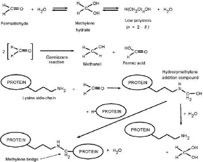
solution formaldehyde forms methylene hydrate. Formation of methylene hydrate
is the first step in formalin fixation .
H2C= O + H2O HOCH2OH
The formed methylene hydrate acts with the side chains of proteins to form
side chains formation is the fundamental and distinct reaction of formaldehyde
fixation.
Formaldehyde reacts with nuclear proteins and nucleic acids [18] and it
also alters nucleotides by interacting with free amino acids. The cross-linking
reactions start at adenine-thymidine (AT)-rich regions of free and naked DNA.
And these reactions increases with increasing temperature. Formaldehyde interact
with C=C and–SH bonds in unsaturated lipids, but it does not react with
carbohydrates. The side chains of lysine, arginine, tyrosine, cysteine, histidine,
serine and threonine are more reactive with methylene hydrate, so they have
REACTIONS OF FORMALDEHYDE WITH PROTEINS:
Fig 2: Reactions of formalin with proteins
COMMONLY USED FORMALIN FORMULAE:
10% Neutral buffered formalin:
It is composed of
Tap water 900 ml
Formalin (37% formaldehyde) 100 ml
Sodium phosphate,monohydrate 4 g
Sodium phosphate, dibasic, anhydrous 6.5 g
Carson’s modified Millonig’s phosphate buffered formalin:
It is composed of
Concentrated formalin l10 ml
Tap water 90 ml
Sodium phosphate, monobasic 1.86 g
Sodium hydroxide 0.42 g
The pH should be 7.2–7.4.
This combination is better for ultra structural preservation than NBF.
Formal (10% formalin), calcium acetate:
It is composed of
Concentrated formalin 100 ml
Tap water 900 ml
Calcium acetate 20 g
It is better in preserving lipids.
Formal saline:
It is composed of
Concentrated formalin 100 ml
Tap water 900 ml
Sodium chloride 9 g
Formal (10% formalin), zinc, unbuffered:
It is composed of
Sodium chloride 4.5 g
Zinc chloride or (zinc sulfate) 1.6 g.
Zinc formalin is an excellent fixative for immunohistochemistry.
Formalin, buffered saline:
It is composed of
Concentrated formalin 100 ml
Tap water 900 ml
Sodium chloride 9 g
Sodium phosphate,dibasic 12 g
Formalin, buffered zinc:
It is composed of 1000 ml of 10% neutral buffered formalin and 1.6 g of
Zinc chloride.
ADVANTAGES OF FORMALIN FIXATION:
It is cheap. Easy to prepare. Relatively stable.
Allows the subsequent application of most staining techniques
without preliminary procedures.
Frozen sections can be prepared from formalin fixed material. Fat staining can easily carried out on tissues fixed in formalin. It penetrates well and does not cause excessive hardening or renders
them brittle.
DISADVANTAGES:
Allergic dermatitis. Irritant to nose.
In tissues unbuffered formalin leads to dark brown artifact
pigment.
It is unsuitable for electron microscopy.
FORMALIN TOXICITY:
Head ache, burning sensation in throat, difficulty in breathing and
exacerbation of asthma are the known toxic effects of formalin.
A study conducted on Finnish women working in a laboratory at least 3
days a week found that there is a significant correlation between spontaneous
abortion and formaldehyde exposure[2].
And another study on Chinese women also reported that abnormal
menstrual cycles in 70% of the women with occupational exposure to
formaldehyde which is 17% in control group[2]. Many researches are going on
through out the world to determine the teratogenic effect of formaldehyde.
In 1987, the U.S. Environmental Protection Agency ( EPA ) classified
formaldehyde as a probable human carcinogen under conditions of unusually
high or prolonged exposure and it limits the permissible exposure level from 1
ppm to 0.75 ppm.
In 2005-06, formaldehyde was found to be seventh most common allergen
IARC ( International Agency for Research on Cancer ) also reported that
there is a strong association between formaldehyde exposure and human
nasopharyngeal carcinoma. And there is successive link between formaldehyde
exposure and myeloid leukemia was established in 2009[1]. With evaluation and
information of all known data led the IARC to reclassify formaldehyde as known
human carcinogen that is from group 2A to group 1 carcinogen. These
statements were reinforced in 2012 by the same agency[1].
In June 2011, the 12th edition National Toxicology program changed the
lining status of formaldehyde from “reasonably anticipated to be human
carcinogen” to “known to be a human carcinogen”.
MEASUREMENT OF FORMALDEHYDE EXPOSURE:
The methods used for the determination of the concentration of
formaldehyde in air are based on spectrophotometry, with sensitivity of 0.01–
0.03 mg/m3 can be achieved. Other methods include colorimetry, polarography,
gas chromatography (GC), fluorimetry, high-performance liquidchromatography
(HPLC), infrared detection and gas detector tubes. Most of the methods require
the formation of a derivative for separation and detection. HPLC is the most
sensitive method (limit of detection, 2 μg/m3 or less). Formaldehyde has been
measured in blood by gas chromatography–mass spectrometry (GC–MS) after
derivatization to pentafluorophenylhydrazone. Formic acid or formate is produced
EXPOSURE LIMITS:
The usual mean concentration during exposure in histopathology laboratory
is 0.5 ppm[44,45,46].
The NIOSH (National Institute of Occupational Safety and Health
Administration) U.S defines
Permissible Exposure Limits as
0.75 ppm time weighted average limit ( for 8 hoursTWA)
2 ppm short term exposure limit ( STEL 15 minutes exposure )[3].
Recommended exposure level :
0.016 ppm TWA to 0.1 ppm for STEL[3].
Individual formaldehyde exposure was monitored by using
Malondialdehyde- deoxyguanosine adducts on leukocytes and the alkylation of
hemoglobin to form a terminal N-methylene valine residue.
NON FORMALIN FIXATIVES:
CARNOY’S FIXATIVE:
It is composed of 6 parts of ethanol, 3 parts of chloroform and one part of
glacial acetic acid. It provides good preservation of nucleic acid in tissues. It is
used for glycogen preservation. RNA stains like methyl green pyronine yields
better results in carnoy’s fixed tissues. However the main drawback is the loss of
high molecular weight RNA. Also it shrinks and harden the tissue, this can be
reduced by avoiding overfixation and processed for low melting point wax
MODIFIED METHACARN:
Components:
6 parts of methanol
3 parts of chloroform
1 part of glacial acetic acid.
Modified methacarn was found to be excellent in preserving tissue RNA[43].
It acts very gently on tissue membranes. As against formalin,
immunohistochemistry can be carried out with lesser duration of incubation,
greater dilution of antibodies and little requirement for antigen retrieval[24].
ZINC BASED FIXATIVES:
It is a mixture of zinc acetate, zinc chloride and calcium chloride in Tris
buffer.
Z 7 contains
Zinc trifluroacetate
Zinc chloride
Zinc acetate
Calcium acetate in Tris-Hcl
The pH is maintained between 6.4 and 6.7.
It has been found to be cost effective, reliable and nontoxic in contrast to
NBF. They are superior in DNA and protein extraction analysis in many types of
tissue and does not need heat pretreatment for antigen retrieval[25]. The RNA,
DNA and protein integrity was found to be good, therefore molecular analysis on
formalin fixed tissues[26]. DNA sequences up to 2.4 kb and RNA fragments up to
362 bp in length could be properly amplified. But zinc fixation also results in
tissue shrinkage and may alter histology.
HOPE fixation (Hepes-glutamic acid buffer mediated organic solvent protection
effect ):
It is a good technique for comprehensive pathological analysis including
immunohistochemistry and molecular pathology. This fixative consists of a
mixture of amino acids at pH 5.8 to 6.4. By diffusion this fixative penetrates
tissues. After fixation, the tissues incubated in acetone at 0-40ºC for dehydration.
Next the specimens are transferred into low melting point paraffin wax and
embedded. HOPE fixed sections show formalin like morphology and give a very
good preservation of proteins and antigenic structures for analysis by
immunohistochemical and enzyme histochemical techniques. Fairly good amount
of good quality DNA and RNA can be extracted from these specimens even after
a period of five years[27]. These specimens are also suitable for molecular analysis
by PCR, RT-PCR and in-situ hybridization even after five years[28]. Absent
cross-linking and better yield of nucleic acids shows that HOPE fixation can emerge to
be an alternative method for tissue banking.
Acetone-methyl benzoate-xylene(AMeX) fixative:
It gives good morphology and good quality of high molecular weight DNA
[29]. This method, tissues were fixed in acetone at -200˚C for overnight. Then
Extraction of good quality of higher molecular weight DNA is seen in this
technique. RNA extraction is also comparable to fresh frozen tissues [30,31].
Universal molecular fixative (UMFIX):
It is composed of methanol and polyethylene glycol. It is always combined
with microwave assisted rapid tissue processing. UMFIX is greatly useful for
amplifying small amplicons by RT-PCR in small biopsies. It was introduced by
Vincek et al recently to extract high molecular weight RNA from laser captured
micro dissected samples. These samples are prepared from paraffin embedded
blocks used for histologic diagnosis[32]. Tissue can also be fixed at normal room
temperature with UMFIX. Immunoreactivity is similar to formalin fixed tissue.
The quality and amount of mRNA and DNA extracted from UMFIX fixed tissues
is also comparable to frozen sections.
FINEFIX:
It is composed of ethanol (65- 75% w/v), polyvinyl alcohol, glycerol and
monomeric carbohydrates. The special feature of ethanol based fixatives is that
they do not form covalent bonds between proteins; they remove the water
molecules which surrounds proteins and as a result, protein coagulation occurs and
enzymatic functions are brought to halt, thereby making the tissue amenable to
conventional proteomic techniques. The major problem in proteomic analysis in
fresh tissues is the difficulty in getting a homogenous population of cells. This is
because normal structures are often intermixed with pathological structures. In
FineFIX fixed, paraffin embedded tissues the morphology is preserved well. This
small cluster of specific cell types. The integrity of DNA and RNA is well
preserved, and hence FineFIX treated tissue aids in better DNA (Formalin upto
350 bp, FineFIX over 2400 bp) and RNA (formalin between 100 - 200 bases,
FineFIX upto 600 bases) analysis. The proteins which are obtained from samples
treated with FineFIX are of a quality similar to those got from fresh frozen sections
thus helping extraction of proteins and conventional proteomic analysis[33]. It fixes
the tissue in a shorter duration and so the histological artefacts seen in alcohol
based fixative are absent.
RCL 2:
It is formalin free, alcohol based fixative. Its constituents are ethanol, acetic
acid and complex carbohydrate. Tissue fixed by RCL2 can be kept at room
temperature. They may be kept at -20˚C when ahigh molecular quality is needed.
Morphology, quality and immunoreactivity is comparable to formalin fixed tissue.
Quality of RNA and protein profile obtained from RCL2 fixed and paraffin
embedded tissue is similar to frozen tissues[34].
UPM:
It is consists of ethanol , methanol, 2 propanol and formalin.
PAGA:
It contains polyethylene glycol, acetic acid, ethanol and glycerol. Nuclear
details are better preserved. However shrinkage artifacts are evident while using
ALTERNATIVES TO FORMALIN FIXATION:
Macroscopic analysis:
Cathy.B.Moelans et al in their study, they found that tissues fixed in RCL
2 and Finefix were of a lighter colour when compared to NBF fixed specimens.
Tissues fixed in Finefix and F-solv were rigid, however tissues fixed by RCL2
was soft and slippery, making cutting a difficult task[3].
Cristina Zanini et al reported that the colour change in tissues fixed by
ZBF, Z7, PAGA,, RCL2 and CellBlock (alternative fixatives) was not the same
as in formalin. There was no difficulty encountered in tissue handling and
sectioning when tissues were fixed in RCL2[1]. Mahdiieh Ghoddosi et al also
reported that tissues fixed in RCL2 were suitable for microtomy.
Fixation time:
Penetration speed of Finefix and RCL2 slightly faster than
NBF-Cathy.B.Moelans et al[3].
Morphological analysis:
Cristina Zanini et al in their study found that an increased affinity for dyes
to tissue sections. Greater affinity for eosin was observed in alcohol and zinc based
fixatives compared to formalin. Alcohol based fixatives showed better
preservation of nuclear details. However shrinkage artifacts are observed and that
was more common when alcohol is at a concentration higher than 50%. Zinc based
fixatives also had shrinkage artifacts[1].
Cathy B Molens et al in their study, “ Formalin Substitute Fixatives
Analysis” tested three new fixatives (F-Solv, RCL2 and FineFIX) in comparison
with neutral buffered formalin. They observed the physical qualities of the
sections (section thickness, disruption, cracking and adhesion) was comparable in
all fixatives with NBF having the best score. Quality of tissue preservation was
assessed based on nuclear and cytoplasmic features, extracellular components and
tissue specific features. It was best for NBF and FineFIX came last. Quality of
staining was assessed based on the following elements, uniformity of staining,
appearance of nucleus, cytoplasm and extracellular components. It was found to
be good in alcohol based fixatives which had results similar to NBF[3].
L.Benerini Gatta et al did a study, “Application of alternative fixatives to
formalin in diagnostic pathology”. Forty specimens were fixed for one day in
various fixatives including formalin, Bouin and Hollande fixatives and Greenfix,
CyMol and UPM. They reported that UPM, Greenfix and Cymol gave
morphological information which was akin to that obtained using formalin. No
differences was found in cell architecture , cytoplasmic and nuclear morphology.
Bouin fixative showed higher resolution in the nucleus and nuclear matrix and the
background was lightly stained. Colour contrast analysis showed hematoxylin
intensity is lower in Bouin, UPM, and Cymol fixation. Eosin affinity is higher in
all the alternate fixatives[4].
Ghoddosi M et al.,did a study “RCL 2: A potential formalin substitute for
tissue fixation in routine pathological specimens.” They have studied 49 cases
observed that more than 90% cases in their study received good score for
morphological features. RCL2 fixed tissues provided better cytoplasmic
assessment[40].
Daniel Groelz et al, did a study “Non-formalin fixative versus
formalin-fixed tissue: a comparison of histology and RNA quality.” They found that there
was higher eosinophilic staining in PAX gene fixed tissues that facilitates better
cytoplasmic details. Nuclear staining was good in PAX gene fixation and was
similar to formalin fixation. Red cell lysis was found to be characteristic of PAX
gene fixed tissues and limited the assessment of vascular congestion. However
distinction between various cells in the mucosa in stomach is better in PAX
gene[39].
M. Kap et al in their study, “. Histological assessment of PAXgene tissue
fixation and stabilization reagents,” reported that PAX gene fixed tissue show
increased eosinophilia but not sufficiently to limit diagnosis[38].
Histochemical analysis:
L.Benerini Gatta et al in their study- bronchial wall stained with PAS and
tissues fixed with formalin showed good staining and clear cartilage identification.
Greenfix, UPM, Cymol and Hollande allowed to fix secretion.
With alcian blue, Hollande was found to be the best. Tissues fixed using
Bouin, UPM and Cymol were showing alcian blue staining which spread into
cartilage and matrix. UPM and Cymol fixed tissues showed very selective staining
With Masson trichrome stain, formalin showed good results with regard to
colour, contrast and positivity. Greenfix and Hollande were also better as they
showed greater definition. While tissues fixed in Bouin showed lesser definition
for trichrome staining.
High iron diamine staining, Greenfix showed results equivalent to formalin.
Whereas UPM and Cymol were better than Bouin and Hollande.
Cathy B.Molens et al, they studied histochemical property with PAS ,
PASD, Alcian blue, Azan, G & S and Jones stain were scored 0 to 2
(0-insufficient staining, 1- intermediate and 2- optimal staining). For all stains NBF
offered good results with a score of 100%. This was followed by FineFIX which
gave 93% and was followed next by RCL2 with 89%.
Immunohistochemistry analysis:
Cathy B.Molens et al , they studied cytokeratin AE 1/3, CAM 52, CD45,
ER, PR, S100,Chromogranin A, p63 and vimentin. Results were scored 0 to 2.
Overall NBF scored 100% followed by RCL2 70% without pretreatment, FineFIX
68%, F solv 60%. Chromogranin A was optimal for all fixatives. S100 was
suboptimal for all alternatives. RCL2 and FineFIX resulted in less than adequate
staining of ER regardless of pre-treatment[3].
Nadji et al showed 33% (23 out of 70 total antibodies) of clinical antibodies
showed better results using UMFIX than standard NBF[41].
Van Essen et al did a study by using 85 in vitro clinical antibodies. They
L. Benerini et al studied PANCK, CD31, Ki 67, S100 and CD68 in ovarian
cancer specimen, they observed that immunoreactions with tissue fixed in 10%
NBF for pancytokeratin resulted very good positive diffuse as well as dense
staining. Greenfix produced very good diffuse positivity while better focal
positivity was obtained with Hollande fixation. Bouin produced weaker focal
positivity. UPM gave selective membrane staining while Cymol was less selective.
For Ki 67, Greenfix produced intense positivity while Bouin and Hollande
fixation gave weaker positive staining than formalin.
For CD 31, colour intensity was weaker with Bouin and Cymol.
CD 68 staining was found to be superior with alternative fixatives
compared to formalin. However the alternative fixatives showed greater
background staining than 10% NBF except UPM which gave less background
staining[4].
Ghoddosi M et al showed that there was no statistical difference between
tissues fixed using formalin and RCL2 in CD34 and Ki67 staining. Modifications
in routine IHC protocols may be required for certain antibodies for tissues fixed in
DNA and RNA extraction:
Ghoddosi M et al demonstrated extraction of genomic DNA showed higher
DNA extraction from specimens fixed in RCL 2 ( 6 to 7 times) than in formalin
fixed tissues[40].
Cristina Zanini et al demonstrated nucleic acid extraction were superior to
10% NBF with regards to quality and amount of nucleic acid extracted from
MATERIALS AND METHODS
The study was done after getting approval from Institutional Ethical
Committee of Tirunelveli Medical College, Tirunelveli. The study was carried out
in the Department of Pathology, Tirunelveli Medical College and Hospital,
Tirunelveli.
Study design:
Cross sectional study.
Study location:
Tissue materials subjected to histopathological examination from
Tirunelveli medical college were utilized for this study.
Sample size:
100 cases and 3 fixatives.
Duration of study:
October 2014 to June 2016.
Inclusion criteria:
Tissue materials collected from MOT (Main operation theatre) and GOT
(Gynecology operation theatre) of Tirunelveli medical college.
Part of the tissue materials were utilized for this study.
Exclusion criteria:
Autolysed specimens,
Materials required:
Absolute ethyl alcohol Concentrated formalin Glycerin
Methylene blue
Sodium Dihydrogen Phosphate Monohydrate Sodium chloride
Anhydrous disodium hydrogen phosphate Distilled water.
METHODOLOGY:
Minimal formalin containing fixatives were prepared with varying
concentrations of formalin, ethanol, glycerin and hypotonic saline. The pH of the
fixatives were maintained under 7.2 to 7.4. Ethanol as a dehydrant fixative, it will
produce cell shrinkage. To overcome this, hypotonic saline was added. Glycerin
was added to minimize evaporation. Methylene blue was added to monitor the
colour of fixatives and subsequent dehydrants and to avoid the tendency to smell
the solutions. The prepared solutions were light blue in colour(Fig. 3).
Fixation was done by three different combinations of proposed components
at 3 different fixation times. Multiple human tissue materials of varying sites (skin,
visceral organs,lymphnodes etc.,) and lesions ( carcinoma, sarcoma and
inflammatory lesions ) were utilized. Tissue slices were immediately fixed in the
Fixative 1:
It is composed of the following components. Formalin–7%
Ethanol - 20% Glycerin - 5%
Methylene Blue- 0.05% 0.7% hypotonic saline Buffer
Sodium dihydrogen phosphate monohydrate - 4g
Anhydrous disodium hydrogen phosphate - 6g
Fixative 2:
Fixative 2 is prepared with the following chemicals. Formalin–6%
Ethanol - 30% Glycerin - 5%
Methylene Blue- 0.05% 0.7% hypotonic saline Buffer
Sodium dihydrogen phosphate monohydrate - 4g
Fixative 3:
Fixative 3 is prepared with the following ingredients. Formalin–5%
Ethanol - 40% Glycerin - 5%
Methylene Blue- 0.05% 0.7% hypotonic saline Buffer
Sodium Dihydrogen Phosphate Monohydrate - 4g
Anhydrous disodium hydrogen phosphate - 6g
Fixation of tissues in above solutions were done at 10, 8 and 7 hours in all the 3
compound fixatives. Tissue processing was done in following steps;
70% ethanol - 1 hour
80% ethanol - 1 hour
90% ethanol - 1 hour
100% ethanol–2 hours
Xylene I - 30 minutes
Xylene II - 30 minutes
Wax I - 1 hour
Wax II - 2 hours.
Processed tissues were embedded in paraffin wax. Then the sections were
Hematoxylin and eosin staining technique:
Preparation of Hematoxylin solution:
Hematoxylin 2.5 g
Potassium alum 50 g
Sodium iodate 0.5 g
Absolute ethanol 25 cc
Glacial acetic acid - 20 cc
Distilled water - 500cc
Hematoxylin is first dissolved in absolute alcohol, and then it is added to
alum. The alum should be previously dissolved in warm distilled water. The
mixture is made to boil and then sodium iodate is carefully added. The stain is
rapidly cooled and when the solution becomes cold,acetic acid is added to it. The
stain is now ready for immediate use.
Preparing Eosin:
Eosin Y - 1 g
95% ethanol - 80 cc
Glacial acetic acid - 0.2 cc
Distilled water - 20 cc
Dissolve 1g Eosin Y in 20 ml of distilled water and add 80 ml 95% ethanol
Staining procedure:
1. Xylene 3 changes–2 minutes each.
2. 90%, 80%, 70% alcohol–10 dips each.
3. Bring sections to water.
4. Harris hematoxylin–15 minutes.
5. Rinse in clean water.
6. Differentiate using 1% acid alcohol.
7. Rinse in clean water.
8. 0.5% lithium carbonate–until blue.
9. Wash in clean water.
10. Eosin–15 seconds to 2 minutes depending on age of eosin.
11. Rinse in clean water.
12. Dehydrate in absolute alcohol - change twice, dip 10 times each.
13. Xylene–change twice and dip each 10 times.
14. Mount using DPX mountant.
Stained slides were studied under light microscope. Fixation artifacts, staining
characteristics, architecture, nuclear and cytoplasmic details were analysed by two
independent pathologists. Nuclear, cytoplasmic and architectural features were
scored between 0- 3. Score 3 was given to nuclear, cytoplasmic and architectural
features of all the tissues fixed in 24 hours conventional 10% NBF that is
considered as absolute fixation.
pattern whether fine, coarse, granular/ reticular pattern and mitotic figures. Score
3 was given to tissues fixed in compound fixatives with similar nuclear features to
tissues fixed in conventional 10% NBF. Score 2 was given to sections with 1 to 2
less defined nuclear features. Score 1 was given to sections with more than 2 less
defined nuclear details. Score 0 was given to sections with poor preservation of
details which was unsuitable for diagnosis.
Cytoplasmic features were assessed by colour of cytoplasm, abundance,
cytoplasmic granules and mucin differentiation. Score 3 was given to tissues fixed
in compound fixatives with similar cytoplasmic features to tissues fixed in
conventional 10% NBF i.e. absolute fixation. Score 2 was given to sections with
cytoplasmic shrinkage with less prominent cytoplasmic granules and considered
as suboptimal fixation. Score 1 was given to sections with more than 2 less defined
cytoplasmic details. Score 0 was given to sections with poor preservation of
details which was unsuitable for diagnosis.
Architectural features were assessed based on shrinkage artifacts,
distortion, cracking and formalin pigments. Score 3 was given to tissues fixed in
compound fixatives with similar architectural features to tissues fixed in
conventional 10% NBF i.e.optimal fixation. Score 2 was given to sections with 1
to 2 less defined architectural features. Score 1 was given to sections with more
than 2 less defined nuclear details. Score 0 was given to sections with poor
Scoring system:
3-Good fixation
2- Sub-optimal
1- poor
0- Unsuitable.
The fixation time and amount of reagents used in each fixation were
evaluated and compared with conventional fixation procedures. The results were
tabulated and analyzed with Mann-Whitney U test and Wilcoxon signed rank test.
P value <0.05 is considered as statistically significant.
Schiff test:
The concentration of formaldehyde vapor in our compound fixatives
was compared with conventional 10% NBF by using schiff’s reagent. No .1
whatman filter paper was soaked in schiff’s reagent and dried in air. Two glass
beakers of 9 cm in length and 7.5 cm in diameter were taken and labeled as beaker
A and beaker B. 10 ml of 10% NBF was poured into beaker A and 10 ml of fixative
2 was poured into beaker B. Both beakers were closed by whatman paper( schiff’s
reagent soakd) and allowed to stand. The time taken for the filter papers to change
OBSERVATION AND RESULTS
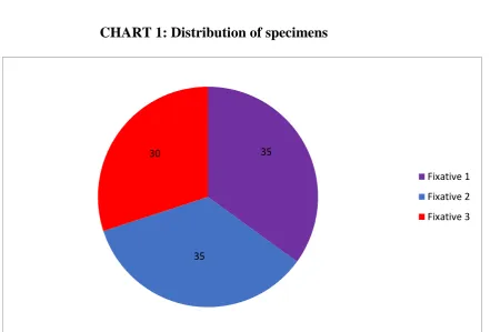
A total of 100 specimens were included in this study. Among them 35
specimens were fixed in fixative 1, another 35 were fixed in fixative 2 and 30
[image:57.595.97.539.427.726.2]specimens were fixed in fixative 3.
TABLE 1: Distribution of specimens
FIXATIVE NO. OF
SPECIMENS(n=100)
PERCENTAGE (%)
Fixative 1 35 35
Fixative 2 35 35
Fixative 3 30 30
CHART 1: Distribution of specimens
35
35 30
Distribution of specimens in fixative 1:
Totally 35 specimens were fixed in fixative 1. Among them 14 (40%) were
uterus and cervix followed by breast 7 cases(20%), thyroid 5 (14.29%),
gastrointestinal tract specimens 3 ( 8.58%), soft tissue 2 (5.71%), lymph node 2
(5.71%) and other ovary (1) and testis (1).
TABLE 2: Distribution of specimens in fixative 1
Specimen No. of cases( n=35 ) Percentage
Breast 7 20
Thyroid 5 14.29
Gastrointestinal tract 3 8.58
Uterus & cervix 14 40
Soft tissue 2 5.71
Lymph node 2 5.71
CHART 2: Distribution of specimens in fixative 1
Distribution of specimens in fixative 2:
35 specimens were fixed in fixative 2. Out of 35, 11 (31.43%) were uterus
and cervix, breast 8 (22.86%), thyroid 7 ( 20% ), gastrointestinal tract 3 ( 8.5%
), soft tissue 2 ( 5.71% ), lymph nodes 2 ( 5.71% ) and others were otongue (1)
and ovary (1).
20
14.29
8.58
40
5.71 5.71 5.71
0 5 10 15 20 25 30 35 40 45
Breast Thyroid GIT Uterus &
cervix Soft tissue Lymphnode Others
TABLE 3:Distribution of specimens in fixative 2
Specimen No of cases Percentage ( % )
Breast 8 22.86
Thyroid 7 20
Gastrointestinal tract 3 8.5
Uterus & cervix 11 31.43
Soft tissue 2 5.71
Lymph node 2 5.71
Others 2 5.71
CHART 3: Distribution of specimens in fixative 2
22.86
20
8.5
31.43
5.71 5.71 5.71
0 5 10 15 20 25 30 35
Breast Thyroid GIT Uterus Soft tissue Node Others
Distribution of specimens in fixative 3:
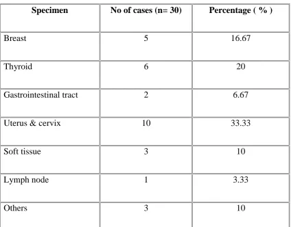
Out of 30 specimens fixed in fixative 3, uterus and cervix were 10 ( 33.33%
) followed by thyroid 6 ( 20% ), breast 5 (16.67% ), soft tissue 3( 10% ),
gastrointestinal tract 2 (6.67% ), lymph node 1( 3.33% ) and others were testis(1),
[image:61.595.100.525.276.608.2]lung (1) and kidney (1) .
TABLE 4: Distribution of specimens in fixative 3
Specimen No of cases (n= 30) Percentage ( % )
Breast 5 16.67
Thyroid 6 20
Gastrointestinal tract 2 6.67
Uterus & cervix 10 33.33
Soft tissue 3 10
Lymph node 1 3.33
CHART 4: Distribution of specimens in fixative 3
MACROSCOPIC APPEARANCE:
Tissues fixed in our newer compound fixatives were light blue in colour. Texture of tissues after fixation was same as tissues fixed in conventional
10% NBF.
There was no difficulty in cutting and sectioning.
EVALUATION OF HISTOMORPHOLOGICAL FEATURES:
The stained slides were analyzed for fixation artifacts, staining
characteristics, architecture, nuclear and cytoplasmic details.
COMPARISON OF NUCLEAR FEATURES:
Nuclear features were assessed based on following features, nuclear and
nucleolar preservation, nucleus size, regularity of nuclear membrane,chromatin
pattern whether fine, coarse, granular/ reticular pattern and mitotic figures .Score
16.67
20
6.67
33.33
10
3.33
10
Breast Thyroid GIT Utreus
&cervix soft tissue Node others
1 were given to sections with nuclear shrinkage, less prominent nucleolus and
mitotic figures and poorly defined chromatin pattern and it is considered as
suboptimal fixation.
Comparison of fixative 1 and conventional 10% NBF:
Nuclear features of tissues fixed in fixative 1 was compared with
conventional formalin fixed tissues. At 10 hours fixation in fixative 1, all the 35
specimens scored 3. At 8 hours fixation, only one specimen (lymph node) showed
nuclear shrinkage compared to 10% NBF and got score 2. All the other 34
specimens received score 3. At 7 hours fixation more than half the cases showed
nuclear shrinkage, less prominent nucleoli and mitotic figures and they got score
2.
TABLE 5: Comparison of Fixative 1 and conventional 10% NBF
FIXATIVES
SCORE
P value*
3 2 1 0
No. of
Sp
ecimens
10% NBF 35 nil nil nil n/a
Fixative 1,10 Hrs 35 nil nil nil n/a
Fixative 1, 8Hrs 34 1 nil nil 0.325
Fixative1,7 Hrs 15 20 nil nil <0.0001
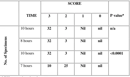
There is no significant difference between 10% NBF and fixative 1 at 10
and 8 hours fixation. So fixative 1 is as equal as 10% NBF in preserving nuclear
features at 10 and 8 hours fixation. There is significant difference at 7 hours
fixation on comparing to conventional formalin , so it is suboptimal in preserving
nuclear details.
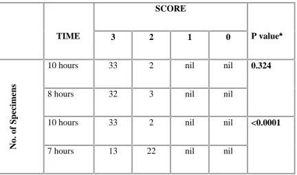
Comparison of fixative 2 and conventional 10% NBF:
Nuclear features of tissues fixed in fixative 2 was compared with
conventional 10% NBF fixed tissues. At 10 and 8 hours fixation in fixative 2, two
to three cases showed less defined chromatin pattern , less prominent nucleolus
and nuclear shrinkage compared to NBF and received score 2. All other specimens
scored 3 and comparable with NBF. More than half the tissues fixed at 7 hours
fixation got score 2.
TABLE 6: Comparison of fixative 2 and conventional 10% NBF
FIXATIVES
SCORE
P value*
3 2 1 0
No. of Sp
ecimens
10% NBF 35 nil nil nil n/a
Fixative 2, 10 Hrs 33 2 nil nil 0.160
Fixative 2, 8Hrs 32 3 nil nil 0.083
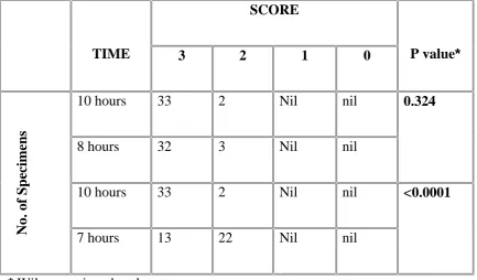
The above table shows there is no significant difference between 10% NBF
and fixative 2 at 10 and 8 hours fixation. So fixative 2 is as equal as 10% NBF in
preserving nuclear features at 10 and 8 hours fixation. There is significant
difference at 7 hours fixation. So nuclear details are not well preserved in 7 hours
fixation.
Comparison of fixative 3 and conventional 10% NBF:
Nuclear details of tissues fixed in fixative 3 was compared with
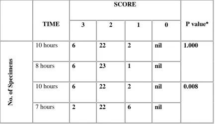
conventional 10% NBF fixed tissues. Most of the specimens fixed in fixative 3
showed nuclear shrinkage, less defined nuclear membrane and chromatin pattern,
less prominent nucleolus in all the 3 fixation hours. Few sections ( 2 to 6 cases )
showed even more shrinkage and poorly preserved chromatin got score 1.
TABLE 7:Comparison of fixative 3 and conventional 10% NBF
FIXATIVES
SCORE
P value*
3 2 1 0
No. of Sp
ecimens
10% NBF 30 nil nil nil n/a
Fixative 3, 10 Hrs 6 22 2 nil <0.0001
Fixative 3, 8Hrs 6 23 1 nil <0.0001
Fixative 3, 7 Hrs 2 22 6 nil <0.0001
The above table shows there is significant difference in all three different
fixation times compared to conventional 10% NBF. So fixative 3 is inferior in
preserving nuclear details compared to conventional formalin .
Comparison of fixative 1 and fixative 2:
Nuclear details were compared between fixative 1 and 2. Almost all of the
cases fixed in both fixative 1 and 2 at 10 and 8 hours fixation received score 3 as
they preserved well defined chromatin pattern , distinct nuclear membrane,
nucleoli and mitotic figures. At 7 hours both fixatives received score 2 because
they showed less prominent above nuclear details.
TABLE 8: Comparison of fixative 1 and fixative 2
TIME FIXATIVES
SCORE
3 2 1 0
P
value
*
No. of Sp
ecimens
10 hours
Fixative 1 35 nil nil nil 0.513
Fixative 2 33 2 nil nil
8 hours
Fixative 1 34 1 nil nil 0.505
Fixative 2 32 3 nil nil
*Mann whitney U test
This table shows there is no significant difference between fixative 1 and 2
in all three fixation times. So both fixatives are equal in nuclear features
preservation at 3 different fixation times.
Comparison of fixative 1 and fixative 3:
Nuclear details were compared between fixative 1 and 3 in the following
table. At 10 and 8 hours fixation, almost all of the specimens fixed in fixative 1
scored 3 as they preserved nuclear details well whereas more than half of the
tissues fixative fixative 3 showed nuclear shrinkage and less defined chromatin
pattern and they got score 2. At 7 hours fixation , 20 cases fixed in fixative 1 got
score 2 as they showed less prominent nucleoli and mitotic figures. 6 cases in
TABLE 9: Comparison of fixative 1 and fixative 3
TIME FIXATIVES
SCORE
P value*
3 2 1 0
No. of Sp
ecimens
10 hours
Fixative 1 35 nil nil nil
<0.0001
Fixative 3 6 22 2 nil
8 hours
Fixative 1 34 1 nil nil
<0.0001
Fixative 3 6 23 1 nil
7 hours
Fixative 1 15 20 nil nil
<0.0001
Fixative 3 2 22 6 nil
*Mann whitney U test
There is significant difference between fixative 1 and 3 in all three fixation
times. So fixative 1 is superior to fixative 3 in preserving nuclear details.
Comparison of fixative 2 and fixative 3:
Nuclear details were compared between fixative 2 and 3. At 10 and 8 hours
fixation, almost all of the specimens fixed in fixative 2 scored 3 as they preserved
nuclear details well whereas more than half of the tissues fixative fixative 3