i EVALUATION OF ANTI E-FAECALIS EFFICACY AND PENETRATION DEPTH OF CURCUMIN LONGA MODIFIED SEALER-AN IN VITRO CLSM
STUDY
Dissertation submitted to
THE TAMIL NADU DR. M.G.R. MEDICAL UNIVERSITY
In partial fulfillment for the degree of
MASTER OF DENTAL SURGERY
BRANCH – IV
CONSERVATIVE DENTISTRY AND ENDODONTICS
ii
ENDORSEMENT BY THE H.O.D. PRINCIPAL / THE HEAD OF THE INSTITUTION
This is to certify that Dr.NISHAN .A, Post Graduate student (2014–2017) in the Department of Conservative Dentistry and Endodontics, K.S.R. Institute of Dental Science and Research, has done this dissertation titled“EVALUATION OF ANTI E-FAECALIS EFFICACY AND PENETRATION DEPTH OF CURCUMIN LONGA MODIFIED SEALER-AN IN VITRO CLSM STUDY”under our guidance and
supervision in partial fulfillment of the regulations laid down by TheTamil Nadu Dr. M.G.R. Medical University, Chennai – 600 032 for M.D.S., (Branch – IV) CONSERVATIVE DENTISTRY AND ENDODONTICS degree examination.
Seal & Signature of H.O.D.
Dr.K. SIVAKUMAR.,M.D.S
PROFESSOR
Seal & Signature of Principal
Dr.G.S.KUMAR.,M.D.S
iii
CERTIFICATE BY THE GUIDE
This is to certify that the dissertation titled“EVALUATION OF ANTI E-FAECALIS EFFICACY AND PENETRATION DEPTH OF CURCUMIN LONGA MODIFIED SEALER-AN IN VITRO CLSM STUDY”is a bonafide research work
done by Dr.NISHAN.A in partial fulfillment of the requirements for the degree of
MASTER OF DENTAL SURGERY in the speciality of CONSERVATIVE DENTISTRY AND ENDODONTICS
DR.SIVA KUMAR.M.D.S PROFESSOR&HOD
K.S.R. INSTITUTE OF DENTAL SCIENCE AND RESEARCH TIRUCHENGODE
Date:
iv
DECLARATION BY THE CANDIDATE
TITLE OF DISSERTATION
EVALUATION OF ANTI E-FAECALIS EFFICACY AND PENETRATION DEPTH OF CURCUMIN LONGA MODIFIED SEALER-AN IN VITRO CLSM STUDY
PLACE OF STUDY
K.S.R. Institute of Dental Science and Research
DURATION OF THE COURSE 3 Years
NAME OF THE GUIDE Dr. Sivakumar.K
HEAD OF THE DEPARTMENT Dr. Sivakumar.K
I hereby declare that no part of the dissertation will be utilized for gaining financial assistance for research or other promotions without obtaining prior permission of the Principal, K.S.R. Institute of Dental Science and Research, Tiruchengode. In addition, I declare that no part of this work will be published either in print or in electronic media without the guide who has been actively involved in dissertation. The author has the right to publish this study solely with the prior permission of the Principal, K.S.R. Institute of Dental Science and Research, Tiruchengode.
v
Acknowledgement
I ex pres s m y sincere thanks t o Chairman Thi ru. Lion . Dr. K.S . Rangasamy,MJF.,Princi pal Dr. G.S . Ku mar, M.D.S.,the Head of the Departm ent of C ons ervative Dent ist r y and Endodonti cs , Dr. Sivaku mar Kailas am, M.D.S., KSR Institut e of Dental S cience and Res earch, Thi ruchengode, for permitti ng me to pursue this cours e and avai l t he facili ties of t his coll ege.
With gratitude I thank m y Profess or Dr. Siva Ku mar, M.D.S., m y guide, for his support and v aluabl e guidance t hrough the j ourne y of m y cours e and m ain dis s ert ati on.
M y si ncere t hanks to Dr. K. Karthick , M.D.S ., R eader, Departm ent of Cons ervative Dentistr y a nd Endodonti cs , for his const ant guidance and i mmens e support wit h utm os t pat ience duri ng M.D.S. course and m ain diss ert ation.
I would als o like to engrave here, m y heart felt respects and t hanks to all m y t eachers in the departm ent,Dr.S ebeena Mathew , Dr. Harik aran M.D.S.,Dr. Deep a, M.D.S., Dr.B oopath i M.D.S . and other st aff m embers for thei r cons tant encouragem ent i n all aspects of m y career as an MDS student .
I would also li ke t o t hank m y fat her M.Ab ul Fazi l ,m y mother
vi I ex tend m y heart felt thanks to m y colleagues Dr.S reed ev.C.P and
Dr.Iswarya.R.Raju for thei r hel p, advi ce and s upport .
I t hank t he librari an Mr. R. Madeshwaran, M.A., B.Ed ., M.Phi l.,
for his val uable work in helping me t o access and co ll ect the art icl es and text books for this di ssert ation.
I l ike to thank Dr. Malini ,V Clin bio,S ri Ram achandra Universit y for her i deas an d guidance during confocal l as er scanning micros cope imagi ng.
I li ke to t hank Dr.Prathaban Munis amy ,Department of biot echnol ogy,KSR Art s and S cience C ollege for his i deas and gui dance in inoculation and culturi ng of E.faecalis.
I ex tend m y thanks to Spy Prin ters, Erode, for thei r hel p in com piling the diss ert ation, pri nting and bi nding.
vii
TABLE OF CONTENTS
SL NO. TITLE PAGE NO.
1. INTRODUCTION 1
2. AIMS AND OBJECTIVES 4
3. REVIEW OF LITERATURE 5
4. MATERIALS AND METHODS 14
5. RESULTS 25
6. DISCUSSION 48
7. SUMMARY 53
8. CONCLUSION 54
9. BIBILIOGRAPHY 55
viii
LIST OF FIGURES
SL NO. TITLE PAGE NO.
1. Schematic representation of specimen preparation for sealer placement and viewing under CLSM for evaluating anti bacterial efficacy
19
2. X Smart Plus and Protaper files. 20
3. Irrigating Solutions and Guttapercha cones 20
4. Teeth samples used in the study for analyzing depth of penetration
21
5. Hydro alcoholic extract of Curcumin
21
6. Sample preparation after placement of zinc oxide eugenol sealer for evaluating anti bacterial efficacy
22
7.
Sample preparation after the placement of curcumin modified MTA sealer for evaluating anti bacterial
efficacy 22
8. MTA sealer used in the study 23
ix 10. Confocal laser scanning microscope(CLSM) 24
11. Anti E.faecalis efficacy of curcumin modified MTA sealer at 7 days
28
12. Anti E.faecalis efficacy of curcumin modified MTA sealer at 21 days
29
13 Anti E.faecalis efficacy of curcumin modified MTA sealer extract at 45 days
30
14 Anti E.faecalis efficacy of zinc oxide eugenol sealer at 7 days
31
15 Anti E.faecalis efficacy of zinc oxide eugenol sealer at 21 days
32
16 Anti E.faecalis efficacy of zinc oxide eugenol sealer at 45 days
33
17 Depth of penetration of zinc oxide eugenol sealer (ZOE) 34
18
Depth of penetration of curcumin modified MTA sealer(MTAC)
x
LIST OF TABLES
S.No. Name of Table Page No.
1.
Depth of penetration(µm) of zinc oxide eugenol sealer and
curcumin modified MTA sealer into dentinal tubules 25
2.
Relative area percentage of dead cell volume in dentinal tubules
treated by curcumin modified MTA sealer 26
3. Relative area percentage of dead cell volume in dentinal tubules
treated by zinc oxide eugenol sealer 27
4. mean and standard deviation of depth of penetration 36
5. Comparison of depth of penetration 37
6.
Mean and Standard deviation of anti E.faecalis efficacy at 7 days
38
7.
Mean and Standard deviation of anti E.faecalis efficacy at 21
days 38
8.
Mean and Standard deviation of anti E.faecalis efficacy at 45
days 39
9.
Comparison of Anti E.faecalis efficacy of Zinc Oxide Eugenol
at 7 days and 21 days 40
10.
Comparison of Anti E.faecalis efficacy of Zinc Oxide Eugenol
at 21 days and 45 days 40
11. Comparison of Anti E.faecalis efficacy of Zinc Oxide Eugenol
at 45 days and 7 days 41
12. Comparison of Anti E.faecalis efficacy of curcumin modified
xi 13.
Comparison of Anti E.faecalis efficacy of curcumin modified
MTA sealer at 21 days and 45 days 42
14. Comparison of Anti E.faecalis efficacy of curcumin modified
MTA sealer at 45 days and 7 days 43
15. Comparison of Anti E.faecalis efficacy at 7 days 44
16.
Comparison of Anti E.faecalis efficacy at 21 days
44
17. Comparison of Anti E.faecalis efficacy at 45 days 45
LIST OF BAR DIAGRAMS
SL NO.
TITLE
PAGE NO.
1
Depth of penetration of root canal sealers in µm
46
2
Anti bacterial efficacy of sealers
1
INTRODUCTION
Microorganisms are primary etiologic agents in pulpal and periapical diseases 1,2. The purpose of endodontic therapy is to eliminate bacteria and their by-products from the infected root canal system and prevent subsequent reinfection3. Chemomechanical preparation (instrumentation and irrigation) and intracanal medicaments significantly reduce microorganisms inside the infected root canal. However, it is virtually impossible to completely eliminate all microbes from root canal system in every case 4. Hence the
use of endodontic obturation materials and sealers with antimicrobial activity is considered beneficial in further reducing the concentration of residual microorganisms4,5. Root canal sealers with antimicrobial activity can help to improve the success rate of endodontic treatment and are especially advantageous in clinical situations where there is persistent or recurrent infection 6 . The persistence of microorganisms in dentinal
tubules, lateral canals and apical ramifications after root canal treatment has been reported 7-9. If the filling provides a good seal, it will only impair the exit of bacteria entrapped in the root canal system. However, to eradicate the remaining microorganisms, the antimicrobial activity of the sealer could play an important role .10-11
The need for a biocompatible material that induces the formation of mineralized tissue and also has suitable flow rate and manipulation, led to the development of MTA-based root canal sealers.12
2 The curcumin longa (turmeric) is extensively used as a spice, food preservative and coloring material in India, China, South East Asia. It has been used in traditional medicine for the treatment of numerous diseases. Curcumin(diferuloylmethane), the main yellow bioactive component of turmeric has been shown to have a wide spectrum of biological actions including anti microbial, anti inflammatory and anti oxidant activities.15-19 Various studies have shown the antimicrobial effects of extracts of roots of curcumin longa on various micro organisms.20-23
Enterococcus faecalis is often used in research that aims to evaluate the antimicrobial properties of endodontic materials. It seems to play a significant role in the etiology of persistent periradicular lesions 24. Enterococcus faecalis possesses several virulence factors that contribute to its ability to survive the effects of conventional root canal therapy25. Besides, this Gram-positive facultative anaerobe is able to invade dentine
tubules and bind to collagen 26.
To achieve the goal of thorough canal obturation not only must the tissue debris and contaminates be removed, but also the filling materials and techniques used to place them must achieve a high level of adaptability to the cleaned root canal space and dentin walls, including penetration into the dentinal tubules if possible.27
3 obtained using this model and viability staining with Confocal Laser Scanning Microscopy (CLSM) have shown reproducible data on dentin disinfection in different conditions, including different biofilm age 33, length of disinfectant exposure 34, and combinations of disinfecting agents .35
4
AIM
To evaluate the anti E.faecalis efficacy and depth of penetration of curcumin modified sealer using Confocal Laser Scanning Microscope(CLSM).
OBJECTIVES
The main objective is to
Evaluate the anti E.faecalis efficacy of curcumin modified sealer(MTAC) and zinc oxide eugenol(ZOE).
5
REVIEW OF LITERATURE
Ørstavik in 1996 recommended the use of endodontic sealer with antibacterial properties to decrease or avoid future growth of the remaining microorganisms6.
Spanberg in 1973 stated that to eradicate the remaining microorganism in the root canal ,the anti microbial activity of sealer could play an important role10.
Peters in 2001 found out that even after chemo mechanical preparation bacterias were left inside root canal. Depending on the host– parasite equilibrium and the nutrition available after root filling, these bacteria may be of importance in recalcitrant apical periodontitis7.
6 intracanal infection after instrumentation, antimicrobial irrigation, and obturation. The microbes were located in inaccessible recesses and diverticula of instrumented main canals, the intercanal isthmus, and accessory canals, mostly as biofilms.
Love in 2001 postulated that a virulence factor of E. faecalis in failed endodontically treated teeth may be related to the ability of E.faecalis cells to maintain the capability to invade dentinal tubules and adhere to collagen in the presence of human serum26.The aim of this study was to identify a possible mechanism that would explain
how E.faecalis could survive and grow within dentinal tubules and reinfect an obturated root canal. Cells of Streptococcus gordonii DL1,Streptococcus mutans NG8 ,or E.faecalis JH2-2 were grown in brain heart infusion broth containing various amounts of human serum for 56 days. The ability of three species to invade dentin and bind to immobilized type 1 collagen in the presence of human serum was assessed by dentine invasion and microtitre well experiments. All three species remained viable over the period of the experiment when grown in human serum. Cells of all three bacteria were able to invade dentine and bind to immobalized collagen. Human serum inhibited dentine invasion and collagen adhesion by S.gordonii DL1 and S.mutans NG8 whilst dentine invasion by E.faecalis JH2-2 was reduced in the presence of serum , but not inhibited , and binding to collagen was enhanced.
7
Gomes in 2006 found out that E.faecalis was detected as frequently in teeth with necrotic pulp as in teeth with failing endodontic treatment when a Polymerize Chain Reaction PCR analysis was used37. The objective of this study was to investigate the presence of Enterococcus faecalis in endodontic infections by culture and polymerase chain reaction analyses. Microbial samples were obtained from 50 teeth with untreated necrotic pulps (primary infection) and from 50 teeth with failing endodontic treatment (secondary infection). Culture techniques were used including serial dilution, plating, incubation, and biochemical identification. For PCR detection, samples were analyzed using a species-specific primer of the 16S rDNA and the downstream intergenic spacer region. Culture and PCR detected the test species in 23 of 100 and 79 of 100 of the teeth, respectively. E faecalis was cultured from 2 (4%) of 50 necrotic canals and from 21 (42%) of 50 root-treated canals. PCR detection identified the target species in 41 (82%) and 38 (76%) of 50 primary and secondary infections respectively.
Kayaoglu and Orstavik in 2004 stated that the most-cited virulence factors are aggregation substance, surface adhesins, sex pheromones, lipoteichoic acid, extracellular superoxide production, the lytic enzymes such as gelatinase and hyaluronidase, and the toxin, cytolysin. Each of them may be associated with various stages of an endodontic infection as well as with periapical inflammation38 .
8 shown that the perturbation of FtsZ functions by natural compounds and chemical agents leads to inhibition of bacterial proliferation39.
Prasanna Neelakantan in 2013 found out that Sodium hypochlorite (3%) showed maximum antibacterial activity against E.faecalis biofilm formed on the tooth substrate, followed by curcumin and CHX(chlorhexidine). Considering the potential for undesirable properties of NaOCl, the use of herbal alternatives in endodontics might prove to be advantageous40 . To evaluate the antimicrobial efficacy of curcumin against
Enterococcus faecalis biofilm formed on tooth substrate in vitro. Sodium hypochlorite (NaOCl) and chlorhexidine (CHX) served as standards for comparison. Biofilms of E.faecalis were formed on instrumented, extracted human teeth (n = 96). At the end of the 2nd day, 2nd week and 8th week, specimens were treated for 30 min with one of the test solutions or saline (control) and the surviving colony-forming units (CFU/mL) was recorded. Results were analyzed by Kruskal-Wallis test and Dunnet test for pair-wise comparison with Bonferroni correction (p = 0.05). Only NaOCl showed complete eradication of bacteria at all time periods. In the 2-day and 2nd week biofilms, curcumin and NaOCl showed complete inhibition, which was significantly lower than the CFU recovered in the CHX and saline groups (p < 0.05). In 8 week biofilms, samples treated with curcumin showed 553 ± 137.6 CFU/mL, which was significantly higher than NaOCl (0 CFU/mL), but significantly lower than CHX (2551 ± 129.8) and saline control (1.42 × 10¹¹ ± 2.12 × 10¹⁰; p < 0.05).
Mandroli in 2013 stated that curcumin has the potential to be developed into medicament for the treatment of various endodontic diseases41.Aim of the study was to
9 gingivalis (ATCC 33277), Prevotella intermedia (ATCC 25611), Enterococcus faecalis (ATCC 29212) from the stock were revived by plating on blood agar medium. Isolated colonies were transferred to sterile Brain Heart Infusion (BHI) broth and once again incubated overnight. The growth concentration was adjusted to 5 X 10⁵ organisms / ml by using 0.5 McFarland’s turbidity standard. MIC was determined, by serial broth dilution of curcumin to 500, 250,125, 62.5, 31.25, 16, 8, 4, 2, 1 μg /ml. respectively. The tubes were then incubated for 24 hours at 370C. The last tube with clear supernatant was
considered to be without any growth and taken as MIC value. Mean MIC values of curcumin were as follows: S. mutans (333.33 μg /ml), A. viscosus (167.67 μg /ml), L. casei (125 μg /ml), P. gingivalis (125 μg /ml), and P. intermedia (208.33 μg /ml). There was no action against E. faecalis.
Mithra Hegde in 2012 found out that the extracts of Curcuma longa showed antimicrobial activity against the tested organisms63 .
Mishra in 2005 stated that curcumin glycine bioconjugates has anti bacterial and anti fungal properties17.
10 from turmeric as a potential antiseptic in prevention and treatment of antibacterial infections has been suggested.
Orstavik in 2005 stated that root canal sealer is essential to not only assist in filling but should also penetrate into small inaccessible areas such as dentinal tubules42.
Tanomaru Filho M in 2007 stated that the ability to penetrate into the dentinal tubules may be beneficial to control or kill bacteria that are located there43.
Suresh Chandra in 2012 stated that the advantages of a deeper sealer penetration are their potential antibacterial effects and entombing the viable bacteria within tubules by isolating them from potential nutrient sources. The potential for bacteria to colonize dentinal tubules has been well established44.The aim of this in vitro study was to evaluate the depth of penetration of 4 different endodontic resin sealers into the radicular dentinal tubules with the aid of confocal microscopy. Methods: Eighty single-rooted teeth were instrumented and divided into 4 groups composed of 20 teeth each. The samples were obturated with AH Plus, RealSeal, EndoRez, and RoekoSeal resin sealers respectively. The core material in all the groups was Resilon. The teeth were sectioned at the coronal, middle and apical thirds and viewed under confocal microscope to determine the depth of penetration of the sealer into the dentinal tubules. Results: The results showed that the maximum penetration was exhibited by RealSeal resin sealer, followed by AH Plus, RoekoSeal, and EndoRez. The coronal third showed the maximum penetration, followed by middle third and least at the apical third.
11 growth. This investigation compared the antibacterial effects of amalgam, zinc oxide-eugenol, Super EBA and a mineral trioxide aggregate on nine facultative bacteria such as Streptococcus faecalis, Streptococcus mitis, Streptococcus mutans, Streptococcus salivarius, Lactobacillus species, Staphylococcus aureus, Staphylococcus epidermidis, Bacillus subtilis, and Escherichia coli B and seven strict anaerobic bacteria, Prevotella (Bacteroides) buccae, Bacteroides fragilis, Prevotella (Bacteroides) intermedia, Prevotella (Bacteroides) melaninogenica, Fusobacterium necrophorum, Fusobacterium nucleatum, and Peptostreptococcus anaerobius. After growing these bacteria on solid media, freshly mixed and 24-h set test materials were placed on the surface of these inoculated media and incubated in the appropriate atmosphere for 24 to 48 h at 37 0C. Impregnated discs with the Super EBA liquid were used as positive controls. The antibacterial effects of each material were measured in millimeters and the data were analyzed using one-way and two-way analysis of variance and Scheffé tests to determine the statistical differences between the antibacterial effects of the test materials. Impregnated discs with Super EBA liquid caused varying degrees of growth inhibition for both facultative and strict anaerobic bacteria. Both types of amalgam had no antibacterial effect against any of the bacteria tested in this study. Mineral trioxide aggregate had an antibacterial effect on some of the facultative bacteria and no effect on any of the strict anaerobic bacteria. Zinc oxide-eugenol and Super EBA pastes had some antibacterial effects on both types of bacteria tested.
I.M.Saleh in 2004 and Zhang in 2007 stated that MTA based sealers doesn’t
have anti bacterial properties against E.faecalis46,47 .
Al –Hezaimi in 2009 stated that the origin of MTA as well as the type of preparation may affect its antimicrobial characteristics14.The antimicrobial effects of 4
12 and 2 gray-colored (GMTA-1, GMTA-2), against C. albicans and E. faecalis were
assessed in vitro. Minimal inhibitory concentration (MIC) for each preparation was
determined using the tube dilution test and Sabouraud agar media for C. albicans and
brain heart infusion media for E. faecalis. Broth tubes were prepared and divided into
experimental and control groups. Aliquots of each of the tested microorganisms were
taken from a stock culture and added to each experimental and positive control group.
All groups were incubated at 37°C and evaluated for turbidity at 24-hrs, 48-hrs, and
72-hour time periods. Samples of 0.1 mL from each of the experimental and control tubes
were subcultured on agar or brain heart infusion plates to confirm visible signs of
bacterial or fungal growth. MIC of MTA against the 2 microorganisms tested varied
among the 4 preparations tested. WMTA-1 and WMTA-2 inhibited C. albicans growth
at concentrations of 3.125 mg/10 mL and 25 mg/10 mL, respectively, and statistically
significant differences were found between 1 and 2 (P<.001).
WMTA-1 and WMTA-2 inhibited E. faecalis growth at concentrations of WMTA-12.5 mg/WMTA-10 mL and 50
mg/10 mL, respectively, and statistically significant differences were found between
WMTA-1 and WMTA-2 (P<.001). GMTA-1 and GMTA-2 inhibited E. faecalis growth
at concentrations of 12.5 mg/10 mL and 3.125 mg/10 mL, respectively, and statistically
significant differences were found between GMTA-1 and GMTA-2 (P<.001). Both
GMTA-1 and GMTA-2 inhibited C. albicans growth at a concentration of 3.125 mg/10
mL and no statistical differences were found between the preparations. Subculture of the
broth tubes in agar or brain heart infusion plates confirmed the turbidity test result.
Estrela in 2000 stated that MTA sealer did not show effective inhibition against E. faecalis13.
13 the endodontic sealers: N-Rickert, Sealapex, AH Plus, Mineral Trioxide Aggregate (MTA) and portland cement. The Agar diffusion method was used in plates previously inoculated with the following microorganisms: C. albicans, S. aureus, E. faecalis, E. coli. The diameters of microbial inhibition zones were measured after 24 hours of incubation in kiln at 37°C. According to the methodology used, it was possible to conclude that only the sealers AH Plus and N-Rickert presented antimicrobial activity against C. albicans, S. aureus, and E. coli; no antimicrobial activity in MTA, Sealapex and portland cement was observed. N-Rickert presented the largest inhibition zones varying from 8 to 18 mm and the microorganism E. faecalis was resistant against all sealers tested.
Sipert in 2005 reported that MTA demonstrated antimicrobial activity against E. faecalis49 .
14
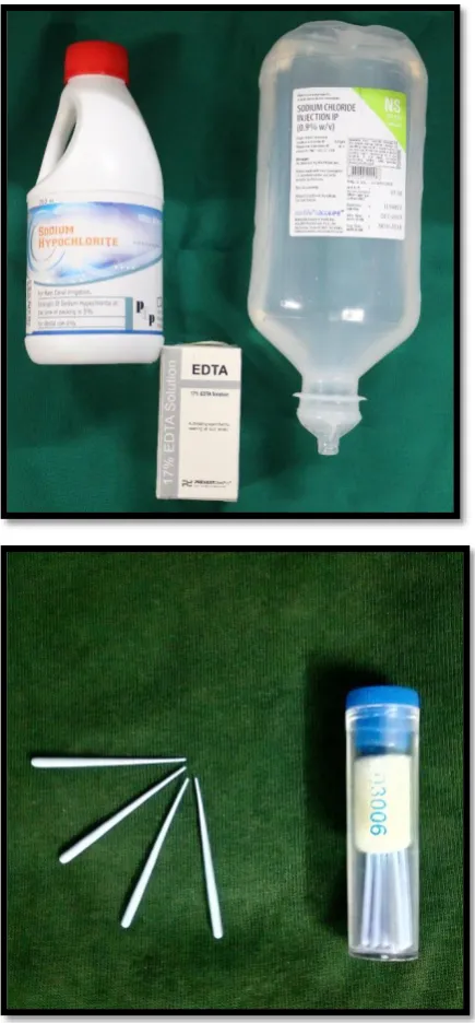
ARMAMENTARIUM USED
1. Extracted single canal tooth 2. 3% thymol solution
3. Diamond disc and mandrel 4. Airotor handpiece
5. Micromotor hand piece(NSK) 6. K files
7. Saline
8. 5.25 % sodium hypochlorite solution 9. Disposable syringe
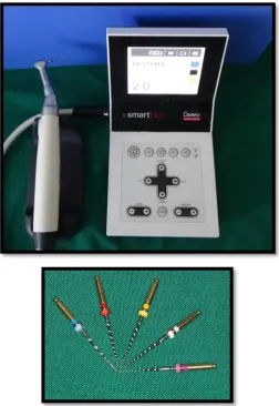
10. X smart (dentsply maillefer) 11. Protaper rotary files
12. Protaper gutta-percha F3 size 13. MTA plus
14. Scaler unit(satelec) 15. E.faecalis(ATCC 29212) 16. Rhodamine B dye 17. Laminar flow chamber
15
SOURCE OF DATA
Extracted single canal tooth has been collected from Department of Oral and Maxillofacial Surgery in KSR Institute of Dental Science and Research. Study was conducted in Department of Conservative Dentistry and Endodontics, KSR Institute of Dental Science and Research. Bacterial inoculation and biofilm development was done in Department of Biotechnology,KSR Arts and Science College. Confocal Laser Scanning Microscopic imaging has been done in Vclin bio research centre, Sri Ramachandra University,Chennai.
INCLUSION CRITERIA
Uniradicular teeth
Teeth with single root canal
EXCLUSION CRITERIA
Multi radicular teeth
Teeth with multiple canals
Teeth with ribbon shaped canals.
Teeth with any anomalies.
16
MATERIALS AND METHODS
Anti Bacterial Efficacy
Dentin Specimen Preparation
45 single rooted teeth were collected from the Department of oral surgery ,KSR Institute of Dental Science and Research. According to a previously described protocol 90 semi cylindrical halves were fractured and shaped into 4x4x2 mm in size. The cementum layer on the external dentin surface was removed so that the thickness of samples remains same. The smear layer on both sides of the specimen was removed by immersion in 5% NaOCl and 6% citric acid each for four minutes in an ultrasonic bath. The prepared dentin specimens with their canals side up were placed inside filter tube.
Dentin Infection of E.Faecalis
E.faecalis ATCC 29212 was used as the test organism and grown in air at 370C overnight on BHI(brain heart infusion) agar plates. The bacteria were harvested and suspended in BHI broth. Cell density was spectrophotometrically standardized into 3×10⁶colony forming unit/ml.
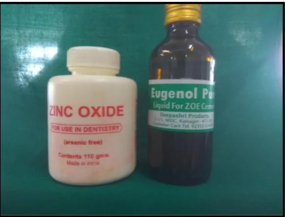
17 The dentin specimens were taken out of each tube rinsed in sterile water for 1 minute and air dried. The outer surface of the specimens were sealed with nail varnish. The 90 infected dentin halves with 3 week old E.faecalis biofilms were randomly allocated to 2 major groups. Group 1(n=45) is zinc oxide eugenol(ZOE) and group 2(n=45) is curcumin modified MTA sealer(MTAC).
Sealer placement
Sealer was placed on the dentin surface of root canal wall. All dentin samples were placed in 100% humidity for 7,21,45 days(n=15) after which the sealer was scraped off.
CLSM Examination
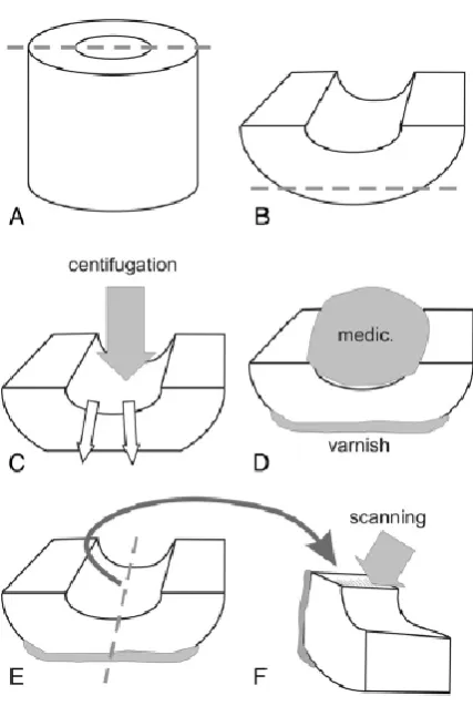
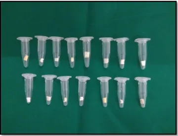
Two semi cylindrical dentin halves of each group were examined by viability staining and CLSM. After scraping of sealers the dentin halves were rinsed in sterile water and vertically fractured through the root canal into 2 halves to expose a fresh surface of longitudinally fractured dentinal tubules for CLSM examination as previously described 32 (fig.1).
18 The confocal laser scanning microscope data were processed by imaris 7.2 software . The thresholds of the red and green fluorescence were manually set according to their respective fluorescence intensity and kept consistent for each sample. Live/dead ratio were automatically calculated by the software.The relative area percentage of red fluorescence to added (green and red) fluorescence indicated the proportion of killed cells. The relative area percentage after exposure to different sealers were subjected to mean and standard deviation and unpaired t test were used to isolate and compare the results at a significance level of p<0.05.
Depth of penetration
30 maxillary single rooted teeth stored in thymol solution were used in this study. The coronal portion was cut at the cementum level .Root canal preparation was done upto 0.5 mm short of working length up to protaper F3. The hand piece used was an electric engine at 250 rpm. Irrigation procedures were accomplished by using 2 ml of 5% sodium hypochlorite for each file used. To remove smear layer 3 ml of 6% citric acid was used. Finally the root canals were flushed with distilled water and canals dried with sterile paper points. F3 master cone was selected and sealers were mixed with rhodamine B dye and then obturated.
Group 1(n=15) zinc oxide eugenol(ZOE)
Group 2 (n=15) curcumin modified MTA sealer(MTAC)
19
CLSM Examination
[image:30.595.229.443.373.692.2]The dentin segments were examined on a confocal laser scanning microscope .The respective absorption and emission wave lengths for the rhodamine B were 540 nm. Dentin samples were analyzed at 20 x for depth of penetration into tubules. The canal wall served as the starting point and sealer penetration into dentinal tubules were measured to a maximum depth of 1000 microns. Statistical significance for the mean depth of penetration of root canal sealers were determined using unpaired t test. The level of significance was set at p<0.05.
20
ARMAMENTARIUM
22
Figure 4: Teeth samples used in the study for analyzing depth of penetration
[image:33.595.158.470.373.604.2]23
Figure 6: sample preparation after placement of zinc oxide eugenol sealer for evaluating anti bacterial efficacy
[image:34.595.149.510.71.345.2]
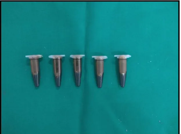
24 Figure 8: MTA sealer used in the study
[image:35.595.168.459.411.633.2]26
RESULTS
Table 1: Depth of penetration(µm) of zinc oxide eugenol sealer and curcumin modified MTA sealer into dentinal tubules
sample ZOE MTAC
1 187.41 234.99
2 237.92 196.75
3 221.65 278.33
4 187.41 241.25
5 194.79 288.43
6 221.65 238.94
7 237.92 262.21
8 221.65 275.27
9 237.8 279.93
10 194.79 234.94
11 187.41 196.77
12 237.8 278.33
13 221.65 241.25
14 194.79 275.26
27
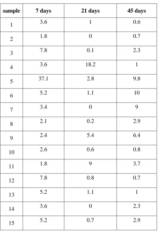
Table 2:Relative area percentage of dead cell volume in dentinal tubules treated by curcumin modified MTA sealer
sample 7 days 21 days 45 days
1 3.6 1 0.6
2 1.8 0 0.7
3 7.8 0.1 2.3
4 3.6 18.2 1
5 37.1 2.8 9.8
6 5.2 1.1 10
7 3.4 0 9
8 2.1 0.2 2.9
9 2.4 5.4 6.4
10 2.6 0.6 0.8
11 1.8 9 3.7
12 7.8 0.8 0.7
13 5.2 1.1 1
14 3.6 0 2.3
28
Table 3: Relative area percentage of dead cell volume in dentinal tubules treated by zinc oxide eugenol sealer
sample 7 days 21 days 45 days
1 18.1 8.2 2.9
2 31.6 3 0.1
3 48 5 0.1
4 15.9 7 0.6
5 8.2 0.3 0.9
6 7 5 1.6
7 0.6 18.1 1
8 8.5 31.6 1.9
9 9.4 48 1.2
10 12.6 0.8 1.6
11 8 0.1 0.3
12 17.4 5.5 0.8
13 15.2 15.9 0.1
14 2.9 9.3 0.8
29
30
31
32
33
34
35
36
37
STATISTICAL ANALYSIS
DESCRIPTIVE STATISTICS
ZOE MTAC Total
Numb er
15 15
30
Mean 214.8373 254.0693
Median 221.6500 262.2100
Std. Deviation 21.21507 30.50071
38
*Unpaired T – test for depth of penetration
Group ZOE MTAC P value*
Mean 214.8373 254.0693 0.0003 SD 21.2151 30.5007
SEM 5.4777 7.8752
N 15 15
39
Mean and Standard deviation of anti E.faecalis efficacy
ZOE MTAC
Number 15 15
Total 30
Mean 13.5667 6.2133
Median 9.4000 3.6000
[image:50.595.228.452.175.340.2]Std. Deviation 12.43369 8.75858
Table 6 :Anti E.faecalis efficacy at 7 days
ZOE MTAC
Number 15 15
Total 30
Mean 10.5733 2.7333
Median 5.5000 .8000
Std. Deviation 13.36148 4.94349
[image:50.595.229.452.465.628.2]40
ZOE MTAC
Number 15 15
Total 30
Mean .9667 3.6067
[image:51.595.229.452.147.312.2]Median .8000 2.3000 Std. Deviation .77889 3.46358
41
Unpaired T – test for anti E.faecalis efficacy of Zinc oxide eugenol
Group ZOE 7 DAYS ZOE 21 DAYS P value*
Mean 13.567 10.573 0.5305
SD 12.434 13.361
SEM 3.210 3.450
[image:52.595.180.449.173.317.2]N 15 15
Table 9 :Comparison of Anti E.faecalis efficacy of Zinc Oxide Eugenol at 7 days and 21 days
Group ZOE 21 DAYS ZOE 45 DAYS P value*
Mean 10.573 0.967 0.0096
SD 13.361 0.779
SEM 3.450 0.201
N 15 15
[image:52.595.175.453.433.579.2]42
Group ZOE 45 DAYS ZOE 7 DAYS P value*
Mean 0.967 13.567 0.0005
SD 0.779 12.434
SEM 0.201 3.210
[image:53.595.177.447.71.218.2]N 15 15
43
Unpaired T – test for anti E.faecalis efficacy of Curcumin modified
MTA sealer
Group MTAC 7 DAYS MTAC 21 DAYS P Value*
Mean 6.213 2.733
0.1910
SD 8.759 4.943
SEM 2.261 1.276
[image:54.595.165.461.143.295.2]N 15 15
Table 12 :Comparison of Anti E.faecalis efficacy of curcumin modified MTA sealer at 7 days and 21 days
Group MTAC 21 DAYS MTAC 45 DAYS P Value*
Mean 2.733 3.607
0.5797
SD 4.943 3.464
SEM 1.276 0.894
N 15 15
[image:54.595.162.462.402.553.2]44
Group MTAC 45 DAYS MTAC 7 DAYS P Value*
Mean 3.607 6.213
0.2929
SD 3.464 8.759
SEM 0.894 2.261
[image:55.595.165.460.71.221.2]N 15 15
45 *
Unpaired T – test for anti E.faecalis efficacy between Zinc Oxide
Eugenol and Curcumin modified MTA sealer
Group ZOE 7 DAYS MTAC 7 DAYS P value*
Mean 13.567 6.213 0.0716
SD 12.434 8.759
SEM 3.210 2.261
[image:56.595.174.449.189.336.2]N 15 15
Table 15 :Comparison of Anti E.faecalis efficacy at 7 days
Group ZOE 21 DAYS MTAC 21 DAYS P value*
Mean 10.573 2.733 0.0420
SD 13.361 4.943
SEM 3.450 1.276
N 15 15
[image:56.595.171.456.464.611.2]46
Group ZOE 45 DAYS MTAC 45 DAYS P value*
Mean 0.967 3.607 0.0075
SD 0.779 3.464
SEM 0.201 0.894
N 15 15
47
Line diagram 1: Depth of penetration of root canal sealers in µm
Line diagram 2 : Anti bacterial efficacy of sealer 190 200 210 220 230 240 250 260 ZOE MTAC 0 2 4 6 8 10 12 14 16
7 DAYS 21 DAYS 45 DAYS
ZOE
48
DEPTH OF PENETRATION
The results of the present study showed that the curcumin modified MTA sealer (MTAC) had significant greater depth of penetration than ZOE. (p value is <.05;unpaired t test)
ANTI E.FAECALIS EFFICACY
The results of the present study showed that there was significantly greater anti bacterial efficacy at 7 days for ZOE than MTAC. There was significantly greater anti bacterial efficacy at 21 days for ZOE than MTAC (p value<0.05;unpaired t test). During 45 days time period , there was significantly greater anti bacterial efficacy for MTAC than ZOE.(p value<0.05;unpaired t test).
Zinc Oxide Eugenol Group (ZOE)
When comparing within groups there was no statistical significant difference between 7 days and 21 days.(p value >0.05;unpaired t test).The 21 days group showed significantly greater anti bacterial efficacy when compared to 45 days.(p value<0.05). The 7 days group showed significantly greater anti bacterial efficacy when compared to 45 days group.(p value<0.05).
Curcumin modified MTA sealer Group (MTAC)
49
DISCUSSION
The major goal of root canal filling is to prevent any interchange between the oral cavity, the root canal system and the peri radicular tissues, thus providing a barrier to canal infection and re infection.
The ideal outcome in root canal obturation is to have a high volume of gutta-percha and a minimal volume of sealer within the body of the root canal space53,54and enhanced penetration into the canal irregularities and dentinal tubules. The penetration of sealers into dentinal tubules may be biologically beneficial, because laboratory studies have shown that endodontic sealers can exert antibacterial effects against bacteria in infected dentinal tubules3. Bacterial penetration into dentinal tubules may reach
100-1,000 µm and it can be enhanced by the absence of smear layer28. Many species seen in
the infection of the root canal have the propensity to penetrate deeply into the dentinal tubules, such as facultative and anaerobic species26,even close to the dentinal-cementum junction55. Even though there is no direct evidence to support the efficacy of sealer penetration into the tubules to kill bacteria in vivo, this achievement would seem to be reasonable especially in teeth with long standing necrotic pulps and chronic apical periodontitis. Even if the bacteria that may remain in the dentinal tubules were not killed, the sealer would serve as a reasonable blocking agent that may prevent bacterial repopulation or inactivate them in the tubules if some level of leakage in the main body of the obturated root canal occurs46.
50 and from the periodontium66,67. Hence the depth of penetration of 2mm section from apical third of the sample was analysed.
Confocal laser scanning microscopy offers advantages compared with scanning electron microscopy and other methodologies to evaluate penetration and interfacial adaptation of root canal sealers. Visualization of the depth of penetration and adaptation of the sealers in horizontal sections is evident at low magnifications by the presence of rhodamine B fluorescence in dentinal tubules. Therefore, a panoramic vision of sealer adaptation into the root canal and dentinal tubules can be easily confirmed at higher magnifications. As indicated in earlier studies using CLSM, labeling with rhodamine B is essential to observe the extent of sealer adaptation and penetration56,57 .
The intensity of the fluorescence in dentinal tubules is related to the quantity of sealer inside the dentinal tubule. A higher fluorescence was related to complete obturation of dentinal tubules, whereas a lower fluorescence corresponded to partial or incomplete obturation of the dentinal tubule lumen46.
The detection of bacteria in the current study was done by using viability staining and CLSM. Viability staining has become a widely used method in measuring bacterial killing in biofilms59-61, allowing for the measurement of the proportion of killed bacteria
in each specimen, which has not been possible at the same level of sensitivity using culture methods and colony-forming unit counting. Parmar et al found that green and red fluorescent bacteria were visible within the dentinal tubules of infected root sections when examined by CLSM62. When using CLSM, it is inevitable that background
51 from dentin as shown in a previous study32 . A strong fluorescent signal from bacteria allows the use of low gain settings during confocal laser scanning microscopic scanning, minimizing the interference from background fluorescence.
Enterococcus faecalis, an opportunistic, facultative anaerobe is associated with persistent apical periodontitis in endodontically treated teeth and is highly prevalent in failed root filled teeth68. Survival and virulence factors of E.faecalis endures prolonged periods of nutritional deprivation, binds to dentin and proficiently invades dentinal tubules, alters host responses, suppresses the action of lymphocytes, possesses lytic enzymes, cytolysin, aggregation substance, pheromones, and lipoteichoic acid36. Apart from this, E. faecalis utilizes serum as a nutritional source, resists intracanal medicaments, maintains pH homeostasis and competes with other cells.
Orstavik stated that an ideal root canal sealer should have anti microbial property6.Hence, the anti bacterial efficacy of sealer against E.faecalis is an important consideration in the selection criteria of a sealer.
The MTA sealer (MTA plus) was selected in this study due to its potential physical properties which is an another criteria for selection of a sealer.64
52 11.5 or greater50. As the pH shown by the above mentioned sealer was between 11 and 12, it can be assumed that its alkalinity was not enough to make the environment unsuitable for the survival of that microorganism. Its proton pump is probably the key factor in its resistance to alkaline agents36 .
Curcuminoids,a biomolecule present in turmeric showed antibacterial efficacy along with other medicinal properties63.Components of turmeric are named curcuminoids (curcumin or diferuloylmethane, demethoxycurcumin and bisdemethoxycurcumin).
These components are polyphenols with a strong antioxidant function58.Curcumin, the
most important fraction, is responsible for the biological activities of turmeric. It has been
hypothesized that curcumin inhibits the assembly of a protein-filamenting
temperature-sensitive mutant Z (FtsZ) protofilaments and also increases the GTPase activity of FtsZ.
The perturbation of the GTPase activity of FtsZ assembly is lethal to bacteria 39.
Antimicrobial components have been incorporated in root canal sealers to prevent regrowth of residual bacteria, and different inhibitory effects have been reported for various types of sealers 65.
Hence hydroalcoholic extract of curcumin was prepared as mentioned in a previous study63 and mixed with MTA sealer to impart anti bacterial property.
The present study was performed with MTA sealer mixed with hydro alcoholic extract of curcumin and zinc oxide eugenol sealer to assess the anti E.faecalis efficacy and depth of penetration of both sealers. However, unmodified MTA sealer has not been included in the present study.
53 and the pH of MTA sealer goes upto 1170 which might have disintegrated curcumin molecule and thus imparting less anti bacterial efficacy. But at 45 days, MTAC showed better efficacy when compared to ZOE. There was no statistically significant difference between 3 time periods of MTAC. But ZOE showed significant decrease in anti bacterial efficacy over the time periods. This could be the reason for the better result for anti bacterial efficacy of MTAC at 45 days. This is in accordance to a previous study done by Zhang et al where he found out that all the sealers including zinc oxide eugenol lost its anti bacterial property after 7 days except sealapex and endorez 4. Eugenol is the anti microbial component present in zinc oxide eugenol sealer71,72. Anti microbial components should be released from the sealer matrix to be effective73. But the anti microbial efficacy was lost as the material set74.
.When depth of penetration was taken into consideration MTAC showed
statistically significant difference from ZOE. This may be explained due to the fact that bioceramic sealer has better depth of penetration which is in accordance with the study done by Mcmichael75 where all bioceramic sealers showed greater than 80% of sealer penetration into dentinal tubules.
54
SUMMARY
The present study was conducted in the department of conservative dentistry and endodontics, KSR institute of dental science and research.90 single rooted teeth were selected and sectioned as per the protocol mentioned by Ma et al. MTA sealer was mixed with hydroalcoholic extract of curcumin(n=45) and was placed inside the canal. Antibacterial efficacy was evaluated under CLSM (Confocal laser scanning microscope) comparing with zinc oxide eugenol.(n=45) at three different time periods(7,21 and 45 days)[n=15] using LIVE/DEAD staining procedure.
30 single rooted teeth was selected and obturated using curcumin modified MTA sealer(MTAC)(n=15) and zinc oxide eugenol(ZOE)(n=15). Rhodamine B dye was mixed with sealers to evaluate the depth of penetration under CLSM
The findings of the present study can be summarized as follows.
1) Regarding anti bacterial efficacy against E.faecalis there was no statistically significant difference for MTAC at 7, 21 and 45 days. While ZOE showed statistically significant decrease in antibacterial efficacy through out the time period from 7to 45 days.
55
CONCLUSION
The following inference has been derived from this study. Although curcumin modified MTA sealer showed no statistically significant difference at 7, 21 and 45 days, the anti bacterial efficacy remained same through out the time period , where as the anti bacterial efficacy of zinc oxide eugenol(ZOE) was diminishing significantly from 7 to 45 days.
56
BIBLIOGRAPHY
1. Kakehashi S, Stanley HR, Fitzgerald RJ. The effects of surgical exposures of
dental pulps in germ-free and conventional laboratory rats. Oral Surg, Oral Med,
Oral Path. 1965 Sep 30;20(3):340-9.
2. Sundqvist G. Ecology of the root canal flora. J endod. 1992 Sep 30;18(9):427-30.
3. Saleh IM, Ruyter IE, Haapasalo M, Ørstavik D. Survival of Enterococcus faecalis
in infected dentinal tubules after root canal filling with different root canal sealers
in vitro. Int Endod J. 2004 Mar 1;37(3):193-8.
4. Zhang H, Shen Y, Ruse ND, Haapasalo M. Antibacterial activity of endodontic
sealers by modified direct contact test against Enterococcus faecalis. J endod.
2009 Jul 31;35(7):1051-5.
5. Baer J, Maki JS. In vitro evaluation of the antimicrobial effect of three endodontic
sealers mixed with amoxicillin. J endod. 2010 Jul 31;36(7):1170-3.
6. Orstavik, D.,. Antibacterial properties of endodontic materials. Int endod J,
1988, 21(2), p.161.
7. Peters LB, Wesselink PR, Buijs JF, Van Winkelhoff AJ. Viable bacteria in root
dentinal tubules of teeth with apical periodontitis. J Endod. 2001 Feb
28;27(2):76-81.
8. Nair PN, Henry S, Cano V, Vera J. Microbial status of apical root canal system
of human mandibular first molars with primary apical periodontitis after
“one-visit” endodontic treatment. Oral Surg, Oral Med, Oral Path, Oral Rad, and
Endod. 2005 Feb 28;99(2):231-52.
9. Sjögren U, Figdor D, Persson S, Sundqvist G. Influence of infection at the time
of root filling on the outcome of endodontic treatment of teeth with apical
57
10. Spangberg L, Engström B, Langeland K. Biologic effects of dental materials: 3.
Toxicity and antimicrobial effect of endodontic antiseptics in vitro. Oral Surg,
Oral Med, Oral Path. 1973 Dec 31;36(6):856-71.
11. Nawal RR, Parande M, Sehgal R, Naik A, Rao NR. A comparative evaluation of
antimicrobial efficacy and flow properties for Epiphany, Guttaflow and AH‐Plus
sealer. Int Endod J. 2011 Apr 1;44(4):307-13.
12. Morgental RD, Vier‐Pelisser FV, Oliveira SD, Antunes FC, Cogo DM, Kopper
PM. Antibacterial activity of two MTA‐based root canal sealers. Int Endod J.
2011 Dec 1;44(12):1128-33.
13. Estrela C, Baummann LL, Estrela CRA, Silva RS, Pe´cora JD Antimicrobial and chemical study of MTA, Portland cement, calcium hydroxide paste, Sealapex and Dycal.Braz Dent J 2000 11, 3–9.
14. Al-Hezaimi K, Al-Shalan TA, Naghshbandi J, Simon JH, Rotstein I. MTA
preparations from different origins may vary in their antimicrobial activity. Oral
surg,Oral Med, Oral Path,Oral Rad and Endod . 2009 May 31;107(5):e85-8.
15. Ruby AJ, Kuttan G, Babu KD, Rajasekharan KN, Kuttan R. Anti-tumour and
antioxidant activity of natural curcuminoids. Cancer letters. 1995 Jul
20;94(1):79-83.
16. Dorai T, Aggarwal BB. Role of chemopreventive agents in cancer therapy.
Cancer letters. 2004 Nov 25;215(2):129-40.
17. Mishra S, Narain U, Mishra R, Misra K. Design, development and synthesis of
mixed bioconjugates of piperic acid–glycine, curcumin–glycine/alanine and
curcumin–glycine–piperic acid and their antibacterial and antifungal properties.
58
18. Panchatcharam M, Miriyala S, Gayathri VS, Suguna L. Curcumin improves
wound healing by modulating collagen and decreasing reactive oxygen species.
Mol Cell Biol.. 2006 Oct 1;290(1-2):87-96.
19. Gupta KK, Bharne SS, Rathinasamy K, Naik NR, Panda D. Dietary antioxidant
curcumin inhibits microtubule assembly through tubulin binding. FEBS Journal.
2006 Dec 1;273(23):5320-32.
20. Singh R, Chandra R. Mridula Bose and Pratibha Mehta Luthra. Current Science.
2002 Sep 25;83(6):737.
21. Niamsa N, Sittiwet C. Antimicrobial activity of Curcuma longa aqueous extract.
J of Pharmacology and Toxicology. 2009;4(4):173-7.
22. Kim KJ, Yu HH, Cha JD, Seo SJ, Choi NY, You YO. Antibacterial activity of
Curcuma longa L. against methicillin‐resistant Staphylococcus aureus.
Phytotherapy Res. 2005 Jul 1;19(7):599-604.
23. Park BS, Kim JG, Kim MR, Lee SE, Takeoka GR, Oh KB, Kim JH. Curcuma
longa L. constituents inhibit sortase A and Staphylococcus aureus cell adhesion
to fibronectin. J Agric Food Chem. 2005 Nov 16;53(23):9005-9.
24. Gomes BP, Pinheiro ET, Sousa EL, Jacinto RC, Zaia AA, Ferraz CC, de
Souza-Filho FJ. Enterococcus faecalis in dental root canals detected by culture and by
polymerase chain reaction analysis. Oral surg,Oral Med, Oral Path,Oral Rad and
Endod . 2006 Aug 31;102(2):247-53.
25. Kayaoglu G, Ørstavik D. Virulence factors of Enterococcus faecalis: relationship
to endodontic disease. Crit Rev Oral Bio & Med. 2004 Sep 1;15(5):308-20..
26. Love RM. Enterococcus faecalis–a mechanism for its role in endodontic failure.
59
27. Gutmann JL. Adaptation of injected thermoplasticized gutta‐percha in the
absence of the dentinal smear layer. Int Endod J. 1993 Mar 1;26(2):87-92.
28. Haapasalo M, Ørstavik D. In vitro infection and of dentinal tubules. J Den Res.
1987 Aug 1;66(8):1375-9.
29. Nagayoshi M, Kitamura C, Fukuizumi T, Nishihara T, Terashita M.
Antimicrobial effect of ozonated water on bacteria invading dentinal tubules. J
Endod. 2004 Nov 30;30(11):778-81.
30. Heling I, Chandler NP. The antimicrobial effect within dentinal tubules of four
root canal sealers. J Endod. 1996 May 31;22(5):257-9.
31. Zapata RO, Bramante CM, de Moraes IG, Bernardineli N, Gasparoto TH, Graeff
MS, Campanelli AP, Garcia RB. Confocal laser scanning microscopy is
appropriate to detect viability of Enterococcus faecalis in infected dentin. J
Endod. 2008 Oct 31;34(10):1198-201.
32. Ma J, Wang Z, Shen Y, Haapasalo M. A new noninvasive model to study the
effectiveness of dentin disinfection by using confocal laser scanning microscopy.
J Endod. 2011 Oct 31;37(10):1380-5.
33. Wang Z, Shen Y, Haapasalo M. Effectiveness of endodontic disinfecting
solutions against young and old Enterococcus faecalis biofilms in dentin canals.
J Endod. 2012 Oct 31;38(10):1376-9.
34. Du T, Wang Z, Shen Y, Ma J, Cao Y, Haapasalo M. Effect of long-term exposure
to endodontic disinfecting solutions on young and old Enterococcus faecalis
biofilms in dentin canals. J Endod. 2014 Apr 30;40(4):509-14.
35. Wang Z, Shen Y, Ma J, Haapasalo M. The effect of detergents on the antibacterial
60
36. Stuart CH, Schwartz SA, Beeson TJ, Owatz CB. Enterococcus faecalis: its role
in root canal treatment failure and current concepts in retreatment. J Endod. 2006
Feb 28;32(2):93-8.
37. Gomes BP, Pinheiro ET, Sousa EL, Jacinto RC, Zaia AA, Ferraz CC, de
Souza-Filho FJ. Enterococcus faecalis in dental root canals detected by culture and by
polymerase chain reaction analysis. Oral surg,Oral Med, Oral Path,Oral Rad and
Endod . 2006 Aug 31;102(2):247-53.
38. Kayaoglu G, Ørstavik D. Virulence factors of Enterococcus faecalis: relationship
to endodontic disease. Crit Rev Oral Biol & Med. 2004 Sep 1;15(5):308-20.
39. Rai D, Singh JK, Roy N, Panda D. Curcumin inhibits FtsZ assembly: an attractive
mechanism for its antibacterial activity. Biochem J. 2008 Feb 15;410(1):147-55.
40. Neelakantan P, Subbarao C, Sharma S, Subbarao CV, Garcia-Godoy F, Gutmann
JL. Effectiveness of curcumin against Enterococcus faecalis biofilm. Acta Odont
Scand. 2013 Nov 1;71(6):1453-7.
41. Mandroli, Praveenkumar S; Bhat, Kishor, An in-vitro evaluation of antibacterial activity of curcumin against common endodontic bacteria J App Pharma Sci Oct 2013 Volume 3 Issue 10 Pages 106-108
42. Ørstavik DA. Materials used for root canal obturation: technical, biological and
clinical testing. Endod Topics. 2005 Nov 1;12(1):25-38.
43. Tanomaru-Filho M, Tanomaru JM, Barros DB, Watanabe E, Ito IY. In vitro
antimicrobial activity of endodontic sealers, MTA-based cements and Portland
cement. J oral sci. 2007;49(1):41-5.
44. Chandra SS, Shankar P, Indira R. Depth of penetration of four resin sealers into
radicular dentinal tubules: a confocal microscopic study. J Endod. 2012 Oct
61
45. Torabinejad M, Hong CU, Ford TP, Kettering JD. Antibacterial effects of some
root end filling materials. J Endod. 1995 Aug 31;21(8):403-6.
46. Ordinola-Zapata R, Bramante CM, Graeff MS, del Carpio Perochena A, Vivan
RR, Camargo EJ, Garcia RB, Bernardineli N, Gutmann JL, de Moraes IG. Depth
and percentage of penetration of endodontic sealers into dentinal tubules after
root canal obturation using a lateral compaction technique: a confocal laser
scanning microscopy study. Oral surg,Oral Med, Oral Path,Oral Rad and Endod
. 2009 Sep 30;108(3):450-7.
47. Zhang H, Shen Y, Ruse ND, Haapasalo M. Antibacterial activity of endodontic
sealers by modified direct contact test against Enterococcus faecalis. J Endod.
2009 Jul 31;35(7):1051-5..
48. Miyagak DC, Carvalho EM, Robazza CR, Chavasco JK, Levorato GL. In vitro
evaluation of the antimicrobial activity of endodontic sealers. Braz oral Res. 2006
Dec;20(4):303-6.
49. Sipert CR, Hussne RP, Nishiyama CK, Torres SA. In vitro antimicrobial activity
of fill canal, sealapex, mineral trioxide aggregate, Portland cement and endorez.
Int Endod J. 2005 Aug 1;38(8):539-43.
50. McHugh CP, Zhang P, Michalek S, Eleazer PD. pH required to kill Enterococcus
faecalis in vitro. J Endod. 2004 Apr 30;30(4):218-9.
51. Al-Hezaimi K, Al-Shalan TA, Naghshbandi J, Simon JH, Rotstein I. MTA
preparations from different origins may vary in their antimicrobial activity. Oral
surg,Oral Med, Oral Path,Oral Rad and Endod . 2009 May 31;107(5):e85-8.
52. Holt DM, Watts JD, Beeson TJ, Kirkpatrick TC, Rutledge RE. The anti-microbial
62
mineral trioxide aggregate mixed with sterile water or 2% chlorhexidine liquid. J
Endod. 2007 Jul 31;33(7):844-7.
53. Peters DD. Two-year in vitro solubility evaluation of four gutta-percha sealer
obturation techniques. J Endod. 1986 Dec 31;12(4):139-45.
54. De-Deus G, Coutinho-Filho T, Reis C, Murad C, Paciornik S. Polymicrobial
leakage of four root canal sealers at two different thicknesses. J Endod. 2006 Oct
31;32(10):998-1001.
55. Peters LB, Wesselink PR, Buijs JF, Van Winkelhoff AJ. Viable bacteria in root
dentinal tubules of teeth with apical periodontitis. J Endod. 2001 Feb
28;27(2):76-81.
56. D’Alpino PH, Pereira JC, Svizero NR, Rueggeberg FA, PashleyDH .Use of fluorescent compounds in assessing bonded resin based restorations: a literature review. J Dent 2006;34:623-34
57. Bitter K, Paris S, Martus P, Schartner R, Kielbassa AM. A confocal laser scanning
microscope investigation of different dental adhesives bonded to root canal
dentine. Int Endod J. 2004 Dec 1;37(12):840-8.
58. Chattopadhyay I, Biswas K, Bandyopadhyay U, Banerjee RK. Turmeric and
curcumin: Biological actions and medicinal applications. Curr Sci. 2004 Jul
10;87(1):44-53.
59. Hamama HH, Yiu CK, Burrow MF. Viability of intratubular bacteria after
chemomechanical caries removal. J Endod. 2014 Dec 31;40(12):1972-6..
60. Shen Y, Stojicic S, Haapasalo M. Bacterial viability in starved and revitalized
biofilms: comparison of viability staining and direct culture. J Endod. 2010 Nov
63
61. Shen Y, Stojicic S, Haapasalo M. Antimicrobial efficacy of chlorhexidine against
bacteria in biofilms at different stages of development. J Endod. 2011 May
31;37(5):657-61..
62. Parmar D, Hauman CH, Leichter JW, McNaughton A, Tompkins GR. Bacterial
localization and viability assessment in human ex vivo dentinal tubules by
fluorescence confocal laser scanning microscopy. Int Endod J. 2011 Jul
1;44(7):644-51.
63. Hegde MN, Shetty S, Yelapure M, Patil A. Evaluation of antimicrobial activity
of aqueous and hydro-alcoholic Curcuma longa extracts against endodontic
pathogens. IOSR J Pharma. 2012 Mar;2(2):192-8.
64. Zhang W, Li Z, Peng B. Ex vivo cytotoxicity of a new calcium silicate–based
canal filling material. Int Endod J. 2010 Sep 1;43(9):769-74.
65. Lai CC, Huang FM, Yang HW, Chan Y, Huang MS, Chou MY, Chang YC.
Antimicrobial activity of four root canal sealers against endodontic pathogens.
Clin oral invest. 2001 Dec 1;5(4):236-9.
66.Cohen S, Burns EC. Pathways of the Pulp. 5th ed. St Louis: Mosby-Year Book,
Inc; 1991. p.215-6.
67. Solomon C, Chalfin H, Kellert M, Weseley P. The endodontic-periodontal lesion:
a rational approach to treatment. The J Amer Dent Assoc. 1995 Apr
30;126(4):473-9.
68. Zehnder M, Guggenheim B. The mysterious appearance of enterococci in filled
root canals. Int Endod J. 2009 Apr 1;42(4):277-87.
69. Zhao C, Liu Z, Liang G. Promising curcumin-based drug design: mono-carbonyl
64
70. Morgental RD, Vier‐Pelisser FV, Oliveira SD, Antunes FC, Cogo DM, Kopper
PM. Antibacterial activity of two MTA‐based root canal sealers. Int Endod J.
2011 Dec 1;44(12):1128-33.
71. Mickel AK, Nguyen TH, Chogle S. Antimicrobial activity of endodontic sealers
on Enterococcus faecalis. J Endod. 2003 Apr 30;29(4):257-8.
72. Abdulkader A, Duguid R, Saunders EM. The antimicrobial activity of endodontic
sealers to anaerobic bacteria. Int Endod J. 1996 Jul 1;29(4):280-3.
73. Siqueira JF, Favieri A, Gahyva SM, Moraes SR, Lima KC, Lopes HP.
Antimicrobial activity and flow rate of newer and established root canal sealers.
J Endod. 2000 May 31;26(5):274-7.
74. AlShwaimi E, Bogari D, Ajaj R, Al-Shahrani S, Almas K, Majeed A. In Vitro
Antimicrobial Effectiveness of Root Canal Sealers against Enterococcus faecalis:
A Systematic Review. J Endod. 2016 Nov 30;42(11):1588-97.
75. . McMichael GE, Primus CM, Opperman LA. Dentinal Tubule Penetration of