0022-538X/06/$08.00⫹0 doi:10.1128/JVI.80.1.322–331.2006
Copyright © 2006, American Society for Microbiology. All Rights Reserved.
Inactivation of Prions by Acidic Sodium Dodecyl Sulfate
David Peretz,
1,2†‡ Surachai Supattapone,
1,2†§ Kurt Giles,
1,2† Julie Vergara,
1Yevgeniy Freyman,
1Pierre Lessard,
1Jiri G. Safar,
1,2David V. Glidden,
3Charles McCulloch,
3Hoang-Oanh B. Nguyen,
1Michael Scott,
1,2㛳
Stephen J. DeArmond,
1,4and Stanley B. Prusiner
1,2,5*
Institute for Neurodegenerative Diseases1and Departments of Neurology,2Epidemiology and Biostatistics,3Pathology,4and
Biochemistry and Biophysics,5University of California San Francisco, San Francisco, California 94143
Received 14 March 2005/Accepted 16 September 2005
Prompted by the discovery that prions become protease-sensitive after exposure to branched polyamine dendrimers in acetic acid (AcOH) (S. Supattapone, H. Wille, L. Uyechi, J. Safar, P. Tremblay, F. C. Szoka, F. E. Cohen, S. B. Prusiner, and M. R. Scott, J. Virol. 75:3453–3461, 2001), we investigated the inactivation of prions by sodium dodecyl sulfate (SDS) in weak acid. As judged by sensitivity to proteolytic digestion, the disease-causing prion protein (PrPSc
) was denatured at room temperature by SDS at pH values of <4.5 or >10.
Exposure of Sc237 prions in Syrian hamster brain homogenates to 1% SDS and 0.5% AcOH at room temper-ature resulted in a reduction of prion titer by a factor of ca. 107
; however, all of the bioassay hamsters eventually developed prion disease. When various concentrations of SDS and AcOH were tested, the duration and temperature of exposure acted synergistically to inactivate both hamster Sc237 prions and human sporadic Creutzfeldt-Jakob disease (sCJD) prions. The inactivation of prions in brain homogenates and those bound to stainless steel wires was evaluated by using bioassays in transgenic mice. sCJD prions were more than 100,000 times more resistant to inactivation than Sc237 prions, demonstrating that inactivation procedures validated on rodent prions cannot be extrapolated to inactivation of human prions. Some procedures that significantly reduced prion titers in brain homogenates had a limited effect on prions bound to the surface of stainless steel wires. Using acidic SDS combined with autoclaving for 15 min, human sCJD prions bound to stainless steel wires were eliminated. Our findings form the basis for a noncorrosive system that is suitable for inactivating prions on surgical instruments, as well as on other medical and dental equipment.
Prions are infectious proteins that cause fatal neurodegen-erative illnesses, including Creutzfeldt-Jakob disease (CJD) in humans, bovine spongiform encephalopathy (BSE), and scrapie in sheep (37, 60, 97). In mammals, prions are com-prised solely of the disease-causing isoform of the prion pro-tein (PrP), designated PrPSc. PrPScis formed from the cellular
precursor PrPCby a process involving a profound
conforma-tional change. While PrPCis a protein with three␣-helices and
little-sheet, PrPScis rich in-sheet structure. It seems likely
that the infectious prion monomer consists of a trimer of PrPSc
molecules based on an ionizing radiation target size of 55 kDa and electron crystallography studies (3, 7, 30, 99). Limited proteolysis of PrPScresults in N-terminal truncation to form
PrP 27-30, which retains infectivity and polymerizes into amy-loid fibrils (49, 67). Electron crystallography combined with molecular modeling suggests that both PrPSc and PrP 27-30
contain a-helix (30, 99). The conformation and extraordinar-ily small size of the prion are probably responsible for its extreme resistance to inactivation.
Reports in the mid-1960s on the resistance of prions to inactivation by both ionizing and UV radiation served to ac-centuate the mysterious nature of the infectious agent causing scrapie of sheep (2, 3). Two decades earlier, the resistance of the scrapie agent to inactivation by formalin was recognized when more than 1,500 sheep, immunized against looping-ill virus with a formalin-treated vaccine contaminated by the scrapie agent, developed scrapie several years after vaccination (29). With the transmission of the scrapie agent to mice and later Syrian hamsters (16, 46), studies were undertaken to define conditions for inactivation. The results of numerous studies designed to probe the molecular nature of the scrapie agent and define conditions for inactivation concluded that protein denaturants were effective at reducing infectivity titers but that complete inactivation required extremely harsh con-ditions, such as 5 h of autoclaving at 134°C or treatment with 2 N NaOH (65, 66). It is important to note that these condi-tions, on which current guidelines are based, were determined for the rodent strains, before it was known that prion strains may exhibit different stabilities to denaturation by heat, as well as chaotropes (56, 91). Defining conditions for inactivation of prions is an important undertaking in view of the human forms of prion disease that were elucidated by studies demonstrating the experimental transmission of prions from patients who died of kuru or CJD to apes and monkeys (26, 27). Radiation inactivation of CJD prions and studies of human PrP argue that human prions, like those causing scrapie and BSE, are resistant to inactivation (10, 28). More recently, the number of cases of iatrogenic CJD; the transmission of BSE prions from
* Corresponding author. Mailing address: Institute for Neurodegen-erative Diseases, 513 Parnassus Ave., HSE-774, San Francisco, CA 94143-0518. Phone: (415) 476-4482. Fax: (415) 476-8386. E-mail: stanley @ind.ucsf.edu.
† D.P., S.S., and K.G. contributed equally to this study. ‡ Present address: Chiron Corp., Emeryville, CA 94608-2916. § Present address: Department of Biochemistry, Dartmouth Medi-cal School, Hanover, NH 03755.
㛳Present address: Department of Zoology, University College, Belfield, Dublin 4, Ireland.
322
on November 8, 2019 by guest
http://jvi.asm.org/
cattle to humans, causing variant (v) CJD (97, 98); and the probable transmission of vCJD by blood transfusion (45, 55) highlight the pressing need for effective prion decontamina-tion.
In the course of studies on the expression of PrP genes in prion-infected cultured cells, we found that branched poly-amine dendrimers rendered PrPScsusceptible to degradation
(83). This enhanced susceptibility to degradation could be mimicked in vitro by incubating prions with polyamine den-drimers at pH⬃3.5 (84). Intrigued by the ability of weak acids such as acetic acid (AcOH) in combination with dendrimers to render prions susceptible to proteolytic degradation, we ex-plored prion stability upon exposure to a variety of protein denaturants under weakly acidic conditions. Of all the deter-gents and chaotropes examined, sodium dodecyl sulfate (SDS) combined with AcOH proved to be the most potent reagent for the inactivation of prions. This finding was unexpected since SDS at neutral pH exhibits only a modest ability to inactivate prions in our experience (63). The experiences of others are noteworthy: 3% SDS at neutral pH has been reported to destroy prion infectivity in brain homogenates when samples were boiled or autoclaved (36, 87, 88). However, prion infectivity in macer-ated brain samples survived boiling for 15 min in 5% SDS at neutral pH (90). These findings suggest that SDS solutions at neutral pH, even when exposed to high temperatures, cannot be used for the complete inactivation of prion infectivity.
As described here, acidic SDS was superior to all other protein denaturants examined. The PrPSc molecule or a higher-order
multimer such as a trimer is susceptible to denaturation by acidic SDS. To study the inactivation of prions by acidic SDS, Sc237 and sCJD prions from Syrian hamsters and humans, respectively, were used. Sc237 prions originated in sheep with scrapie and were isolated on passage from rats to Syrian ham-sters (46). sCJD prions were from a patient who did not have any PRNP gene mutations and appeared to have developed prion disease spontaneously. Both immunoblotting and bioas-says in rodents were used to assess the inactivation of prions in brain homogenates by acidic SDS, as well as those adhering to a steel surface (100). Our studies identified conditions under which it is possible to inactivate all detectable prion infectivity by a combination of acidic SDS and 15 min of autoclaving.
MATERIALS AND METHODS
Inocula.sCJD was confirmed by histopathology, immunohistochemistry, and
detection of human PrPScby Western blotting. Genomic sequencing of the open
reading frame revealed no mutations and methionine homozygosity at position 129. The Sc237 hamster prion strain was a gift from Richard Marsh and was repeatedly passaged in golden Syrian hamsters (LVG:Lak) purchased from Charles River Laboratory (Wilmington, MA).
Preparation of brain homogenates and acidic buffers.Crude brain homoge-nates (10% [wt/vol]) in calcium- and magnesium-free phosphate-buffered saline (PBS) were prepared by repeated extrusion through syringe needles of succes-sively smaller size, as previously described (78). Nuclei and debris were removed
by centrifugation at 1,000⫻gfor 5 min. Incubations of brain homogenates with
various solutions were performed with continuous shaking at 100 cycles/min. Glycine buffer was made as a 1 M stock titrated to pH 3.0, whereas AcOH and peracetic acid were added directly without adjustment. In other experiments, 10% (wt/vol) brain homogenates were prepared in calcium- and magnesium-free PBS with two 3.2-mm stainless steel beads using the Mini-BeadBeater-8 appa-ratus (BioSpec, Bartlesville, OK) for two cycles of 45 s each and then placed on
ice. Homogenates were either precleared at 500⫻gfor 5 min (see Fig. 2B) or
used without centrifugation, and the protocol was performed without shaking (see Fig. 1C and 2A and Table 1 to Table 4). The final pH value for each sample
was measured directly on parallel, uninfected samples with a calibrated pH electrode (Radiometer, Copenhagen, Denmark) during each experiment and is provided in the appropriate figure legends.
PrPScdetection by immunoblotting.PrPScin neutralized samples was
mea-sured by limited proteinase K (PK) digestion and immunoblotting as described previously (83). After incubations, an equal volume of 4% Sarkosyl–100 mM 4-(2-hydroxyethyl)piperazine-1-ethanesulfonic acid (HEPES; pH 7.5)–200 mM NaCl was added to neutralize each sample. Protease digestion was performed
with 20g of PK/ml (Invitrogen, Carlsbad, CA) for 1 h at 37°C. Digestions were
terminated by the addition of 8l of 0.5 M phenylmethylsulfonyl fluoride in
absolute ethanol (Roche, Indianapolis, IN). Digested samples were then mixed
with equal volumes of 2⫻SDS sample buffer. All samples were boiled for 5 min
prior to electrophoresis. SDS-polyacrylamide gel electrophoresis (PAGE) was performed on 1.5-mm 12% polyacrylamide gels (39). After electrophoresis, Western blotting was performed as previously described (78). Membranes were blocked with 5% nonfat milk protein in PBST (calcium- and magnesium-free PBS plus 0.1% Tween 20) for 1 h at room temperature. Blocked membranes
were incubated with 1g of recombinant, humanized antibody fragments (Fab)
D13 (Fig. 1 and 2) or D18 (Fig. 1B)/ml. After incubation with the primary Fab,
membranes were washed 3⫻10 min in PBST, incubated with horseradish
peroxidase-labeled, anti-human Fab secondary antibody (ICN) diluted 1:5,000 in PBST for 45 min at room temperature, and washed again four times for 10 min each time in PBST. After enhanced chemiluminescent (ECL) detection (Amer-sham Bioscience, Piscataway, NJ) for 1 to 5 min, blots were sealed in plastic covers and exposed to ECL Hypermax film (Amersham). Films were processed automatically in a Konica film processor.
Preparation and bioassay of prion-coated stainless steel wires. Four-millime-ter segments of 3-0 stainless steel suture wire (Ethicon, Cornelia, GA) were coated with prions and bioassayed by a modification of a procedure described previously (100). Wire segments were incubated with 10% prion-infected brain homogenate in PBS at room temperature for 16 h in a 10-cm petri dish, washed five times for 10 min each time at room temperature with PBS, followed by a
10-min wash with H2O, and air dried in a ducted class II, type B2 biosafety
cabinet (Baker, Sanford, ME) overnight. Coated wires (10 to 15) were incubated
with 1 ml of H2O, SDS, AcOH, or an SDS-AcOH solution at different
temper-atures for various durations. After incubation, wires were washed briefly in PBS and implanted into the right cerebral hemisphere. Mice were premedicated with buprenorphine hydrochloride (Buprenex; Reckitt Benckiser Healthcare, Berk-shire, United Kingdom), anesthetized using isoflurane (AErrane; Baxter Health-care, Deerfield, IL), and kept immobilized in a stereotaxic apparatus. A 1-cm skin incision was performed under aseptic conditions and a 0.9-mm bore hole was drilled through the skull, ca. 1 mm caudal and 1.2 mm right of the skull reference point bregma. The stainless steel wire was inserted into the brain with forceps. The skin was then closed by using surgical glue (Nexaband; Abbott Laboratories, Abbott Park, IL). The wires remained embedded in the brains of the mice for the duration of the experiment.
Bioassay for prion infectivity in brain homogenates.Brain homogenates were diluted 1:10 into sterile, calcium- and magnesium-free PBS plus 5 mg of bovine serum albumin/ml. Brain homogenates treated with various solutions, at final concentrations of 2.5%, were diluted 1:10 (see Tables 1 and 3) or 1:25 (see Tables 2 and 4). New, sterile, individually packaged needles, syringes, and tubes were used. All work was carried out in laminar flow hoods to avoid cross-contamination.
Brain homogenates containing hamster Sc237 prions were bioassayed in ham-sters or transgenic (Tg) mice expressing Syrian hamster (SHa) PrP, designated Tg7 mice. Brain homogenates containing human sCJD prions were bioassayed in mice expressing a chimeric mouse-human PrP transgene designated MHu2M(M165V, E167Q), in which the most rapid incubation times are ca. 120 days (38); for simplicity, these mice are designated Tg22372 mice. For negative
controls, brain homogenates of uninoculatedPrnp0/0mice were used. Hamsters
and weanling mice were inoculated intracerebrally with 50 and 30l,
respec-tively, of diluted samples. Inoculation was carried out with a 26-gauge, disposable hypodermic needle inserted into the right parietal lobe. After inoculation, mice were examined daily for neurologic dysfunction. Standard diagnostic criteria were used to identify animals affected by prion disease (13, 62). Animals whose deaths were imminent were sacrificed, and their brains were removed for histo-logical and biochemical analysis.
Survival analysis.Prion incubation periods in experimental models have been
reported historically as the mean incubation period⫾the standard error of the
mean. This approach assumes that the data are normally distributed, which is a reasonable approximation for high-titer samples. When prion titers are low, as in prion inactivation studies, the distribution of incubation periods becomes asym-metric, and not all animals succumb to disease. In such cases, the calculation of
on November 8, 2019 by guest
http://jvi.asm.org/
a mean underestimates the incubation period because mice that do not become ill are excluded. Applying survival analysis methods overcomes these issues and also incorporates previously censored data on animals that die without showing clinical signs of prion disease (asymptomatic on the last inspection). We calcu-lated median incubation periods and their standard error based on survival curves calculated by the method of Kaplan and Meier (34).
The effects of a treatment protocol on two different prion strains were
ana-lyzed by separate Cox models (19) with terms for log10dilution and inactivation
and compared by stratification (33). All calculations were performed with Stata 8 (Stata Corp., College Station, TX).
RESULTS
Prions in SHa brain homogenates were chosen for our initial inactivation studies for three reasons: (i) SHa prions are the most well characterized with respect to physical properties, (ii) the titers of prions in SHa brain are 10- to 100-fold higher than those found in the brains of other species, and (iii) high-titer samples produce disease in⬃70 days in hamsters and 45 days in Tg7 mice. The high titers of prions in SHa brain homoge-nates and short incubation times create a large range over which measurements can be made, and thus, provide the most sensitive system available for evaluating low levels of infectivity.
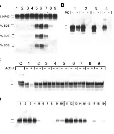
Acidic SDS denatures PrPSc
.To examine whether solutions of 1 to 4% SDS could denature PrPSc more effectively at
different pH values, we used 50 mM sodium acetate and Tris acetate buffers to maintain the pH between 3.5 and 10.0 for homogenates prepared from the brains of Syrian hamsters infected with Sc237 prions. Our studies demonstrated that aqueous solutions of ⱖ1% SDS could denature PrPSc
com-pletely at pH values ofⱕ4.5 orⱖ10.0, as judged by immuno-blotting for PrP after the samples were subjected to limited proteolysis (Fig. 1A). We also found that acidic solutions other than AcOH enable SDS to denature PrPScin Sc237-infected
brain homogenate: PrPScwas denatured in the presence of 1%
SDS plus either 0.5% AcOH (pH 3.6), 50 mM glycine (pH 3.7), or 0.2% peracetic acid (pH 3.4) for 15 min at 37°C (Fig. 1B). Acidic SDS denatured PrPScin Sc237-infected brain
homog-enate in 30 min at room temperature, as judged by Western blotting (Fig. 1C, paired lanes 1). We next investigated whether dilute acid in the presence of detergents other than SDS could also denature PrPSc. We incubated Sc237-infected brain
ho-mogenates with various detergents, in the presence or absence of AcOH, for 30 min at room temperature. The detergents cholic acid, deoxycholic acid, Triton X-100, NP-40, Tween 20, CTAB (cetyltrimethylammonium bromide), Zwittergent, and CHAPS {3-[(3-cholamidopropyl)-dimethylammonio]-1-pro-panesulfonate} failed to denature PrPSc at low pH (Fig. 1C,
paired lanes 2 to 9, respectively). We next tested the ability of alkyl sulfates and alkyl sulfonates of various alkyl chain lengths to denature PrPScin the presence of 0.5% AcOH. Alkyl
sul-fates with alkyl chains of 9 to 12 carbon atoms and alkyl sulfonates with alkyl chains of 10 to 13 carbon atoms denatured PrPScsubstantially in the presence of dilute acid (Fig. 1D).
Physicochemical properties of PrPSc
[image:3.585.302.538.81.358.2]treated with acidic SDS. To investigate whether the inactivation of prions by acidic SDS is reversible, we added a neutralization buffer containing the nonionic detergent NP-40 to a sample of Sc237-infected brain homogenate initially exposed to 1% SDS and 0.5% AcOH for 15 min at 37°C. We then dialyzed the neutralized sample con-taining mixed micelles for 8 h at 4°C to remove the SDS. This
FIG. 1. Western blots of prion-infected brain homogenates treated with different detergents and at different pH values. For all panels, apparent molecular masses based on the migration of protein stan-dards are 30 and 27 kDa (top and bottom markers, respectively). (A) Samples of 1% brain homogenate containing Sc237 prions were incubated for 15 min at 37°C with the indicated detergent at the following pH values: 3.5 (lane 1), 4.0 (lane 2), 4.5 (lane 3), 5.0 (lane 4), 6.0 (lane 5), 7.0 (lane 6), 8.0 (lane 7), 9.0 (lane 8), and 10.0 (lane 9). Sodium acetate buffers (50 mM) were used to maintain pH values 3 to 6 (lanes 1 to 5), and 50 mM Tris acetate buffers were used to maintain pH values 7 to 10 (lanes 6 to 9). After incubation, all samples were neutralized by the addition of an equal volume of 4% Sarkosyl–100 mM HEPES (pH 7.5)–200 mM NaCl and subjected to limited PK digestion (20g/ml for 1 h at 37°C). (B) Samples of 1% brain homog-enate containing Sc237 prions were incubated for 15 min at 37°C in 1% SDS plus 50 mM Tris acetate (pH 7.0) (paired lane 1), 0.5% AcOH (pH 3.6) (paired lane 2), 50 mM glycine (pH 3.7) (paired lane 3), or 0.2% peracetic acid (pH 3.4) (paired lane 4). After incubations, sam-ples were neutralized and then left undigested (⫺) or digested with PK (⫹) as described in panel A. (C) Samples of 2.5% brain homogenate containing Sc237 prions were incubated with 2% detergent either alone (⫺) or with (⫹) 1% AcOH at room temperature for 30 min. Anionic (paired lanes 1 to 3), nonionic (paired lanes 4 to 6), cationic (paired lane 7), and zitterionic (paired lanes 8 to 9) detergents were used. Lane assignments are as follows: lane 1, SDS; lane 2, cholic acid; lane 3, deoxycholic acid; lane 4, Triton X-100; lane 5, NP-40; lane 6, Tween 20; lane 7, CTAB; lane 8, Zwittergent; lane 9, CHAPS. All paired samples (lanes 1 to 9) were neutralized by the addition of 280 l of 2% Sarkosyl–200 mM HEPES (pH 7.5)–100 mM NaCl and subjected to limited PK digestion (4g for 1 h at 37°C). C, undigested control. (D) Samples of 1% brain homogenate containing Sc237 prions were incubated with 0.5% AcOH (pH 3.6) and either 1% alkyl sulfates (lanes 1 to 10), NP-40 (lanes 11 and 12), or alkyl sulfonates (lanes 13 to 19) for 2 h at 37°C. Alkyl sulfate detergents with backbones of 5 to 14 carbon atoms (lanes 1 to 10, respectively) were tested. Alkyl sulfo-nate detergents with backbones of 6 (lane 13), 7 (lane 14), and 9 to 13 carbon atoms (lanes 15 to 19, respectively) were used. All samples were subjected to limited PK digestion.
on November 8, 2019 by guest
http://jvi.asm.org/
procedure did not restore the protease-resistance of PrPScthat
had been denatured by acidic SDS (Fig. 2A, paired lanes 2). To investigate further the biophysical changes in PrPSc
mol-ecules induced by exposure to acidic SDS, we performed ul-tracentrifugation. We found that PrPScmolecules become
sol-uble in acidic SDS (Fig. 2B).
Inactivation of Syrian hamster prions by acidic SDS. To determine whether denaturation of PrPSccaused by acidic SDS
correlates with a reduction in prion infectivity, we incubated Sc237-infected brain homogenates for 2 h with different buffers at various temperatures and inoculated the treated samples intracerebrally into Syrian hamsters. Inoculation of a control brain homogenate containing ⬃109 ID
50units/ml (one ID50
unit is the infectious dose causing illness in 50% of inoculated animals) at neutral pH without detergent caused disease in all animals in 84 ⫾ 0.4 days (Fig. 3). Animals inoculated with samples exposed to either 0.5% AcOH or 1% SDS alone for 2 h at room temperature developed disease in 79⫾0.9 days and 91⫾0.4 days, respectively (Fig. 3). In contrast, exposure of the inoculum to a solution of 1% SDS and 0.5% AcOH for 2 h at room temperature prolonged the median incubation time to 200⫾2.3 days (Fig. 3). Similar results were obtained when samples were incubated at 37°C (data not shown).
The prolonged incubation times found after acidic SDS treatment of hamster brain homogenates indicate that the prion titers were reduced from ⬃109 ID
50units/ml to⬍100
ID50 units/ml. These titers were calculated from standard
curves that relate the incubation time to the dose of prions in the inoculum (61, 62). That the prions were not completely
inactivated by the procedure used is clear from the survival curves: all of the hamsters used for bioassay eventually devel-oped disease (Fig. 3). Although the data argue that SDS and dilute acid act synergistically to diminish prion infectivity, com-plete inactivation necessitated modification of the protocol.
Bioassays in transgenic mice.Although these initial studies proved promising, we were concerned that extensive bioassays in hamsters would be complicated by the relatively short life span of these animals (69); we therefore turned to Tg mice expressing high levels of SHaPrP, designated Tg7 mice (70, 78). Uninoculated Tg7 mice remain healthy for more than 500 days, while those inoculated with⬃109ID
50units/ml of prions
develop neurologic dysfunction in approximately 45 days. To make our studies more clinically relevant, we used crude brain homogenates, rather than homogenates precleared by low-speed centrifugation.
A 10% (wt/vol) Sc237-infected brain homogenate was seri-ally diluted, and each dilution was inoculated into Tg7 mice. As the infected brain homogenate is diluted, the percentage of mice succumbing to prion disease decreases (Fig. 4A) and the median incubation period lengthens. These median time points were interpolated with an exponential decay curve to highlight this relationship (Fig. 4A). A similar curve was developed relating the length of the incubation time and the size of the inoculum dose for bioassays of human sCJD prions with Tg22372 mice (Fig. 4B).
[image:4.585.311.529.68.226.2]Inactivation of Syrian hamster prions by acidic SDS at elevated temperatures.In an attempt to destroy the residual prion infectivity found by initial bioassay (Fig. 3), we increased the concentrations of SDS to 2% and of AcOH to 1% and raised the treatment temperature to 65°C (Table 1). A solution composed of 4% SDS and 2% AcOH was added directly to an equal volume of sample containing a 5% (wt/vol) prion-infected crude brain homogenate, mixed, and incubated with-out additional agitation.
[image:4.585.45.283.68.155.2]FIG. 2. Physical characterization of Sc237 prions denatured by acidic SDS. (A) A 0.5-ml sample of 1% prion-infected brain homog-enate was incubated in 1% SDS–0.5% AcOH (pH 3.6) for 15 min at 37°C and then neutralized by the addition of 0.5 ml of 2% NP-40–100 mM HEPES (pH 7.5)–200 mM NaCl. A 50-l aliquot was removed for analysis, and the remainder of the sample was dialyzed two times against 2 liters of 1% NP-40–50 mM HEPES (pH 7.5)–100 mM NaCl for 8 h at 4°C (molecular mass cutoff of 10 kDa). Sample before (paired lane 1) and after (paired lane 2) dialysis. Samples were undi-gested (⫺) or subjected (⫹) to limited PK digestion (20g/ml for 1 h at 37°C). The apparent molecular masses based on the migration of protein standards are 30 and 27 kDa. (B) Samples of 1% prion-infected brain homogenate were incubated at room temperature for 15 min with PBS (lanes 1, 4, 7, and 10), 50 mM Tris (pH 7.0) with 1% SDS (lanes 2, 5, 8, and 11), or 1% SDS–0.5% AcOH (lanes 3, 6, 9, and 12). After incubation, samples were centrifuged at 100,000⫻gfor 1 h at 4°C. Supernatant fractions (lanes 1 to 6) were removed and neutralized with an equal volume of 2⫻neutralization buffer (200 mM NaCl, 4% Sarkosyl, 400 mM HEPES [pH 7.5]) and subjected to limited PK digestion (PK-protein [1:50], for 1 h at 37°C) as indicated. Pellet fractions (lanes 7 to 12) were resuspended in 1⫻neutralization buffer and subjected to limited PK digestion as indicated. All samples were mixed with an equal volume of 2⫻SDS sample buffer. The apparent molecular masses based on the migration of protein standards are 36 and 29 kDa.
FIG. 3. Effect of acidic SDS and temperature on prion infectivity. Samples of 1% brain homogenate infected with Sc237 prions contain-ing⬃109ID
50units/ml were incubated with continuous shaking for 2 h
in the specified solution at room temperature. After incubation, sam-ples were diluted 1:10 into calcium- and magnesium-free PBS plus 5 mg of bovine serum albumin/ml, and 50-l aliquots were inoculated into eight Syrian hamsters each, and survival curves were calculated. Symbols:‚, 50 mM sodium acetate (pH 7.0);F, 0.5% AcOH (pH 3.6);
Œ, 1% SDS and 50 mM sodium acetate (pH 7.0);E, 1% SDS and 0.5% AcOH (pH 3.6).
on November 8, 2019 by guest
http://jvi.asm.org/
Inoculation of a control Sc237-infected brain homogenate at neutral pH without detergent produced disease in Tg7 mice with a median incubation time of 46 days (Table 1). Although uninoculated Tg7 mice remained healthy for 500 days, some animals can develop a nontransmissible neuromyopathy after
this time (96). Since mice are inoculated at 8 to 10 weeks of age, bioassays in Tg7 mice were terminated after 400 days to exclude the possibility of illness not caused by prions. After exposure of Sc237-infected brain homogenates to 2% SDS and 1% AcOH at 65°C for 30 min, the inoculum produced prion disease in some of the mice, with significantly prolonged incu-bation times. Increasing the exposure time of the inoculum to 2 h or 18 h resulted in no animals becoming ill 400 days after inoculation.
Acidic SDS inactivates Syrian hamster prions bound to steel wire.To study the inactivation of prions on surfaces (100), we soaked stainless steel wires in 10% brain homogenates con-taining Sc237 prions for 16 h at room temperature. The prion-coated wires were implanted into the parietal lobes of Tg7 mice and produced disease in 52⫾0.3 days. Treatment of the wires at 65°C for 30 min with 2% SDS and 1% AcOH in-creased the incubation time to 82⫾0.7 days after implantation into the brains of Tg7 mice; treatment at 65°C for 2 h further lengthened the incubation time, but most of the animals still developed disease in fewer than 400 days. Only treatment at 65°C for 18 h resulted in no animals succumbing to disease before 400 days (Table 1).
Inactivation of human prions by acidic SDS. Because dif-ferent prion strains exhibit distinct resistances to inactivation by chaotropic salts and heat (56, 57, 81, 89, 91), we investigated the inactivation of sCJD prions by acidic SDS. Brain homog-enates were prepared from an sCJD patient homozygous for methionine at residue 129, who carried no mutations in the
PRNPgene and whose unglycosylated PrP 27-30 migrated at 21 kDa on SDS-PAGE (53). These human prions, designated sCJD(MM1), are the most common strain found in sCJD pa-tients (52, 53). We either inoculated brain homogenate con-taining sCJD(MM1) prions intracerebrally or implanted wires that were coated with the homogenate into Tg22372 mice (38). It is noteworthy that, like the human PrPScinoculum, the PrP
[image:5.585.58.272.73.373.2]transgene in Tg22372 mice encodes methionine at position 129. Sporadic CJD(MM1) prions in brain homogenates or on steel wires were treated with 2% SDS and 1% AcOH at 65°C (Table 1). For brain homogenates, acidic SDS treatment for 30 min at 65°C prolonged the incubation times from 131⫾ 0.7 days to 266⫾8.5 days. Increasing the time of exposure (2 h or 18 h) to acidic SDS at 65°C lengthened the incubation time (⬎500 days) and decreased the proportion of Tg mice devel-oping disease (⬃25%). Studies of sCJD-coated steel wires pro-longed incubation times only slightly, and removal of infectivity on these surfaces was less efficient than in brain homogenates.
FIG. 4. Proportion and incubation period of transgenic mice suc-cumbing to disease with each dilution of inoculum. Ten percent brain homogenates were serially diluted 10-fold, and each dilution was inocu-lated into 12 mice. Bars represent the percentage of mice that showed clinical signs of disease when inoculated with the indicated log10dilution
of brain homogenate. The incubation period, the number of days from inoculation to the manifestation of clinical symptoms, was also measured for these mice. When at least half of the animals became ill, the median incubation period was determined by Kaplan-Meier statistics and plotted against log10dilution (datum point⫾the standard error). Values were
interpolated with the best fit to an exponential decay curve. (A) Sc237 prions bioassayed in Tg(SHaPrP)7 mice; (B) sCJD prions bioassayed in Tg(MHu2M, M165V, E167Q)22372 mice.
TABLE 1. Inactivation of prions by 2% SDS plus 1% AcOH at 65°Ca
Condition(s)
Hamster Sc237 prions in Tg7 mice Human sCJD prions in Tg23372 mice
Homogenate Wire Homogenate Wire
IP (days) % (n⫽10) IP (days) % (n⫽10) IP (days) % (n⫽10) IP (days) % (n⫽10)
Negative control ⬎400 0 ⬎400 0 ⬎500 0 ⬎500 0
Positive control 46⫾0.2 100 52⫾0.3 100 131⫾0.7 100 215⫾0.9 100
2% SDS–1% AcOH, 30 min ⬎400 26 82⫾0.7 100 266⫾8.5 74 354⫾1.6 86
2% SDS–1% AcOH, 2 h ⬎400 0 269⫾3.2 68 ⬎500 26 ⬎500 44
2% SDS–1% AcOH, 18 h ⬎400 0 ⬎400 0 ⬎500 25 ⬎500 25
aMedian incubation period (IP)⫾the standard error and percentage of animals succumbing to prion disease were calculated by using Kaplan-Meier analysis.
on November 8, 2019 by guest
http://jvi.asm.org/
[image:5.585.40.543.620.715.2]The same treatment protocol (exposure to 2% SDS and 1% AcOH for 30 min at 65°C) resulted in large differences in the level of inactivation for the two prion strains studied (Table 1). Because the titer of Sc237 prions is 10- to 100-fold higher than sCJD prions in brain homogenates (Fig. 4), data were analyzed by a stratified Cox regression (33) in order to quantify differ-ences between hamster and human prions. Taking the ratios of coefficients in the Cox model, it is possible to relate the effect of an inactivation procedure to an approximately equivalent dilution. Applying the stratified Cox regression to the 30-min exposure to acidic SDS, we estimate a 9.0 log10reduction in
infectivity for Sc237 prions and a 3.8 log10reduction for sCJD
prions. The difference in inactivation between the two prion strains is 5.2 log10(95% confidence intervals, 3.7 to 6.8 log10).
This analysis argues that sCJD prions in human brain homog-enates are 105-fold more resistant to inactivation than Sc237
prions in SHa brain homogenates.
Two-step inactivation protocol.Concerned that exposure to acid results in the aggregation of proteins and that such aggre-gates might protect PrPScfrom acidic SDS–mediated
denatur-ation, we developed a two-step protocol: SDS at neutral pH is used to disperse proteins prior to exposure to AcOH. In these studies, exposure to 4% neutral SDS was performed at 65°C for 30 min, followed by acidic SDS (4% SDS and 1% AcOH) at 65°C for 30 min, 2 h, or 18 h (Table 2). All two-step inac-tivation studies were completed in three independent sets (n⫽
8) and, since no significant differences were observed, the sets were combined for analysis.
SHa brain homogenates (10% [wt/vol]) containing Sc237 prions were treated by using the two-step acidic SDS protocol. Untreated homogenates produced neurologic dysfunction af-ter 55 days in Tg7 mice (Table 2), whereas the two-step pro-cedure at 65°C for 30 min completely abolished prion infectiv-ity. In experiments with Sc237 prion-coated wires, untreated control wires produced disease in 63⫾0.1 days after implan-tation. In contrast to homogenates, exposure of prion-coated wires to the two-step procedure at 65°C for 30 min did not completely inactivate prion infectivity.
Human brain homogenates (10% [wt/vol]) containing sCJD (MM1) prions were treated by using the two-step acidic SDS protocol as described above. Untreated homogenates pro-duced neurologic dysfunction in 139 ⫾ 0.4 days in Tg22372 mice (Table 2). Exposure of these homogenates to the two-step procedure at 65°C for 30 min substantially reduced prion
in-fectivity, while extending the time to 18 h completely abolished infectivity. In three separate experiments with sCJD prion-coated wires, untreated control wires produced disease in 181⫾1.2 days after implantation. In contrast to homogenates, exposure of prion-coated wires to the two-step procedure at 65°C, even for 18 h, did not inactivate prion infectivity, with the majority of Tg22372 mice still succumbing to disease.
Acidic SDS and autoclaving abolish prion infectivity. Al-though exposure of Sc237 and sCJD(MM1) prions to 2% SDS and 1% AcOH at 65°C for 2 h was sufficient to destroy more than 99.99% of the infectivity in brain homogenates, complete inactivation was not achieved (Table 1). Also, although the two-step procedure using 4% neutral SDS, followed by a com-bination of 4% SDS and 1% AcOH, further diminished infec-tivity levels, residual infecinfec-tivity could still be measured, partic-ularly on the surfaces of steel wires coated with sCJD(MM1) prions (Table 2).
To determine whether autoclaving for a brief time in the presence of acidic SDS could eliminate prion infectivity, we exposed Sc237 and sCJD(MM1) prions in brain homogenates or on wire surfaces to 121°C in the presence or absence of acidic SDS (Table 3). Neither Sc237 nor sCJD(MM1) prions in brain homogenates or on wire surfaces were detectable by bioassay in Tg mice after exposure to 121°C for 15 min in the presence of 2% SDS and 1% AcOH (Table 3). Similarly, sam-ples first exposed to 4% neutral SDS followed by a combina-tion of 4% SDS and 1% AcOH at 134°C were devoid of prion infectivity (Table 4). The efficacy of acidic SDS-mediated in-activation of prions is strikingly documented by how many prions survived autoclaving at 121°C or 134°C for 15 min, 30 min, or 2 h in the absence of acidic SDS (Tables 3 and 4). The survival of sCJD(MM1) prions bound to steel wires after au-toclaving for 2 h at 134°C is especially alarming (Table 4).
To quantify the log10reduction in prion titer from these
procedures, we used a Cox proportional-hazards model based on the serial dilution data (Fig. 4) and derived partial-likeli-hood ratios based on 95% confidence intervals. Treatment of sCJD prions in brain homogenate for 30 min at 121°C (Table 3) results in a 6.8 log10reduction (lower 95%
confi-dence interval, 5.3 log10). If no animals succumb to disease, the
[image:6.585.45.537.82.202.2]extent of inactivation cannot be quantified. However, it is still possible to determine a lower 95% confidence interval. For example, treatment of sCJD prions in brain homogenate with the two-step protocol for 15 min at 134°C (Table 4) gives a
TABLE 2. Inactivation of prions by 4% SDS plus 1% AcOH at 65°Ca
Condition(s)
Hamster Sc237 prions in Tg7 mice Human sCJD prions in Tg23372 mice
Homogenate Wire Homogenate Wire
IP (days) % (n⫽24) IP (days) % (n⫽24) IP (days) % (n⫽24) IP (days) % (n⫽24)
Negative control ⬎400 0 ⬎400 0 ⬎500 0 ⬎500 0
Positive control 55⫾0.0 100 63⫾0.1 100 139⫾0.4 100 181⫾1.2 100
ddH2O, 18 h 55⫾0.1 100 67⫾0.2 94 179⫾4.5 62 208⫾3.8 100
1% AcOH, 18 h 60⫾0.4 100 63⫾0.1 100 172⫾0.8 100 196⫾1.9 100
4% SDS, 18 h 77⫾0.4 100 106⫾3.1 62 ⬎500 32 223⫾1.8 79
4% SDS–1% AcOH, 30 min ⬎400 0 ⬎400 14 ⬎500 5 278⫾15 71
4% SDS–1% AcOH, 2 h ⬎400 0 ⬎400 25 ⬎500 13 259⫾6.4 64
4% SDS–1% AcOH, 18 h ⬎400 0 ⬎400 0 ⬎500 0 379⫾30 60
aMedian incubation period (IP)⫾the standard error and percentage of animals succumbing to prion disease were calculated by using Kaplan-Meier analysis.
on November 8, 2019 by guest
http://jvi.asm.org/
lower 95% confidence interval of 6.1 log10reduction in
infec-tivity.
The incubation periods for prion-coated wires cannot be converted into titers since the prions seem to be bound tightly to the surface of the wire (95) and it is not possible to remove them for measurement.
DISCUSSION
The differences in the inactivation profiles of prions and viruses provided the first clues that the scrapie agent was not a slow virus as had been widely thought (6, 23, 41, 48, 58, 59, 80). Mechanism of acidic SDS inactivation.From the results of the studies described here, exposure to acidic SDS appears to denature PrPSc(Fig. 1). Although SDS between pH 4.5 and pH
10.0 at room temperature is a poor denaturant, it becomes an excellent denaturant at a pH ofⱕ4.5 andⱖ10 (Fig. 1). More-over, weak acids are poor denaturants for PrPScexcept in the
presence of SDS.
Denaturation of PrPScby SDS at pH 4 was dependent on the
concentration of SDS, the time of exposure, and the temper-ature. Equivalent levels of denaturation seemed to be achieved by inversely varying the time and temperature; the time of exposure for complete inactivation of prions was 15 min when a temperature ofⱖ121°C was used (Tables 3 and 4).
In our initial experiments, denaturation of PrPScwas
mea-sured by the loss of protease-resistance of PrP 27-30 and a decrement in prion infectivity. Immunodetection of PrP 27-30 by using Western blotting has a dynamic range of⬃100-fold, whereas bioassays measure prions over an⬃109-fold range as
described above. In determining the mechanism of acidic
SDS-mediated denaturation of PrPSc, immunoassays are useful but
only prolonged bioassays are adequate to assess whether or not complete inactivation of prion infectivity has been achieved.
Stabilities of different prion strains.The strain phenotype of the prion is enciphered in the conformation of PrPSc(9, 43, 57,
94), and different strains display distinct conformational sta-bilities as reflected in their resistance to denaturation by chao-tropes and heat (36, 43, 57, 77, 81, 91). Besides the strain of prion, the sequence of PrP, determined by the last host in which the prion was passaged, influences the conformation of PrPSc and hence its stability (79). The passage of prions
through different species may lead to new prion strains with properties different from those of the parental strain (21, 35, 38, 54, 57, 72).
Conformational stability profiles, as measured by sensitivity to denaturation by GdnHCl gave half-maximal (Gdn1/2) values
of 1.5 and 1.8 M for Sc237 prions (56, 57) compared to a Gdn1/2value of 1.8 M for sCJD(MM1) prions (101). However,
the resistances of these strains to denaturation by acidic SDS are significantly different (Table 1 to Table 4), implying that inactivation protocols validated on one strain cannot be ex-trapolated to another. Exploring the spectrum of strain con-formations with respect to inactivation by acidic SDS should be facilitated by the availability of synthetic prions, especially since the MoSP1 strain is the most stable strain studied to date (42, 43).
[image:7.585.42.541.81.194.2]Limitations on the measurement of prion infectivity. Bioas-says for prions are most effective when high-titer samples are being assessed. Under these circumstances, the incubation times are the shortest and the variance among the animals is
TABLE 3. Inactivation of prions by 2% SDS plus 1% AcOH at 121°Ca
Condition(s)
Hamster Sc237 prions in Tg7 mice Human sCJD prions in Tg23372 mice
Homogenate Wire Homogenate Wire
IP (days) % (n⫽10) IP (days) % (n⫽10) IP (days) % (n⫽10) IP (days) % (n⫽10)
Positive control 46⫾0.2 100 62⫾0.3 100 146⫾0.4 100 207⫾1.1 100
Untreated, 15 min 344⫾20 73 160⫾7.3 100 221⫾1.0 100 ⬎500 22
Untreated, 30 min ⬎400 12 ⬎400 20 ⬎500 0 ⬎500 0
Untreated, 2 h ⬎400 12 ⬎400 0 ⬎500 0 414⫾15 73
2% SDS–1% AcOH, 15 min ⬎400 0 ⬎400 0 ⬎500 0 ⬎500 0
2% SDS–1% AcOH, 30 min ⬎400 0 ⬎400 0 ⬎500 12 ⬎500 0
2% SDS–1% AcOH, 2 h ⬎400 0 ⬎400 0 ⬎500 0 ⬎500 0
aMedian incubation period (IP)⫾the standard error and percentage of animals succumbing to prion disease were calculated by using Kaplan-Meier analysis.
TABLE 4. Inactivation of prions by 4% SDS plus 1% AcOH at 134°Ca
Condition(s)
Hamster Sc237 prions in Tg7 mice Human sCJD prions in Tg23372 mice
Homogenate Wire Homogenate Wire
IP (days) % (n⫽24) IP (days) % (n⫽24) IP (days) % (n⫽24) IP (days) % (n⫽24)
Positive control 55⫾0.0 100 63⫾0.1 100 139⫾0.5 100 181⫾1.2 100
Untreated, 15 min ⬎400 5 96⫾0.6 87 ⬎500 0 218⫾4.1 73
Untreated, 30 min ⬎400 0 262⫾10 55 ⬎500 0 242⫾2.8 63
Untreated, 2 h ⬎400 0 ⬎400 9 ⬎500 0 ⬎500 46
4% SDS–1% AcOH, 15 min ⬎400 0 ⬎400 0 ⬎500 0 ⬎500 0
4% SDS–1% AcOH, 30 min ⬎400 0 ⬎400 4 ⬎500 0 ⬎500 0
4% SDS–1% AcOH, 2 h ⬎400 0 ⬎400 0 ⬎500 0 ⬎500 0
aMedian incubation period (IP)⫾the standard error and percentage of animals succumbing to prion disease were calculated by using Kaplan-Meier analysis.
on November 8, 2019 by guest
http://jvi.asm.org/
[image:7.585.43.538.601.715.2]the smallest. As the incubation times lengthen, the variance increases. In studies of prion inactivation, it is possible to measure a reduction in titer over a range of⬃109-fold with
Sc237 prions using either Syrian hamsters or Tg(SHaPrP) mice. Inactivation studies performed with sCJD(MM1) prions using Tg mice permit measurements of prion infectivity over a range of⬃106-fold.
It is clear that the best measurements can be obtained when the incubation times are short, as is the case for high-titer samples. For example, Tg7 mice develop neurologic dysfunc-tion ⬃45 days after inoculation with high-titer samples of Sc237 prions (Fig. 4A).
Prion inactivation with acidic SDS. From the foregoing studies, acidic SDS provides for the first time a means of completely inactivating prions under relatively gentle condi-tions. The need for noncorrosive inactivation of prions is clearly illustrated by cases of iatrogenic CJD in which prion-contaminated neurosurgical equipment seems to have spread prions from one patient to another (8, 11, 14, 15, 20, 31). This concern has been heightened in Britain, where the finding of high titers of vCJD prions in lymphoid tissues worries author-ities that any surgical procedure could result in the spread of prions from one patient to another (18, 32). In Britain, 15 young people, all of whom later developed vCJD, donated blood that was transfused into 48 recipients. Recently, two of these recipients have been identified, in whom it is likely that vCJD prions were transmitted from the blood of the young donors (45, 55).
Conventional hospital disinfectants including ethylene ox-ide, propriolactone, hydrogen peroxox-ide, iodophors, peracetic acid, chaotropes, and phenolics have little effect on prion in-fectivity (89), although some modified cleaning reagents have been suggested to have utility in diminishing prion titers (25, 71). In addition, prions are resistant to inactivation by UV irradiation, aldehyde fixation, boiling, standard gravity auto-claving at 121°C, and detergent solubilization (1, 5, 22, 24, 40, 64, 89).
Although prolonged proteolytic digestion diminishes prion titers (17, 47, 68), complete inactivation of prion infectivity is difficult to achieve enzymatically. As with any enzymatic reac-tion, protease-catalyzed hydrolysis of PrPSc becomes
increas-ingly less efficient as the concentration of the substrate PrPSc
decreases (44, 50). Moreover, one or more substances in crude suspensions may inhibit an enzyme or mixture of enzymes used to digest PrPSc.
Currently recommended protocols for prion decontamina-tion include either (i)⬎2% available chlorine of sodium hy-pochlorite for 2 h, (ii) 2 M NaOH for 1 h, or (iii) autoclaving at 134°C for 4.5 h (4, 12, 63, 66, 73, 74, 76, 82, 92, 93). Each of these protocols has important limitations: sodium hypochlorite and NaOH are corrosive at the concentrations required to inactivate prions; NaOH did not inactivate CJD prions com-pletely in some reports (85, 86); and extended autoclaving at high temperature is deleterious to many materials. Currently, some high-risk surgical instruments are soaked in 2 N NaOH for 1 h, rinsed with water, and autoclaved at 134°C for 1 h (75), while many other such instruments are discarded.
In the studies reported here, sCJD(MM1) prions on steel wires were not completely inactivated by autoclaving at either 121 or 134°C in the absence of acidic SDS (Tables 3 and 4); this
contrasts with Sc237 prions that were completely inactivated by autoclaving alone (Tables 3 and 4). It is noteworthy that the prion-coated wires were not subjected to any procedures that might reduce the level of prions, such as washing, shaking, scrub-bing, and sonicating. Such cleaning procedures are known to reduce substantially the titers of many different pathogens (74).
The data presented here document the efficacy of acidic SDS combined with autoclaving for complete inactivation of human and hamster prions in brain homogenates and on the surface of steel wires (Tables 3 and 4). Acidic SDS combined with auto-claving should supplant routine autoauto-claving used to sterilize medical and dental equipment. For equipment such as fiber optic instruments that cannot be autoclaved, submerging such equipment in acidic SDS at 65°C will substantially reduce prion infectivity but not completely eliminate it (Table 2). Acidic SDS at room temperature may also find application in the inactivation of equipment and surfaces. Sc237 prion infectivity was reduced more than 99.99% in studies conducted at room temperature (Fig. 3).
Concluding remarks.The results of inactivation studies de-scribed here argue that acidic SDS combined with autoclaving be applied immediately for sterilization of surgical and dental instruments. Because SDS is both a detergent and a protein denaturant, it should prove especially apt for sterilizing instru-ments with complex shapes, serrations, locks, bores, and crev-ices. Whether other compounds such as urea that are known to denature PrPSc (51, 63, 64) will prove useful in combination
with a weak acid is unknown.
In addition to inactivating prions on the surfaces of surgical instruments and diagnostic equipment, acidic SDS may prove useful in the sterilization of dental and ophthalmologic instru-ments, as well as the cleaning of operating theaters and invasive diagnostic suites. Acidic SDS is likely to find use in the cleansing and sterilization of equipment used in the production of biophar-maceuticals. In addition, acidic SDS might be applied in the cleaning of abattoirs, meat-processing plants, butcher shops, kitchens, and wherever mammalian products are prepared for human consumption. Acidic SDS might also be used on equip-ment used in the rendering of offal. Whether acidic SDS or a biodegradable formulation such as acidic urea can be incorpo-rated into some products derived from rendered offal remains to be determined.
In summary, it is important to recognize that procedures routinely used in medical and dental settings do not inactivate prions. That being the case, it may be prudent to institute acidic-SDS protocols as configured in the studies presented here. Acidic SDS combined with autoclaving completely inac-tivated prion infectivity, even on steel surfaces. Inactivation of human and hamster prions mediated by acidic SDS occurs rapidly and can be achieved without boiling or autoclaving. Acidic SDS is noncorrosive and offers other practical advan-tages that make it suitable for widespread use.
ACKNOWLEDGMENTS
This study was supported by grants from the National Institutes of Health (AG02132, AG10770, and AG021601). S.S. was supported by the Burroughs Wellcome Fund Career Development Award and an NIH Clinical Investigator Development Award.
We thank the staff at the Hunters Point animal facility.
K.G., J.G.S., and S.B.P. have financial interests in InPro Biotech-nology, Inc.
on November 8, 2019 by guest
http://jvi.asm.org/
REFERENCES
1.Alper, T.1985. Scrapie agent unlike viruses in size and susceptibility to
inactivation by ionizing or ultraviolet radiation. Nature317:750.
2.Alper, T., W. A. Cramp, D. A. Haig, and M. C. Clarke.1967. Does the agent
of scrapie replicate without nucleic acid? Nature214:764–766.
3.Alper, T., D. A. Haig, and M. C. Clarke.1966. The exceptionally small size
of the scrapie agent. Biochem. Biophys. Res. Commun.22:278–284.
4.Baron, H., J. Safar, D. Groth, S. J. DeArmond, and S. B. Prusiner.1999.
Biosafety issues in prion diseases, p. 743–777.InS. B. Prusiner (ed.), Prion
biology and diseases. Cold Spring Harbor Laboratory Press, Cold Spring Harbor, N.Y.
5.Bellinger-Kawahara, C., J. E. Cleaver, T. O. Diener, and S. B. Prusiner.
1987. Purified scrapie prions resist inactivation by UV irradiation. J. Virol.
61:159–166.
6.Bellinger-Kawahara, C., T. O. Diener, M. P. McKinley, D. F. Groth, D. R. Smith, and S. B. Prusiner.1987. Purified scrapie prions resist inactivation by procedures that hydrolyze, modify, or shear nucleic acids. Virology
160:271–274.
7.Bellinger-Kawahara, C. G., E. Kempner, D. F. Groth, R. Gabizon, and S. B. Prusiner.1988. Scrapie prion liposomes and rods exhibit target sizes of
55,000 Da. Virology164:537–541.
8.Bernoulli, C., J. Siegfried, G. Baumgartner, F. Regli, T. Rabinowicz, D. C. Gajdusek, and C. J. Gibbs, Jr.1977. Danger of accidental person-to-person
transmission of Creutzfeldt-Jakob disease by surgery. Lanceti:478–479.
9.Bessen, R. A., and R. F. Marsh.1994. Distinct PrP properties suggest the molecular basis of strain variation in transmissible mink encephalopathy.
J. Virol.68:7859–7868.
10.Bockman, J. M., D. T. Kingsbury, M. P. McKinley, P. E. Bendheim, and S. B. Prusiner.1985. Creutzfeldt-Jakob disease prion proteins in human
brains. N. Engl. J. Med.312:73–78.
11.Brown, P., M. Preece, J. P. Brandel, T. Sato, L. McShane, I. Zerr, A. Fletcher, R. G. Will, M. Pocchiari, N. R. Cashman, J. H. d’Aignaux, L. Cervenakova, J. Fradkin, L. B. Schonberger, and S. J. Collins.2000. Iatrogenic
Creutzfeldt-Jakob disease at the millennium. Neurology55:1075–1081.
12.Budka, H., A. Aguzzi, P. Brown, J.-M. Brucher, O. Bugiani, J. Collinge, H. Diringer, F. Gullotta, M. Haltia, J.-J. Hauw, J. W. Ironside, H. A. Kretzschmar, P. L. Lantos, C. Masullo, M. Pocchiari, W. Schlote, J. Tateishi, and R. G. Will.1995. Tissue handling in suspected Creutzfeldt-Jakob disease (CJD) and other human spongiform encephalopathies (prion
diseases). Brain Pathol.5:319–322.
13.Carlson, G. A., D. T. Kingsbury, P. A. Goodman, S. Coleman, S. T. Marshall, S. DeArmond, D. Westaway, and S. B. Prusiner.1986. Linkage of
prion protein and scrapie incubation time genes. Cell46:503–511.
14.Centers for Disease Control.1997. Creutzfeldt-Jakob disease associated with cadaveric dura mater grafts—Japan, January 1979-May 1996. Morb.
Mortal. Wkly. Rep.46:1066–1069.
15.Centers for Disease Control.1985. Fatal degenerative neurologic disease in patients who received pituitary derived human growth hormone. Morb.
Mortal. Wkly. Rep.34:359–360.
16.Chandler, R. L.1961. Encephalopathy in mice produced by inoculation
with scrapie brain material. Lanceti:1378–1379.
17.Cho, H. J.1983. Inactivation of the scrapie agent by pronase. Can. J. Comp.
Med.47:494–496.
18.Collins, S., M. G. Law, A. Fletcher, A. Boyd, J. Kaldor, and C. L. Masters.
1999. Surgical treatment and risk of sporadic Creutzfeldt-Jakob disease: a
case-control study. Lancet353:693–697.
19.Cox, D. R.1972. Regression models and life-tables. J. R. Stat. Soc. Ser. A
Method.34:187–220.
20.de Silva, R., and T. Esmonde.1994. Iatrogenic transmission of
Creutzfeldt-Jakob disease: an update. CNS Drugs2:96–101.
21.Dickinson, A. G.1976. Scrapie in sheep and goats, p. 209–241.InR. H. Kimberlin (ed.), Slow virus diseases of animals and man. North-Holland Publishing, Amsterdam, The Netherlands.
22.Dickinson, A. G., and D. M. Taylor.1978. Resistance of scrapie agent to
decontamination. N. Engl. J. Med.299:1413–1414.
23.Diener, T. O., M. P. McKinley, and S. B. Prusiner.1982. Viroids and prions.
Proc. Natl. Acad. Sci. USA79:5220–5224.
24.Ernst, D. R., and R. E. Race.1993. Comparative analysis of scrapie agent
inactivation methods. J. Virol. Methods41:193–202.
25.Fichet, G., E. Comoy, C. Duval, K. Antloga, C. Dehen, A. Charbonnier, G. McDonnell, P. Brown, C. I. Lasmezas, and J. P. Deslys.2004. Novel
meth-ods for disinfection of prion-contaminated medical devices. Lancet364:
521–526.
26.Gajdusek, D. C., C. J. Gibbs, Jr., and M. Alpers. 1966. Experimental
transmission of a kuru-like syndrome to chimpanzees. Nature209:794–796.
27.Gibbs, C. J., Jr., D. C. Gajdusek, D. M. Asher, M. P. Alpers, E. Beck, P. M. Daniel, and W. B. Matthews.1968. Creutzfeldt-Jakob disease (spongiform
encephalopathy): transmission to the chimpanzee. Science161:388–389.
28.Gibbs, C. J., Jr., D. C. Gajdusek, and R. Latarjet.1978. Unusual resistance to ionizing radiation of the viruses of kuru, Creutzfeldt-Jakob disease. Proc.
Natl. Acad. Sci. USA75:6268–6270.
29.Gordon, W. S.1946. Advances in veterinary research. Vet. Res.58:516–520. 30.Govaerts, C., H. Wille, S. B. Prusiner, and F. E. Cohen.2004. Evidence for
assembly of prions with left-handed-helices into trimers. Proc. Natl. Acad.
Sci. USA101:8342–8347.
31.Healy, D. L., and J. Evans.1993. Creutzfeldt-Jakob disease after pituitary
gonadotrophins. Br. J. Med.307:517–518.
32.Hilton, D. A., A. C. Ghani, L. Conyers, P. Edwards, L. McCardle, D. Ritchie, M. Penney, D. Hegazy, and J. W. Ironside.2004. Prevalence of lymphoreticular
prion protein accumulation in UK tissue samples. J. Pathol.203:733–739.
33.Holt, J. D., and R. L. Prentice.1974. Survival analyses in twin studies and
matched pair experiments. Biometrika61:17–30.
34.Kaplan, E. L., and P. Meier.1958. Nonparametric estimation from
incom-plete observations. J. Am. Stat. Assoc.53:457–481.
35.Kimberlin, R. H., and C. A. Walker.1979. Pathogenesis of scrapie: agent multiplication in brain at the first and second passage of hamster scrapie in
mice. J. Gen. Virol.42:107–117.
36.Kimberlin, R. H., C. A. Walker, G. C. Millson, D. M. Taylor, P. A. Robertson, A. H. Tomlinson, and A. G. Dickinson.1983. Disinfection studies with two
strains of mouse-passaged scrapie agent. J. Neurol. Sci.59:355–369.
37.Kong, Q., W. K. Surewicz, R. B. Petersen, W. Zou, S. G. Chen, P. Gambetti, P. Parchi, S. Capellari, L. Goldfarb, P. Montagna, E. Lugaresi, P. Piccardo, and B. Ghetti.2004. Inherited prion diseases, p. 673–775.InS. B. Prusiner (ed.), Prion biology and diseases, 2nd ed. Cold Spring Harbor Laboratory Press, Cold Spring Harbor, N.Y.
38.Korth, C., K. Kaneko, D. Groth, N. Heye, G. Telling, J. Mastrianni, P. Parchi, P. Gambetti, R. Will, J. Ironside, C. Heinrich, P. Tremblay, S. J. DeArmond, and S. B. Prusiner.2003. Abbreviated incubation times for human prions in mice expressing a chimeric mouse—human prion protein
transgene. Proc. Natl. Acad. Sci. USA100:4784–4789.
39.Laemmli, U. K.1970. Cleavage of structural proteins during the assembly of
the head of bacteriophage T-4. Nature227:680–685.
40.Latarjet, R.1979. Inactivation of the agents of scrapie, Creutzfeldt-Jakob
disease, and kuru by radiations, p. 387–408.InS. B. Prusiner and W. J.
Hadlow (ed.), Slow transmissible diseases of the nervous system, vol. 2. Academic Press, Inc., New York, N.Y.
41.Latarjet, R., B. Muel, D. A. Haig, M. C. Clarke, and T. Alper. 1970. Inactivation of the scrapie agent by near monochromatic ultraviolet light.
Nature227:1341–1343.
42.Legname, G., I. V. Baskakov, H.-O. B. Nguyen, D. Riesner, F. E. Cohen, S. J. DeArmond, and S. B. Prusiner.2004. Synthetic mammalian prions.
Science305:673–676.
43.Legname, G., H.-O. B. Nguyen, I. V. Baskakov, F. E. Cohen, S. J. DeArmond, and S. B. Prusiner.2005. Strain-specified characteristics of mouse synthetic
prions. Proc. Natl. Acad. Sci. USA102:2168–2173.
44.Lineweaver, H., and D. Burk.1934. The determination of enzyme
dissoci-ation constants. J. Am. Chem. Soc.56:658–666.
45.Llewelyn, C. A., P. E. Hewitt, R. S. Knight, K. Amar, S. Cousens, J. Mackenzie, and R. G. Will. 2004. Possible transmission of variant
Creutzfeldt-Jakob disease by blood transfusion. Lancet363:417–421.
46.Marsh, R. F., and R. H. Kimberlin.1975. Comparison of scrapie and transmissible mink encephalopathy in hamsters. II. Clinical signs,
pathol-ogy, and pathogenesis. J. Infect. Dis.131:104–110.
47.McKinley, M. P., D. C. Bolton, and S. B. Prusiner.1983. A protease-resistant protein is a structural component of the scrapie prion. Cell
35:57–62.
48.McKinley, M. P., F. R. Masiarz, and S. B. Prusiner.1981. Reversible
chemical modification of the scrapie agent. Science214:1259–1261.
49.McKinley, M. P., R. K. Meyer, L. Kenaga, F. Rahbar, R. Cotter, A. Serban, and S. B. Prusiner.1991. Scrapie prion rod formation in vitro requires both
detergent extraction and limited proteolysis. J. Virol.65:1340–1351.
50.Michaelis, L., and M. I. Menton.1913. Die kinetic der invertinwirkung.
Biochem. Z.49:333–369.
51.Millson, G. C., G. D. Hunter, and R. H. Kimberlin.1976. The
physico-chemical nature of the scrapie agent, p. 243–266.InR. H. Kimberlin
(ed.), Slow virus diseases of animals and man. American Elsevier, New York, N.Y.
52.Palmer, M. S., A. J. Dryden, J. T. Hughes, and J. Collinge.1991. Homozy-gous prion protein genotype predisposes to sporadic Creutzfeldt-Jakob
disease. Nature352:340–342.
53.Parchi, P., A. Giese, S. Capellari, P. Brown, W. Schulz-Schaeffer, O. Windl, I. Zerr, H. Budka, N. Kopp, P. Piccardo, S. Poser, A. Rojiani, N. Streichem-berger, J. Julien, C. Vital, B. Ghetti, P. Gambetti, and H. Kretzschmar.1999. Classification of sporadic Creutzfeldt-Jakob disease based on molecular and
phenotypic analysis of 300 subjects. Ann. Neurol.46:224–233.
54.Pattison, I. H.1966. The relative susceptibility of sheep, goats and mice to
two types of the goat scrapie agent. Res. Vet. Sci.7:207–212.
55.Peden, A. H., M. W. Head, D. L. Ritchie, J. E. Bell, and J. W. Ironside.2004.
Preclinical vCJD after blood transfusion in aPRNPcodon 129 heterozygous
patient. Lancet364:527–529.
56.Peretz, D., M. Scott, D. Groth, A. Williamson, D. Burton, F. E. Cohen, and S. B. Prusiner.2001. Strain-specified relative conformational stability of the
scrapie prion protein. Protein Sci.10:854–863.
on November 8, 2019 by guest
http://jvi.asm.org/
57.Peretz, D., R. A. Williamson, G. Legname, Y. Matsunaga, J. Vergara, D. Burton, S. J. DeArmond, S. B. Prusiner, and M. R. Scott.2002. A change in the conformation of prions accompanies the emergence of a new prion
strain. Neuron34:921–932.
58.Prusiner, S. B.2004. Development of the prion concept, p. 89–141.InS. B. Prusiner (ed.), Prion biology and diseases, 2nd ed. Cold Spring Harbor Laboratory Press, Cold Spring Harbor, N.Y.
59.Prusiner, S. B.1982. Novel proteinaceous infectious particles cause scrapie.
Science216:136–144.
60.Prusiner, S. B. (ed.).2004. Prion biology and diseases, 2nd ed. Cold Spring Harbor Laboratory Press, Cold Spring Harbor, N.Y.
61.Prusiner, S. B., S. P. Cochran, D. E. Downey, and D. F. Groth.1981. Determination of scrapie agent titer from incubation period measurements
in hamsters, p. 385–399.InJ. W. Streilein, D. A. Hart, J. Stein-Streilein,
W. R. Duncan, and R. E. Billingham (ed.), Hamster immune responses in infectious and oncologic diseases. Plenum Press, Inc., New York, N.Y. 62.Prusiner, S. B., S. P. Cochran, D. F. Groth, D. E. Downey, K. A. Bowman,
and H. M. Martinez.1982. Measurement of the scrapie agent using an
incubation time interval assay. Ann. Neurol.11:353–358.
63.Prusiner, S. B., D. Groth, A. Serban, N. Stahl, and R. Gabizon.1993. Attempts to restore scrapie prion infectivity after exposure to protein
de-naturants. Proc. Natl. Acad. Sci. USA90:2793–2797.
64.Prusiner, S. B., D. F. Groth, S. P. Cochran, F. R. Masiarz, M. P. McKinley, and H. M. Martinez.1980. Molecular properties, partial purification, and assay by incubation period measurements of the hamster scrapie agent.
Biochemistry21:4883–4891.
65.Prusiner, S. B., D. F. Groth, M. P. McKinley, S. P. Cochran, K. A. Bowman, and K. C. Kasper. 1981. Thiocyanate and hydroxyl ions inactivate the
scrapie agent. Proc. Natl. Acad. Sci. USA78:4606–4610.
66.Prusiner, S. B., M. P. McKinley, D. C. Bolton, K. A. Bowman, D. F. Groth, S. P. Cochran, E. M. Hennessey, M. B. Braunfeld, J. R. Baringer, and M. A. Chatigny.1984. Prions: methods for assay, purification and
characteriza-tion, p. 293–345.InK. Maramorosch and H. Koprowski (ed.), Methods in
virology, vol. 8. Academic Press, Inc., New York, N.Y.
67.Prusiner, S. B., M. P. McKinley, K. A. Bowman, D. C. Bolton, P. E. Bendheim, D. F. Groth, and G. G. Glenner.1983. Scrapie prions aggregate
to form amyloid-like birefringent rods. Cell35:349–358.
68.Prusiner, S. B., M. P. McKinley, D. F. Groth, K. A. Bowman, N. I. Mock, S. P. Cochran, and F. R. Masiarz.1981. Scrapie agent contains a
hydro-phobic protein. Proc. Natl. Acad. Sci. USA78:6675–6679.
69.Prusiner, S. B., J. Safar, and S. J. DeArmond.2004. Bioassays of prions,
p. 143–186.InS. B. Prusiner (ed.), Prion biology and diseases, 2nd ed. Cold
Spring Harbor Laboratory Press, Cold Spring Harbor, N.Y.
70.Prusiner, S. B., M. Scott, D. Foster, K.-M. Pan, D. Groth, C. Mirenda, M. Torchia, S.-L. Yang, D. Serban, G. A. Carlson, P. C. Hoppe, D. Westaway, and S. J. DeArmond. 1990. Transgenetic studies implicate interactions
between homologous PrP isoforms in scrapie prion replication. Cell 63:
673–686.
71.Race, R. E., and G. J. Raymond.2004. Inactivation of transmissible
spon-giform encephalopathy (prion) agents by environ LpH. J. Virol.78:2164–
2165.
72.Ridley, R. M., and H. F. Baker.1996. To what extent is strain variation evidence for an independent genome in the agent of the transmissible
spongiform encephalopathies? Neurodegeneration5:219–231.
73.Rosenberg, R. N., L. L. White, III, P. Brown, D. C. Gajdusek, J. J. Volpe, J. Posner, and P. J. Dyck.1986. Precautions in handling tissues, fluids, and other contaminated materials from patients with documented or suspected
Creutfeldt-Jakob disease. Ann. Neurol.19:75–77.
74.Rutala, W. A.1996. APIC guideline for selection and use of disinfectants.
Am. J. Infect. Control.24:313–342.
75.Rutala, W. A., and D. J. Weber.2001. Creutzfeldt-Jakob disease:
recom-mendations for disinfection and sterilization. Clin. Infect. Dis.32:1348–
1356.
76.Safar, J., D. Groth, S. J. DeArmond, and S. B. Prusiner.1999. Prions,
p. 134–147.InJ. Richmond and R. W. McKinney (ed.), Biosafety in
mi-crobiological and biomedical laboratories, 4th ed. U.S. Department of Health and Human Services Public Health Service, Centers for Disease Control and Prevention and National Institutes of Health, Washington, D.C.
77.Safar, J., H. Wille, V. Itri, D. Groth, H. Serban, M. Torchia, F. E. Cohen, and S. B. Prusiner.1998. Eight prion strains have PrPScmolecules with
different conformations. Nat. Med.4:1157–1165.
78.Scott, M., D. Foster, C. Mirenda, D. Serban, F. Coufal, M. Wa¨lchli, M.
Torchia, D. Groth, G. Carlson, S. J. DeArmond, D. Westaway, and S. B. Prusiner.1989. Transgenic mice expressing hamster prion protein produce
species-specific scrapie infectivity and amyloid plaques. Cell59:847–857.
79.Scott, M. R., D. Peretz, H.-O. B. Nguyen, S. J. DeArmond, and S. B. Prusiner.2005. Transmission barriers for bovine, ovine, and human prions
in transgenic mice. J. Virol.79:5259–5271.
80.Setlow, R. B.2002. Shedding light on proteins, nucleic acids, cells, humans,
and fish. Mutat. Res.511:1–14.
81.Somerville, R. A., R. C. Oberthu¨r, U. Havekost, F. MacDonald, D. M. Taylor, and A. G. Dickinson.2002. Characterization of thermodynamic diversity between transmissible spongiform encephalopathy agent strains
and its theoretical implications. J. Biol. Chem.277:11084–11089.
82.Steelman, V. M.1999. Prion diseases–an evidence-based protocol for
in-fection control. AORN J.69:946–954; 956–967.
83.Supattapone, S., H.-O. B. Nguyen, F. E. Cohen, S. B. Prusiner, and M. R. Scott.1999. Elimination of prions by branched polyamines and implications
for therapeutics. Proc. Natl. Acad. Sci. USA96:14529–14534.
84.Supattapone, S., H. Wille, L. Uyechi, J. Safar, P. Tremblay, F. C. Szoka, F. E. Cohen, S. B. Prusiner, and M. R. Scott.2001. Branched polyamines
cure prion-infected neuroblastoma cells. J. Virol.75:3453–3461.
85.Tamai, Y., F. Taguchi, and S. Miura.1988. Inactivation of the
Creutzfeldt-Jakob disease agent. Ann. Neurol.24:466–467.
86.Tateishi, J., T. Tashima, and T. Kitamoto. 1988. Inactivation of the
Creutzfeldt-Jakob disease agent. Ann. Neurol.24:466.
87.Tateishi, J., T. Tashima, and T. Kitamoto.1991. Practical methods for chemical inactivation of Creutzfeldt-Jakob disease pathogen. Microbiol.
Immunol.35:163–166.
88.Taylor, D. M.1999. Inactivation of prions by physical and chemical means.
J. Hosp. Infect.43:S69–S76.
89.Taylor, D. M.2000. Inactivation of transmissible degenerative
encephalo-pathy agents: a review. Vet. J.159:10–17.
90.Taylor, D. M., K. Fernie, I. McConnell, and P. J. Steele.1999. Survival of scrapie agent after exposure to sodium dodecyl sulphate and heat. Vet.
Microbiol.67:13–16.
91.Taylor, D. M., K. Fernie, P. J. Steele, I. McConnell, and R. A. Somerville.
2002. Thermostability of mouse-passaged BSE and scrapie is independent of host PrP genotype: implications for the nature of the causal agents.
J. Gen. Virol.83:3199–3204.
92.Taylor, D. M., S. L. Woodgate, and M. J. Atkinson.1995. Inactivation of the bovine spongiform encephalopathy agent by rendering procedures. Vet.
Rec.137:605–610.
93.Taylor, D. M., S. L. Woodgate, A. J. Fleetwood, and R. J. G. Cawthorne.
1997. Effect of rendering procedures on the scrapie agent. Vet. Rec.141:
643–649.
94.Telling, G. C., P. Parchi, S. J. DeArmond, P. Cortelli, P. Montagna, R. Gabizon, J. Mastrianni, E. Lugaresi, P. Gambetti, and S. B. Prusiner.1996. Evidence for the conformation of the pathologic isoform of the prion
protein enciphering and propagating prion diversity. Science 274:2079–
2082.
95.Weissmann, C., M. Enari, P. C. Klohn, D. Rossi, and E. Flechsig.2002.
Transmission of prions. Proc. Natl. Acad. Sci. USA99:16378–16383.
96.Westaway, D., S. J. DeArmond, J. Cayetano-Canlas, D. Groth, D. Foster, S.-L. Yang, M. Torchia, G. A. Carlson, and S. B. Prusiner.1994. Degen-eration of skeletal muscle, peripheral nerves, and the central nervous
sys-tem in transgenic mice overexpressing wild-type prion proteins. Cell76:
117–129.
97.Will, R. G., M. P. Alpers, D. Dormont, and L. B. Schonberger. 2004.
Infectious and sporadic prion diseases, p. 629–671.InS. B. Prusiner (ed.),
Prion biology and diseases, 2nd ed. Cold Spring Harbor Laboratory Press, Cold Spring Harbor, N.Y.
98.Will, R. G., J. W. Ironside, M. Zeidler, S. N. Cousens, K. Estibeiro, A. Alperovitch, S. Poser, M. Pocchiari, A. Hofman, and P. G. Smith.1996. A
new variant of Creutzfeldt-Jakob disease in the UK. Lancet347:921–925.
99.Wille, H., M. D. Michelitsch, V. Gue´nebaut, S. Supattapone, A. Serban, F. E. Cohen, D. A. Agard, and S. B. Prusiner.2002. Structural studies of the scrapie prion protein by electron crystallography. Proc. Natl. Acad. Sci.
USA99:3563–3568.
100.Zobeley, E., E. Flechsig, A. Cozzio, M. Enari, and C. Weissmann.1999. Infectivity of scrapie prions bound to a stainless steel surface. Mol. Med.
5:240–243.
101.Zou, W. Q., J. Zheng, D. M. Gray, P. Gambetti, and S. G. Chen.2004. Antibody to DNA detects scrapie but not normal prion protein. Proc. Natl.
Acad. Sci. USA101:1380–1385.