0022-538X/06/$08.00⫹0 doi:10.1128/JVI.80.9.4356–4362.2006
Copyright © 2006, American Society for Microbiology. All Rights Reserved.
Characterization of Transcription Factor Binding to Human
Papillomavirus Type 16 DNA during Cellular Differentiation
Andrew Carson and Saleem A. Khan*
Department of Molecular Genetics and Biochemistry, University of Pittsburgh School of Medicine, Pittsburgh, Pennsylvania
Received 21 December 2005/Accepted 6 February 2006
Human papillomaviruses (HPVs) require terminal differentiation of the host cell to produce infectious virions. The process of viral maturation involves a variety of changes in the expression/activity of host proteins that lead to high-level replication of the viral genome and expression of the late viral genes. Although the late promoter regions of HPV type 16 (HPV-16) are still not fully characterized, differentiation-dependent regula-tion of viral genes is thought to involve changes in the binding of host cell transcripregula-tion factors to the viral promoter and regulatory regions. Currently, very little is known about specific cellular transcription factors involved in this process. We used the Panomics TransSignal protein/DNA array to identify changes in the levels of cellular transcription factors during methylcellulose-induced differentiation of W12 (20863) cells containing HPV-16. We then identified the differentially expressed transcription factors that specifically bind to HPV-16 DNA, including the known promoter and regulatory regions. We have validated the results obtained from the Panomics array by Western blot analysis. Furthermore, by chromatin immunoprecipitation assays, we have shown that many of the transcription factors identified in the above screen bind to the HPV-16 promoter/ regulatory sequences in vivo and that the level of this binding is increased during differentiation. This approach identified approximately 30 transcription factors that specifically bind to HPV-16 sequences and may be involved in regulating HPV-16 transcription during differentiation. Although some of these transcription factors have previously been suggested to be involved in HPV-16 transcription, a number of them represent novel viral DNA-host protein interactions.
Human papillomaviruses (HPVs) are small, nonenveloped, double-stranded DNA tumor viruses that are associated with more than 99% of cervical cancers worldwide (reviewed in references 9, 12, and 17). More than 100 types of HPVs have been identified to date. Certain mucosatropic HPVs, such as HPV types 6 and 11 (HPV-6 and -11), are rarely seen in cervical cancers, whereas other types, such as HPV-16, -18, -31, and -45, are associated with a much higher risk of cervical malignancy (9, 12, 17). The HPV genome often integrates into the host chromosome at the viral E2 locus in these malignan-cies, and the resulting cells do not express the viral regulatory protein E2 (9, 12, 17). The loss of E2 activity results in the overexpression of the high-risk HPV-16 E6 and E7 oncopro-teins, which promote cell growth by a variety of mechanisms including the inactivation of the functions of the cellular tumor suppressor proteins p53 and pRB (8, 30). Even though HPV-positive cancer cells do not produce infectious virions, under-standing the life cycle of HPVs, including the regulation of viral gene expression, is fundamental to a better understanding of HPV-associated malignances. Upon infection of the human host, HPVs gain access to the basal epithelium, and early genes are transcribed as soon as 8 h postinfection (23). The early genes E1 and E2 are required for viral replication (35), allow-ing viral DNA to be maintained episomally at low copy num-bers in the basal epithelium (9, 12, 17). Virion production requires terminal differentiation of the host cell, during which the virus replicates to high copy numbers and produces the
capsid proteins L1 and L2, and mature virions are formed (9, 12, 17). The virus that is shed can then reinfect the basal epithelium or spread to new hosts. Many regulated changes in viral gene expression are thought to occur during cellular dif-ferentiation, viral DNA amplification, and virion production.
The HPV-16 early promoter, p97, is involved in the tran-scription of E6, E7, and other viral genes (9, 12, 17). Ubiqui-tous transcription factors (TFs) such as AP-1, NF1, Oct1, and SP1 have been shown to activate transcription from the p97 promoter (2, 5, 7, 13). The YY1 transcription factor has been shown to both activate and repress transcription from this promoter (10). Also, expression from the p97 promoter has been shown to increase slightly during cellular differentiation (2, 14, 37). The p670 promoter, which likely corresponds to the late HPV-16 promoter, is also thought to be activated during differentiation, resulting in the expression of the late genes L1 and L2 (14, 27). A number of promoters in HPV-16 have been identified with transcription start sites between positions 200 to 700 that may also play a role in late gene expression. Some of these promoters have been shown to be upregulated during differentiation in HPV-16 (14, 27), while others are downregu-lated during differentiation of closely redownregu-lated viruses (24). A few differentiation-specific transcription factors, such as CDP (1, 22, 25) and EPOC-1 (37), have also been identified and may play a role in transcriptional regulation of papillomaviruses during differentiation. Other specific transcription factors, such as Sox5 and SRY, have previously been shown to regulate transcription of closely related HPVs (34). The ratios of SP1 and SP3 factors are also known to change during differentia-tion, and this in turn may also regulate HPV-16 gene expres-sion (3). Linker scanning analysis of the early promoter of the high-risk HPV-31 has shown that many TFs and cis-acting * Corresponding author. Mailing address: Department of Molecular
Genetics and Biochemistry, University of Pittsburgh School of Medicine, Room East 1240 Biomedical Science Tower, Pittsburgh, PA 15261. Phone: (412) 648-9025. Fax: (412) 624-1401. E-mail: khan@pitt.edu.
4356
on November 8, 2019 by guest
http://jvi.asm.org/
sequences are important in differentiation-dependent induc-tion of gene expression (31, 32). Similarly, a deleinduc-tion analysis of the late promoter of HPV-31 has revealed many regions and protein factors that are also important in the activation of the late promoter during differentiation (34). It has also been shown that activation of the late HPV-31 promoter during differentiation is independent of viral genome amplification (4, 34).
In this study, we have investigated the changes in the levels of cellular transcription factors during differentiation to iden-tify factors that specifically bind to the HPV DNA. For this, we used a Panomics TransSignal array containing 345 binding sites for transcription factors. By competition analysis, we identified the transcription factors that specifically bind to the HPV-16 DNA, including the upstream regulatory region as well as the region containing the late promoter. We found that the bind-ing activities of 36 factors that interact with HPV sequences are altered during differentiation. We confirmed these results by Western blot analysis and also showed by chromatin immu-noprecipitation (ChIP) assays that many of the above tran-scription factors bind to the HPV-16 DNA in vivo. Our data identified several differentially expressed transcription factors that were previously known to bind to HPV-16 DNA during differentiation, as well as a number of novel factors that were not previously known to interact with HPV-16 DNA.
MATERIALS AND METHODS
Cell line.The HPV-16 cervical cell line 20863 (a clonal population of W12 generated from a low-grade squamous intraepithelial lesion) (11) containing episomal HPV-16 DNA was obtained from the laboratories of Margaret Stanley and Paul Lambert (Medical Research Council, United Kingdom, and University of Wisconsin, respectively). The cell line was maintained in E medium with 10% fetal bovine serum in the presence of murine 3T3 J2 fibroblast feeder cells previously treated with mitomycin C (11). The 20863 cell line was induced to differentiate by treatment with 1.68% methylcellulose and 10M C8 for 48 h. Before DNA isolation, fibroblast feeders were removed by use of phosphate-buffered saline with 0.5 mM EDTA (Gibco BRL).
Panomics array.To investigate the relative binding of human TFs to their unique consensus sequences, we used the TransSignal protein/DNA combo array with spin column preparation (catalog no. MA1215) from Panomics (Redwood City, CA). Array analysis was performed as per the manufacturer’s instructions using nuclear extracts from 20863 cells before and after differentiation. Nuclear extracts were prepared using the Panomics nuclear extraction kit (catalog no. AY2002) as per the manufacturer’s instructions. Ten micrograms of nuclear extract from 20863 cells isolated before and after differentiation was incubated with 10l of TransSignal probe mix (Panomics) containing 345 biotin-labeled double-stranded DNA oligonucleotides. TFs bound to the double-stranded oli-gonucleotides were recovered by using the Panomics spin column. The biotin-labeled oligonucleotides specifically bound to the TFs were eluted and hybridized to the TransSignal array membrane containing oligonucleotides (representing 345 consensus binding sites for TFs) overnight at 42°C. The blots were then washed and incubated with a horseradish peroxidase (HRP)-conjugated streptavidin according to the manufacturer’s instructions. The resulting spots were visualized on Kodak (Rochester, NY) X-Omat Blue XB-1 film and scanned using a Ty-phoon scanner. Spots were identified and quantified using ImageQuant software, and the data were compared using Excel software. In competition experiments, either 20g of pUC19/HPV-16 DNA or 4 g of a 1.7-kb PstI fragment of HPV-16 (nucleotides [nt] 7005 to 7816 and nt 1 to 875, which includes all the known promoters) was mixed with 5l of the TransSignal probe mixture, fol-lowed by incubation with the nuclear extracts made from differentiated 20863 cells. This represents approximately a 20-fold molar excess of each competitor over the oligonucleotides in the probe mixture (Panomics, personal communi-cation). The resulting array was then compared to the data obtained using differentiated 20863 nuclear extracts with no competitor HPV-16 DNA. The signals obtained in the presence of the above competitors were then compared with those obtained in the absence of any competitor. Since signal strength varies moderately in each array due to a variety of factors, the total signal strength for each array obtained using ImageQuant software was normalized to those of
other arrays. On average, the variation in total signal strength between arrays was less than 1.4-fold. Only factors found to be differentially bound upon differen-tiation and competed off by both full-length HPV DNA and HPV promoter regions are reported.
Sequence analysis.In order to further characterize potential TF binding sites identified by the Panomics array, known TF binding sites were obtained from the TRANSFAC database (36) and compared to the HPV-16 sequences. The bind-ing sites reported by TRANSFAC facilitated the identification of putative TF binding sites in the HPV-16 regulatory regions and also helped in the design of primer sequences used in the ChIP assays.
ChIP assays.Chromatin immunoprecipitation (ChIP) assays were performed using the Upstate (Lake Placid, NY) chromatin immunoprecipitation assay kit according to the manufacturer’s instructions. Briefly, 106cells were trypsinized
and cross-linked using formaldehyde and were sonicated with 16 pulses at setting 7 on an Ultrasonics model W-225R sonicator (Plainview, NY). This generated DNA fragments with an average length of approximately 500 bp. Lysates were then incubated with a salmon sperm DNA/protein A-agarose slurry to remove cell debris and nonspecific proteins, followed by centrifugation. An aliquot of the supernatant was used as a “no-immunoprecipitation” control in the ChIP assays. This supernatant was then incubated overnight with 20l of the appropriate primary antibodies with gentle agitation. All antibodies were purchased from Santa Cruz Biotechnology (Santa Cruz, CA). Complexes were then immunopre-cipitated with protein A-agarose and washed as per the manufacturer’s instruc-tions. Proteins were uncross-linked from DNA by heat and removed using phe-nol-chloroform extraction. DNA was then recovered by ethanol-precipitation and subjected to PCR analysis using various HPV-16-specific primer sets. Primer pair I amplifies a region from nt 7054 to nt 7221, which is upstream of the established p97 promoter and includes the NFATx site. Primer pair II amplifies a region from nt 7661 to nt 7853, which includes part of the p97 promoter and is close to the NF1 and Pax5 sites. Primer pair III amplifies a region from nt 267 to nt 440, which may be involved in late gene expression and is near two C/EBP␣ sites and one C-Myb site. Primer pair IV amplifies a region from nt 651 to 816, which may be involved in late gene expression and is in the vicinity of WT1 and Stat5 binding sites.-Actin primers amplify a region of the-actin open reading frame with no known transcription factor binding activity. The sequences of the various primer pairs were as follows: primer pair I (HPV nt 7054 to 7221), 5⬘-GATTGAAGGCCAAACCAAAA-3⬘and 5⬘-CAAGCACATACAAGCACA TACAAA-3⬘; primer pair II (HPV nt 7661 to 7853), 5⬘-TAAATCACTATGCG CCAACG-3⬘and 5⬘-CACACACCCATGTGCAGTTT-3⬘; primer pair III (HPV nt 267 to 440), 5⬘-GAGATGGGAATCCATATGCTG-3⬘and 5⬘-CAGGACAC AGTGGCTTTTGA-3⬘; primer pair IV (HPV nt 651 to 816), 5⬘-CAGCTCAG AGGAGGAGGATG-3⬘ and 5⬘-GCCCATTAACAGGTCTTCCA-3⬘; -actin primer pair, 5⬘-GCCATGTACGTTGCTATCCA-3⬘and 5⬘-AGGAAGGAAGG CTGGAAGAG-3⬘.
Quantitative PCR analysis of DNA obtained from these ChIPs was performed using the QuantiTect SYBR Green PCR kit from QIAGEN (Valencia, CA) as per the manufacturer’s instructions. As a control, HPV primer set I was used to amplify HPV DNA from the “no-immunoprecipitation” samples described above by using the QuantiTect SYBR Green PCR kit. The signals from ChIP amplifications were then normalized to this control HPV amplification to ac-count for unequal cell ac-counts, lysis efficiency, or other differences in sample processing. The annealing temperature for all PCRs was 58°C.
Western blot analysis.Western blot analyses were performed as described previously (29). Whole-cell lysates were prepared from 20863 cells by incubation in a buffer containing 50 mM Tris-HCl, pH 8.0, 150 mM NaCl, 1% Triton X-100, with 2 mM Na3VO4and 50 mM NaF added as phosphatase inhibitors, and one
pellet of Complete protease inhibitor cocktail (Roche, Mannheim, Germany) per 10 ml. Lysates were cleared by centrifugation for 1 min at 7,000 rpm in a refrigerated microcentrifuge and quantified by Bradford assay. All antibodies were purchased from Santa Cruz Biotechnology, with the exception of glyceral-dehyde-3-phosphate dehydrogenase (GAPDH), which was purchased from Chemicon (Temecula, CA). Primary rabbit polyclonal antibodies were used at the following dilutions: anti-C/EBP␣, 1:1,000; anti-C-Myb, 1:200; anti-NF1, 1:1,000; anti-NFATx (NFATc3), 1:200; anti-Stat5, 1:100; anti-WT1, 1:1,200. Pri-mary mouse monoclonal antibodies for Pax5 were used at a dilution of 1:200, those for GAPDH at a dilution of 1:30,000, and those for keratin K10 at a dilution of 1:1,000. All primary antibodies were incubated for 2 h in TBS-T (10 mM Tris-HCl, pH 7.5, 150 mM NaCl, 0.05% Tween 20) with various blocking agents at room temperature. C/EBP␣, C-Myb, GAPDH, Stat5, WT1, and keratin K10 primary antibodies were incubated in 5% dry milk. Anti-NFATx antibodies were incubated in 1% dry milk, while anti-NF1 and anti-Pax5 antibodies were incubated in the presence of 5% bovine serum albumin. Following primary antibody incubation, blots were successively washed three times with TBS-T for
on November 8, 2019 by guest
http://jvi.asm.org/
5, 10, and 15 min with gentle agitation. An HRP-conjugated anti-rabbit second-ary antibody was then added at a dilution of 1:3,000 for all blots utilizing rabbit primary antibodies, including anti-C/EBP␣, C-Myb, GAPDH, anti-NF1, anti-NFATx (NFATc3), anti-Stat5, and anti-WT1. An HRP-conjugated anti-mouse secondary antibody was added at a dilution of 1:5,000 to Pax5, GAPDH, and keratin K10 blots, since they utilized mouse primary antibodies. The blots were incubated for 1 h at room temperature in TBS-T containing the same blocking agent as that used with the primary antibodies. Blots were again successively washed for 5, 10, and 15 min, and proteins were visualized using the ECL Plus Western Blotting detection system (Amersham, Little Chalfont, Buck-inghamshire, United Kingdom) as per the manufacturer’s instructions.
RESULTS
The 20863 cell line containing HPV-16 can be differentiated
by methylcellulose and C8 treatment.We confirmed the
pres-ence of episomal HPV-16 DNA in the W12 clone 20863 by isolation of the Hirt fraction (19) followed by Southern blot analysis (data not shown). To differentiate 20863 cells, we used a combination of methylcellulose and C8 treatment. Methyl-cellulose is an established method to differentiate keratino-cytes (11). C8 is a synthetic diacylglycerol that activates the protein kinase C pathway to induce a more complete differen-tiation (31, 32). These treatments have been used together to induce differentiation in HPV-positive cells (31, 32). Treat-ment of the 20863 cell line with methylcellulose and C8 re-sulted in induction of the differentiation marker keratin K10, as shown by Western blot analysis (Fig. 1), demonstrating that differentiation has been induced.
Identification of transcription factors with altered expres-sion/activity during differentiation that bind to HPV-16 DNA.
We utilized a Panomics TranSignal array to compare the bind-ing of TFs present in nuclear extracts of undifferentiated and differentiated 20863 cells to oligonucleotides representing 345 different consensus sequences. This analysis identified binding increases of ⬎2-fold for 64 consensus sequences and a de-creases of ⬎2-fold for 86 sequences (data not shown). This corresponds to changes in the binding activities of 148 different TFs upon differentiation (the ISRE and CBF/NF-Y factors have two binding sites each on the array).
In order to identify TFs that show differences in their activ-ities during differentiation and that also bind to HPV-16 se-quences, a series of competition assays were performed. In one experiment, a pUC19 plasmid containing the full-length HPV-16 genome (35) was added in a 20-fold molar excess over the target oligonucleotides during incubation with the nuclear extracts. The binding of TFs to the exogenously added HPV-16 DNA is expected to compete with their binding to the bio-tinylated target oligonucleotides. This would result in a loss/ reduction in signal obtained upon hybridization of the labeled
oligonucleotides to the array (see Materials and Methods). The binding of 48 transcription factors to the oligonucleotides was competed off (⬎4-fold reduction in probe binding to the array). These data suggested that upon differentiation, the binding of 38 TFs to cloned HPV-16 is increased while that of 9 TFs is decreased. We also used a 1.7-kb PstI fragment of HPV-16 (nt 7005 to 7816 and nt 1 to 875) that includes both the p97 and late promoters of HPV-16 as a competitor in the above assays. The data obtained from the array showed that the above fragment competed for the binding of 31 TFs that showed increased binding and 5 that showed decreased binding to the probe oligonucleotides upon differentiation. The cloned pUC19/HPV-16 DNA contains both specific and “nonspecific” sequences, and the “nonspecific” sequences also serve as a negative control in these experiments. Note that most of the TFs competed off by pUC19/HPV-16 DNA were also
com-TABLE 1. Changes in transcription factor binding to HPV-16 promoters during cellular differentiation
Transcription factor Fold increase
a
or decrease
Increased binding during differentiation
AP-2 ... 6.39
ATF2...100
C/EBP␣... 7.61 CACC ...100
CBF/NF-Y...100
C-Myb ...100
CP1 ...100
CPIB ...100
EBP40, -45 ... 10.1 ETF...100
GAG ... 5.87 GBF1, -2, -3, HY5 ...100
GKLF ... 3.03 H4TF-1 ...100
HIF-1 ... 51.4 KTP...100
Lactoferrin BP...100
LCR-F1 ...100
MT-box ... 5.75 MTF...100
NF1 ...100
NFAtx ...100
NF-kB ... 3.83 Pax5...100
PCF ... 2.03 PEBP ...100
RFX1, 2, 3 ... 20.3 RIPE3a ...100
RORE... 2.56 Snail ...100
Stat5 ... 3.83 TFE3L ...100 WT1 ... 25.3 YY1... 2.38
Decreased binding during differentiation
AP-1 ... 2.93 GRE... 14.6 MyoG... 6.76 Pbx1 ... 2.04 TREF1, -2 ... 33.0
aWhen a TF was absent in undifferentiated cells but present upon
[image:3.585.106.222.68.131.2]differen-tiation, an infinity value was assigned for the fold increase by the software. The infinity value has been arbitrarily assigned a value of 100.
FIG. 1. 20863 cells express the differentiation marker keratin K10 upon treatment with methylcellulose (MC) and C8. Cell lysates were prepared from treated or untreated cells and subjected to Western blot analysis using anti-keratin K10 antibodies.
on November 8, 2019 by guest
http://jvi.asm.org/
[image:3.585.305.538.291.689.2]peted off by the known HPV-16 regulatory regions present in the 1.7-kb PstI fragment. This provides a validation of the technique and shows that most of the TFs specifically interact with the HPV-16 regulatory regions. Based on the above ex-periments, the TFs that specifically bind to a region of HPV-16 that contains known promoter regions are listed in Table 1.
The HPV-16 promoter regions have identifiable consensus sequences for novel TFs whose expression is altered during
differentiation.Many TFs identified in the above experiments
were previously not known to bind to the HPV-16 promoters and may represent novel interactions. Therefore, we analyzed the sequence of the PstI fragment of HPV-16 (nt 7005 to 7816 and nt 1 to 875) for the presence of consensus binding sites for known human TFs in the TRANSFAC database (36). A num-ber of binding sites for both TFs that are known to interact with HPV-16 DNA and novel factors that represent previously unknown interactions with the viral DNA were identified in this region, including several NF1 binding sites, two sites for C/EBP␣, and single sites for C-Myb, Stat5, Pax5, WT1, and NFATx (Fig. 2).
The levels of several transcription factors that bind to
HPV-16 sequences are increased during differentiation. The
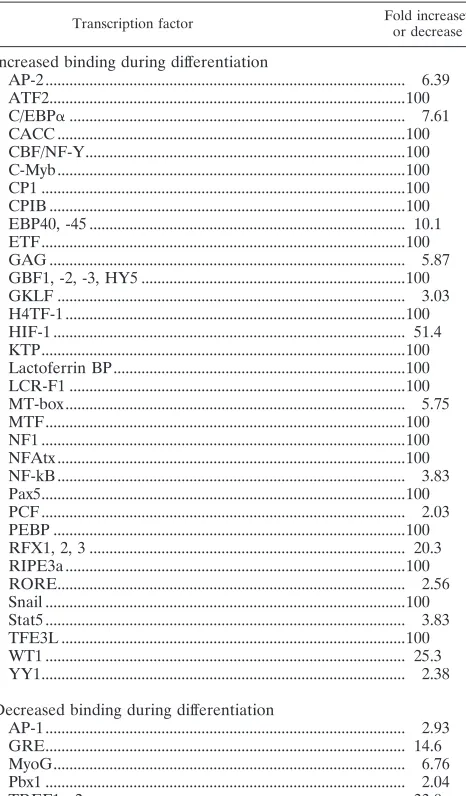
Panomics TranSignal array detects changes in the binding of known transcription factors to their binding sites. While this would generally indicate altered expression of TFs during dif-ferentiation, it may also be due to changes in the activities of TFs (for example, due to protein modifications). Therefore, we also carried out Western blot analyses for a few representative TFs to determine if the increases in their binding activities during differentiation as determined by using the Panomics array are correlated with increased protein levels. Figure 3 is a representation of three independent experiments and shows that the levels of C-Myb, NF1, Pax5, and WT1 were dramati-cally increased upon differentiation, while the levels of C/EBP␣ and NFATx showed only a modest increase. These results showed that the expression of several TFs that bind to HPV-16 promoter/ regulatory regions is upregulated during differentiation.
Binding of transcription factors to the HPV-16 DNA in vivo.
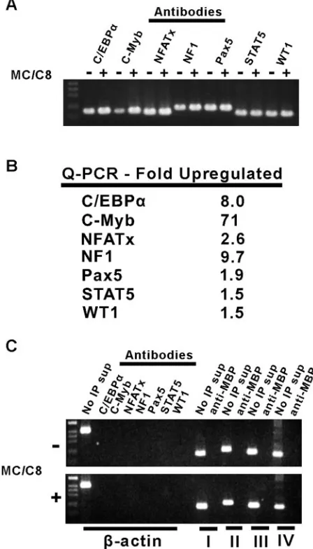
ChIP experiments were carried out to determine whether some of the TFs identified in the above screen bind to the HPV-16 DNA in vivo. For this purpose, proteins bound to DNA in the 20863 cell line with or without differentiation were cross-linked with formaldehyde, and specific DNA-protein complexes were isolated using antibodies against selected transcription factors as described in Materials and Methods. The DNA bound to
[image:4.585.112.469.72.160.2]proteins was recovered, and the bound HPV sequences were identified by standard PCR analysis as shown in Fig. 4A. In order to quantify the amount of HPV DNA present, the above samples were also analyzed by quantitative PCR using primer pairs near the predicted binding sites for the above transcrip-tion factors (see Fig. 2). The relative amounts of bound DNA are expected to represent the relative levels of specific TFs bound to the viral DNA in differentiated versus undifferenti-ated cells. The data, which are averages of three experiments, showed that C/EBP␣, C-Myb, and NF1 bound to the HPV-16 DNA in differentiated cells to substantially higher levels than in undifferentiated cells, with increases of 8-, 71-, and 9.7-fold, respectively (Fig. 4B). NFATx, Pax5, Stat5, and WT1 showed more-moderate increases in binding to HPV sequences upon differentiation (2.6-, 1.9-, 1.5-, and 1.5-fold, respectively). In control experiments, the use of template DNA immunopre-cipitated using antibodies specific to various TFs failed to am-plify the-actin gene (Fig. 4C). In another control, we used anti-maltose binding protein (MBP) antibodies in the above experiments, and the “mock-precipitated” DNA was used as the template for PCR amplification using HPV-16 primer pairs I, II, III, and IV, described above. No amplified DNA was obtained (Fig. 4C). The above ChIP assays identified TFs that bind to HPV-16 regulatory regions in vivo and also revealed FIG. 2. Putative TF binding sites in the HPV-16 promoters and upstream regions (nt 7005 to 7904 and nt 1 to 875). TFs found to bind to HPV-16 sequences in this study are indicated by asterisks. The binding sites for some TFs previously shown to bind to HPV-16 are also indicated. The p97 and p670 promoters are indicated, along with the locations of the primers used for the PCR and quantitative PCR for which results are shown in Fig. 4.
FIG. 3. Western blot analysis of TFs in differentiated and undiffer-entiated cells. Whole-cell lysates were subjected to Western blot anal-ysis using the appropriate antibodies as described in Materials and Methods. MC, methylcellulose.
on November 8, 2019 by guest
http://jvi.asm.org/
[image:4.585.361.482.523.682.2]the TFs whose binding to the viral DNA is increased upon differentiation.
DISCUSSION
We have used W12 clone 20863 to identify changes in TF binding to HPV-16 promoter/regulatory regions during host cell differentiation using the Panomics TransSignal array. The 20863 cell line harbors HPV-16 episomally and can be induced
to differentiate (Fig. 1). Our analysis identified changes (ⱖ 2-fold difference) in the binding of TFs to 150 consensus se-quences during differentiation out of the 345 sese-quences present on the array. These changes in TF binding may either represent altered expression, activity, or both. Competition experiments using the Panomics TransSignal arrays revealed 38 TFs present in differentiated 20863 cells with increased binding to the pUC19/ HPV-16 DNA, while 9 TFs in differentiated cells showed a de-crease in binding to this DNA (Table 1). Since TF consensus sequences may also be present randomly in the HPV-16 genome or the vector pUC19 sequence and may have no role in transcrip-tion, we also used a 1.7-kb PstI fragment of HPV-16 (nt 7005 to 7816 and nt 1 to 875) which contains all the known viral promoter/ regulatory elements as a competitor in the above experiments. These results identified 31 TFs with increased binding to the above 1.7-kb region, while 5 TFs showed reduced binding upon differentiation (Table 1). These TFs were a subset of those iden-tified when the pUC19/HPV-16 DNA was used as the competitor. We then analyzed the above 1.7-kb region for the presence of putative binding sites for known TFs using the TRANSFAC da-tabase (36). A majority, but not all, of the TFs identified by the Panomics array have identifiable binding sites in this region (Fig. 2 and data not shown). The putative binding sites of many of the above TFs are located in regions associated with late promoters, and these factors may play a role in controlling late gene expres-sion. These poorly characterized promoters (with transcription start sites located between nt 200 and 700 of the HPV-16 ge-nome) are thought to be dramatically upregulated during host cell differentiation compared to the slight upregulation of the p97 promoter (14, 27). A plausible hypothesis is that this upregulation of the late promoters is due to increased binding of stimulatory TFs (including those identified in this study) during differentia-tion. It is interesting, therefore, that while approximately similar numbers of TFs show increased and decreased binding to their consensus sequences during differentiation (65 and 85, respec-tively), the vast majority of TFs that interact with the regulatory regions of HPV-16 show increased binding activity during differ-entiation (Table 1). Many of these factors are known to stimulate transcription and may play a role in the upregulation of late promoters during the viral life cycle.
[image:5.585.49.273.75.467.2]Several of the TFs found in this study to be upregulated during differentiation, such as C/EBP␣and NF1, have previ-ously been shown to affect HPV-16 transcription (2, 7, 10, 15). Such known factors provide a validation of the approach we have used in this study, which also identified several TFs not previously implicated in the regulation of HPV gene expres-sion. Our data also expand the knowledge of previously known interactions between HPVs and TFs. For C/EBP␣, this repre-sents the first in vivo evidence that this factor binds to the HPV-16 DNA and the first demonstration that this binding and protein expression increase during differentiation. The dramatically increased DNA binding activity of NF1 during differentiation is most likely due to a significant increase in its expression, as shown by Western blot analysis (Fig. 3). NF1 has been shown to upregulate the p97 promoter through its mul-tiple binding sites located in this region (2, 7). Many other TFs such as Stat5 have been loosely associated with HPV infection or HPV-related malignancies but were not known to be di-rectly involved in transcription of the viral genome (6, 16, 21, 26). The activity of Stat5 protein was consistently increased in FIG. 4. ChIP and quantitative PCR (Q-PCR) analysis of TF binding.
(A) Nonquantitative PCR analysis of HPV-16 DNA recovered from im-munoprecipitates from 20863 cell lysates using the indicated antibodies.⫺ and⫹, absence and presence, respectively, of methylcellulose (MC) and C8. (B) Relative increases in the binding of various TFs to HPV-16 DNA in 20863 cells upon differentiation. (C) Control experiments demonstrat-ing the specificity of the antibodies used in the ChIP assays. In the left half of each panel, a region of the-actin open reading frame with no known TF binding activity was PCR amplified from ChIP samples using the indicated antibodies. The right half of each panel shows that the HPV-16 PCR primer pairs I, II, III, and IV (see Materials and Methods) do not amplify DNA from material obtained after immunoprecipitation by using the control anti-MBP antibodies. No IP sup, supernatant obtained prior to immunoprecipitation.
on November 8, 2019 by guest
http://jvi.asm.org/
both oligonucleotide binding, as determined by the Panomics array, and in vivo binding to the HPV-16 genome, as assayed by ChIP (Table 1 and Fig. 4). Our results further support a role for Stat5 in HPV gene expression during viral maturation or virus-induced carcinogenesis.
We have identified several TFs in this study and provide the first in vivo evidence for their binding to HPV-16 DNA as well as for changes in their binding to the viral genome during differentiation. For example, binding of C-Myb, Pax5, NFATx, and WT1 to the HPV-16 regulatory region represents novel interactions. The Wilms’ tumor gene product (WT1) is known to be associated with tumor progression (20), and its interac-tion with the HPV-16 promoter regions could represent an additional mechanism by which this factor may promote car-cinogenesis. TFs such as C-Myb, NFATx, and Pax5 are known to play important roles in development and/or differentiation (18, 28, 33). Expression of the C-Myb and Pax5 protein was not detectable in the 20863 epithelial cells before differentiation but was induced during differentiation, as shown by Western blot analysis (Fig. 3). While Western blot analysis showed a limited increase in the levels of NFATx during differentiation (Fig. 3), its DNA binding activity was significantly increased as determined by the Panomics array and ChIP assays (Table 1 and Fig. 4). This could be due to changes in nuclear local-ization, since NFATx activity is known to be regulated in this manner (33). It is likely that HPV-16 and possibly other HPVs have adapted to utilize many cellular transcription factors involved in differentiation and development to reg-ulate their gene expression during host cell differentiation in epithelial cells.
The relative changes in TF levels or activity during differ-entiation as identified by the Panomics array, ChIP assay, and Western blot analysis were variable. These variations may rep-resent differences in the sensitivity of the assays or increased activity resulting from protein modifications and/or subcellular localization, or they may be due to some unknown factors. The fact that we have validated our data by carrying out detailed analyses of a select number of TFs identified using the Panomics array increases the confidence that other TFs identified using this array may also interact with HPV-16 sequences and their expres-sion/activity may also be altered during differentiation.
Taken together, our results as well as those of previous studies by other investigators suggest that HPVs may have evolved to utilize many changes in transcription factor binding and expression during differentiation so as to achieve tight regulation of the late viral genes. Many such factors are in-volved in differentiation, development, and/or carcinogenesis and should be targets for further study. In the future, we plan to confirm interactions of additional TFs with HPVs and to determine the functional significance of many of the novel transcription factors in HPV gene expression and HPV-asso-ciated disease.
ACKNOWLEDGMENTS
This work was supported by Public Health Service grant GM-51861 from the National Institute of General Medical Sciences to S.A.K. A.C. was supported by a National Science Foundation Graduate Fellowship.
REFERENCES
1.Ai, W., E. Toussaint, and A. Roman.1999. CCAAT displacement protein binds to and negatively regulates human papillomavirus type 6 E6, E7, and E1 promoters. J. Virol.73:4220–4229.
2.Apt, D., T. Chong, Y. Liu, and H. U. Bernard.1993. Nuclear factor I and epithelial cell-specific transcription of human papillomavirus type 16. J. Vi-rol.67:4455–4463.
3.Apt, D., R. M. Watts, G. Suske, and H. U. Bernard.1996. High Sp1/Sp3 ratios in epithelial cells during epithelial differentiation and cellular trans-formation correlate with the activation of the HPV-16 promoter. Virology
224:281–291.
4.Bodily, J. M., and C. Meyers.2005. Genetic analysis of the human papillo-mavirus type 31 differentiation-dependent late promoter. J. Virol.79:3309– 3321.
5.Chan, W. K., T. Chong, H. U. Bernard, and G. Klock.1990. Transcription of transforming genes of the oncogenic human papillomavirus type-16 is stim-ulated by tumor promoters through AP1 binding sites. Nucleic Acids Res.
18:763–769.
6.Chang, Y. E., and L. A. Laimins.2000. Microarray analysis identifies inter-feron-inducible genes and Stat-1 as major transcriptional targets of human papillomavirus type 31. J. Virol.74:4174–4182.
7.Chong, T., D. Apt, B. Gloss, M. Isa, and H. U. Bernard.1991. The enhancer of human papillomavirus type 16: binding sites for the ubiquitous transcrip-tion factors Oct-1, NFA, TEF-2, NF1, and AP1 participate in epithelial cell-specific transcription. J. Virol.65:5933–5943.
8.Ciccolini, F., G. Di Pasquale, F. Carlotti, L. Crawford, and M. Tommasino.
1994. Functional studies of E7 proteins from different HPV types. Oncogene
9:2633–2638.
9.Dell, G., and K. Gaston.2001. Contributions in the domain of cancer re-search: review of human papillomaviruses and their role in cervical cancer. Cell. Mol. Life Sci.58:1923–1942.
10.Dong, X. P., F. Stubenrauch, E. Beyer-Finkler, and H. Pfister.1994. Preva-lence of deletions of YY1-binding sites in episomal HPV 16 DNA from cervical cancers. Int. J. Cancer58:803–808.
11.Fehrmann, F., and L. A. Laimins.2003. Human papillomaviruses: targeting differentiating epithelial cells for malignant transformation. Oncogene22:
5201–5207.
12.Flores, E. R., and P. F. Lambert.1997. Evidence for a switch in the mode of human papillomavirus type 16 DNA replication during the viral life cycle. J. Virol.71:7167–7179.
13.Gloss, B., and H. U. Bernard.1990. The E6/E7 promoter of human papilloma-virus type 16 is activated in the absence of E2 proteins by a sequence-aberrant SP1 distal element. J. Virol.64:5577–5584.
14.Grassmann, K., B. Rapp, H. Maschek, K. U. Petry, and T. Iftner.1996. Identification of a differentiation-inducible promoter in the E7 open reading frame of human papillomavirus type 16 (HPV-16) in raft cultures of a new cell line containing high copy numbers of episomal HPV-16 DNA. J. Virol.
70:2339–2349.
15.Hadaschik, D., K. Hinterkeuser, M. Oldak, H. J. Pfister, and S. Smola-Hess.
2003. The papillomavirus E2 protein binds to and synergizes with C/EBP factors involved in keratinocyte differentiation. J. Virol.77:5253–5265. 16.Havard, L., S. Rahmouni, J. Boniver, and P. Delvenne.2005. High levels of
p105 (NFKB1) and p100 (NFKB2) proteins in HPV16-transformed kerati-nocytes: role of E6 and E7 oncoproteins. Virology331:357–366.
17.Hebner, C. M., and L. A. Laimins.2005. Human papillomaviruses: basic mechanisms of pathogenesis and oncogenicity. Rev. Med. Virol.16:83–97. 18.Hirokawa, S., H. Sato, I. Kato, and A. Kudo.2003. EBF-regulating Pax5
transcription is enhanced by STAT5 in the early stage of B cells. Eur. J. Immunol.7:1824–1829.
19.Hirt, B.1967. Selective extraction of polyoma DNA from infected mouse cell cultures. J. Mol. Biol.26:365–369.
20.Lee, S. B., and D. A. Haber.2001. Wilms tumor and the WT1 gene. Exp. Cell Res.264:74–99.
21.Nees, M., J. M. Geoghegan, T. Hyman, S. Frank, L. Miller, and C. D. Woodworth.2001. Papillomavirus type 16 oncogenes downregulate expres-sion of interferon-responsive genes and upregulate proliferation-associated and NF-B-responsive genes in cervical keratinocytes. J. Virol.75:4283– 4296.
22.O’Connor, M. J., W. Stunkel, C. H. Koh, H. Zimmermann, and H. U. Bernard.2000. The differentiation-specific factor CDP/Cut represses tran-scription and replication of human papillomaviruses through a conserved silencing element. J. Virol.74:401–410.
23.Ozbun, M. A.2002. Human papillomavirus type 31b infection of human keratinocytes and the onset of early transcription. J. Virol.76:11291–11300. 24.Ozbun, M. A., and C. Meyers.1999. Two novel promoters in the upstream regulatory region of human papillomavirus type 31b are negatively regulated by epithelial differentiation. J. Virol.73:3505–3510.
25.Pattison, S., D. G. Skalnik, and A. Roman.1997. CCAAT displacement protein, a regulator of differentiation-specific gene expression, binds a neg-ative regulatory element within the 5⬘end of the human papillomavirus type 6 long control region. J. Virol.71:2013–2022.
on November 8, 2019 by guest
http://jvi.asm.org/
26.Rocha-Zavaleta, L., C. Huitron, J. R. Caceres-Cortes, J. A. Alvarado-Moreno, A. Valle-Mendiola, I. Soto-Cruz, B. Weiss-Steider, and R. Rangel-Corona. 2004. Interleukin-2 (IL-2) receptor-␥signalling is activated by c-Kit in the absence of IL-2, or by exogenous IL-2 via JAK3/STAT5 in human papillomavirus-associated cervical cancer. Cell. Signal. 16:1239– 1247.
27.Rosenstierne, M. W., J. Vinther, C. N. Hansen, M. Prydsoe, and B. Norrild.
2003. Identification and characterization of a cluster of transcription start sites located in the E6 ORF of human papillomavirus type 16. J. Gen. Virol.
84:2909–2920.
28.Rushton, J. J., L. M. Davis, W. Lei, X. Mo, A. Leutz, and S. A. Ness.2003. Distinct changes in gene expression induced by A-Myb, B-Myb and c-Myb proteins. Oncogene22:308–313.
29.Sambrook, J., E. F. Fritsch, and T. Maniatis.1989. Molecular cloning: a laboratory manual, 2nd ed. Cold Spring Harbor Laboratory Press, Cold Spring Harbor, N.Y.
30.Scheffner, M., J. M. Huibregtse, R. D. Vierstra, and P. M. Howley.1993. The HPV-16 E6 and E6-AP complex functions as a ubiquitin-protein ligase in the ubiquitination of p53. Cell75:495–505.
31.Sen, E., S. Alam, and C. Meyers.2004. Genetic and biochemical analysis of
cisregulatory elements within the keratinocyte enhancer region of the
hu-man papillomavirus type 31 upstream regulatory region during different stages of the viral life cycle. J. Virol.78:612–629.
32.Sen, E., J. L. Bromberg-White, and C. Meyers.2002. Genetic analysis ofcis
regulatory elements within the 5⬘region of the human papillomavirus type 31 upstream regulatory region during different stages of the viral life cycle. J. Virol.76:4798–4809.
33.Serfling, E., F. Berberich-Siebelt, S. Chuvpilo, E. Jankevics, S. Klein-Hessling, T. Twardzik, and A. Avots.2000. The role of NF-AT transcription factors in T cell activation and differentiation. Biochim. Biophys. Acta1498:1–18. 34.Spink, K. M., and L. A. Laimins.2005. Induction of human papillomavirus
type 31 late promoter requires differentiation but not DNA amplification. J. Virol.79:4918–4926.
35.Sverdrup, F., and S. A. Khan.1994. Replication of human papillomavirus (HPV) DNAs supported by the HPV type 18 E1 and E2 proteins. J. Virol.
68:505–509.
36.Wingender, E., X. Chen, R. Hehl, H. Karas, I. Liebich, V. Matys, T. Meinhardt, M. Pruss, I. Reuter, and F. Schacherer.2000. TRANSFAC: an integrated system for gene expression regulation. Nucleic Acids Res.
28:316–319.
37.Yukawa, K., K. Butz, T. Yasui, H. Kikutani, and F. Hoppe-Seyler.1996. Regulation of human papillomavirus transcription by the differentiation-dependent epithelial factor Epoc-1/skn-1a. J. Virol.70:10–16.