Welcome To GetPedia.com :
The Online Information Resource.
Search GetPedia
Business
Advertising
Branding
Business Management
Business Ethics
Careers, Jobs & Employment
Customer Service
Marketing
Networking
Network Marketing
Pay-Per-Click Advertising
Presentation
Public Relations
Sales
Sales Management
Sales Telemarketing
Sales Training
Small Business
Strategic Planning
Entrepreneur
Negotiation Tips
Team Building
Top Quick Tips
Internet & Businesses Online
Affiliate Revenue
Blogging, RSS & Feeds
Domain Name
E-Book
E-commerce
Email Marketing
Ezine Marketing
Ezine Publishing
Forums & Boards
Internet Marketing
Online Auction
Search Engine Optimization
(SEO)
Spam Blocking
Streaming Audio & Online
Music
Traffic Building
Video Streaming
Web Design
Web Development
Web Hosting
Web Site Promotion
Finance
Credit
Currency Trading
Debt Consolidation
Debt Relief
Loan
Insurance
Investing
Mortgage Refinance
Personal Finance
Real Estate
Taxes
Stocks & Mutual Fund
Structured Settlements
Leases & Leasing
Wealth Building
Communications
Broadband Internet
Mobile & Cell Phone
VOIP
Video Conferencing
Satellite TV
Reference & Education
Book Reviews
College & University
Psychology
Science Articles
Food & Drinks
Coffee
Cooking Tips
Recipes & Food and Drink
Wine & Spirits
Home & Family
Crafts & Hobbies
Elder Care
Holiday
Home Improvement
Home Security
Interior Design & Decorating
Landscaping & Gardening
Babies & Toddler
Pets
Parenting
Pregnancy
News & Society
Dating
Divorce
Marriage & Wedding
Political
Relationships
Religion
Sexuality
Computers & Technology
Computer Hardware
Data Recovery & Computer
Backup
Game
Internet Security
Personal Technology
Software
Arts & Entertainment
Casino & Gambling
Humanities
Humor & Entertainment
Language
Music & MP3
Philosophy
Photography
Poetry
Shopping & Product Reviews
Book Reviews
Fashion & Style
Health & Fitness
Acne
Aerobics & Cardio
Alternative Medicine
Beauty Tips
Depression
Diabetes
Exercise & Fitness
Fitness Equipment
Hair Loss
Medicine
Meditation
Muscle Building & Bodybuilding
Nutrition
Nutritional Supplements
Weight Loss
Yoga
Recreation and Sport
Fishing
Golf
Martial Arts
Motorcycle
Self Improvement & Motivation
Attraction
Coaching
Creativity
Dealing with Grief & Loss
Finding Happiness
Get Organized - Organization
Leadership
Motivation
Inspirational
Positive Attitude Tips
Goal Setting
Innovation
Spirituality
Stress Management
Success
Time Management
Writing & Speaking
Article Writing
Book Marketing
Copywriting
Public Speaking
Writing
Travel & Leisure
Aviation & Flying
Cruising & Sailing
Outdoors
Vacation Rental
Cancer
Breast Cancer
Mesothelioma & Asbestos
Cancer
Copyright © 2006
GetPedia | Links
Works!
Search GetPedia
Demystified Series
Advanced Statistics Demystified Algebra DemystifiedAnatomy Demystified Astronomy Demystified Biology Demystified Biotechnology Demystified Business Statistics Demystified Calculus Demystified
Chemistry Demystified College Algebra Demystified Differential Equations Demystified Digital Electronics Demystified Earth Science Demystified Electricity Demystified Electronics Demystified
Environmental Science Demystified Everyday Math Demystified
Geometry Demystified Math Proofs Demystified
Math Word Problems Demystified Microbiology Demystified
Physics Demystified Physiology Demystified Pre-Algebra Demystified Precalculus Demystified Probability Demystified
Project Management Demystified Quantum Mechanics Demystified Relativity Demystified
MICROBIOLOGY DEMYSTIFIED
TOM BETSY, D.C.
JIM KEOGH
McGRAW-HILL
Copyright © 2005 by The McGraw-Hill Companies, Inc. All rights reserved. Manufactured in the United States of America. Except as permitted under the United States Copyright Act of 1976, no part of this publication may be reproduced or distributed in any form or by any means, or stored in a database or retrieval system, without the prior written permission of the publisher.
0-07-147134-0
The material in this eBook also appears in the print version of this title: 0-07-144650-8.
All trademarks are trademarks of their respective owners. Rather than put a trademark symbol after every occurrence of a trademarked name, we use names in an editorial fashion only, and to the benefit of the trademark owner, with no intention of infringement of the trademark. Where such designations appear in this book, they have been printed with initial caps. McGraw-Hill eBooks are available at special quantity discounts to use as premiums and sales promotions, or for use in corporate training programs. For more information, please contact George Hoare, Special Sales, at george_hoare@mcgraw-hill.com or (212) 904-4069.
TERMS OF USE
This is a copyrighted work and The McGraw-Hill Companies, Inc. (“McGraw-Hill”) and its licensors reserve all rights in and to the work. Use of this work is subject to these terms. Except as permitted under the Copyright Act of 1976 and the right to store and retrieve one copy of the work, you may not decompile, disassemble, reverse engineer, reproduce, modify, create derivative works based upon, transmit, distribute, disseminate, sell, publish or sublicense the work or any part of it without McGraw-Hill’s prior consent. You may use the work for your own noncommercial and personal use; any other use of the work is strictly prohibited. Your right to use the work may be terminated if you fail to comply with these terms.
THE WORK IS PROVIDED “AS IS.” McGRAW-HILL AND ITS LICENSORS MAKE NO GUARANTEES OR WARRANTIES AS TO THE ACCURACY, ADEQUACY OR COMPLETENESS OF OR RESULTS TO BE OBTAINED FROM USING THE WORK, INCLUDING ANY INFORMATION THAT CAN BE ACCESSED THROUGH THE WORK VIA HYPERLINK OR OTHERWISE, AND EXPRESSLY DISCLAIM ANY WARRANTY, EXPRESS OR IMPLIED, INCLUDING BUT NOT LIMITED TO IMPLIED WARRANTIES OF MERCHANTABILITY OR FITNESS FOR A PARTICULAR PURPOSE. McGraw-Hill and its licensors do not warrant or guarantee that the functions contained in the work will meet your requirements or that its operation will be uninterrupted or error free. Neither McGraw-Hill nor its licensors shall be liable to you or anyone else for any inaccuracy, error or omission, regardless of cause, in the work or for any damages resulting there from. McGraw-Hill has no responsibility for the content of any information accessed through the work. Under no circumstances shall McGraw-Hill and/or its licensors be liable for any indirect, incidental, special, punitive, consequential or similar damages that result from the use of or inability to use the work, even if any of them has been advised of the possibility of such damages. This limitation of liability shall apply to any claim or cause whatsoever whether such claim or cause arises in contract, tort or otherwise.
������������
Want to learn more?
I would like to dedicate this book to my wife, Shelley,
and my two babies, Juliana and Thomas,
for their encouragment during the writing of this book
and the continued joy they bring to my life.
Dr. Tom Betsy
This book is dedicated to Anne, Sandy, Joanne,
Amber-Leigh Christine, and Graaf,
without whose help and support
this book could not have been written.
Introduction
xiii
Acknowledgments
xix
CHAPTER 1
The World of the Microorganism
1
Types of Microorganisms
2
What Is a Microorganism?
4
What’s in a Name: Naming and Classifying
5
How Small Is a Microorganism?
7
Your Body Fights Back
7
History of the Microscope
9
How Do Organisms Appear?
10
Germ Theory
12
Vaccination
15
Killing the Microorganism
16
Quiz
19
CHAPTER 2
The Chemical Elements of Microorganisms
23
Everything Matters
24
Chemical Elements and the Atom
24
A Dinner Table of Elements:
The Periodic Table
27
The Glowing Tale of Isotopes
27
Around They Go: Electronic Configuration
29
Before James There Was Bond . . .
Chemical Bond
29
Decoding Chemical Shorthand
31
vii
I Just Want to See Your Reaction
31
Molarity: Hey, There’s a Mole Amongst Us
36
An Unlikely Pair: Inorganic and Organic
37
The Blueprint of Protein Synthesis
42
The Power House: ATP
44
Quiz
44
CHAPTER 3
Observing Microorganisms
47
Size Is a Matter of Metrics
47
Here’s Looking at You
51
What Big Eyes You Have: Magnification
52
The Microscope
54
Preparing Specimens
60
Quiz
64
CHAPTER 4
Prokaryotic Cells and Eukaryotic Cells
67
Prokaryotic Cells
68
Eukaryotic Cells
78
Quiz
83
CHAPTER 5
The Chemical Metabolism
87
Riding the Metabolism Cycle
87
Catabolic and Anabolic: The Only Reactions
You Need
88
A Little Give and Take: Oxidation-Reduction
88
Making Power: ATP Production
89
What’s Your Name: Naming and
Classifying Enzymes
89
Brewing Up Protein
90
The Magic of Enzymes: Enzyme Activities
90
The Krebs Cycle
94
Fermentation
97
Other Catabolic Pathways
97
Photosynthesis
98
CHAPTER 6
Microbial Growth and Controlling
Microbial Growth
103
Chemical Requirements for Microbial
Growth
104
Culture Media
105
Phases of Growth
109
Measurements of Microbial Growth
110
Establishing Bacterial Numbers
by Indirect Methods
111
Controlling Microbial Growth
112
Microbial Death Rates
113
Action of Antimicrobial Agents
113
Chemical Agents That Control Microbial
Growth
114
Quiz
116
CHAPTER 7
Microbial Genetics
119
Genetics
120
DNA Replication: Take My Genes, Please!
120
Protein Synthesis
123
Controlling Genes: You’re Under My Spell
124
Mutations: Not a Pretty Copy
125
Quiz
127
CHAPTER 8
Recombinant DNA Technology
131
Genetic Engineering: Designer Genes
132
Gene Therapy: Makes You Feel Better
134
DNA Fingerprinting: Gotcha
134
Recombinant DNA Technology and Society:
Too Much of a Good Thing
136
Quiz
137
CHAPTER 9
Classification of Microorganisms
139
Taxonomy: Nothing to Do with the IRS
139
Nomenclature of Taxonomy: Name Calling
140
Classification: All Natural
146
Quiz
148
CHAPTER 10
The Prokaryotes: Domains Archaea and
Bacteria
151
Archaea
152
Aerobic/Microaerophilic, Motile,
Helical/Vibroid, Gram-Negative Bacteria
154
Gram-Negative Aerobic Rods and Cocci
155
Facultatively Anaerobic Gram-Negative Rods 156
Anaerobic Gram-Negative Cocci and Rods
158
Rickettsias and Chlamydias
158
Mycoplamas
159
Gram-Positive Cocci
160
Endospore-Forming Gram-Positive Rods
and Cocci
162
Regular Nonsporing Gram-Positive Rods
163
Irregular Nonsporing Gram-Positive Rods
164
Mycobacteria
164
Nocardia Forms
165
Quiz
165
CHAPTER 11
The Eukaryotes: Fungi, Algae, Protozoa,
and Helminths
167
Fungi
168
Algae
171
Protozoa
174
Helminths
178
Quiz
182
CHAPTER 12
Viruses, Viroids, and Prions
185
Viruses
185
Viroids
192
Prions
193
CHAPTER 13
Epidemiology and Disease
197
What Is Epidemiology?
197
Classification of Disease
198
Infection Sites
199
Disease Transmission
201
The Development of Disease
203
Epidemiological Studies
204
Control of Communicable Diseases
205
Nosocomial Infections
206
Who Is Susceptible?
206
Prevention and Control of Nosocomial
Infections
207
Quiz
207
CHAPTER 14
Immunity
211
What Is Immunity?
211
Types of Immunity
213
A Closer Look at Antigens
214
A Closer Look at Binding
214
B Cells
216
Lasting Immunity
218
Antibodies Used for Diagnosing Diseases
218
Chemical Messengers
218
T Cells
219
Macrophages and Natural Killer Cells
220
Quiz
221
CHAPTER 15
Vaccines and Diagnosing Diseases
223
What Is a Vaccine?
223
Types of Vaccines
224
Developing a Vaccine
225
Diagnosing Diseases
226
Quiz
228
CHAPTER 16
Antimicrobial Drugs
231
Chemotherapeutic Agents: The Silver Bullet
231
Antimicrobial Activity: Who to Attack?
233
The Attack Plan
235
Exploring Antimicrobial Drugs
237
Chemotherapy Tests
246
Quiz
247
Final Exam
249
Answers to Quiz and Exam Questions
265
xiii
When you hear the words “germ,” “bacteria,” and “virus” you might cringe, run-ning for the nearest sink to wash your hands. These words may bring back mem-ories of when you caught a cold or the flu—never a pleasant experience. Germs, bacteria, viruses and other microscopic organisms are called microorganisms, or microbes for short. And as you’ll learn throughout this book, some microbes cause disease while others help fight it.
Think for a moment. Right now there are thousands of tiny microbes living on the tip of your finger in a world that is so small that it can only be visited by using a microscope. In this book we’ll show you how to visit this world and how to interact with these tiny creatures that call the tip of your finger home.
The microscopic world was first visited in the late 1600s by the Dutch mer-chant and amateur scientist Antoni van Leeuwenhoek. He was able to see living microorganisms by using a single-lens microscope. We’ve come a long way since Van Leeuwenhoek’s first visit. Today scientists are able to see through
some microbes and study the organelles that bring them to life.
It wasn’t until the Golden Age of Microbiology between 1857 and 1914 when scientists such as Louis Pasteur and Robert Koch made a series of discoveries that rocked the scientific community. During this period scientists identified microbes that caused diseases, learned how to cure those diseases, and then pre-vented them from occurring through the use of immunization.
Scientists were able to achieve these remarkable discoveries by using cultur-ing techniques to grow colonies of microbes in the laboratory. Once microbes could be grown at will, scientists focused their experiments on ways to slow that growth and stop microbes in their tracks—killing the microbe and curing the dis-ease caused by the microbe.
Culturing microbes is central to the study of microbiology. You’ll be using many of the same culturing techniques described in this book to colonize microbes in your college laboratory. We provide step-by-step instructions on how to do this.
xiii
You would find it difficult to live without the aid of microbes. For example, living inside your intestines are colonies of microorganisms. Just this thought is enough to make your skin crawl. As frightful as this thought might be, however, these microbes actually assist your body in digesting food. That is, you might have difficulty digesting some foods if these microbes did not exist.
Microbes in your intestines are beneficial to you as long as they remain in your intestines. However, you’ll become very ill should they decide to wander into other parts of your body. Don’t become too concerned—these microbes tend to stay at home unless your intestines are ruptured as a result of trauma.
By the end of this book you’ll learn about the different types of microbes, how to identify them by using a microscope, and how to cultivate colonies of microbes.
A Look Inside
Microbiology can be challenging to learn unless you follow the step-by-step approach that is used in Microbiology Demystified.Topics are presented in an order in which many students like to learn them—starting with basic compo-nents and then gradually moving on to those that are more complex.
Each chapter follows a time-tested formula that explains topics in an easy-to-read style. You can then compare your knowledge with what you’re ex-pected to know by taking chapter tests and the final exam. There is little room for you to go adrift.
CHAPTER 1: THE WORLD OF THE MICROORGANISM
You’ll begin your venture into the microscopic world of microbes by learning the fundamentals. These are the terms and concepts that all students need to understand before they can embark on more advanced topics, such as cultivat-ing their own microbes.
CHAPTER 2: THE CHEMICAL ELEMENTS
OF MICROORGANISMS
Chemistry is a major factor in microbiology because microbes are made up of chemical elements. Scientists are able to destroy microbes by breaking them down into their chemical elements and then disposing of those elements.
Before you can understand how this process works, you must be familiar with the chemical principles related to microbiology. You’ll learn about these chemi-cal principles in this chapter.
CHAPTER 3: OBSERVING MICROORGANISMS
“Wash the germs from your hands!” That was the cry of every mom who knew that hand washing is the best way to prevent sickness. Most kids balked at hand washing simply because they couldn’t see the germs on their hands.
We’ll show you how to see germs and other microbes in this chapter by using a microscope. You’ll learn everything you need to know to bring microbes into clear focus so you can see with a microscope what you can’t see with the naked eye.
CHAPTER 4: PROKARYOTIC CELLS AND EUKARYOTIC
CELLS
It is time to get down and personal with two common microbe cells. These are prokaryotic cells and eukaryotic cells. These names are probably unfamiliar to you, but they won’t be by the time you’re finished reading this chapter.
Prokaryotic cells are bacteria cells and eukaryotic cells are cells of animals, plants, algae, fungi, and protozoa. Each carries out the six life processes that all living things have in common. In this chapter you’ll learn about how prokary-otes and eukaryprokary-otes carry out these life processes.
CHAPTER 5: THE CHEMICAL METABOLISM
“It’s my slow metabolism! That’s why I can’t shed a few pounds.” This is a great excuse for being unable to lose weight, but the reason our metabolisms are slow is because we tend not to exercise enough.
In this chapter, you’ll learn about the biochemical reactions that change food into energy—collectively called metabolism—and how the cell is able to con-vert nutrients into energy.
CHAPTER 6: MICROBIAL GROWTH
AND CONTROLLING MICROBIAL GROWTH
You and microbes need nutrients to grow—chemical nutrients such as carbon, hydrogen, nitrogen, and oxygen. However, not all microbes need the same chemical nutrients. For example, some require oxygen while others can thrive in an oxygen-free environment.
You’ll learn in this chapter how to classify microbes by the chemical nutri-ents they need to survive. You’ll also learn how to use this knowledge to grow microbes and control their growth in the laboratory.
CHAPTER 7: MICROBIAL GENETICS
Just like us, microbes inherit genetic traits from their species’ previous genera-tions. Genetic traits are instructions on how to everything to stay alive. Some in-structions are passed along to the next generation while other inin-structions are not. In this chapter you’ll learn how microorganisms inherit genetic traits from previous generations of microorganisms. Some of these traits show them how to identify and process food, how to excrete waste products, and how to reproduce.
CHAPTER 8: RECOMBINANT DNA TECHNOLOGY
Who we are and what we are going to be is programmed into our genes. The same is true for microbes. This genetic information is encoded into DNA by the linking of nucleic acids in a specific sequence.
Genetic information can be reordered in a process called genetic engineering. You’ll learn about genetic engineering and how to recombinant DNA using DNA technology in this chapter.
CHAPTER 9: CLASSIFICATION OF MICROORGANISMS
There are thousands of microbes and no two are identical, but many have simi-lar characteristics. Microbiologists have spent years carefully observing microbes and organizing them into groups by their similarities.
CHAPTER 10: THE PROKARYOTES:
DOMAINS ARCHAEA AND BACTERIA
Bacteria are one of the most common microbes that you encounter. Some bacte-ria cause disease and other bactebacte-ria help you live by aiding in digestion. There are many different kinds of bacteria; however, all bacteria can be grouped into four divisions based on the characteristics of their cell walls.
Each division is further divided into sections based on other characteristics, such as oxygen requirements, motility, shape, and Gram-stain reaction. In this chapter, you’ll learn how to use these divisions and sections to identify bacteria.
CHAPTER 11: THE EUKARYOTES:
FUNGI, ALGAE, PROTOZOA, AND HELMINTHS
In this chapter you’ll take a close look at the kingdoms of fungi, protista, and animalia. These are microbes that are commonly known as fungi, algae, proto-zoa, and helminths.
Eukaryotes are a type of microbe and are different from bacteria and viruses. However, they, too, are beneficial to us. They supply food, remove waste, and cure disease (in the form of antibiotics). And as bacteria, some eukaryotes also cause disease.
CHAPTER 12: VIRUSES, VIROIDS, AND PRIONS
Probably one of the most feared microbes is a virus because often there is little or nothing that can be done to kill it. Once you’re infected, you can treat the symptoms, such as a runny nose and watery eyes, but otherwise you must let the virus run its course.
Did you ever wonder why this is case? If so, then read this chapter for the answer and learn what a virus is, how viruses live, and which diseases they cause.
CHAPTER 13: EPIDEMIOLOGY AND DISEASE
It’s flu season and you can only hope that you don’t become infected—otherwise you’ll have ten days of chills, sneezing, and isolation. No one will want to come close to you for fear of catching the flu.
In this chapter, you’ll learn about diseases like the flu and how diseases are spread. You’ll also learn how to take simple precautions to control and prevent the spread of diseases.
CHAPTER 14: IMMUNITY
Inside your body there is a war going on. An army of B cells, T cells, natural killer cells, and other parts of your immune system are on the defense. These cells seek microbes to rip apart before any of them can give you a runny nose, cough, or that dreaded feverish feeling.
The immune system is your body’s defense mechanism: Its “soldiers” sur-round, neutralize, and destroy foreign invaders before they can do harm. In this chapter you’ll learn about your immune system and how it gives you daily pro-tection against invading microbes.
CHAPTER 15: VACCINES AND DIAGNOSING DISEASES
Think about this: Each year millions of people pay their doctor to inject them with the flu virus. On the surface that may not make sense, but after reading this chapter you’ll find that it makes perfect sense because this injection is actually a vaccination against the flu.
A vaccine prevents you from catching a certain disease because it has ele-ments of that disease, triggering your body to create antibodies to the disease. You’ll learn about vaccines and antibodies in this chapter.
CHAPTER 16: ANTIMICROBIAL DRUGS
“Doc, give me a pill to knock out whatever is causing me to be sick!” All of us say this whenever we come down with an illness. All we want is a magic pill that makes us feel better. Sometimes that magic pill—or injection—contains a microbe that seeks out and destroys pathogenic microbes, which are disease-causing microbes.
I would like to acknowledge my mentor, colleague, and friend Professor Robert Highley for all his encouragement and technical support in the production of this book; my friend Ms. Joan Sisto for all her hours of computer work; and my friend and coauthor Professor Jim Keogh for asking me to help write this book. Thank you.
DR. TOMBETSY
Professor Robert Highley has done a magnificent job as technical editor on this project. His diligence and attention to detail has made Microbiology Demystified
a rewarding addition to every microbiology student’s library.
JIMKEOGH
xix
ACKNOWLEDGMENTS
1
CHAPTER
1
The World of the
Microorganism
Microbiology is the study of microorganisms, which are tiny organisms that live around us and inside our body. An organismis a living thing that ingests and breaks down food for energy and nutrients, excretes undigested food as waste, and is capable of reproduction. You are an organism and so are dogs, cats, insects, and other creatures that you see daily.
A microorganism is simply a very, very small organism that you cannot see with your naked eye, but you sure feel its effect whenever your eyes fill with water and mucus flows like an open faucet from your nose. You call it a head cold. Actually, you are under siege by an army of microorganisms attacking membranes inside your body. Watery eyes and a runny nose are ways that you fight microorganisms by flushing them out of your body.
Microorganisms are a key component of biological warfare along with chem-icals that disrupt homeostasis. The anthrax attack that followed the 9/11 terror-ist attacks clearly illustrated how a dusting of anthrax in an envelope can be lethal to people in an office building. Anthraxis a disease caused by the micro-organism Bacillus anthracis, a bacterium that forms endospores and infiltrates
the body by ingestion, skin contact, and inhalation. An endosporeis a bacterium in a dormant state that forms within a cell.
Fortunately, incidents of biological attacks using microorganisms have been infrequent. However, there are thousands of microorganisms all around us that can be just as deadly and debilitating as the microorganisms used in warfare throughout history.
Types of Microorganisms
UNFRIENDLY MICROORGANISMS
An infection is caused by the infiltration of a disease-causing microorganism known as a pathogenic microorganism. Some pathogenic microorganisms infect humans, but not other animals and plants. Some pathogenic microorganisms that infect animals or plants also infect humans.
[image:24.532.202.360.459.611.2]Pathogenic microorganisms make headlines and play an important role in his-tory. Legendary gunfighter John “Doc” Holliday is famous for his escapades in the Wild West. He dodged countless bullets, showing that he was the best of the best when it came to gun fighting. Yet Mycobacterium tuberculosistook down Doc Holliday quietly, without firing a shot. Mycobacterium tuberculosisis the bacterium that causes tuberculosis (Fig. 1-1). This bacterium affects the lung tis-sue when droplets of respiratory secretions or particles of dry sputum from a per-son who is infected with the disease are inhaled by an uninfected perper-son.
Yersinia pestis nearly conquered Europe in the fourteenth century with the help of the flea. Yersinia pestis is the microorganism that caused the Black Plague (Fig. 1-2) and killed more than 25 million Europeans. You might say that
Yersinia pestislaunched a sneak attack. First, it infected fleas that were carried into populated areas on the backs of rats. Rodents traveled on ships and then over land in search of food. Fleas jumped from rodents and bit people, trans-mitting the Yersinia pestismicroorganism into the person’s blood stream.
In an effort to prevent the spread of Yersinia pestis, sailors entering Sicily’s seaports had to wait 40 days before leaving the ship. This gave time for sailors to exhibit the symptoms of the Black Plague if the Yersinia pestis microorgan-ism had infected them. Sicilians called this quarantenaria. Today we know it as quarantine. Sailors who did not exhibit these symptoms were not infected and free to disembark.
Campers and travelers sometimes become acquainted with Giardia lamblia, Escherichia coli, or Entameba histolyticawhenever they visit tropical countries. Travelers who become infected typically do not die but come down with a bad case of diarrhea.
FRIENDLY MICROORGANISMS
Not all microorganisms are pathogens. In fact many microorganisms help to maintain homeostasis in our bodies and are used in the production of food and other commercial products. For example, flora are microorganisms found in our intestines that assist in the digestion of food and play a critical role in the formation of vitamins such as vitamin B and vitamin K. They help by breaking down large molecules into smaller ones.
CHAPTER 1
The World of the Microorganism
3
What Is a Microorganism?
Microorganisms are the subject of microbiology,which is the branch of science that studies microorganisms. A microorganism can be one cell or a cluster of cells that can be seen only by using a microscope.
Microorganisms are organized into six fields of study: bacteriology, virology, mycology, phycology, protozoology, and parasitology.
BACTERIOLOGY
Bacteriology is the study of bacteria. Bacteria are prokaryotic organisms. A
prokaryoticorganism is a one-celled organism that does not have a true nucleus. Many bacteria absorb nutrients from their environment and some make their own nutrients by photosynthesis or other synthetic processes. Some bacteria can move freely in their environment while others are stationary. Bacteria occupy space on land and can live in an aquatic environment and in decaying matter. They can even cause disease. Bacillus anthracis is a good example. It is the bac-terium that causes anthrax.
VIROLOGY
[image:26.532.232.360.498.608.2]Virologyis the study of viruses. Avirusis a submicroscopic, parasitic, acellular entity composed of a nucleic acid core surrounded by a protein coat. Parasitic acellularmeans that a virus receives food and shelter from another organism and is not divided into cells. An example of a virus is the varicella-zostervirus (Fig. 1-3), which is the virus that causes chickenpox in humans.
MYCOLOGY
Mycologyis the study of fungi. A fungus is a eukaryoticorganism, often micro-scopic, that absorbs nutrients from its external environment. Fungi are not pho-tosynthetic. Aeukaryotic microorganismis a microorganism whose cells have a nucleus, cytoplasm and organelles. These include yeasts and some molds. Tinea pedis,better known as athlete’s foot, is caused by a fungus.
PHYCOLOGY
Phycologyis the study of algae. Algaeare eukaryotic photosyntheticorganisms that transform sunlight into nutrients using photosynthesis. Aeukaryotic photo-synthetic microorganism is a microorganism whose cells have a nucleus, nuclear envelope, cytoplasm, and organelles and that is able to carry out photosynthesis.
PROTOZOOLOGY
Protozoology is the study of protozoa, animal-like single-cell microorganisms that can be found in aquatic environments. Many obtain their food by engulfing or ingesting smaller organisms. Protozoa are found in aquatic and terrestrial environments. An example is Amoeba proteus.
PARASITOLOGY
Parasitologyis the study of parasites. Aparasiteis an organism that lives at the expense of another organism or host. Parasites that cause disease are called
pathogens. Examples of parasites are bacteria, viruses, protozoa, and many ani-mals such as worms, flatworms, and arthropods (insects).
What’s in a Name: Naming and Classifying
Carl Linnaeus developed the system for naming organisms in 1735. This system is referred to as binominal nomenclature. Each organism is assigned two latin-ized names because Latin or Greek was the traditional language used by schol-ars. The first name is called the genus. The second name is called the specific
epithet, which is the name of the species, and is not capitalized. The genus and the epithet appear underlined or italicized.
The name itself describes the organism. For example, Staphylococcus aureus
is a very common bacterium. Staphylococcusis the genus and aureusis the epi-thet. In this case, the genus describes the appearance of the cells. Staphylomeans a clustered arrangement of the cells and coccussignifies that the cells are spheres. In other words, this means a cluster of sphere-like cells. Aureus is the Latin word for golden, which means that the cluster of sphere-like cells has a golden hue.
Sometimes an organism is named for a researcher, as is the case with
Escherichia coli (Fig. 1-4), better known as E. coli. The genus is Escherichia, which is named for Theodor Escherich, a leading microbiologist. The epithet is
coli, which implies that the bacterium lives in the colon (large intestine). Organisms were classified into either the animal kingdom or the plant king-dom before the scientific community discovered microorganisms in the seven-teenth century. It was at that time when scientists realized that this classification system was no longer valid.
Carl Woese developed a new classification system that arranged organisms according to their molecular characteristics and then cellular characteristics. However, it wasn’t until 1978 when scientists could agree on the new system for classifying organisms, and it took 12 years after this agreement before the new system was published.
Woese devised three classification groups called domains. A domain is larger than a kingdom. These are:
Domains
[image:28.532.248.338.94.193.2]•
Eubacteria: Bacteria that have peptidoglycan cell walls. (Peptidoglycan is the molecular structure of the cell walls of eubacteria which consists of N-acetylglucosamine, N-acetylmuramic acid, tetrapeptide, side chain and murein.)•
Archaea: Prokaryotes that do not have peptidoglycan cell walls.•
Eucarya: Organisms from the following kingdoms:Kingdoms
•
Protista (Note: This is in the process of changing.)—algae, protozoa, slime molds.•
Fungi—one-celled yeasts, multicellular molds, and mushrooms.•
Plantae—moss, conifers, ferns, flowering plants, algae.•
Animalia—insects, worms, sponges, and vertebrates.How Small Is a Microorganism?
Microorganisms are measured using the metric system, which is shown in Table 1.1 In order to give you some idea of the size of a microorganism, let’s compare a microorganism to things that are familiar to you.
German shepherd 1 meter
Human gamete (egg) from a female ovary 1 millimeter A human red blood cell 100 micrometers A typical bacterium cell 10 micrometers
A virus 10 nanometers
An atom 0.1 nanometer
Your Body Fights Back
Immunologyis the study of how an organism specifically defends itself against infection by microorganisms. When a microorganism such as the bacterium
Streptococcus pyogenes, which can cause strep throat, invades your body, white blood cells engulf the bacterial cells and digest it in an immune response called phagocytosis. Phagocytosisis the ability of a cell to engulf and digest solid mate-rials by the use of pseudopodsor “false feet.”
Phagocytosis was discovered in 1880 by Russian zoologist Elie Metchnikoff, who was one of the first scientists to study immunology. Metchnikoff studied the body’s defense against disease-causing agents and invading microorganisms. He
discovered that leukocytes (white blood cells) defended the body by engulfing and eating the invading microorganism.
DRUGS: SEND IN THE CAVALRY
Invading microorganisms activate your body’s immune system. It is at this point when you experience a fever and feel sick. In an effort to help your immune sys-tem, physicians prescribe drugs called antibiotics that contain one or more antimicrobial agents that combat bacteria. An antimicrobial agent is a substance that specifically inhibits and destroys the attacking microorganism.
One of the most commonly used antimicrobial agent is penicillin. Penicillin
is made from Penicillium, which is a mold that secretes materials that interfere with the synthesis of the cell walls of bacteria causing “lysis,” or destruction of the cell wall, and kills the invading microorganism.
IMMUNITY: PREVENT ING A MICROORGANISM AT TACK
Our bodies have a wide range of body responses in the fight against pathogens. These responses are referred to as nonspecific resistance. Resistanceis the abil-ity of the body to ward off disease. The lack of resistance is called susceptibil-ity. When your immune system is compromised you become susceptible to pathogens invading your body where they divide into colonies causing disease, making you sick.
Generally, your first line of defense is to use mechanical and chemical means to prevent a pathogen from entering your body. Skin is the primary mechanical means to fight pathogens; it acts as a barrier between the pathogen and the inter-nal structures of your body. Mucous membranes are another mechanical barrier;
Unit Fraction of Standard English Equivalent
meter (m) 3.28 feet
centimeter (cm) 0.01 m = 10−2 0.39 inch
millimeter (mm) 0.001 m = 10−3 0.039 inch micrometer (µm) 0.000001 m = 10−6 0.000039 inch
nanometer (nm) 0.000000001 m = 10−9 0.000000039 inch
they move the pathogen using tears and saliva (to flush it) and mucus cilia in the respiratory track to physically move it. Urination, defecation, and vomiting are other mechanical means to combat a pathogen by forcefully removing the pathogen from your body.
Chemical means attack a pathogen by changing its pH properties. Skin sebum is an important defense. Sebum is a thick substance secreted by the sebaceous glands; it consists of lipids and cellular debris that have a low pH, enabling it to chemically destroy a pathogen.
Sweat contains the enzyme lysozyme, which attacks the cell walls of bacte-ria. Hyaluronic acid found in areolar connective tissue sets up a chemical barrier that restricts a pathogen to a localized area of the body. Likewise, gastric juice and vaginal secretions have a low pH that is a natural barrier to many kinds of pathogens.
History of the Microscope
Diseases are less baffling today than they were centuries ago, when scientists and physicians were clueless as to what causes disease. Imagine for a moment that a close relative had taken ill. One day she was well and the next day she was sick for no apparent reason. Soon she was dead if her body couldn’t fight the ill-ness. You couldn’t see whatever attacked her—and neither could the doctor.
ZACHARIAS JANSSEN
In 1590, Zacharias Janssen developed the first compound microscope in Middleburg, Holland. Janssen’s microscope consisted of three tubes. One tube served as the outer casing and contained the other two tubes. At either ends of the inner tubes were lenses used for magnification. Janssen’s design enabled scientists to adjust the magnification by sliding the inner tubes. This enabled scientists to enlarge the image of a specimen three and nine times the specimen’s actual size.
ROBERT HOOKE
In 1665, Robert Hooke,an English scientist, popularized the use of the compound microscope when he placed the lenses over slices of cork and viewed little boxes that he called cells. It was his discovery that led to the development of cell the-ory in the nineteenth century by Mathias Schleiden, Theodor Schwann, and Rudolf Virchow. Cell theory states that all living things are composed of cells.
ANTONI VAN
LEEUWENHOEK
Hooke’s experiments with a crude microscope inspired Antoni van Leeuwenhoek to further explore the micro world. Van Leeuwenhoek, an amateur lens grinder, improved Hooke’s microscope by grinding lenses to achieve magnification. His microscope required one lens. With his improvement, van Leeuwenhoek became the first person to view a living microorganism, which he called Animalcules.
This discovery took place during the 1600s, when scientists believed that organisms generated spontaneously and did not come from another organism. This sounds preposterous today; however, back then scientists were just learning that a cell was the basic component of an organism.
How Do Organisms Appear?
FRANCESCO REDI
Italian physician Francesco Redi developed an experiment that demonstrated that an organism did not spontaneously appear. He filled jars with rotting meat. Some jars he sealed and others he left opened. Those that were open eventually contained maggots, which is the larval stage of the fly. The other jars did not con-tain maggots because flies could not enter the jar to lay eggs on the rotting meat.
His critics stated that air was the ingredient required for spontaneous gen-eration of an organism. Air was absent from the sealed jar and therefore no sponta-neous generation could occur, they said (Fig. 1-5). Redi repeated the experiment except this time he placed a screen over the opened jars. This prevented flies from entering the jar. There weren’t any maggots on the rotting meat.
[image:32.532.239.343.533.614.2]Until that time scientists did not have a clue about how to fight disease. However, Redi’s discovery gave scientists an idea. They used Redi’s findings to conclude that killing the microorganism that caused a disease could prevent the disease from occurring. A new microorganism could only be generated by
Fig. 1-5. No spontaneous generation occurred in the sealed jar.
the reproduction of another microorganism. Kill the microorganism and you won’t have new microorganisms, the theory went—you could stop the spread of the disease. Scientists called this the Theory of Biogenesis. The Theory of Bio-genesis states that a living cell is generated from another living cell.
LOUIS PASTEUR
Although the Theory of Biogenesis disproved spontaneous generation, sponta-neous generation was hotly debated among the scientific community until 1861 when Louis Pasteur, a French scientist, resolved the issue once and for all. Pasteur showed that microorganisms were in the air. He proved that sterilized medical instruments became contaminated once they were exposed to the air.
Pasteur came to this conclusion by boiling beef broth in several short-necked flasks. Some flasks were left open to cool. Other flasks were sealed after boil-ing. The opened flasks became contaminated with microorganisms while no microorganisms appeared in the closed flasks. Pasteur concluded that airborne microorganisms had contaminated the opened flasks.
In a follow-up experiment, Pasteur placed beef broth in an open long-necked flask. The neck was bent into an S-shape. Again he boiled the beef broth and let it cool. The S-shaped neck trapped the airborne microorganisms (see Fig. 1-6).
[image:33.532.163.319.388.602.2]CHAPTER 1
The World of the Microorganism
11
The beef broth remained uncontaminated even after months of being exposed to the air. The very same flask containing the original beef broth exists today in Pasteur Institute in Paris and still shows no sign of contamination. Pasteur’s experiments validated that microorganisms are not spontaneously generated.
Based on Pasteur’s findings, a concerted effort was launched to improve sterilization techniques to prevent microorganisms from reproducing. Pasteur-ization, one of the best-known sterilization techniques, was developed and named for Pasteur. Pasteurization kills harmful microorganisms in milk, alco-holic beverages, and other foods and drinks by heating it enough to kill most bacteria that cause spoilage.
JOHN TYNDALL AND FERDINAND COHN
The work of John Tyndall and Ferdinand Cohn in the late 1800s led to one of the most important discoveries in sterilization. They learned that some microorgan-isms are resistant to certain sterilization techniques. Until their discovery, scien-tists had assumed that no microorganism could survive boiling water, which became a widely accepted method of sterilization. This was wrong. Some ther-mophiles resisted heat and could survive a bath in boiling water. This meant that there was not one magic bullet that killed all harmful microorganisms.
Germ Theory
Until the late 1700s, not much was really known about diseases except their impact. It seemed that anyone who came in contact with an infected person contracted the disease. A disease that is spread by being exposed to infection is called a contagious disease. The unknown agent that causes the disease is called a contagion. Today we know that a contagion is a microorganism, but in the 1700s many found it hard to believe something so small could cause such devastation.
ROBERT KOCH
people who simply inhaled the air around an infected animal were likely to inhale the anthrax bacterium spores and come down with the disease. Koch’s investigations into anthrax led him to discover how microorganisms work.
Anthrax is caused by Bacillus anthracis(Fig. 1-7), which is a bacterial type of microorganism consisting of one cell. Bacillus anthracis,whether in a dor-mant or an active state, is called a spore. A spore is not infectious. However, under the right conditions, the Bacillus anthracisspores germinate and enter the active state and rapidly multiply and become infectious.
The question that Koch raised is: Would taking active Bacillus anthracisfrom one animal and injecting it into a healthy animal cause the healthy animal to come down with anthrax? If so, then he could prove that a microorganism was actually the cause of disease.
Bacillus anthracis was present in the blood of infected animals, so Koch removed a small amount of blood and injected it into a healthy animal. The ani-mal came down with anthrax. He repeated the experiment by removing a sani-mall amount of blood from the newly infected animal and gave it to another healthy animal. It, too, came down with anthrax.
Koch expanded his experiment by cultivating Bacillus anthracison a slice of potato. He then exposed the potato to the right blend of air, nutrients, and tem-perature. Koch took a small sample of his homegrown Bacillus anthracis and injected it into a healthy animal. The animal came down with anthrax.
Based on his findings, Koch developed the Germ Theory. The Germ Theory
states that a disease-causing microorganism should be present in animals infected by the disease and not in healthy animals. The microorganism can be cultivated away from the animal and used to inoculate a healthy animal. The
[image:35.532.174.305.91.234.2]CHAPTER 1
The World of the Microorganism
13
healthy animal should then come down with the disease. Samples of a micro-organism taken from several infected animals are the same as the original microorganism from the first infected animal.
Four steps used by Koch to study microorganisms are referred to as Koch’s Postulates. Koch’s Postulates state:
1. The microorganism must be present in the diseased animal and not present in the healthy animal.
2. Cultivate the microorganism away from the animal in a pure culture.
3. Symptoms of the disease should appear in the healthy animal after the healthy animal is inoculated with the culture of the microorganism.
4. Isolate the microorganism from the newly infected animal and culture it in the laboratory. The new culture should be the same as the microorganism that was cultivated from the original diseased animal.
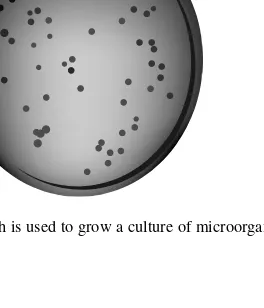
[image:36.532.225.357.438.592.2]Koch’s work with anthrax also developed techniques for growing a culture of microorganisms. He eventually used a gelatin surface to cultivate microorgan-isms. Gelatin inhibited the movement of microorganmicroorgan-isms. As microorganisms reproduced, they remained together, forming a colony that made them visible without a microscope. The reproduction of microorganisms is called colonizing. The gelatin was replaced with agar that is derived from seaweed and still used today. Richard Petri improved on Koch’s cultivating technique by placing the agar in a specially designed disk that was to later be called the Petri dish (Fig. 1-8). It, too, is still used today.
Vaccination
The variola microorganism was one of the most feared villains in the late 1700s. The variola virus causes smallpox. If variola didn’t kill you, it caused pus-filled blisters that left deep scars that pitted nearly every part of your body. Cows were also susceptible to a variation of variola called cowpox. Milkmaids who tended to infected cows contracted cowpox and exhibited immunity to the smallpox virus.
EDWARD JENNER
Edward Jenner, an English physician, discovered something very interesting about both smallpox and cowpox in 1796. Those who survived smallpox never contracted smallpox again, even when they were later exposed to someone who was infected with smallpox. Milkmaids who contracted cowpox never caught smallpox even though they were exposed to smallpox.
Jenner had an idea. He took scrapings from a cowpox blister found on a milk-maid and, using a needle scratched the scrapings into the arm of James Phipps, an 8-year-old. Phipps became slightly ill when the scratch turned bumpy. Phipps recovered and was then exposed to smallpox. He did not contract smallpox because his immune system developed antibodies that could fight off variola and vaccinia.
Jenner’s experiment discovered how to use our body’s own defense mecha-nism to prevent disease by inoculating a healthy person with a tiny amount of the disease-causing microorganism. Jenner called this a vaccination, which is an extension of the Latin word vacca(cow). The person who received the vaccina-tion became immuneto the disease-causing microorganism.
ELIE METCHNIKOFF
Elie Metchnikoff, a nineteenth-century Russian zoologist, was interested by Jenner’s work with vaccinations. Metchnikoff wanted to learn how our bodies react to vaccinations by exploring our body’s immune system. He discovered that white blood cells (leukocytes) engulf and digest microorganisms that invade the body. He called these cells phagocytes, which means “cell eating.” Metch-nikoff was one of the first scientists to study the new area of biology called
immunology, the study of the immune system.
Killing the Microorganism
Great strides were made during the late 1800s in the development of antiseptic techniques. It began with a report by Hungarian physician Ignaz Semmelweis on a dramatic decline in childbirth fever when physicians used antiseptic tech-niques when delivering babies. Infections become preventable through the use of antiseptic techniques.
JOSEPH LISTER
Joseph Lister, an English surgeon, developed one of the most notable antiseptic techniques. During surgery he sprayed carbolic acid over the patient and then bandaged the patient’s wound with carbolic acid–soaked bandages. Infection following surgery dramatically dropped when compared with surgery performed without spraying carbolic acid. Carbolic acid,also known as phenolwas one of the first surgical antiseptics.
PAUL EHRLICH
Antiseptics prevented microorganisms from infecting a person, but scientists still needed a way to kill microorganisms after they infected the body. Scientists needed a magic bullet that cured diseases. At the turned of the nineteenth cen-tury, Paul Ehrlich, a German chemist, discovered the magic bullet. Ehrlich blended chemical elements into a concoction that, when inserted into an infected area, killed microorganisms without affecting the patient. Today we call Ehrlich’s concoction a drug. Ehrlich’s innovation has led to chemotherapy using synthetic drugs that are produced by chemical synthesis.
ALEXANDER FLEMING
Scientists from all over set out to use Ehrlich’s findings to find drugs that could make infected patients well again. One of the most striking breakthroughs came in 1929 when Alexander Fleming discovered Penicillin notatum, the organ-ism that synthesizes penicillin. Penicillium notatum is a fungus that kills the
Fleming grew cultures of Staphylococcus aureus, a bacterium, in the laboratory. He was also conducting experiments with Penicillium notatim, a mold. By acci-dent the Staphylococcus aureus was contaminated with the Penicillium notatum, causing the Staphylococcus to stop reproducing and die. Penicillium notatum be-came one of the first antibiotics. An antibioticis a substance that kills bacteria.
A summary of the scientists and their contributions can be found in Table 1-2.
CHAPTER 1
The World of the Microorganism
17
Fig. 1-9. Penicillium notatumis a fungus that kills the staphylococcus aureus.
Table 1-2. Scientists and Their Contributions
Year Scientist Contribution
1590 Zacharias Janssen Developed the first compound microscope. 1590 Robert Hooke Observed nonliving plant tissue of a thin slice of
cork.
1668 Francesco Redi Discovered that microorganisms did not spontane-ously appear. His contribution led to the finding that killing the microorganism that caused the disease could prevent the disease.
1673 Antoni van Invented the single-lens microscope, grinding the Leeuwenhoek microscope lens to improve magnification. First
person to view a living organism.
Year Scientist Contribution
1850s Mathias Schleiden, Developed cell theory. Theodore Schwann,
Rudolf Virchow
1847 Ignaz Semmelweis Reported a dramatic decline in childbirth fever after physicians used antiseptic techniques when deliver-ing babies.
1864 Louis Pasteur Discovered that microorganisms were everywhere, living on organisms and in nonliving things such as air. His work led to improved sterilization techniques called pasteurization. One of the founders of bacteriology.
1867 Joseph Lister Reduced infections after surgery by spraying car-bolic acid over the patient before bandaging the wound. This was the first surgical antiseptic. 1876 Robert Koch Discovered how microorganisms spread contagious
diseases by studying anthrax. Developed the Germ Theory. Developed techniques for cultivating microorganisms.
1870s John Tyndall, Discovered that some microorganisms are resistant to Ferdinand Cohn certain sterilization techniques. One of the founders
of bacteriology.
1884 Elie Metchnikoff Discovered that white blood cells (leukocytes) engulf and digest microorganisms that invade the body. Coined the word phagocytes. Founded the branch of science called immunology.
1887 Richard Petri Developed the technique of placing agar into a spe-cially designed dish to grow microorganisms, which was later called the Petri dish.
1890 Paul Ehrlich Developed the first drug to fight disease-causing microorganisms that had already entered the body. 1928 Alexander Fleming Discovered Penicillium notatum,the fungus that
kills staphylococcus aureus,a microorganism that is a leading cause of infection.
Quiz
1. What is a microorganism?
(a) A microorganism is a small organism that takes in and breaks down food for energy and nutrients, excretes unused food as waste, and is capable of reproduction.
(b) A microorganism is a small organism that causes diseases only in plants.
(c) A microorganism is a small organism that causes diseases only in animals.
(d) A microorganism is a term that refers to a cell.
2. What is a pathogenic microorganism? (a) A microorganism that multiplies (b) A microorganism that grows in a host (c) A microorganism that is small
(d) A disease-causing microorganism
3. Name the parts of this microorganism using the nomenclature system:
Mycobacterium tuberculosis.
(a) A bacterium is a one-cell organism that does not have a distinct nucleus.
(b) Mycobacterium is the presemous and tuberculosis is the specific postsemous.
(c) Mycobacteriumis the epithet and tuberculosisis the specific genus. (d) Mycobacteriumis the genus and tuberculosisis the specific epithet.
4. Why is a bacterium called a prokaryotic organism?
(a) A bacterium is a one-cell organism that does not have a distinct nucleus.
(b) A bacterium is a one-cell organism that has a distinct nucleus. (c) A bacterium is a multicell organism that does not have a distinct
nucleus.
(d) A bacterium is a multicell organism that has a distinct nucleus.
5. Why is a fungus called a eukaryotic microorganism?
(a) Fungus has cells that have a nucleus, nuclear envelope, cytoplasm, and organelles.
(b) Fungus has cells that have a nucleus and no nuclear envelope. (c) Fungus has cells that have a nucleus, nuclear envelope, cytoplasm,
but no organelles.
(d) Fungus has cells that have no nucleus, no nuclear envelope, no cyto-plasm, and no organelles.
6. What is Archaea?
(a) Archaea is a classification for organisms that have two nuclei. (b) Archaea is a classification for organisms that use phagocytosis. (c) Archaea is a classification of an organism that identifies prokaryotes
that do not have peptidoglycan cell walls.
(d) Archaea is a classification of an organism that identifies prokaryotes that have peptidoglycan cell walls.
7. What is phagocytosis?
(a) The ability of a cell to reproduce.
(b) The ability of a cell to move throughout a microorganism.
(c) The ability of a cell to engulf and digest solid materials by use of pseudopods, or “false feet.”
(d) The ability of a cell to change shape.
8. What is a compound microscope? (a) A microscope that has one lenses.
(b) A microscope that has two sets of lenses: an ocular lens and an eye-piece.
(c) A microscope whose lenses are concave. (d) A microscope whose lenses are convex.
9. What is Germ Theory?
(a) Germ Theory states that a disease-causing microorganism should be present in animals infected by the disease and not in healthy animals. (b) Germ Theory states that a disease-causing microorganism should be
present in healthy animals and not in infected animals.
(c) Germ Theory states that a disease-causing microorganism should be destroyed.
(d) Germ Theory states that a disease-causing microorganism cannot be destroyed.
(b) Edward Jenner discovered how to create vaccinations to trigger the body’s immune system to develop antibodies that fight micro-organisms.
(c) Edward Jenner discovered the compound microscope.
(d) Edward Jenner discovered the compound nomenclature system.
2
CHAPTER
23
The Chemical
Elements of
Microorganisms
No doubt you’re asking yourself what chemistry has to do with microbiology since they seem to be two different branches of science. The simple answer is that microorganisms are made up of chemicals, as is every organism—and all matter. Remember that matter is anything that occupies space and has mass.
You might say that an organism is a chemical processing plant where things are broken down into chemical elements; these chemical elements are then re-arranged to form new things. You do this every time you ingest food. Food is a group of chemical compounds. The digestive process rearranges these digested chemical compounds into new substances that provide you with energy and nutrients that are necessary for you to live. Some microorganisms called auto-trophic organismsmanufacture their own food. Microorganisms that derive energy from other microorganisms (food) are called heterotopic organisms.
When you catch a cold or become infected by pathogenic microorganisms, your body is no longer in homeostasis. You feel rotten, but what’s really hap-pening is that the microorganism is disrupting your chemical processing plant’s normal operation. Some microorganisms prevent necessary chemical processing from occurring. Other microorganisms cause your chemical processing plant to execute different processes designed to fight the microorganism attack and return your body to homeostasis—then your body is back to normal.
As you can see, chemistry is a crucial component of microbiology. It is for this reason that we begin the study of microorganisms with a close look at chemistry.
Everything Matters
Anything that takes up space and has mass is matter. The chair you’re sitting on is matter. You are matter. And so are the microorganisms crawling over you and the chair. All nonliving and living things are matter because they take up space and have mass.
Matter is anything that takes up space and has mass. It is easy to envision something taking up space, but what is mass?
Massis the amount of matter a substance or an object contains. A common mis-conception is that mass is the weightof a substance. It is true that the more there is of a substance, the more it weighs. However, weight is the force of gravity act-ing on mass and is calculated as weight =mass ×gravity. A trip to the moon will clarify the difference between mass and weight: You have the same mass on earth as you do on the moon, but you weigh more on the earth than you do on the moon because the earth has six times the gravitational force of the moon.
Chemical Elements and the Atom
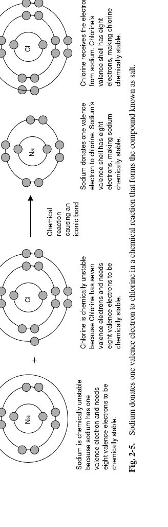
Everything including you is composed of chemical elements.Achemical ele-ment,sometimes simply referred to as an element, is a substance that cannot be broken down into simpler substances by a chemical process. All matter is a com-bination of chemical elements.
A chemical element is made up of atoms. An atomis the smallest particle of an element; it cannot be further decomposed into smaller chemical substance (Fig. 2-1). In the early 1800s, John Dalton developed the Atomic Theory, which explains the relationship between an element and an atom. The Atomic Theory