Treatment of gastrointestinal stromal
tumor: focus on imatinib mesylate
Omar S Din Penella J Woll
Academic Department of Clinical Oncology, University of Sheffield, Weston Park Hospital, Sheffield, UK
Correspondence: Omar Din
Weston Park Hospital, Whitham Road, Sheffi eld, UK
S10 2SJ
Tel +1 0114 2265000 Email omarsdin@hotmail.com
Abstract: Gastrointestinal stromal tumor (GIST) is a rare primary neoplasm of the gastrointestinal tract, mesentery, or omentum. In the past, surgery has been the only effective treatment. The diagnosis and treatment of GIST has been revolutionized over the past decade, since expression of the receptor tyrosine kinase KIT was shown to occur on these tumors. Muta-tions in this proto-oncogene commonly cause constitutive activation of the KIT tyrosine kinase receptor, an important factor in the pathogenesis of the disease. The development of specifi c tyrosine kinase inhibitors, such as imatinib mesylate, has led to a breakthrough in the treatment of advanced GIST. Treatment with this drug has led to signifi cant improvements in survival, with overall response rates in excess of 80%. Side effects are common, but usually manageable. The success of this drug has led to further trials investigating its use in the pre- and postoperative situation. This review summarizes the current knowledge of GIST and imatinib treatment and possible future developments.
Keywords: imatinib, STI-571, GIST, gastrointestinal, stromal, tumor
Introduction
Gastrointestinal stromal tumor (GIST) is a rare tumor, accounting for less than 1% of primary gastrointestinal (GI) neoplasms. It is, however, the commonest non-epithelial tumor of the gastrointestinal tract. The median age of diagnosis is approximately 60 years, with the annual incidence estimated at 10–20 cases per million (Nilsson et al 2005). It is very rare in children and affects males and females equally. GIST is mainly a disease of the GI tract, mesentery, and omentum. Most commonly, it originates in the stomach (60%), followed by the small intestine (30%), the colon and rectum (5%), and the oesophagus (5%) (Van der Zwan and DeMatteo 2005). Many are found incidentally at surgery or autopsy. GIST can be classifi ed into different risk groups. At presentation, only 44% are overtly malignant or high risk, while 32% are of low or very low risk (Nilsson et al 2005). Although the exact pathogenesis is not fully known, it is thought to originate from the same lineage as the interstitial cells of Cajal. These are pacemaker cells of the GI tract, which are phenotypically similar to GIST cells (Kindblom et al 1998).
stain positive for CD117, making it a very useful marker for diagnosis (Miettinen and Lasota 2001). This has led to the development of the targeted therapy imatinib mesylate (STI-571; Glivec®, Novartis, Basel, Switzerland). This drug
inhibits several tyrosine kinase receptors with varying affi n-ity, including KIT, the BCR-ABL fusion protein, and the platelet derived growth factor receptor (PDGFR) (Heinrich et al 2000, De Giorgi and Verweij 2005).
Presentation
Because GIST was only recently recognized as a separate clinicopathological entity, the literature prior to 2000 did not give an accurate account of the clinical behavior of GIST.
GIST can present in many ways. Thirty percent are diagnosed incidentally on a pathological or autopsy resec-tion specimen (Nilsson et al 2005). Small tumors may be asymptomatic and GISTs can grow to a large size before producing any symptoms. This may be because GISTs grow by displacing adjacent structures rather than invading them. Presenting symptoms can therefore include non-specifi c GI symptoms such as nausea, vomiting, dyspepsia, abdominal pain, distension, or change in bowel behavior. Less com-monly, there may be symptoms of obstruction, bleeding, or rupture into the peritoneal cavity.
Despite radical resection with clear margins, 40%–80% recur within the abdominal cavity. However, the majority of recurrences are solitary and thus may be resectable. The most common sites of metastases are the peritoneum and liver, whereas lymph node metastases are relatively rare. In contrast to leiomyosarcomas, pulmonary and bone metastases occur late and are uncommon.
Rare familial cases of GIST with a mutated KIT have been recognized. This may be as part of the Carney triad of gastric GIST, functioning extra-adrenal paraganglioma and pulmonary chondroma. This mainly affects young women and was fi rst described in by Carney in 1977 (Carney et al 1977). Although it is thought to be hereditary (young age and multiple specifi c tumors), no genetic abnormality has been identifi ed. GIST has also been reported in association with neurofi bromatosis type 1 (Von Reckinghausen’s disease) (Ishida et al 1996).
GIST can range in size from less than 1 cm to over 30 cm in diameter. Various investigations may be used in the diagnosis of GIST. Gastric tumors are often detected by endoscopy. Macroscopically, primary GIST is usually a well-circumscribed submucosal vascular tumor, protrud-ing into the lumen. Computed tomography (CT) is useful to assess the extent of the primary disease and to assess
for the presence of metastatic disease. Magnetic resonance imaging (MRI) may provide further soft tissue delineation. Positron emission tomography (PET) with the tracer 18-fl uorodeoxyglucose (18-FDG) demonstrates intense uptake, but may not distinguish GIST from other malignancies. Its main use is in demonstrating the presence or absence of metastases.
Smaller tumors are usually treated by excision biopsy. For larger lesions, the use of preoperative biopsy is controversial. This is because there is a risk of tumor rupture, hemorrhage, and perforation of viscera (Joensuu et al 2002). The diagnos-tic yield from endoscopic biopsy is approximately 50%.
Diagnosis
Macroscopically, GIST has the appearance of a friable unen-capsulated mass arising from the muscle layer, rather than the epithelium. Central necrosis is often present in larger lesions (D’Amato et al 2005). Microsopically, most GISTs consist of a uniform population of spindle cells (70%). Some are characterized as epithelioid type (20%), which typically arise in the stomach (Miettinen et al 2002). The remaining 10% consist of a mixture of these two morphologies. The spindle cells are arranged in short fasicles or whorls, with prominent nuclear palisading. Either type may contain cur-vilinear collections of extracellular collagen called skeinoid fi bres (Corless et al 2004). The eosinophilic subtype consists of rounded cells with eosinophilic or clear cytoplasm (Van der Zwan and DeMatteo 2005).
growth factor receptor alpha (PDGFRα) mutations are found in 5%–10% GISTs (Sihto et al 2005).
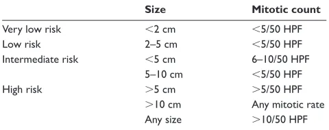
Many efforts have been made to produce a reliable prog-nostic classifi cation (Table 1) (Fletcher et al 2002). Mitotic count and tumor size have been shown to be very important. A large study examined 1765 tumors for prognostic markers (Miettinen et al 2005). Tumours less than 10 cm with less than 5 mitoses per 50 high powered fi elds (HPFs) had only a 2%–3% risk of metastases. Conversely, the metastatic rate for tumors greater than 10 cm, with greater than 5 mitoses per 50 HPFs, was as high as 86%. Non-gastric primary tumor location and male gender may also be independent adverse prognostic factors (Rutkowski et al 2007).
Evidence is now emerging that mutational analysis can help to predict prognosis. A recent study showed improved survival in those with KIT exon 11 point mutation compared to deletions in gastric GISTs. The same was true with com-parison with point mutations from other locations (Steigen et al 2007). This is discussed further below.
Pathogenesis
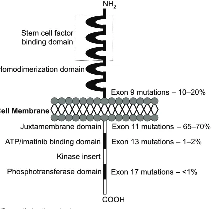
GIST cells are phenotypically very similar to the KIT positive interstitial cells of Cajal of the GI tract. They are pacemaker cells which regulate motility and their development depends on cellular signalling regulated by the KIT protein (Miet-tinen et al 2002). The KIT proto-oncogene on chromosome 4 (4q11–q12) encodes for the KIT protein, which is also expressed on mast cells, germ cells, and hemopoietic cells. Stem cell factor is the ligand for this receptor and binding activates cell growth, differentiation, and survival. In GIST, ligand independent activation occurs due to the presence of an activating mutation, which was fi rst described by Hirota and colleagues. They showed that GIST cells were able to grow without their ligand in vitro and in nude mice (Hirota et al 1998). Since then, many more gain-of-function muta-tions have been identifi ed (Sanborn and Blanke 2005). It has been shown that the site of mutation has a prognostic importance. The most frequent are exon 11 (65%–70%), 9 (10%–20%), 13 (1%–2%) and 17 (1%) (Figure 1). Muta-tions of the juxtamembrane domain (exon 11) of the KIT receptor are the most frequently seen, where small in-frame deletions and insertions or point mutations are responsible for ligand-independent receptor dimerization. These mutations are most commonly found in GIST of the stomach. Dele-tions and inserDele-tions tend to affect the fi rst part of the exon (especially codons 557 to 559), whereas point muations are limited to just 4 codons (557, 559, 560, and 576) (Corless et al 2004). Exon 9 mutations are located in the extracellular
domain of the KIT receptor, whereas exons 13 and 17 encode for the intracellular part of the receptor (kinase 1 domain and activation loop respectively).
Some studies have suggested a more aggressive clinical behaviour in tumors with exon 11 mutations (Ernst et al 1998; Lasota et al 1999; Taniguchi et al 1999; Li et al 2000). Others have suggested that KIT mutations were not identifi ed more commonly in higher grade GISTs (Rubin et al 2001) and that mutations of exon 11 were common in histologically benign GISTs. So there is continuing controversy as to whether exon 11 mutations confer aggressive behavior or not. As imatinib targets the ATP (adenosine triphosphate) binding site of the KIT receptor (encoded by exon 11), tumors with exon 11 mutations treated with imatinib have a signifi cantly better partial response rate, event free and overall survival compared to exon 9 or no detectable mutation. A partial response rate of up to 83% has been achieved, showing how sensitive these tumors can be (Heinrich et al 2003a; Corless et al 2004; Debiec-Rychter et al 2006).
Not all GISTs demonstrate KIT mutation. An important study of 40 KIT-negative GISTs demonstrated PDGFRα activating mutations in 14 (35%) (Heinrich et al 2003b). The authors also showed that the mechanisms of cytogenetic progression and signal transduction were similar to those seen in KIT positive GISTs. These two mutational events are mutually exclusive of each other. Other studies have confi rmed similar fi ndings (Hirota et al 2003; Wardelmann et al 2004). Activating mutations of PDGFRα do not appear in other tumor types (Burger et al 2005; Sihto et al 2005).
[image:3.612.320.560.77.172.2]KIT or PDGFRα mutations occur early in the pathogenesis of GIST, as they are found even in very small tumors. Progres-sion of GIST has been shown to be related to various chromo-somal aberrations (Gunawan et al 2002). The most common chromosomal aberration seen in malignant GIST are losses of 14q32 and 22q11.The loss of the long arm of chromosome
Table 1 Proposed approach for defi ning risk of aggressive behavior in GISTs
Size Mitotic count
Very low risk 2 cm 5/50 HPF
Low risk 2–5 cm 5/50 HPF
Intermediate risk 5 cm 6–10/50 HPF
5–10 cm 5/50 HPF
High risk 5 cm 5/50 HPF
10 cm Any mitotic rate
Any size 10/50 HPF
Reproduced with permission from Fletcher CD, Berman JJ, Corless C, et al. 2002. Diagnosis of gastrointestinal stromal tumors: A consensus approach. Hum Pathol, 33:459–65. Copyright© 2002 Elsevier.
22 is particularly associated with progression to a borderline or malignant lesion (Debiec-Rychter et al 2001).
Surgery
Surgery remains the standard initial management for all localized GISTs. The tumor should be removed en bloc, with a clear margin. The pseudocapsule should be removed and not penetrated. Therefore, a wedge resection (stomach) or segmental resection (intestine) is required. If neighboring structures are involved, en-bloc resection should still be con-templated. Studies have evaluated the outcomes of surgery, comparing complete versus incomplete resections. In a series of 200 GISTs, median survival was 66 months for complete resection compared with 22 months for incomplete resection or unresectable disease (DeMatteo et al 2000). The complete resection rate was approximately 85%. The optimum surgical
margin has not been clarifi ed. Lymph node dissection or biopsy is not recommended mainly due the pattern of spread of GISTs: lymph node metastases are rare.
More recently, there has been a move to laparoscopic surgery, particularly for gastric GISTs. One series of 50 consecutive patients showed this approach was associated with low morbidity and short hospitalization. All resections had clear margins and the long term disease free survival was 92% (Novitsky et al 2006). In a second series of 22 clinically suspected GISTs, similar fi ndings were shown, with only one case of recurrence (Berindoague et al 2006).
Spontaneous tumor rupture, or rupture during surgery, increases the risk of peritoneal recurrence and is an adverse prognostic factor. Overall 5-year survival rates for primary resected disease are in the order of 50%–55%. One series has reported recurrence rates of up to 90% after surgical resection
NH
2COOH
Juxtamembrane domain Exon 11 mutations – 65
ATP/imatinib binding domain
Kinase insert
Phosphotransferase domain
Homodimerization domain
Stem cell factor
binding domain
Exon 9 mutations – 10
Exon 13 mutations – 1
Exon 17 mutations – <1%
NH
2COOH
Juxtamembrane domain Exon 11 mutations – 65–70%
ATP/imatinib binding domain
Kinase insert
Phosphotransferase domain
Homodimerization domain
Stem cell factor
binding domain
Exon 9 mutations – 10–20%
Exon 13 mutations – 1–2%
Exon 17 mutations – <1%
[image:4.612.84.511.54.471.2]Cell Membrane
(Ng et al 1992). Most recurrences occurred within the fi rst 2 years of resection. Prognostic factors for recurrence are shown in Table 2.
There is no standard follow up regimen for resected GIST. As recurrent disease can be treated by surgery or imatinib, active follow up is warranted. Suggested follow up protocols have been developed according to risk groups. For example, a high risk patient should have a CT scan every 3–4 months for 3 years, then every 6 months to 5 years. For low risk, a CT scan every 6 months for 5 years is acceptable (Blay et al 2005).
The commonest sites of metastases are the liver and intraperitoneum. There is some evidence that metastatectomy can improve survival in selected patients (Chen et al 1998). Those that benefi t most have a disease free interval of greater than 12 months, well differentiated histology and an isolated liver metastasis. Surgery can be used for palliation, but with the advent of imatinib, it has largely been superseded.
Chemotherapy
Interpretation of results from previous studies is diffi cult as it is likely that many tumors classifi ed as leiomyosarcomas were actually GIST. Given this reservation, the effi cacy of chemo-therapy is low, with response rates less than 10% (Dematteo et al 2002). Doxorubicin and ifosfamide have limited activity in GIST compared to other soft tissue sarcomas. One study of 26 patients with GIST showed 38.4% to express P-glycoprotein and 35.4% expressed MRP1 (multi drug resistance protein). These levels were signifi cantly higher than for soft tissue leiomyosarcoma. This may explain the poor response of GIST to standard chemotherapy (Plaat et al 2000).
Radiotherapy
Radiation therapy has little role to play in the management of GIST. Most tumors are not amenable to treatment because of their location and close proximity to vital organs. GISTs are thought to be relatively resistant. Nevertheless, radio-therapy can be successfully used in patients with advanced disease to control bleeding or other troublesome symptoms.
Hepatic artery embolization
This technique may provide palliation in patients with GIST metastatic to the liver. Due to the vascular nature of GIST, occluding the supplying artery may be effective. There has been more interest in chemoembolization, which allows increased local drug delivery, but reduced systemic effects due to high fi rst pass metabolism in the liver. Two small studies have shown limited effi cacy with responses lasting 8–12 months (Mavligit et al 1995; Rajan et al 2001).
Development of imatinib mesylate
Imatinib was developed as a tyrosine kinase receptor inhibi-tor. In 1989, a family of compounds called tyrphostins were shown to have specifi city for the epidermal growth factor receptor. Further developments led to the identifi cation of 2-phenylaminopyrimidine compounds as having the most promising inhibitory activity against receptor tyrosine kinases. This led to the development of imatinib, which was shown to inhibit the intracellular kinases ABL and BCR-ABL fusion protein in chronic myeloid leukemia (CML) cells (Druker et al 1996), but was subsequently found to have comparable activity against the KIT receptor (wild type and mutant) and PDGFR (Carroll et al 1997; Heinrich et al 2000).
Imatinib is a competitive antagonist of the adenosine triphosphate (ATP) binding site. It blocks the transfer of phosphate groups from ATP to tyrosine residues of the sub-strates. This causes interruption of the downstream signalling process that leads to cell proliferation, including MAP kinase and Akt (Heinrich et al 2000).
Pharmacology of imatinib mesylate
Pharmacokinetic studies of imatinib in healthy volunteers, patients with CML and GIST have shown good oral absorp-tion and a bioavailability of 98%, regardless of the prepara-tion (soluprepara-tion, capsule or tablet) or dosage strength (100 mg, 400 mg) (Peng et al 2005). Once absorbed, it binds to serum proteins (mainly albumin and alpha 1-acid glycoprotein) and peak concentrations are reached 4 hours after administration (D’Amato et al 2005).
The main circulating metabolite is an N-demethylated piperazine derivative which accounts for 16% of the AUC (area under the curve) for imatinib. This, with imatinib accounts for most of the activity, but there are a number of smaller metabolites. CYP3A4 is the major cytochrome P450 involved in imatinib metabolism. Thus, drugs that are co-administered may alter the pharmacokinetics. Erythro-mycin, fl uconazole, and rifampicin have shown inhibition of imatinib metabolism. Imatinib increases exposure to simvastatin. Alprazolam, caffeine, clindamycin, clonazepam, cortisol, ethinyl oestradiol, and verapamil may cause toxic
Table 2 Prognostic factors for recurrence Tumor size 5 cm
effects when given with imatinib. St. John’s wort increases imatinib clearance by 43%. Patients should avoid excessive amounts of paracetamol as both are metabolized by CYP3A4 (D’Amato et al 2005; Novartis 2005).
The terminal elimination half-life is approximately 18 hours, which allows for once a day dosage. Excretion is via the feces and urine, the majority being the metabolites. In patients with GIST, steady state exposure was 1.5 fold higher than that observed in CML for the same 400 mg/day doseage. This mainly seems to depend on albumin, white blood count and bilirubin. Hepatic and renal dysfunction and liver metastases may cause variable and reduced metabolism of the drug. Age, race, sex, and bodyweight do not signifi -cantly affect the pharmacokinetics of the drug (Peng et al 2005; Novartis 2005).
Preclinical studies of imatinib
Druker and colleagues fi rst recognized the BCR-ABL protein to be an excellent target for imatinib as the BCR-ABL muta-tion is present in the vast majority of patients with CML. They showed that imatinib inhibited proliferating myeloid cell lines specifi cally. 95% reductions in concentrations of the abnormal protein were seen. Little toxicity was seen in normal bone marrow cells. Very rapidly in vitro results were tested in vivo, with signifi cant effects of imatinib seen in nude mice and then humans with chronic phase CML (Savage and Antman 2002; Druker et al 1996).
The early success of imatinib in chronic phase CML led investigators to assess its effects on c-KIT receptor tyrosine kinase activity (Heinrich et al 2000). C-KIT autophosphory-lation, activation of mitogen activated protein (MAP) kinase and activation of Akt were all inhibited in a c-KIT expressing cell line. In a cell line with an activating mutation of c-KIT, more potent inhibitory effects occurred as compared to the wild-type receptor. These fi ndings led to the use of imatinib in the fi rst patient in 2001 as discussed below (Joensuu et al 2001).
Phase I studies
Following on from the preclinical studies, imatinib was tested in a phase I study in patients with Philadelphia chromosome positive CML (Druker et al 2001). This and subsequent studies established that a continuous dosing schedule of 400 mg/day was safe and effective in CML. The fi rst GIST patient to receive imatinib was a 50-year-old woman, with KIT-positive metastatic GIST associated with a mutation in exon 11. She had progressive metastatic disease despite surgery, combination cytotoxic chemotherapy, thalidomide
and interferon alpha. She was treated with 400 mg of imatinib per day and response was evaluated using 18-FDG PET (18-fl uorodeoxyglucose positron emission tomography) and CT scanning. After 1 month, the patient had a complete metabolic response and by 8 months many of the liver metastases had disappeared or reduced in size (Joensuu et al 2001).
A formal phase I study was conducted by the EORTC (European Organisation for Research and Treatment of Can-cer) Soft Tissue and Bone Sarcoma Group (van Oosterom et al 2001). 40 patients (36 with GIST and 4 with non-GIST soft tissue sarcoma) were recruited between August and December 2000. The dose groups of imatinib were 400 mg once daily, 300 mg twice daily, 400 mg twice daily, and 500 mg twice daily. Dose limiting toxic effects were nausea, vomiting, oedema and skin rash which were seen at a dose of 500 mg twice daily. The maximum tolerated dose recom-mended was 400 mg twice daily. No responses were seen in non-GIST sarcomas. Among the GIST patients, responses were seen at all dose levels. Fifty-four percent achieved a partial response, 37% had stable disease, and 5% had pro-gressive disease. After 9 months of therapy, 82% of GIST patients continued to obtain clinically important benefi ts. In the update, the most common adverse effects for those continuing on therapy were periorbital oedema (40%) and peripheral oedema (37.5%) (van Oosterom et al 2002).
Phase II studies
After the success of the initial studies in GIST and CML, fur-ther studies were conceived. The fi rst trial was a US – Finland trial, which was a multicentre, randomized, open label phase II study (Demetri et al 2002). One hundred and forty-seven patients with unresectable or metastatic GIST were randomly assigned to receive 400 mg or 600 mg of imatinib daily, between July 2000 and April 2001. For those who progressed on 400 mg daily, the dose was increased to 600 mg daily. After a median follow up of 21 months, 66% achieved a partial response and 17% had stable disease. No signifi cant differences were seen between the two dose groups. 14% died of progressive disease. Median survival had not been reached, but 85% were estimated to be alive at 19 months. The trial was not powered to distinguish between effi cacy at the two dose levels. However a third of those who progressed on the lower dose sustained a subsequent partial response on crossover (Blanke et al 2004).
51 patients participated, 27 of whom had confi rmed GIST, 24 with other soft tissue sarcomas. In the GIST group, 4% had complete remission, 67% partial remission, 18% stable disease, and 11% progressive disease. In contrast, there were no objective responses in the non-GIST sarcoma group. Among GIST patients, the median time to onset of response was 113 days. 73% of GIST patients were free from disease progression at 1 year. In terms of toxicity, the most frequent adverse effects were anemia (92%), oedema, particularly periorbital (84%), rash (69%), and fatigue (76%). Granulo-cytopenia was less common at 48%. These studies led to the registration of imatinib for the treatment of locally advanced or metastatic GIST in 2002.
A Japanese phase II randomized study evaluated 74 patients with unresectable or metastatic GIST. Patients received either 400 mg daily or 600 mg daily. The results showed slightly lower response rates of 55% (overall) and a partial response rate of 41%. Interestingly, there were more responses in the higher dose group (61% vs 47%), but this did not reach statistical signifi cance. The higher dose group had more interruptions of therapy as well as dose reductions. Grade 3 or 4 neutropenia and dermatitis was also signifi cantly higher. The investigators therefore recommended 400 mg daily as the optimal dosing schedule (Doi et al 2004).
Response assessment
In clinical trials in patients with advanced GIST treated with imatinib, the median time to objective response is about 13 weeks (Demetri et al 2002). However, some patients have a dramatic improvement in symptoms and signs within days, accompanied by evidence of reduced metabolic activity on PET scans. Interestingly, no cases of tumor lysis syndrome have yet been described. The changes following an imatinib response can be quite distinct. Commonly cystic changes develop that could be confused with progressive disease (Linton et al 2006; Ryu et al 2006). Therefore traditional response assessment criteria are not entirely relevant. In the original patient treated with imatinib, biopsy specimens at 1 and 2 months after starting treatment showed a marked decrease in tumor cells as well as myxoid degeneration and scarring. No necrosis or infl ammatory reaction was seen. The tumor cells did not stain for Ki-67 (a proliferation marker) suggesting relatively low activity (Joensuu et al 2001).
Many trials have used CT and 18-FDG PET in the assessment of imatinib response. One study compared CT, PET and dual modality PET/CT monitoring the effects of imatinib therapy in 20 patients. PET/CT identified 282 metastases, CT picked up 249 lesions and PET alone
showed 135. In evaluating disease at 3 and 6 months of therapy, PET provided 100% accuracy for response, com-pared to 57%–60% using CT. PET also detected responses earlier than CT (Antoch et al 2004). In a similar study of 28 assessable patients, the absence of FDG uptake after initial treatment was associated with a better prognosis than those with residual uptake. CT detected more lesions, but was less good at predicting prognosis (Goerres et al 2005). A further study showed PET response was associated with a signifi cantly longer progression free survival (Stroobants et al 2003; King 2005).
Choi and colleagues have proposed new response crite-ria for GISTs. They studied 172 lesions in 40 patients with metastatic disease. Patients were assessed pre-treatment and after 2 months with CT imaging and FDG-PET. A decrease in tumor size of more than 10% or a decrease in tumor density of more than 15% on CT had a specifi city of 100% and a sensi-tivity of 97% in identifying PET responses. This compares to 100% specifi city and 52% sensitivity using RECIST criteria. Thus, RECIST criteria are felt to be a poor indicator of clini-cal benefi t, underestimating the effects of treatment (Choi et al 2007). These new criteria have been further evaluated and correlation has been shown with time to tumor progres-sion and disease specifi c survival. No similar correlation was seen using RECIST criteria (Benjamin et al 2007).
Following initial response to imatinib, resistance often develops. There have been reports of a characteristic new nodule-within-a-mass pattern of recurrence (Shankar et al 2005). In this series of 92 patients treated with imatinib, 39 developed progressive disease and 21 of them showed this pattern. It was the fi rst sign of progression in the majority with this feature. The authors postulate that this pattern represents a localized clone of mutant tumor cells developing resistance to c-KIT inhibition. This is important, as traditional response criteria may not identify the fi rst signs of recurrent disease.
Phase III trials
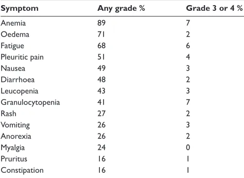
response) was 50% for the 400 mg group and 54% in the 800 mg group. 56% had progression on 400 mg compared to 50% on 800 mg (HR 0.82, 95% CI 0.69–0.98, p = 0.026). Overall survival was 85% at 1 year and 69% at 2 years for the 400 mg group and 86% at 1 year and 74% at 2 years for the 800 mg group. There were more dose reductions and interruptions in the higher dose group. Toxicity was common but manageable. Oedema, anemia, rash, lethargy, nausea, bleeding, diarrhoea, and dyspnoea were statistically more common in the 800 mg group. Table 3 shows the frequency and grade of toxicity for those receiving 400 mg/day.
The North American Sarcoma Intergroup study (S0033) randomized 746 patients to either 400 mg or 800 mg daily of imatinib. Patients randomized to 400 mg daily were dose escalated to 800 mg daily on disease progression. The pri-mary end point was overall survival. Early results presented in abstract in 2003 showed no difference in overall survival or progression free survival, after a median follow up of 14 months. The proportion of patients achieving an objective response or stable disease was similar in both arms (75% (400 mg) vs 73% (800 mg)). Objective response rates were 43% vs 41% respectively (Benjamin et al 2003). In the 2004 update, after a median follow up of 25 months, the median survival had not been reached. 2 year survival for the 400 mg vs 800 mg group was 73% and 78% respectively, 2 year pro-gression free survival was 50% and 53% respectively. Again these were not statistically different. Eighty-eight patients crossed over to the higher dose on disease progression. Of these, 7% demonstrated partial response and 29% achieved stable disease (Rankin et al 2004).
These two studies have shown differing results. The North American study with overall survival as primary end point has to date shown no differences between the two dose groups. In contrast, the EORTC study, with progression free survival as its primary end point, has shown a statistically signifi cant improvement in progression free survival, but requires further follow up to assess overall survival. In both studies, survival results may be infl uenced by the cross over design.
At ASCO 2007, the GIST Meta-analysis Group presented the results of a meta-analysis of these two randomized stud-ies. After a median follow up of 45 months, a small but signifi cant progression free survival advantage was seen in the high dose arm. Overall survival was identical in the two arms (median survival 4.08 years (bid) vs 4.05 years (od), HR 1.00). Compared to KIT exon 11 mutants, wild type KIT, exon 9 and other mutations conferred a poorer prog-nosis. The interaction between dose effect on progression
free survival and prognostic factors was signifi cant for KIT exon 9 mutations, but not for other factors. The heterogeneity between the two trials is still being evaluated (Van Glabbeke et al 2007).
The results of an interesting phase III trial have recently been reported by the French Sarcoma Group (Blay et al 2007). It was a prospective randomized trial comparing continuous and interrupted use of imatinib beyond 1 year of treatment in patients with advanced GIST. Between May 2002 and April 2004, 182 patients were enrolled. Of these, only 58 were eli-gible for randomization after 1 year with controlled disease. 26 patients continued with imatinib, whilst 32 discontinued until progression. Documented progression was seen in 8 of 26 patients in the continuous group and 26 of 32 patients in the interrupted group (p 0.001). However, 24 of the latter subsequently responded to reintroduction of imatinib. There were no differences in survival, resistance or quality of life between the two groups. This study supports the use of continuous imatinib, due to the high level of progression after interruption of therapy. In a further French study, 35 patients free from progression after 3 years of imatinib at 400 mg/day, were randomly offered to continue or interrupt treatment. 9 progressors were reported after a median fol-low up of 5.3 months, all of whom achieved tumor control (at least stable disease) after rechallenge with imatinib. This study continues (Le Cesne et al 2007).
Neoadjuvant and adjuvant imatinib
[image:8.612.304.541.64.237.2]The success of imatinib in controlling locally advanced and metastatic GIST has led to interest in the neoadju-vant and adjuneoadju-vant use of the drug. There have been case reports and series suggesting a role for the neoadjuvant
Table 3 Frequency of side effects (400 mg/day dose)
Symptom Any grade % Grade 3 or 4 %
Anemia 89 7
Oedema 71 2
Fatigue 68 6
Pleuritic pain 51 4
Nausea 49 3
Diarrhoea 48 2
Leucopenia 43 3
Granulocytopenia 41 7
Rash 27 2
Vomiting 26 3
Anorexia 26 2
Myalgia 24 0
Pruritus 16 1
Constipation 16 1
approach (Bauer et al 2005; Loughrey et al 2005; Raut et al 2006). In a series of 90 patients reported by Bauer and col-leagues, 12 were rendered suitable for surgical resection of residual disease. Eleven of these patients achieved a com-plete resection, although most had viable tumor cells in the resected specimen. The median overall survival was 46 (range 4–85) months. This report has therefore shown prolonged survival and a signifi cant rate of subsequent resectability after treatment with imatinib induction. In another series, Raut et al evaluated 69 consecutive patients with advanced GIST treated by surgery whilst receiving imatinib or another tyrosine kinase inhibitor, sunitinib malate (SU11248; Sutent®,
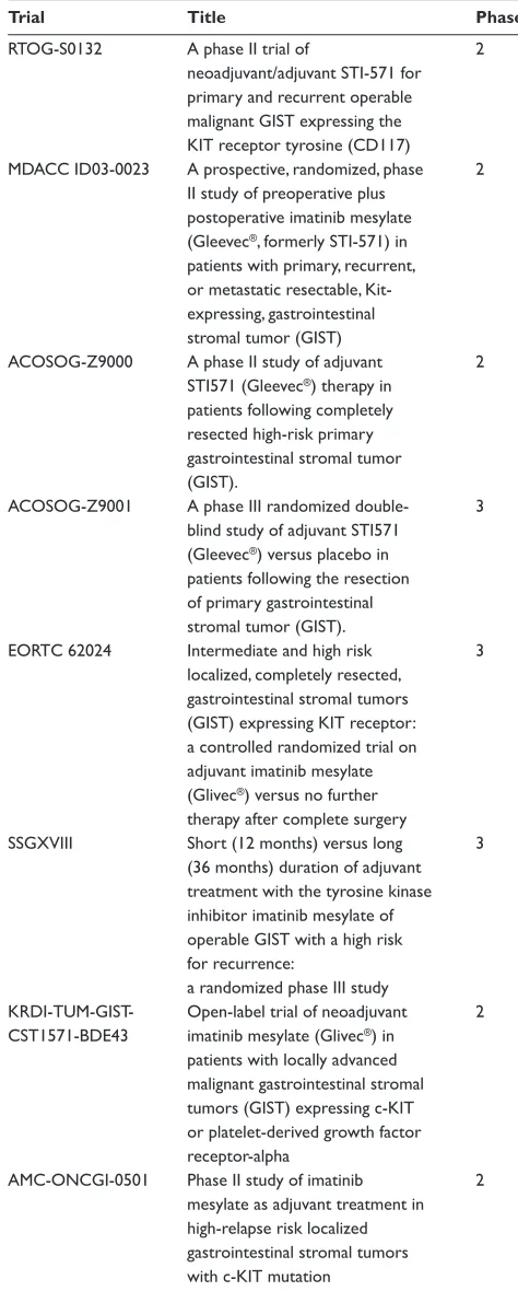
Pfi zer inc, New York). Surgical outcome correlated strongly with pre-surgery disease status. There was no evidence of residual disease in 78% with stable disease before surgery, in 25% with limited progression and in 7% with generalized progression. One-year progression free survival was 80%, 33%, and 0% for stable disease, limited progression and gen-eralized progression respectively. One-year overall survival was 95%, 86%, and 0% for the same groups. These reports suggest a role for primary imatinib therapy and surgery for advanced GIST. Selected patients may experience prolonged survival after this aggressive initial approach. However, the actual benefi t of imatinib followed by surgery still requires further evaluation in prospective clinical trials. Such trials are underway and are shown in Table 4. Neoadjuvant imatinib is not recommended where a change in tumor size will not affect surgery (Blay et al 2005). It can, however, be consid-ered where a tumor response could permit function-sparing surgery, eg, rectum or oesophagus.
[image:9.612.318.555.80.670.2]There has also been signifi cant interest in the use of ima-tinib in the adjuvant setting. This stems from the successes of treatment of advanced disease with the rationale that it may be more effective in minimal residual disease and may be curative in this setting. In high risk patients, the rates of recurrence after primary surgery are about 50%. Prospective trials are running to assess the role of imatinib following surgery for high risk GIST and are shown in Table 4 (NCI, 2007). The ACOSOG Z9001 study randomized 708 patients following complete resection of a primary GIST of at least 3 cm diameter, to either 1 year of imatinib 400 mg/day or placebo. Upon recurrence, patients could be crossed over from placebo to imatinib, or their imatinib dose increased to 800 mg/daily. The trial was stopped early after a planned interim analysis, with a median follow up of 1.2 years. The primary endpoint of 1-year relapse free survival was 97% in the imatinib arm and 83% in the placebo arm, with a hazard ratio of 0.325 (p = 0.0000014). It appears the signifi cant benefi t occurs in the fi rst 18 months,
Table 4 Neoadjuvant and adjuvant trials of imatinib in GIST
Trial Title Phase
RTOG-S0132 A phase II trial of 2 neoadjuvant/adjuvant STI-571 for primary and recurrent operable malignant GIST expressing the KIT receptor tyrosine (CD117) MDACC ID03-0023 A prospective, randomized, phase 2
II study of preoperative plus postoperative imatinib mesylate (Gleevec®, formerly STI-571) in
patients with primary, recurrent, or metastatic resectable, Kit-
expressing, gastrointestinal
stromal tumor (GIST)
ACOSOG-Z9000 A phase II study of adjuvant 2 STI571 (Gleevec®) therapy in
patients following completely resected high-risk primary gastrointestinal stromal tumor (GIST). ACOSOG-Z9001 A phase III randomized double- 3
blind study of adjuvant STI571 (Gleevec®) versus placebo in
patients following the resection
of primary gastrointestinal
stromal tumor (GIST).
EORTC 62024 Intermediate and high risk 3 localized, completely resected,
gastrointestinal stromal tumors (GIST) expressing KIT receptor: a controlled randomized trial on adjuvant imatinib mesylate (Glivec®) versus no further
therapy after complete surgery SSGXVIII Short (12 months) versus long 3
(36 months) duration of adjuvant treatment with the tyrosine kinase inhibitor imatinib mesylate of operable GIST with a high risk
for recurrence:
a randomized phase III study
KRDI-TUM-GIST- Open-label trial of neoadjuvant 2 CST1571-BDE43 imatinib mesylate (Glivec®) in
patients with locally advanced malignant gastrointestinal stromal tumors (GIST) expressing c-KIT or platelet-derived growth factor receptor-alpha AMC-ONCGI-0501 Phase II study of imatinib 2
mesylate as adjuvant treatment in high-relapse risk localized gastrointestinal stromal tumors with c-KIT mutation
but then the recurrence rate is similar in both arms. This has brought concern that early treatment may lead to the early development of resistance. Longer follow up required to see if there is an improvement in overall survival, although con-tamination of the placebo group may jeopardise this analysis (DeMatteo et al 2007). The EORTC study (62024) of adjuvant imatinib selects patients on the basis of intermediate or high risk of recurrence, rather than tumor size alone. Patients are randomized to 2 years treatment with imatinib 400 mg/day or observation. The primary endpoint is overall survival, and the study is expected to complete accrual by January 2008.
Imatinib resistance
Two types of resistance to imatinib treatment exist: primary resistance (on initial use of the drug) and secondary resistance (after an initial response). Ten to fi fteen percent of patients are primarily resistant to treatment with imatinib. Fletcher and associates evaluated 3 patients with primary and 13 patients with secondary resistance (Fletcher et al 2003). Four mechanisms of imatinib resistance were identifi ed: 1) Target resistance due to mutation – a new KIT or PDGFRα point mutation and protein activation, superimposed on the original mutation in that gene. 2) KIT genomic amplifi cation with overexpression of the KIT oncoprotein, without a new point mutation. 3) Target modulation – activation of an alternate receptor tyrosine kinase protein, accompanied by loss of KIT oncoprotein expression. 4) Functional resistance – KIT or PDGFRα activation, outside the juxtamembrane hotspot regions, in the absence of a secondary point mutation. All these mechanisms were involved in late resistance, but only 4) was seen in primary resistance. At progression, all GISTs showed activation of essential downstream pathways depen-dent on KIT or PDGFRα oncogenic stimulation in untreated tumors. In PDGFRα exon 18 (90% of all PDGFRα), the point mutations have been shown to be resistant to imatinib. The in-frame deletions are, however, sensitive to imatinib. A further study showed secondary mutations in almost half of patients with acquired resistance (Antonescu et al 2005). These all had a primary exon 11 mutation and mostly exon 17 secondary mutations. Primary resistant tumors did not con-tain secondary mutations. Other studies have also shown new KIT mutations as a cause for secondary resistance to imatinib (Wardelmann et al 2005; Bertucci et al 2006; Wardelmann et al 2006). RNA interference has revealed imatinib resistant GIST cells are still dependent on KIT kinase activity for activation of essential downstream signalling pathways. HSP 90 inhibition has been shown, in preclinical studies, to be a possible solution to imatinib resistance (Bauer et al 2006).
As part of the EORTC phase III study, various co-factors were studied to assess if they played a part in early or late resistance (Van Glabbeke et al 2005). Initial resistance occurred in 12%. This was independently predicted for by the presence of lung metastases and the absence of liver metastases. Predictors of late resistance were high baseline granulocyte count, a non-stomach primary tumor, large primary size, and low initial imatinib dose.
There may also be a pharmacological reason behind ima-tinib resistance (De Giorgi and Verweij 2005). Toxic effects of imatinib decrease over time as does the exposure of the drug (Judson et al 2005). Dose related effects have been seen in some of the clinical studies which may suggest a change in the pharmacokinetics during drug exposure could result in a change in the effi cacy of the drug.
Other clinical studies
There have been many studies in the imatinib era which have been evaluating the effects of imatinib treatment in clinical practice. In the pre-imatinib era, surgery was the mainstay of treatment. For example, 5-year overall survival in surgical series have ranged between 21% and 95%, partly depending on the grade of tumor included (Wu et al 2003, Bucher et al 2006). GISTs of the jejunum and ileum treated surgically have been shown to have a 39% tumor related mortality, which was twice that of gastric GISTs (Miettinen et al 2006). Case series assessing the use of imatinib in advanced disease have shown comparable responses to the trial data, with overall responses in excess of 80% (Schindler et al 2005; Kasper et al 2006,).
the real benefi ts in association with surgery will have to be gauged in the light of several ongoing clinical trials (Wu et al 2003; Benjamin et al 2006).
Future perspectives
The role of imatinib in GIST is now well established. It has become a standard of care for advanced and metastatic disease. Its role in neoadjuvant and adjuvant therapy is cur-rently the subject of clinical trials. Evidence for the use of mutational analysis is increasing. This could be used to give information on tumor aggressiveness and potential response to therapy. Debiec-Rychter and colleagues have shown the presence of KIT exon 9 activating mutations and the absence of detectable KIT or PDGFRα predict for a worse outcome. However, expression of exon 9 KIT oncoprotein was shown to be associated with an improved progression free survival with the higher dose schedule of imatinib. There was no difference according to dose in those with the exon 11 mutation (Debiec-Rychter et al 2006). This suggests that those GISTs with an exon 9 mutation should be commenced on the higher 800 mg/day dose of imatinib. Others could safely be started on 400 mg/day and increased to 800 mg on evidence of progression.
Gene expression patterns have been assessed using DNA microarray techniques. The gene FLJ10261, encoding for the DOG1 protein is specifi cally expressed in GISTs, irrespective of KIT or PDGFRA mutation status (West et al 2004). Its function is not known, although it seems to be fairly specifi c to GIST, rarely being expressed in other soft tissue tumors. It may have a future role in diagnosis, especially in PDGFR mutants failing to express the KIT antigen.
There are now various newer agents being investigated in GIST. Sunitinib, an oral, multitargeted receptor tyrosine kinase inhibitor has shown promise in tumor models. Specifi -cally, it has been shown to inhibit KIT, PDGFR, FMS-like tyrosine kinase 3 and VEGF. Its role has now been inves-tigated in a randomized controlled trial against placebo for imatinib resistant GIST patients (Demetri et al 2006). The study was unblinded early, as sunitinib gave a signifi cantly longer time to tumor progression (27.3 weeks) compared with placebo (6.4 weeks). The difference was statistically signifi -cant (HR 0.33, p 0.0001). However, the trial had a cross-over design and patients who progressed on the placebo arm were offered the active drug. These patients were analysed by intention-to-treat as part of the placebo group, which may have reduced the observed benefi t of the sunitinib treatment. The design of this trial has been questioned (Joensuu 2006). There is evidence that the discontinuation of imatinib may
cause enhanced growth of previously controlled metastases and tumor fl are. This may be because patients on imatinib have high concentrations of stem cell factor. Thus, for those patients discontinuing imatinib, an inferior outcome may have occurred than if they continued on imatinib treatment. As a result, a larger difference in time to progression was seen. The question that needs to be asked now is how does sunitinib compare with continued imatinib use in imatinib resistant patients?
The most common side effect of sunitinib was fatigue. There was no difference in the incidence of grade 3 or 4 toxicity between the groups, which may refl ect the level of disease related symptoms. The rate of hypertension was increased in the sunitinib group and this may be due to the drugs antiangiogenic properties. There is also a small risk of hypothyroidism with long term usuage (Desai et al 2006).
There has been growing interest in the use of VEGF inhibitors such as Bevacizumab (a monoclonal antibody targeted against the VEGF receptor) in advanced GIST. A large phase III trial proposed by SWOG (South Western Oncology Group) will be testing its use with or without imatinib. A phase II study is currently evaluating the use of Sorafenib, a small molecular inhibitor of Raf kinase, PDGFR and VEGFR receptor kinase, in patients previously progressing on imatinib and sunitinib. PTK787/ZK222584, a novel tyrosine kinase inhibitor, is being assessed in another phase II trial. Additional multitargeted tyrosine kinase inhibitors, including AZD2171 and AMG706 are also being investigated. The chemotherapeutic agents docetaxel and gemcitabine in combination are to be given in a proposed phase II trial. Other new agents are PKC 412 (inhibitor of protein kinase C, KIT, PDGFR, and VEGF), BMS-354825 (tyrosine kinase inhibitor of KIT, PDGFR, abl and src), oblimerson sodium (an antisense oligonucleotide inhibiting BCL-2), and CCI 779 (a rapamycin analogue inhibitor of the protein kinase mammalian target of rapamycin). There are a few phase I trials proposed or underway: IPI-504 (an inhibi-tor of heat shock protein 90) and perifosine and sunitinib in combination are two for the future (De Giorgi and Verweij 2005; NCI 2007).
Other drugs that have been tested in GIST and found inactive include ET-743 (trabectedin) and brostallicin.
Conclusion
teams has facilitated multicentre international research, leading to the participation of astonishingly high proportions of patients in high quality clinical trials. The introduction of imatinib mesylate has revolutionized the treatment of patients with locally advanced and metastatic GIST, leading to important gains in quality of life and survival. The process of discovery, development and evaluation of this drug acts as a new paradigm for the introduction of other biologically targeted agents in cancer.
References
Antoch G, Kanja J, Bauer S, et al. 2004. Comparison of PET, CT, and dual-modality PET/CT imaging for monitoring of imatinib (STI571) therapy in patients with gastrointestinal stromal tumors. J Nucl Med, 45:357–65.
Antonescu CR, Besmer P, Guo T, et al. 2005. Acquired resistance to ima-tinib in gastrointestinal stromal tumor occurs through secondary gene mutation. Clin Cancer Res, 11:4182–90.
Bauer S, Hartmann JT, De Wit M, et al. 2005. Resection of residual disease in patients with metastatic gastrointestinal stromal tumors responding to treatment with imatinib. Int J Cancer, 117:316–25.
Bauer S, Yu LK, Demetri GD, et al. 2006. Heat shock protein 90 inhibi-tion in imatinib-resistant gastrointestinal stromal tumor. Cancer Res, 66:9153–61.
Benjamin RS, Blanke CD, Blay JY, et al. 2006. Management of gastro-intestinal stromal tumors in the imatinib era: selected case studies.
Oncologist, 11:9–20.
Benjamin RS, Choi H, Macapinlac HA, et al. 2007. We should desist using RECIST, at least in GIST. J Clin Oncol, 25:1760–4.
Benjamin RS, Rankin C, Fletcher C, et al. 2003. Phase III dose-randomized study of imatinib mesylate (STI571) for GIST: Intergroup S0033 early results. Proc Am Soc Clin Oncol, 22.
Berindoague R, Targarona EM, Feliu X, et al. 2006. Laparoscopic resection of clinically suspected gastric stromal tumors. Surg Innov, 13:231–7. Bertucci F, Goncalves A, Monges G, et al. 2006. Acquired resistance to
imatinib and secondary KIT exon 13 mutation in gastrointestinal stromal tumour. Oncol Rep, 16:97–101.
Blanke C, Joensuu H, Demetri G, et al. 2004. Long-term follow up of advanced gastrointestinal stromal tumor (GIST) patients treated with ima-tinib mesylate. Proc Am Soc Clin Oncol GI Cancer Symposium, 2. Blay JY, Bonvalot S, Casali P, et al. 2005. Consensus meeting for the
management of gastrointestinal stromal tumors. Report of the GIST Consensus Conference of 20–21 March 2004, under the auspices of ESMO.[erratum appears in Ann Oncol. 2005 Jun; 16(6):993 Note: Mac Clure, J [corrected to McClure, J]]. Ann Oncol, 16:566–78.
Blay JY, Le Cesne A, Ray-Coquard I, et al. 2007. Prospective multicentric randomized phase III study of imatinib in patients with advanced gastrointestinal stromal tumors comparing interruption versus continu-ation of treatment beyond 1 year: the French Sarcoma Group. J Clin Oncol, 25:1107–13.
Bucher P, Egger JF, Gervaz P, et al. 2006. An audit of surgical manage-ment of gastrointestinal stromal tumours (GIST). Eur J Surg Oncol, 32:310–4.
Burger H, Den Bakker MA, Kros JM, et al. 2005. Activating mutations in c-KIT and PDGFRalpha are exclusively found in gastrointestinal stromal tumors and not in other tumors overexpressing these imatinib mesylate target genes.[see comment]. Cancer Biol Ther, 4:1270–4.
Carney JA, Sheps SG, Go VL, et al. 1977. The triad of gastric leiomyo-sarcoma, functioning extra-adrenal paraganglioma and pulmonary chondroma. N Engl J Med, 296:1517–8.
Carroll M, Ohno-Jones S, Tamura S, et al. 1997. CGP 57148, a tyrosine kinase inhibitor, inhibits the growth of cells expressing BCR-ABL, TEL-ABL, and TEL-PDGFR fusion proteins. Blood, 90:4947–52.
Chen H, Pruitt A, Nicol TL, et al. 1998. Complete hepatic resection of metastases from leiomyosarcoma prolongs survival. J Gastrointest Surg, 2:151–5.
Choi H, Charnsangavej C, Faria SC, et al. 2007. Correlation of computed tomography and positron emission tomography in patients with meta-static gastrointestinal stromal tumor treated at a single institution with imatinib mesylate: proposal of new computed tomography response criteria. J Clin Oncol, 25:1753–9.
Corless CL, Fletcher JA, Heinrich MC. 2004. Biology of gastrointestinal stromal tumors. J Clin Oncol, 22:3813–25.
D’Amato G, Steinert DM, McAuliffe JC, et al. 2005. Update on the biol-ogy and therapy of gastrointestinal stromal tumors. Cancer Control, 12, 44–56.
DeGiorgi U, and Verweij J. (2005) Imatinib and gastrointestinal stromal tumors: Where do we go from here? Mol Cancer Ther, 4:495–501. Debiec-Rychter M, Lasota J, Sarlomo-Rikala M, et al. 2001. Chromosomal
aberrations in malignant gastrointestinal stromal tumors: correlation with c-KIT gene mutation. Cancer Genet Cytogenet, 128:24–30. Debiec-Rychter M, Sciot R, Le Cesne A, et al. 2006. KIT mutations and
dose selection for imatinib in patients with advanced gastrointestinal stromal tumours. Eur J Cancer, 42:1093–103.
DeMatteo R, Owzar K, Maki R, et al.; ACOSOG AIAGS. 2007. Adjuvant imatinib mesylate increases recurrence free survival (RFS) in patients with completely resected localized primary gastrointestinal stromal tumor (GIST): North American Intergroup Phase III trial ACOSOG Z9001. 2007 ASCO Annual Meeting, 10079.
DeMatteo RP, Heinrich MC, El-Rifai WM, et al. 2002. Clinical manage-ment of gastrointestinal stromal tumors: before and after STI-571.
Hum Pathol, 33:466–77.
DeMatteo RP, Lewis JJ, Leung D, et al. 2000. Two hundred gastrointestinal stromal tumors: recurrence patterns and prognostic factors for survival.
Ann Surg, 231:51–8.
Demetri GD, Van Oosterom AT, Garrett CR, et al. 2006. Effi cacy and safety of sunitinib in patients with advanced gastrointestinal stromal tumour after failure of imatinib: a randomised controlled trial. Lancet, 368:1329–38.
Demetri GD, Von Mehren M, Blanke CD, et al. 2002. Effi cacy and safety of imatinib mesylate in advanced gastrointestinal stromal tumors.
N Engl J Med, 347:472–80.
Desai J, Yassa L, Marqusee E, et al. 2006. Hypothyroidism after sunitinib treatment for patients with gastrointestinal stromal tumors. Ann Intern Med, 145:660–4.
Doi T, Nishida T, Hirota S, et al. 2004. Phase II clinical study of STI571 in Japanese (Jpn) patients (pts) with malignant gastrointestinal stromal tumors (GIST): Results of the B 1201 study. J Clin Oncol, 22:4078.
Druker BJ, Talpaz M, Resta DJ, et al. 2001. Effi cacy and safety of a specifi c inhibitor of the BCR-ABL tyrosine kinase in chronic myeloid leukemia.
N Engl J Med, 344:1031–7.
Druker BJ, Tamura S, Buchdunger E, et al. 1996. Effects of a selective inhibitor of the Abl tyrosine kinase on the growth of Bcr-Abl positive cells. Nat Med, 2:561–6.
Ernst SI, Hubbs AE, Przygodzki RM, et al. 1998. KIT mutation portends poor prognosis in gastrointestinal stromal/smooth muscle tumors.
Lab Invest, 78:1633–6.
Fletcher CD, Berman JJ, Corless C, et al. 2002. Diagnosis of gastrointestinal stromal tumors: A consensus approach. Hum Pathol, 33:459–65. Fletcher JA, Corless CL, Dimitrijevic S, et al. 2003. Mechanisms of
resis-tance to imatinib mesylate (IM) in advanced gastrointestinal stromal tumor (GIST). Proc Am Soc Clin Oncol, 22.
Goerres GW, Stupp R, Barghouth G, et al. 2005. The value of PET, CT and in-line PET/CT in patients with gastrointestinal stromal tumours: long-term outcome of treatment with imatinib mesylate. Eur J Nucl Med Mol Imaging, 32:153–62.
Heinrich MC, Corless CL, Demetri GD, et al. 2003a. Kinase mutations and imatinib response in patients with metastatic gastrointestinal stromal tumor. J Clin Oncol, 21:4342–9.
Heinrich MC, Corless CL, Duensing A, et al. 2003b. PDGFRA activating mutations in gastrointestinal stromal tumors. Science, 299:708–10. Heinrich MC, Griffi th DJ, Druker BJ, et al. (2000) Inhibition of c-kit receptor
tyrosine kinase activity by STI 571, a selective tyrosine kinase inhibi-tor. Blood, 96:925–32.
Hirota S, Isozaki K, Moriyama Y, et al. 1998. Gain-of-function muta-tions of c-kit in human gastrointestinal stromal tumors. Science, 279:577–80.
Hirota S, Ohashi A, Nishida T, et al. 2003. Gain-of-function mutations of platelet-derived growth factor receptor alpha gene in gastrointestinal stromal tumors. Gastroenterology, 125:660–7.
Ishida T, Wada I, Horiuchi H, et al. 1996. Multiple small intestinal stromal tumors with skeinoid fi bers in association with neurofi bromatosis 1 (von Recklinghausen’s disease). Pathol Int, 46:689–95.
Joensuu H. 2006. Sunitinib for imatinib-resistant GIST. Lancet, 368:1303–4.
Joensuu H, Fletcher C, Dimitrijevic S, et al. 2002. Management of malignant gastrointestinal stromal tumours. Lancet Oncol, 3:655–64.
Joensuu H, Roberts PJ, Sarlomo-Rikala M, et al. 2001. Effect of the tyrosine kinase inhibitor STI571 in a patient with a metastatic gastrointestinal stromal tumor. N Engl J Med, 344:1052–6.
Judson I, Ma P, Peng B, et al. 2005. Imatinib pharmacokinetics in patients with gastrointestinal stromal tumour: a retrospective population phar-macokinetic study over time. EORTC Soft Tissue and Bone Sarcoma Group. Cancer Chemother Pharmacol, 55:379–86.
Kasper B, Kallinowski B, Herrmann T, et al. 2006. Treatment of gastrointes-tinal stromal tumor with imatinib mesylate: a retrospective single-center experience in Heidelberg. Dig Dis, 24:207–11.
Kindblom LG, Remotti HE, Aldenborg F, et al. 1998, Gastrointestinal pacemaker cell tumor (GIPACT): gastrointestinal stromal tumors show phenotypic characteristics of the interstitial cells of Cajal. Am J Pathol, 152:1259–69.
King DM. 2005. The radiology of gastrointestinal stromal tumours (GIST).
Cancer Imaging, 5:150–6.
Kosmadakis N, Visvardis EE, Kartsaklis P, et al. 2005. The role of surgery in the management of gastrointestinal stromal tumors (GISTs) in the era of imatinib mesylate effectiveness. Surg Oncol, 14:75–84. Lasota J, Jasinski M, Sarlomo-Rikala M. et al. 1999. Mutations in exon 11
of c-Kit occur preferentially in malignant versus benign gastrointestinal stromal tumors and do not occur in leiomyomas or leiomyosarcomas.
Am J Pathol, 154:53–60.
Le Cesne A, Ray-Coquard I, Bui M, et al. 2007. Continuous versus interrup-tion of imatinib (IM) in responding patients with advanced GIST after three years of treatment: A prospective randomized phase III trial of the French Sarcoma Group. J Clin Oncol, 2007 ASCO Annual Meeting Proceedings, Part I. 25(18S) (June 20 Supplement):10005.
Li SQ, O’Leary TJ, Sobin LH, et al. 2000. Analysis of KIT mutation and protein expression in fi ne needle aspirates of gastrointestinal stromal/ smooth muscle tumors. Acta Cytol, 44:981–6.
Linton KM, Taylor MB, Radford JA. 2006. Response evaluation in gas-trointestinal stromal tumours treated with imatinib: misdiagnosis of disease progression on CT due to cystic change in liver metastases.
Br JRadiol, 79:e40–4.
Loughrey MB, Mitchell C, Mann GB, et al. 2005. Gastrointestinal stromal tumour treated with neoadjuvant imatinib. J Clin Pathol, 58:779–81. Mavligit GM, Zukwiski AA, Ellis LM, et al. 1995. Gastrointestinal
leio-myosarcoma metastatic to the liver. Durable tumor regression by hepatic chemoembolization infusion with cisplatin and vinblastine.
Cancer, 75:2083–8.
Mazur MT, Clark HB. 1983. Gastric stromal tumors. Reappraisal of histo-genesis. Am J Surg Pathol, 7:507–19.
Miettinen M, Lasota J. 2001. Gastrointestinal stromal tumors – defi nition, clinical, histological, immunohistochemical, and molecular genetic features and differential diagnosis. Virchows Arch, 438:1–12.
Miettinen M, Majidi M, Lasota J. 2002. Pathology and diagnostic criteria of gastrointestinal stromal tumors (GISTs): a review. Eur J Cancer, 38:S39–51.
Miettinen M, Makhlouf H, Sobin LH, et al. (2006) Gastrointestinal stromal tumors of the jejunum and ileum: a clinicopathologic, immunohisto-chemical, and molecular genetic study of 906 cases before imatinib with long-term follow-up. Am J Surg Pathol, 30:477–89.
Miettinen M, Sobin LH, Lasota J. 2005. Gastrointestinal stromal tumors of the stomach: a clinicopathologic, immunohistochemical, and molecu-lar genetic study of 1765 cases with long-term follow-up. Am J Surg Pathol, 29:52–68.
Miettinen M, Virolainen M, Maarit Sarlomo R. 1995. Gastrointestinal stromal tumors – value of CD34 antigen in their identifi cation and separation from true leiomyomas and schwannomas. Am J Surg Pathol, 19:207–16.
NCI. 2007. GIST clinical trials search [online]. Accessed 5 May 2007. URL: http://www.cancer.gov/search/ResultsClinicalTrials.aspx?prot ocolsearchid=2096061.
Neuhaus SJ, Clark MA, Hayes AJ, et al. 2005/ Surgery for gastrointestinal stromal tumour in the post-imatinib era. ANZ J Surg, 75:165–72. Ng EH, Pollock RE, MunselL MF, et al. 1992. Prognostic factors infl uencing
survival in gastrointestinal leiomyosarcomas. Implications for surgical management and staging. Ann Surg, 215:68–77.
Nilsson B, Bumming P, Meis-Kindblom JM, et al. 2005. Gastrointestinal stromal tumors: the incidence, prevalence, clinical course, and prog-nostication in the preimatinib mesylate era – a population-based study in western Sweden. Cancer, 103:821–9.
Novartis. 2005. Glivec (imatinib) Summary of Product Characteristics. Novitsky YW, Kercher KW, Sing RF, et al. 2006. Long-term outcomes
of laparoscopic resection of gastric gastrointestinal stromal tumors.
Ann Surg, 243:738–45; discussion 745–7.
Peng B, Lloyd P, Schran H. 2005. Clinical pharmacokinetics of imatinib.
Clin Pharmacokinetics, 44:879–94.
Plaat BE, Hollema H, Molenaar WM, et al. 2000. Soft tissue leiomyo-sarcomas and malignant gastrointestinal stromal tumors: differences in clinical outcome and expression of multidrug resistance proteins.
J Clin Oncol, 18:3211–20.
Rajan DK, Soulen MC, Clark TW, et al. 2001. Sarcomas metastatic to the liver: response and survival after cisplatin, doxorubicin, mitomycin-C, Ethiodol, and polyvinyl alcohol chemoembolization. J Vasc Interv Radiol, 12:187–93.
Rankin C, Von Mehren M, Blanke C, et al. 2004. Dose effect of imatinib (IM) in patients (pts) with metastatic GIST – Phase III Sarcoma Group Study S0033. Proc Am Soc Clin Oncol, 22.
Raut CP, Posner M, Desai J, et al. 2006. Surgical management of advanced gastrointestinal stromal tumors after treatment with targeted systemic therapy using kinase inhibitors. J Clin Oncol, 24:2325–31.
Rubin BP, Singer S, Tsao C, et al. 2001. KIT activation is a ubiquitous feature of gastrointestinal stromal tumors. Cancer Res, 61:8118–21. Rutkowski P, Nowecki ZI, Michej W, et al. 2007. Risk criteria and
prog-nostic factors for predicting recurrences after resection of primary gastrointestinal stromal tumor. Ann Surg Oncol, 14:2018–27. Ryu MH, Lee JL, Chang HM, et al. 2006. Patterns of progression in
gas-trointestinal stromal tumor treated with imatinib mesylate. Jpn J Clin Oncol, 36:17–24.
Sakakura C, Hagiwara A, Soga K, et al. 2006. Long-term survival of a case with multiple liver metastases from duodenal gastrointestinal stromal tumor drastically reduced by the treatment with imatinib and hepatec-tomy. World J Gastroenterol, 12:2793–7.
Sanborn RE, Blanke CD. (2005) Gastrointestinal stromal tumors and the evolution of targeted therapy. Clin Adv Hematol Oncol, 3:647–57.
Savage DG, Antman KH. 2002. Imatinib mesylate – a new oral targeted therapy. N Engl J Med, 346:683–93.
Shankar S, Vansonnenberg E, Desai J, et al. 2005. Gastrointestinal stromal tumor: new nodule-within-a-mass pattern of recurrence after partial response to imatinib mesylate. Radiology, 235:892–8.
Sihto H, Sarlomo-Rikala M, Tynninen O, et al. 2005. KIT and platelet-derived growth factor receptor alpha tyrosine kinase gene mutations and KIT amplifi cations in human solid tumors. J Clin Oncol, 23:49–57. Steigen SE, Eide TJ, Wasag B, et al. 2007. Mutations in gastrointestinal
stromal tumors – a population-based study from Northern Norway.
APMIS, 115:289–98.
Stroobants S, Goeminne J, Seegers M, et al. 2003. 18FDG-Positron emis-sion tomography for the early prediction of response in advanced soft tissue sarcoma treated with imatinib mesylate (Glivec).[see comment].
Eur J Cancer, 39:2012–20.
Takahashi H, Saitoh K, Kishi H, et al. 1995. Immunohistochemical locali-sation of stem cell factor (SCF) with comparison of its receptor c-Kit proto-oncogene product (c-KIT) in melanocytic tumours. Virchows Arch, 427:283–8.
Taniguchi M, Nishida T, Hirota S, et al. 1999. Effect of c-kit muta-tion on prognosis of gastrointestinal stromal tumors. Cancer Res, 59:4297–300.
Van Der Zwan SM, Dematteo RP. 2005. Gastrointestinal stromal tumor: 5 years later. Cancer, 104:1781–8.
Van Glabbeke M, Verweij J, Casali PG, et al. 2005. Initial and late resistance to imatinib in advanced gastrointestinal stromal tumors are predicted by different prognostic factors: a European Organisation for Research and Treatment of Cancer-Italian Sarcoma Group-Australasian Gastro-intestinal Trials Group study. J Clin Oncol, 23:5795–804.
Van Glabbeke MM, Owzar K, Rankin C, et al. (METAGIST), GM-AG. 2007. Comparison of two doses of imatinib for the treatment of unre-sectable or metastatic gastrointestinal stromal tumors (GIST): A meta-analyis based on 1,640 patients (pts). J Clin Oncol, 2007 ASCO Annual Meeting Proceedings,Part I. 25(18S) (June 20 Supplement):10004. Van Oosterom AT, Judson I, Verweij J, et al. 2001. Safety and effi cacy
of imatinib (STI571) in metastatic gastrointestinal stromal tumours: a phase I study. Lancet, 358:1421–3.
Van Oosterom AT, Judson IR, Verweij J, et al. European Organisation for Research and Treatment of Cancer Soft Tissue and Bone Sarcoma. 2002 Update of phase I study of imatinib (STI571) in advanced soft tissue sarcomas and gastrointestinal stromal tumors: a report of the EORTC Soft Tissue and Bone Sarcoma Group. Eur J Cancer, 38:S83–7. Verweij J, Casali PG, Zalcberg J, et al. 2004. Progression-free survival in
gastrointestinal stromal tumours with high-dose imatinib: randomised trial.[see comment]. Lancet, 364:1127–34.
Verweij J, Van Oosterom A, Blay JY, et al. 2003. Imatinib mesylate (STI-571 Glivec, Gleevec) is an active agent for gastrointestinal stromal tumours, but does not yield responses in other soft-tissue sarcomas that are unselected for a molecular target. Results from an EORTC Soft Tissue and Bone Sarcoma Group phase II study.[see comment].
Eur J Cancer, 39:2006–11.
Wardelmann E, Hrychyk A, Merkelbach-Bruse S, et al. 2004. Association of platelet-derived growth factor receptor alpha mutations with gastric primary site and epithelioid or mixed cell morphology in gastrointestinal stromal tumors. J Mol Diagn, 6:197–204.
Wardelmann E, Merkelbach-Bruse S, Pauls K, et al. 2006. Polyclonal evolution of multiple secondary KIT mutations in gastrointestinal stromal tumors under treatment with imatinib mesylate.[see comment].
Clin Cancer Res, 12:1743–9.
Wardelmann E, Thomas N, Merkelbach-Bruse S, et al. 2005. Acquired resistance to imatinib in gastrointestinal stromal tumours caused by multiple KIT mutations. Lancet Oncol, 6:249–51.
Went PT, Dirnhofer S, Bundi M, et al. 2004. Prevalence of KIT expression in human tumors. J Clin Oncol, 22:4514–22.
West RB, Corless CL, Chen X, et al. 2004. The novel marker, DOG1, is expressed ubiquitously in gastrointestinal stromal tumors irrespective of KIT or PDGFRA mutation status. Am J Pathol, 165:107–13. Wu PC, Langerman A, Ryan CW, et al. 2003. Surgical treatment of