MAL promoter hypermethylation as a novel prognostic marker in
gastric cancer
TE Buffart1,4, RM Overmeer1,4, RDM Steenbergen1, M Tijssen1, NCT van Grieken1, PJF Snijders1, HI Grabsch2, CJH van de Velde3, B Carvalho1and GA Meijer*,1
1Department of Pathology, VU University Medical Center, Amsterdam, The Netherlands;2Pathology and tumour biology, Leeds Institute of Molecular Medicine, University of Leeds, Leeds, UK;3Department of Surgery, Leiden University Medical Center, Leiden, The Netherlands
T-lymphocyte maturation associated protein, MAL, has been described as a tumour-suppressor gene with diagnostic value in colorectal and oesophageal cancers, and can be inactivated by promoter hypermethylation. The aim of this study was to analyse the prevalence of MAL promoter hypermethylation and the association with mRNA expression in gastric cancers and to correlate methylation status to clinicopathological data. Bisulphite-treated DNA isolated from formalin-fixed and paraffin-embedded samples of 202 gastric adenocarcinomas and 22 normal gastric mucosae was subjected to real-time methylation-specific PCR (Q-MSP). Two regions within theMALpromoter (M1 and M2) were analysed. In addition, 17 frozen gastric carcinomas and two gastric cancer cell lines were analysed both by Q-MSP and real-time RT – PCR. Methylation of M1 and M2 occurred in 71 and 80% of the gastric cancers, respectively, but not in normal gastric mucosa tissue. Hypermethylation of M2, but not M1, correlated with significantly better disease-free survival in a univariate (P¼0.03) and multivariate analysis (P¼0.03) and with downregulation of expression (P¼0.01). These results indicate that MAL has a putative tumour-suppressor gene function in gastric cancer, and detection of promoter hypermethylation may be useful as a prognostic marker.
British Journal of Cancer(2008)99,1802 – 1807. doi:10.1038/sj.bjc.6604777 www.bjcancer.com Published online 11 November 2008
&2008 Cancer Research UK
Keywords: gastric cancer; promoter hypermethylation; prognostic marker; MAL
Despite the overall decreasing rates of incidence and mortality, gastric cancer remains the second most common cause of cancer death worldwide (Parkinet al, 2005). The only possible curative treatment is surgery, but clinical outcome largely depends on the stage of the disease. Early detection of gastric cancer, before the tumour has metastasised to the lymph nodes, can therefore contribute to reducing deaths from gastric cancer. However, the knowledge on the molecular pathogenesis of gastric cancer and availability of possible biomarkers with clinical value is limited. Further insight in the molecular pathogenesis of gastric cancer will aid the discovery of new markers with high clinical relevance in gastric cancer, which are essential for improving gastric cancer prognosis.
Gastric cancers, like many other solid tumours, are charac-terised by the presence of genetic instability leading to the disruption of many genes, either resulting in their activation (oncogenes) or inactivation (tumour-suppressor genes). One of the common mechanisms of inactivation of tumour-suppressor genes is promoter hypermethylation (Baylinet al, 1998). Gene silencing by promoter hypermethylation has been described in gastric cancer for multiple genes, including hMLH1, involved in DNA mismatch repair,CDH1, involved in cell adhesion and the cell cycle regulatorp16(Fleisher et al, 1999; Estelleret al, 2001; Machado et al, 2001; Carvalho et al, 2003). In addition, promoter
hypermethylation ofCox2has been shown to be an independent prognostic marker in gastric cancer (de Maatet al, 2007).
In other gastrointestinal cancers, that is, colorectal and oesophageal cancer, the T-lymphocyte maturation associated protein MAL, involved in glycolipid-enriched membrane-mediated apical transport, has been described to be inactivated by promoter hypermethylation (Puertollano and Alonso, 1999; Mimori et al, 2003; Kazemi-Noureini et al, 2004; Mori et al, 2006; Lindet al, 2007). Promoter hypermethylation ofMALwas a frequent event in these two cancer types, but infrequent in normal mucosa. As promoter hypermethylation ofMALcould already be detected in cancer precursor lesions, it has been suggested as a tumour-suppressor gene with diagnostic value (Lind et al, 2007; Mimori et al, 2007). To the best of our knowledge, MAL promoter hypermethylation has not yet been shown in gastric cancers. Aim of this study was therefore to analyse promoter hypermethylation of MAL in gastric cancers, its relation to gene silencing and to determine its clinical value as a prognostic marker.
MATERIALS AND METHODS
MaterialTwo hundred and two formalin-fixed and paraffin-embedded (FFPE) gastric adenocarcinoma samples, randomly selected from the Leeds University archive and 22 normal gastric biopsy specimens, randomly selected from the archives of the VU University Medical Center, were included in this study. In addition, 17 snap-frozen gastric cancer tissue samples, obtained from the archives of the department of pathology of the VU Received 3 September 2008; revised 10 October 2008; accepted 20
October 2008; published online 11 November 2008
*Correspondence: Professor Dr GA Meijer, Department of Pathology, VU University Medical Center, PO Box 7057, Amsterdam 1007 MB, The Netherlands; E-mail: ga.meijer@vumc.nl
4
These authors have contributed equally to this work.
www.bjcancer.com
Translat
ional
University Medical Center (Weiss et al, 2003) were included. Patients did not receive chemotherapy, nor radiotherapy. More-over, two gastric cancer cell lines, IPA220 and GP202 (Gartner et al, 1996), kindly provided by Professor Dr R Seruca (IPATIMUP, Porto, Portugal), the cervical cancer cell line SiHA, obtained from the American Type Culture Collection (Manassas, VA, USA), and primary human keratinocytes were included. The study was approved by the Institutional Review Board and was in accordance with medical and ethical guidelines in place in The Netherlands.
Cell culture
Cells were maintained in standard culturing conditions. IPA220 and GP202 were cultured in RPMI (Life Technologies, Breda, The Netherlands) supplemented with 10% fetal calf serum, 100 U ml1 penicillin, 100mg ml1 streptomycin and 2 mmol l1 L-glutamine (Life Technologies) (Gartneret al, 1996). The cervical cancer cell line SiHa was maintained in DMEM (Life Technologies) supple-mented with 10% FCS, 100 U ml1penicillin, 100mg ml1 strepto-mycin and 2 mmol l1 L-glutamine (Life Technologies) (Steenbergenet al, 2004). Primary keratinocytes were cultured in serum-free keratinocyte growth medium (Life Technologies) supplemented with bovine pituitary extract (50mg ml1), epidermal growth factor (5 ng ml1), penicillin (100 U ml1), streptomycin (100mg ml1) and
L-glutamine (2 mmol l1) (Life Technologies) (Steenbergenet al, 1996).
DNA and RNA isolation procedures
DNA of the primary gastric tumour tissues was isolated as described before using a commercially available DNA isolation kit (QIAamp microkit; Qiagen, Hilden, Germany) (Weisset al, 1999; Buffartet al, 2007). Briefly, areas of at least 70% of tumour cells were marked on a 4mm haematoxylin and eosin stained tissue section. Tumour tissue was macro dissected from adjacent serial 10mm sections, using a needle. After deparaffinisation, an over-night incubation at 371C with sodium-thiocyanate (1M) and a proteinase K treatment, DNA was extracted.
DNA and RNA of the 17 snap-frozen gastric carcinoma tissue samples and gastric cancer cell lines, SiHa cervical cancer cell line and primary keratinocytes was isolated using TRIzol reagent (Invitrogen, Breda, The Netherlands) according to the instructions of the manufacturer, with some modifications. Details are described elsewhere (Weisset al, 1999, 2003) (http://www.english. vumc.nl/afdelingen/microarrays/). All DNA and RNA concentra-tions and purities were measured on a Nanodrop ND-1000 spectrophotometer (Isogen, IJsselstein, The Netherlands).
Bisulphite treatment and real-time methylation specific PCR
Of each DNA sample, 500 ng was used for bisulphite treatment using a commercially available DNA modification kit (EZ DNA Methylation Kitt; Zymo Research, Orange, CA, USA).
Real-time methylation specific PCR (Q-MSP) was performed using primer sets targeting two regions within theMALpromoter (i.e. from680 to573 bp (M1) and92 to7 bp (M2) relative to the first ATG). Both regions within the MAL promoter were selected within the CpG island of theMALpromoter. Amplicons were detected and quantified using Taqman probes. The house-keeping geneb-actinwas chosen as a reference for total DNA input measurement.
Q-MSP reactions were carried out in a total reaction volume of 12ml containing 50 ng bisulphite treated genomic DNA, 417 nMof each primer, 208 nM probe and 2 QuantiTect Probe PCR Kit master mix (Qiagen, Westburg, Leusden, The Netherlands). For bothMALM1 and M2 regions, the PCR reaction was performed for 45 cycles (15 s at 951C and 60 s at 591C) with an initial denaturation
of 15 min at 951C. For each Q-MSP a standard curve of serial dilutions of bisulphite-treated DNA (50, 5, 2.5, 0.5 and 0.25 ng) of the SiHa cervical cancer cell line was used. All samples were analysed in duplicate. As a negative control, multiple water samples, unmodified genomic DNA obtained from SiHa cells and unmethylated DNA obtained from primary keratinocytes were included. To determine the relative quantity of methylation, we calculated the ratios betweenMALM1 and MALM2 methylated DNAvsb-actinDNA (average quantity of methylatedMALDNA/ average DNA quantity forb-actin1000).
Real-time RT – PCR
Of each RNA sample, 1mg was reverse transcribed to cDNA using oligo(dT)20 Primer (Invitrogen) with AMV reverse transcriptase (Promega, Leiden, The Netherlands). RT – PCR was performed in a total reaction volume of 25ml, containing 22.5ml master mix and 2.5ml cDNA (25 ng). The master mix contained 12.5ml of SYBR Green PCR master mix (Applied Biosystems, Nieuwerkerk a/d IJssel, The Netherlands) and 0.5mMof each primer. All samples were analysed in duplicate in a 7300 Real-time PCR System (Applied Biosystems). Amplification was performed in 50 cycles of 951C for 15 s and an annealing temperature of 601C for 1 min, with an initial denaturation step of 5 min at 951C. Relative expression levels were determined from the obtained Ct values and the 2DDCt method, using snRNP U1A as reference (Livak and Schmittgen, 2001), and transformed into a ln scale. Primary keratinocytes and the SiHa cervical cancer cell line were used as positive and negative controls respectively. Primer sequences are described earlier (Wismanet al, 2006; de Wildeet al, 2008; Wiltinget al, 2008).
Statistical analysis
Receiver operator characteristic (ROC) curves were analysed for assessing the best cutoff value for methylation, on all FFPE samples, assuming that normal gastric mucosae are unmethylated and gastric carcinomas are methylated. Cutoff points were chosen based on the point on the ROC curve showing 100% specificity. Positivity for each methylated promoter region was consi-dered when a specific sample had a ratio of M1/b-actin1000 or M2/b-actin1000 above the respective cutoff value. A sign test was used for testing significance of differences in frequencies of M1vsM2 methylation.
Mann – Whitney U-test was used to determine significance of differences in expression values between methylated and un-methylated gastric carcinomas. Survival analysis was performed using the Kaplan – Meier method, using the survival length starting from the day of surgery of the primary tumour to the date of death due to gastric cancer (event) or to the last day of clinical follow-up (censored). Differences in survival length were analysed using the log-rank test. Multivariate analysis was performed using a Cox’s proportional hazard regression model in a forward stepwise method for variable selection. Gender, histological type, tumour stage (T-stage) and lymph node stage (N-stage) were entered into the analysis. w2 test was used for calculating differences in methylation status and tissue type, gender of the patient, histological type of the tumour and tumour stage.t-Test was used to evaluate age related differences in methylation status (SPSS 14.0 for Windows, Chicago, IL, USA). P-values below 0.05 were considered to be significant.
RESULTS
FrequentMALpromoter methylation in gastric
carcinomas
The chosen cutoff values of methylation for the M1 and M2 promoter regions, based on the ROC curve analysis, were ratios of
1803
Translational
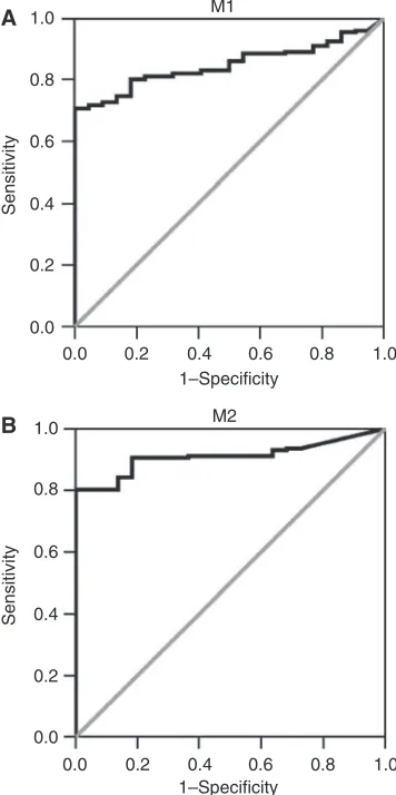
relative methylated DNA quantities of M1/b-actin1000 and M2/b-actin1000 above 95 and 22, respectively, yielding a specificity of 100% for both promoter regions and a sensitivity of 71 and 80% for M1 and M2 promoter regions, respectively. ROC curves for both M1 and M2 promoter regions are shown in Figure 1.
Both gastric cancer cell lines, IPA220 and GP202, showed methylation of both M1 and M2 promoter regions. Of all 202 gastric adenocarcinomas tested, 143 carcinomas (70.8%) showed methylation of the M1 promoter region and 162 carcinomas (80.2%) showed methylation of the M2 promoter region. Methylation prevalences of M1 and M2 promoter regions were significantly different (P¼0.004). Dense methylation, that is, methylation of both M1 and M2 promoter regions, was detected in 133 (65.8%) of the carcinomas. Thirty carcinomas (14.9%) were unmethylated. All normal gastric mucosa samples were unmethylated for both regions. w2 test yielded a signi-ficant difference between methylation status of gastric carci-nomas and normal gastric mucosa tissues (Po0.001). An overview of the methylation status for both promoter regions is given in Table 1.
Correlation ofMAL promoter methylation and survival
Follow-up data was available for 200 out of 202 patients. Only patients without distant metastasis at the time of surgery (M0) were included, leaving 179 patients for the survival analysis. Patients with a gastric carcinoma methylated for the M2 promoter region had a significantly better survival compared with patients with tumours unmethylated for the M2 promoter region (P¼0.03) (Figure 2). No significant correlation was found between M2 promoter methylation and age or gender of the patient, histological type of the tumour, T-stage and N-stage (Table 2). In addition, no significant correlation between M1 promoter methylation status and clinicopathological characteristics, includ-ing survival, was found.
Multivariate analysis revealed only N-stage, T-stage and MAL M2 promoter methylation status, in order of significance, to have independent prognostic value (Table 2).
MALpromoter methylation is associated with reduced
gene expression
Reduced expression of MAL relative to the housekeeping gene snRNP U1A was observed in both gastric cancer cell lines compared with primary keratinocytes that did not show MAL promoter hypermethylation. Of the 17 gastric cancer tissue samples tested for MAL mRNA expression, 11 (64.7%) were methylated for M1, 11 (64.7%) for M2 and 9 (52.9%) samples showed methylation of both regions. Gastric carcinomas with M2 promoter methylation showed significantly lower expression of the MAL gene compared with M2 unmethylated gastric cancers (P¼0.01) (Figure 3, Table 3). For the M1 region, no significant differences in MAL mRNA expression were found between methylated and unmethylated tumours.
DISCUSSION
Gastric cancer is a common disease with generally a poor prognosis (Parkinet al, 2005). Biomarkers can be used to predict prognosis and optimise therapeutic strategies. Hypermethylation of the MAL promoter has been shown in colorectal and oesophageal cancers and MAL has been proposed as a putative tumour suppressor in these cancer types (Mimori et al, 2003; Kazemi-Noureini et al, 2004; Moriet al, 2006). Moreover, it has been proposed as a candidate marker for early detection of these cancers, as methylation of MAL could already be detected in precursor lesions (Lindet al, 2007, 2008; Mimoriet al, 2007). In this study we show thatMALmight have a similar role in gastric cancers, as methylation of MALis detected at high frequency in gastric cancers and not in normal gastric mucosa samples.
In this study, two regions within the MALpromoter, M1 and M2, were analysed. Q-MSP analysis showed methylation of both promoter regions in the two gastric cancer cell lines analysed, indicating that methylation actually occurs in gastric epithelial 1.0
0.8 0.6 0.4 0.2 0.0
1–Specificity 1–Specificity
0.0 0.2 0.4 0.6 0.8 1.0
Sensitivity
Sensitivity
M2
1.0 0.8 0.6 0.4 0.2 0.0 0.0 0.2 0.4 0.6 0.8
[image:3.595.310.560.92.163.2]1.0 M1
Figure 1 Receiver operator characteristics of (A) M1 promoter
methylation and (B) M2 promoter methylation in 202 gastric cancers and 22 normal gastric mucosa samples (FFPE samples only), assuming that normal gastric mucosae are unmethylated and gastric carcinomas are methylated. Chosen cutoff levels for methylation were 95 and 22 for M1 and M2 promoter regions, respectively. This yielded a specificity of 100% for both promoter regions and a sensitivity of 71% for M1 promoter region and 80% for the M2 promoter region.
Table 1 Overview of methylation status of M1 (680 to573 bp) and
M2 (92 to 7 bp) regions within the MAL promoter for 202 gastric carcinoma tissues and 22 normal gastric mucosa tissues
Carcinomas
n¼202 Normal mucosan¼22 P-value
No methylation 30 (14.9%) 22 (100%) o0.001
M1 methylation 143 (70.8%) 0 (0%) o0.001
M2 methylation 162 (80.2%) 0 (0%) o0.001
dense methylation 133 (65.8%) 0 (0%) o0.001
w2 test yielded a significant difference between methylation status of gastric
carcinomas and normal gastric mucosa tissues (Po0.001). 1804
Translat
ional
Table 2 Overview of patient and tumour characteristics of the 179 tumours used in the univariate and multivariate survival analysis
Univariate analysis
Methylated Unmethylated P-value
Age (years) 72 (52 – 96) 71 (54 – 87) NS
Gender
Male 88 (62%) 20 (56%) NS
Female 55 (38%) 16 (44%)
Histological type
Intestinal 100 (70%) 23 (64%) NS
Diffuse 18 (13%) 7 (19%)
Mixed 25 (17%) 6 (17%)
T-stage
T1 10 (7%) 2 (6%) NS
T2 50 (35%) 16 (44%)
T3 78 (55%) 18 (50%)
T4 5 (3%) —
N-stage
N0 41 (29%) 9 (25%) NS
N1 62 (43%) 15 (42%)
N2 32 (22%) 8 (22%)
N3 7 (5%) 4 (11%)
Unknown 1 (1%) —
Multivariate analysis
HR 95% CI P-value
Gender 0.94
Histological type 0.48
T-stage 1.73 1.23 – 2.42 0.001
N-stage 1.52 1.20 – 1.91 0.001
MALM2 methylation 0.59 0.36 – 0.96 0.03
CI¼confidence interval; HR¼hazard ratio. Absolute number and percentages are given for gender, histological type, tumour stage (T-stage) and lymph node status (N-stage). Age is given as mean age and range. None of the clinicopathological characteristics were significantly correlated with M2 promoter methylation status (P¼NS). Multivariate analysis showed that T-stage, N-stage and M2 methylation status are prognostic variables for patient outcome.
Table 3 Relative ln transformed expression values (E) and methylation
status for the M1 and M2 regions within theMALpromoter of the two gastric cancer cell lines and 17 gastric carcinoma tissues
Sample E M1 M2
IPA220 0.003 Methylated Methylated
GP202 0.0003 Methylated Methylated
1 0.35 Methylated Methylated
2 0.24 Methylated Methylated
3 0.06 Methylated Methylated
4 0.72 Methylated Methylated
5 0.10 Methylated Methylated
6 0.05 Methylated Methylated
7 0.77 Methylated Unmethylated
8 0.28 Unmethylated Unmethylated
9 0.19 Unmethylated Unmethylated
10 2.28 Unmethylated Unmethylated
11 0.28 Unmethylated Methylated
12 0.03 Unmethylated Methylated
13 0.03 Methylated Methylated
14 0.04 Methylated Methylated
15 5.56 Unmethylated Unmethylated
16 0.01 Methylated Methylated
17 0.33 Methylated Unmethylated
12 10 8 6 4
Survival (years) 2
0 0.0 0.2 0.4 0.6 0.8 1.0
Cum sur
viv
a
l
P = 0.03 Log rank = 4.96
Methylation No Methylation Censored Censored
n = 70 (143)
[image:4.595.68.253.60.207.2]n = 22 (36)
Figure 2 Kaplan – Meier survival analysis of 179 patients with primary
gastric cancers assessed for the methylation status of the M2 region (92 to7 bp) within theMALpromoter. Patients with primary gastric cancers methylated for the M2 promoter region (n¼143) showed a significantly better survival compared to patients (n¼36) with gastric cancers without M2 promoter methylation (P¼0.03; log rank¼4.96). The number of patients who died of gastric cancer (events) is 70 and 22, respectively.
Methylation No methylation
0 1 2 3 4 5
Expression
6
P = 0.01 M2
Methylation No methylation
0 1 2 3 4 5 6
Expression
P = 0.27 M1
Figure 3 Box plots of the relative expression values of 17 gastric
carcinoma tissue samples methylated and unmethylated for the M1 (A) and M2 (B) promoter regions. Gastric carcinomas methylated for the M2MAL promoter region show significantly lower expression of the MAL gene compared with unmethylated gastric carcinomas (P¼0.01).
1805
Translational
[image:4.595.36.286.341.680.2] [image:4.595.301.551.535.732.2]cells. Earlier studies showed protein expression of MAL by immunohistochemistry in normal gastric mucosa. Strong expres-sion of MAL was observed in parietal and chief cells, but not in muscle and submucosa cells (Marazuela et al, 2003). Gel-based MSP analysis revealed a small subpopulation of unmethylated cells for both the M1 and M2 promoter regions in these cell lines (data not shown). Expression of MALwas hardly detected in both cell lines indicating that methylation is probably the main mechanism of downregulation of this gene. In gastric cancers, methylation of the M2 region was more frequently observed compared with the M1 region (80.2 vs 70.8%). In consistence with what has been described by Lindet al(Lindet al, 2008), an unequal distribution of DNA methylation within the MALpromoter was observed in gastric cancers, with the highest frequencies of methylation in the region closest to the transcription start site. M2 region is located around the transcription start site.
Results of this study show thatMALmay serve as a prognostic marker in gastric cancer, as patients with tumours methylated for the M2 region show significantly better survival compared with patients with tumours unmethylated forMALor methylated only for the M1 region. This survival benefit was independent of other clinicopathological data such as age, gender, histological type of the tumour, tumour stage and lymph node status. Interestingly, also in Hodgkin’s lymphoma patients a similar association was found with a significantly worse survival in patients whose tumours expressed the MAL protein compared with patients with tumours lacking MAL expression (Hsi et al, 2006), indicating a prognostic value ofMALalso in other cancer models.
The finding that inactivation of MAL by methylation gives a better prognosis for the patients may seem contradicting with a putative tumour suppressor function of this gene in gastric cancer. However, this finding has been observed earlier for the mismatch repair genehMLH1, which also has a tumour suppressor function. Inactivation of this gene leads to microsatellite instability of the tumour and patients with microsatellite instable tumours have a better prognosis compared with patients with microsatellite stable tumours (Ribicet al, 2003; Parcet al, 2004; Beghelliet al, 2006). Therefore, tumours without MAL methylation might have a different biology overall, which could relate to poorer clinical
outcome, rather than the outcome being dependent onMALitself. However, this study was performed retrospectively on archival material, and therefore, rather should be considered as hypothesis generating. Actual clinical implementation of MAL promoter hypermethylation as a diagnostic marker requires further valida-tion in a prospective study.
To test the biological relevance ofMALpromoter hypermethy-lation, in a subset of gastric cancers the association betweenMAL promoter hypermethylation and mRNA expression was analysed. Results showed lower expression of MAL in gastric cancers methylated for the M1 or M2 region. The association between promoter methylation and reduced mRNA expression strengthens the biological relevance of MAL methylation and supports a putative role as tumour-suppressor gene. However, correlation between reduced expression and methylation of MAL was only significant for the M2 region, indicating that M2 methylation would have more biological relevance. Two out of 17 gastric cancers showed reducedMALmRNA expression whereas the gene was unmethylated for both promoter regions. This indicates that other regulatory mechanisms ofMALsilencing also exist in gastric cancer, which may include DNA copy number loss, other epigenetic mechanisms, altered expression of transcription factors regulatingMALor microRNAs targeting theMALgene.
In summary, this study shows frequent promoter hypermethyla-tion ofMALin gastric cancers, and not in normal gastric mucosa samples. Promoter hypermethylation of MALis associated with downregulation of its expression, especially when methylation occurs at the M2 region within the promoter. Methylation of this region within the MAL promoter correlates with a significantly better survival of the patients. Altogether, these results pinpoint MALas a putative tumour-suppressor gene with a role in gastric cancer, which may serve as an independent prognostic marker for clinical outcome of gastric cancer patients.
ACKNOWLEDGEMENTS
This study was supported by Dutch Cancer Society, grants KWF 2004-3051 and KWF 2005-3276.
REFERENCES
Baylin SB, Herman JG, Graff JR, Vertino PM, Issa JP (1998) Alterations in DNA methylation: a fundamental aspect of neoplasia.Adv Cancer Res72: 141 – 196
Beghelli S, de MG, Barbi S, Tomezzoli A, Roviello F, Di GC, Vindigni C, Bortesi L, Parisi A, Saragoni L, Scarpa A, Moore PS (2006) Microsatellite instability in gastric cancer is associated with better prognosis in only stage II cancers.Surgery139:347 – 356
Buffart T, Carvalho B, Hopmans E, Brehm V, Klein-Kranenbarg E, Schaaij-Visser T, Eijk P, van Grieken N, Ylstra B, van de Velde CJ, Meijer G (2007) Gastric cancers in young and elderly patients show different genomic profiles.J Pathol211:45 – 51
Carvalho B, Pinto M, Cirnes L, Oliveira C, Machado JC, Suriano G, Hamelin R, Carneiro F, Seruca R (2003) Concurrent hypermethylation of gene promoters is associated with a MSI-H phenotype and diploidy in gastric carcinomas.Eur J Cancer39:1222 – 1227
de Maat MF, van de Velde CJ, Umetani N, de HP, Putter H, van Hoesel AQ, Meijer GA, van Grieken NC, Kuppen PJ, Bilchik AJ, Tollenaar RA, Hoon DS (2007) Epigenetic silencing of cycloo-xygenase-2 affects clinical outcome in gastric cancer.J Clin Oncol25: 4887 – 4894
de Wilde J, De-Castro AJ, Snijders PJ, Meijer CJ, Rosl F, Steenbergen RD (2008) Alterations in AP-1 and AP-1 regulatory genes during HPV-induced carcinogenesis.Cell Oncol30:77 – 87
Esteller M, Corn PG, Baylin SB, Herman JG (2001) A gene hypermethylation profile of human cancer.Cancer Res61:3225 – 3229
Fleisher AS, Esteller M, Wang S, Tamura G, Suzuki H, Yin J, Zou TT, Abraham JM, Kong D, Smolinski KN, Shi YQ, Rhyu MG, Powell SM, James SP, Wilson KT, Herman JG, Meltzer SJ (1999) Hypermethylation of the hMLH1 gene promoter in human gastric cancers with microsatellite instability.Cancer Res59:1090 – 1095
Gartner F, David L, Seruca R, Machado JC, Sobrinho-Simoes M (1996) Establishment and characterization of two cell lines derived from human diffuse gastric carcinomas xenografted in nude mice.Virchows Arch428: 91 – 98
Hsi ED, Sup SJ, Alemany C, Tso E, Skacel M, Elson P, Alonso MA, Pohlman B (2006) MAL is expressed in a subset of Hodgkin lymphoma and identifies a population of patients with poor prognosis.Am J Clin Pathol
125:776 – 782
Kazemi-Noureini S, Colonna-Romano S, Ziaee AA, Malboobi MA, Yazdanbod M, Setayeshgar P, Maresca B (2004) Differential gene expression between squamous cell carcinoma of esophageus and its normal epithelium; altered pattern of mal, akr1c2, and rab11a expression.World J Gastroenterol10:1716 – 1721
Lind GE, Ahlquist T, Kolberg M, Berg M, Eknaes M, Alonso MA, Kallioniemi A, Meling GI, Skotheim RI, Rognum TO, Thiis-Evensen E, Lothe RA (2008) Hypermethylated MAL gene – a silent marker of early colon tumorigenesis.J Transl Med6:13
Lind GE, Ahlquist T, Lothe RA (2007) DNA hypermethylation of MAL: a promising diagnostic biomarker for colorectal tumors.Gastroenterology
132:1631 – 1632 1806
Translat
ional
Livak KJ, Schmittgen TD (2001) Analysis of relative gene expression data using real-time quantitative PCR and the 2(-Delta Delta C(T)) Method.
Methods25:402 – 408
Machado JC, Oliveira C, Carvalho R, Soares P, Berx G, Caldas C, Seruca R, Carneiro F, Sobrinho-Simoes M (2001) E-cadherin gene (CDH1) promoter methylation as the second hit in sporadic diffuse gastric carcinoma.Oncogene20:1525 – 1528
Marazuela M, Acevedo A, Adrados M, Garcia-Lopez MA, Alonso MA (2003) Expression of MAL, an integral protein component of the machinery for raft-mediated pical transport, in human epithelia.J Histochem Cytochem
51:665 – 674
Mimori K, Nishida K, Nakamura Y, Ieta K, Yoshikawa Y, Sasaki A, Ishii H, Alonso MA, Mori M (2007) Loss of MAL expression in precancerous lesions of the esophagus.Ann Surg Oncol14:1670 – 1677
Mimori K, Shiraishi T, Mashino K, Sonoda H, Yamashita K, Yoshinaga K, Masuda T, Utsunomiya T, Alonso MA, Inoue H, Mori M (2003) MAL gene expression in esophageal cancer suppresses motility, invasion and tumorigenicity and enhances apoptosis through the Fas pathway.
Oncogene22:3463 – 3471
Mori Y, Cai K, Cheng Y, Wang S, Paun B, Hamilton JP, Jin Z, Sato F, Berki AT, Kan T, Ito T, Mantzur C, Abraham JM, Meltzer SJ (2006) A genome-wide search identifies epigenetic silencing of somatostatin, tachy-kinin-1, and 5 other genes in colon cancer.Gastroenterology131:797 – 808 Parc Y, Gueroult S, Mourra N, Serfaty L, Flejou JF, Tiret E, Parc R (2004)
Prognostic significance of microsatellite instability determined by immunohistochemical staining of MSH2 and MLH1 in sporadic T3N0M0 colon cancer.Gut53:371 – 375
Parkin DM, Bray F, Ferlay J, Pisani P (2005) Global cancer statistics, 2002.
CA Cancer J Clin55:74 – 108
Puertollano R, Alonso MA (1999) MAL, an integral element of the apical sorting machinery, is an itinerant protein that cycles between the
trans-Golgi network and the plasma membrane. Mol Biol Cell 10: 3435 – 3447
Ribic CM, Sargent DJ, Moore MJ, Thibodeau SN, French AJ, Goldberg RM, Hamilton SR, Laurent-Puig P, Gryfe R, Shepherd LE, Tu D, Redston M, Gallinger S (2003) Tumor microsatellite-instability status as a predictor of benefit from fluorouracil-based adjuvant chemotherapy for colon cancer.N Engl J Med349:247 – 257
Steenbergen RD, Kramer D, Braakhuis BJ, Stern PL, Verheijen RH, Meijer CJ, Snijders PJ (2004) TSLC1 gene silencing in cervical cancer cell lines and cervical neoplasia.J Natl Cancer Inst96:294 – 305
Steenbergen RD, Walboomers JM, Meijer CJ, van der Raaij-Helmer EM, Parker JN, Chow LT, Broker TR, Snijders PJ (1996) Transition of human papillomavirus type 16 and 18 transfected human foreskin keratinocytes towards immortality: activation of telomerase and allele losses at 3p, 10p, 11q and/or 18q.Oncogene13:1249 – 1257
Weiss MM, Hermsen MA, Meijer GA, van Grieken NC, Baak JP, Kuipers EJ, van Diest PJ (1999) Comparative genomic hybridisation.Mol Pathol 52: 243 – 251
Weiss MM, Kuipers EJ, Postma C, Snijders AM, Siccama I, Pinkel D, Westerga J, Meuwissen SG, Albertson DG, Meijer GA (2003) Genomic profiling of gastric cancer predicts lymph node status and survival.
Oncogene22:1872 – 1879
Wilting SM, de WJ, Meijer CJ, Berkhof J, Yi Y, van Wieringen WN, Braakhuis BJ, Meijer GA, Ylstra B, Snijders PJ, Steenbergen RD (2008) Integrated genomic and transcriptional profiling identifies chromosomal loci with altered gene expression in cervical cancer.Genes Chromosomes Cancer47(10):890 – 905
Wisman GB, Nijhuis ER, Hoque MO, Reesink-Peters N, Koning AJ, Volders HH, Buikema HJ, Boezen HM, Hollema H, Schuuring E, Sidransky D, van der Zee AG (2006) Assessment of gene promoter hypermethylation for detection of cervical neoplasia.Int J Cancer119:1908 – 1914
1807

![Poly[tris(μ3 5 aminoisophthalato)diaquadicerium(III)]](data:image/gif;base64,R0lGODlhAQABAIAAAP///wAAACH5BAEAAAAALAAAAAABAAEAAAICRAEAOw==)