This is a repository copy of Experimental validation of plant peroxisomal targeting prediction algorithms by systematic comparison of in vivo import efficiency and in vitro PTS1 binding affinity..
White Rose Research Online URL for this paper: http://eprints.whiterose.ac.uk/84173/
Version: Accepted Version
Article:
Skoulding, NS, Chowdhary, G, Deus, MJ et al. (3 more authors) (2015) Experimental validation of plant peroxisomal targeting prediction algorithms by systematic comparison of in vivo import efficiency and in vitro PTS1 binding affinity. Journal of Molecular Biology, 427 (5). pp. 1085-1101. ISSN 0022-2836
https://doi.org/10.1016/j.jmb.2014.12.003
[email protected] Reuse
Items deposited in White Rose Research Online are protected by copyright, with all rights reserved unless indicated otherwise. They may be downloaded and/or printed for private study, or other acts as permitted by national copyright laws. The publisher or other rights holders may allow further reproduction and re-use of the full text version. This is indicated by the licence information on the White Rose Research Online record for the item.
Takedown
If you consider content in White Rose Research Online to be in breach of UK law, please notify us by
Experimental validation of plant peroxisomal targeting prediction algorithms by systematic comparison ofin vivoimport efficiency andin vitroPTS1 binding affinity
Nicola S. Skoulding#1, Gopal Chowdhary#2,3, Mara J. Deus2, Alison Baker4, Sigrun
Reumann2,5, and Stuart L. Warriner1
#
These authors contributed equally to this work.
1
- School of Chemistry and the Astbury Centre, University of Leeds, Leeds, LS2 9JT, UK.
2
- Centre for Organelle Research (CORE), Faculty of Science and Technology, University
of Stavanger, Richard Johnsons gate 4, N-4021 Stavanger, Norway.
3
- KIIT School of Biotechnology, Campus XI, KIIT University, I-751024 Bhubaneswar,
India.
4
– Centre for Plant Sciences, School of Molecular and Cellular Biology, University of Leeds,
Leeds, LS2 9JT, UK.
5
- Department of Biology, Biocentre Klein Flottbek, University of Hamburg, D-22609
Hamburg, Germany.
Correspondence to Stuart Warriner,[email protected], +44 113 343 6437.
Abstract
Most peroxisomal matrix proteins possess a C-terminal targeting signal type 1 (PTS1). Accurate prediction of functional PTS1 sequences and their relative strength by
computational methods is essential for determination of peroxisomal proteomesin silico, but
has proved challenging, due to high sequence variability of non-canonical targeting signals, particularly in higher plants, and low availability of experimentally validated non-canonical
andin vitrothermodynamic binding of mutated variants within the context of one model targeting sequence. There was broad agreement between the methods for entire PTS1
domains and position-specific single amino acid (aa) residues, including residues upstream of the PTS1 tripeptide. The hierarchy Leu>Met>Ile>Val at the C-terminal position was
determined for all methods but both experimental approaches suggest Tyr is under weighted in the prediction algorithm due to the absence of this residue in the positive training dataset. A combination of methods better defines the score range that discriminates a functional PTS1.In vitrobinding to the PEX5 receptor could discriminate amongst strong targeting signals whilstin vivotargeting assays were more sensitive, allowing detection of weak
functional import signals that were below the limit of detection in the binding assay. Together the data provide a comprehensive assessment of the factors driving PTS1 efficacy and
provide a framework for the more quantitative assessment of the protein import pathway in
higher plants.
Keywords (not in title): PEX5, Fluorescence anisotropy, YFP fusion, peptide, specificity
List of acronyms: aa, amino acid(s); ACX4, acyl-CoA oxidase 4; At, Arabidopsis thaliana; EYFP, enhanced yellow fluorescent protein; Hs, human; PTS1/2, peroxisome targeting signal type 1/2; PWM, position weight matrices; ROS, reactive oxygen species; TPR,
Introduction
Peroxisomes are ubiquitous organelles within eukaryotes, responsible for a wide range of intracellular roles which are critical to cell and organism function. Compared to other cell organelles, peroxisomes are very dynamic and metabolically versatile. For example in cotyledons ofArabidopsis thalianaand other oil seed plants, a major role of peroxisomes is in mobilisation of storage lipids and conversion to carbohydrates to support early
heterotrophic seedling growth. As the cotyledons become photoautotrophic, photorespiration becomes the predominant pathway. Additionally, it is increasingly apparent that peroxisomes are connected into many if not all aspects of plant life, including primary metabolism,
hormone synthesis and signalling of reactive oxygen species (ROS)1. Proteomic studies from different tissues are revealing new and unexpected peroxisomal capabilities, for example in
synthesis of secondary metabolites and in plant defence2; 3; 4; 5. Collectively, these roles are of critical importance for plant fitness and productivity, underscored by the severe, sometimes lethal phenotypes of peroxisome biogenesis mutants6; 7. Different peroxisome functions are determined by their precise enzyme set which in turn reflects the balance between import and turnover of individual proteins and the organelle as a whole.1
Proteins destined for the peroxisomal matrix are typically synthesised in the cytosol with one of two peroxisome targeting signals (PTS1 or PTS2) within their sequence. These are recognised by cytosolic receptors that initiate the import of the cargo protein into the peroxisome. The peroxisome targeting signal type 1 (PTS1) was initially described as a C-terminal motif characterised by the consensus [S/A/C]-[K/R/H]-[L/M]8; 9; 10; 11; 12; 13although it is now known that residues outside the tripeptide also contribute to recognition by the cycling receptor PEX512; 14 13; 15. The PEX5-cargo protein complex interacts with
peroxisomal membrane proteins resulting in translocation of the cargo into the organelle matrix; the receptor is then recycled to the cytosol16. A second targeting signal of
peroxisomal matrix proteins, the PTS2, is located near the N-terminus of cargo proteins and is recognised by a different primary receptor, PEX7. PEX7 acts as an adaptor protein that directly interacts with the so-called long isoform of PEX5 in plants and animals, enabling the two pathways to converge at the peroxisomal membrane16; 17.
membrane and receptor recycling, a process that requires mono-ubiquitination at a conserved N-terminal Cys in mammals and yeast18; 19 20. Since this Cys is conserved in plant PEX5 a similar recycling system most likely operates across eukaryotes. High resolution structures provide molecular level information on the interaction between the C-terminal TPR domain of human and trypanosome PEX5 and model PTS1 peptides21; 22 and full-length PTS1 cargo 23; 24
.
Proteomic analyses of peroxisomes have shown that resident proteins have PTSs that can differ significantly from the simple initial consensus pattern of canonical PTS1 tripeptides2; 3; 4; 5
. However, the technical difficulty of isolating pure peroxisomes makes direct proteomic determination of peroxisomal contents impractical for detailed insight into the variations between species, tissues and as a function of time and environmental stimuli25. A clear
The true potential of bioinformatics lies in the combination and continuous improvement of computational predictions by experimental validations. The definition of a more precise PWM score range for peroxisome import and determining whether the predicted probabilities of PTS1 proteins for peroxisome targeting correlate with import strength and efficiency is important for model development. Novel peroxisomal candidate proteins are typically validated byin vivoexperiments in which the full-length proteins are fused to fluorescent reporters, transiently expressed in plant cells and subsequent cellular localisation is observed 2; 3; 4; 5; 12
Potential drawbacks to this approach are; the effect of introducing a tag, which could potentially mask targeting information; non-physiological levels of expression; and the inability to generate quantitative data. A complementary approach is to explicitly measure the binding constants of putative signals with their receptorin vitroand to use these
thermodynamic parameters to assess if the interactions are strong enough to act as the basis
of cargo recognition and therefore import27; 28. These thermodynamic data provide rapid, robust and quantitative information about the relative affinities of different sequences to a receptor, but are limited by their reduction of protein import complexity to a simplified two-component system, namely the binding of PTS1 peptides to PEX5. Maynard and Berg29 measured affinities of model PTS-1 binding peptides for wild type and mutant human PEX5, and deduced relative free energy contributions of binding for a range of natural human PTS1 sequences and sequences selected from a PTS1 sequence library using Hs PEX5 as bait. This study, which considered predominantly ‘canonical’ PTS1 signals proposed cut-off values for
in vitro affinity that are required for functional PTS-1 signals29. Corresponding studies have not been performed in the plant context and systematic analysis of weaker PTS-1 signals is lacking, making the cross validation ofin vivo, -in silicoandin vitromethods hard to perform reliably.
In this work we report a systematic study of mutagenised putative PTS1 domains, validating
in silicopredictions by the two independent and complementary methods ofin vivotargeting studies andin vitrodetermination of PTS1 peptide affinities. This data set allows; (i) more precise definition of the prediction grey-zone, (ii) validation of the predicted,
position-specific strength of individual PTS1 tripeptide residues, and (iii) validation of the identity and function of targeting enhancing and inhibitory residues located in the eleven residues
PTS1 targeting elements and domains. The comparative data raise intriguing questions regarding how cytosolic plant proteins are able to evolve extremely weak non-canonical PTS1s for peroxisome targeting while competing with native canonical PTS1 proteins for PEX5 binding.
Results
In the PWM-based PTS1 protein prediction model, each of the 20 possible aa residues of the C-terminal 14-aa sequence is assigned a position-specific score that indicates whether a specific residue at a particular sequence position is predicted to enhance (more positive score) or reduce peroxisome targeting (more negative score) and to what extent (Suppl. Table 1). The total prediction score represents the sum of the position-specific PWM scores of the
C-terminal 14 aa residues30. Until now, however, quantitative experimental data validating the predicted targeting efficiency of single PTS1 domain residues (of PTS1 tripeptides or
upstream residues) and of entire PTS1 domains have remained scarce, resulting in a relatively
imprecise definition of the threshold for peroxisome targeting. .
To minimize secondary effects such as aa residue interdependency and secondary structure, an effect analysis of specific single and multiple point mutations introduced either into the PTS1 tripeptide or into the upstream domain is best investigated in the context of one specific constant model sequence. TheZinnia elegansacyl-CoA oxidase 4 (ZeACX4) sequence was considered suitable and representative because (i) the PTS1 domain construct was weakly targeted to peroxisomes in onion epidermal cells, as determined byin vivosubcellular targeting analyses12(and Fig. 1a), (ii) the sequence terminated with a non-canonical, experimentally validated PTS1 tripeptide (SRV>, “>” designates the extreme C
terminus), (iii) the domain upstream to the PTS1 tripeptide contained predicted enhancer elements and (iv) the PTS1 domain had been assigned a relatively low prediction score below threshold in the prediction grey-zone.
Validation of the PTS1 protein prediction model by semi-quantitativein vivosubcellular targeting analyses using mutagenized PTS1 domain constructs
detected in organelle-like punctuate structures that coincided with DsRed-SKL labelled peroxisomes in double transformants (Fig. 1a3,b, Table 112). The peroxisome targeting efficiency of the model sequence was referred to as weak (detectable only after several days). The positive control EYFP-PTS1 (EYFP extended C-terminally by a PTS1 decapeptide terminating with CKI>, Fig. 1c) labelled peroxisomes 18-24 h post transformation (p.t.) (referred to as strong peroxisome targeting) and EYFP alone was cytosolic at all time points (Fig. 1d).
Initial experiments focused on aa mutations at position -1 (The aa residues considered for the PWM model are numbered -1 to -14 with position -1 referring to the C-terminal residue). The PWM prediction score matrix indicates that the six aa residues that have been experimentally determined to occur in plant PTS1 tripeptides at position -1 ([LMIFVY]) possess differential
predicted targeting strengths, ranging from high for Leu (PWM score=0.66) and Met (0.64), followed by Ile (0.33) to weak for Phe, Val and Tyr (-0.09 to -0.016)12, (Suppl. Table 1). Consistent with the increase of the PTS1 prediction score for the mutagenized sequence SR(V-to-I)> (from 0.216 to 0.664, Table 1), peroxisome targeting of the corresponding EYFP construct was detected at all three time points p.t. (18-24 h, 48 h and 7 d), as shown in single transformation without image modifications of brightness and contrast (Fig. 1e). Peroxisome targeting was confirmed in double labelling experiments using DsRed-SKL as peroxisomal marker (Suppl. Fig. 1a). Hence, the single point mutation V-to-I (pos. -1) converted the weak domain into a strong PTS1 domain as predicted (Fig. 1e, Table 1).
Similarly the mutation SR(V-to-M)> significantly enhanced peroxisome targeting from weak to strong efficiency (Fig. 1f, Table 1, Suppl. Fig. 1b). The significantly higher PTS1
prediction score of Met at pos. -1 (PWM score=0.66) compared to Ile (0.33) suggested that both strong PTS1 tripeptides might still differ in peroxisome targeting efficiency if
investigated at sufficiently high resolution. Hence, reporter gene expression and fusion protein targeting was investigated at very early time points (4 h, 8 h, 12 h and 24 h) after biolistic bombardment. While reporter gene expression was hardly detectable until 8 h p.t., EYFP expression and fluorescence became visible 12 h p.t. for both constructs (SRM> and SRI>) without significant differences in cellular fluorescence intensity (Suppl. Fig. 1 d2 and e2). Significant differences in peroxisome targeting, however, could be resolved for both PTS1s. While the reporter fusion terminating with SRI> remained fully cytosolic in all cells
SRM> became clearly detectable in peroxisomes against some yellow fluorescent
background of newly synthesized EYFP and, hence, was assigned very strong peroxisome targeting efficiency (Suppl. Fig. 1 d2). This difference in cytosolic versus peroxisomal targeting was consistently found in nearly all transformed cells and reproducible in independent experiments.
The PTS1 tripeptide alteration SR(V-to-Y) marginally reduced the PTS1 domain prediction score from 0.216 to 0.173 (Table 1). Contrary to the expected maintenance or reduction of weak peroxisome targeting, the SRY> construct targeted peroxisomes with moderate
efficiency, as indicated by the detection of peroxisome targeting 48 h p.t. (Fig. 1g, Suppl. Fig. 1c). To verify the specificity of protein import into peroxisomes in the given experimentalin
vivosystem, we further investigated one predicted deleterious position -1 mutation. The point mutation SR(V-to-K)> reduced the PTS1 domain prediction score slightly by 0.1 (from 0.216 to 0.119, Table 1), and positively charged aa residues have not been identified at position -1 in plant PTS1 tripeptides. Indeed, the reporter fusion terminating with SRK> remained cytosolic even at maximum sensitivity of detecting weak peroxisome targeting (7 d p.t., Fig. 1h).
the reporter fusion terminating with STV> was no longer targeted to peroxisomes (Fig. 1j). These experimental data confirmed the high specificity of peroxisomal protein import in the chosenin vivosystem and assisted in defining experimentally the lower limit of PTS1 domain prediction scores for peroxisome import (PWM score=-0.2, Fig. 2).
Among all 12 possible residues allowed at pos. -3, Ser is assigned the maximum peroxisome targeting strength. The PTS1 tripeptide mutation to PRV>, which reduced the PTS1 domain prediction score from 0.216 to -0.135 (Table 1), abolished any reporter fusion targeting to peroxisomes (Fig. 1k).
Next, the effect of multiple point mutations introduced into the PTS1 tripeptide of the model sequence was investigated. The dual tripeptide mutation from SRV> to SNM> significantly
enhanced peroxisome targeting from weak to moderate strength, as fluorescent peroxisomes became detectable 48 h p.t. (Fig. 1l). The result fully agreed with the significant increase in PTS1 domain prediction score (from 0.216 to 0.523, Table 1). Conversely, the dual tripeptide
mutation from SRV> to SNY> abolished the weak peroxisome targeting of the model sequence (Fig. 1m). The experimental result was fully consistent with the significant decrease in PTS1 domain prediction score (from 0.216 to -0.272, Table 1).
Finally, potential enhancing function of upstream residues on peroxisome targeting was investigated. The upstream domain of the model sequence (VAKTTRP-SRV>) contained two basic residues (Lys, Arg) and one Pro residue, all of which are generally considered to act as targeting enhancing elements in plant PTS1 sequences31; 32. First, the two basic residues (K position -8; R position -5) and one Pro (P) residue (position -4) were exchanged to Gly (G) residues, thereby lowering the PTS1 domain prediction score slightly from 0.216 to 0.073. Similar to the original sequence, the reporter fusion terminating with the mutated decapeptide (VAGTTGG-SRV>) remained detectable in peroxisomes 7 d p.t. (Fig. 1n). In contrast, changing the two basic upstream residues to acidic residues (VAETTDP-SRV>), which further lowered the PTS1 domain prediction score (to 0.011), completely abolished
Posterior probabilities facilitate the interpretation of the absolute prediction scores and quantify the probability for peroxisome targeting, ranging from zero (0% probability) to one (100%), with 0.5 corresponding to the prediction threshold of 50% probability for
peroxisome targeting12. In addition to the initial standard posterior probability12, a so-called balanced probability value has been calculated for the PWM model26by assuming an equal variance of positive (PTS1) and negative (non-PTS1) example sequence scores, which leads to a broader intermediate probability value range and higher targeting probability values for sequences differing from the majority of positive examples, i.e., non-canonical and low-abundance peroxisomal proteins. On the downside of increased sensitivity, the fraction of non-peroxisomal proteins with probability values >50% increases substantially and leads to a higher proportion of false positive predictions. To better visualise the relationship betweenin
vivotargeting andin silicoprediction of targeting signals, the experimentally tested sequences were grouped into four categories (cytosolic, weak, moderate or strong
peroxisomal targeting) and plotted against the PWM score, standard posterior probability and balanced posterior probability scores (Fig. 2, Table 1 and Suppl. Tables 2 & 3). The analysis reveals a clear positive correlation between PWM score and experimentally determined strength of targeting, although there is overlap of scores between categories that can be distinguished experimentally (Fig. 2a, Suppl. Table 2 & 3). the standard posterior probability does not sensitively discriminate between sequences with differentin vivodetermined
targeting strengths (Fig. 2b), the balanced post posterior probability is superior in its
discrimination ability (Fig. 2c), particularly between moderate and cytosolic proteins, which are poorly distinguished using the other methods (correlation matrices are shown in
Supplementary Information Table 3). Some weakly peroxisome-targeted sequences and one moderately peroxisome-targeted sequence (SRY>) fall below the 50% threshold of the balanced posterior probability (Fig. 2, Suppl. Table 2), indicating that iterative approaches combining bioinformatics and experimental research are required in the future to further improve the prediction ability of non-canonical PTS1 sequences
Determination of PTS1 peptide binding affinities to AtPEX5
In order to better understand the thermodynamics of binding between Arabidopsis (At)PEX5 and a range of potential targeting sequences, a series ofin vitroexperiments were performed.
the full length protein (aa 1-728, termed PEX5) and an N-terminal truncation comprising aa 340-728 (PEX5C)17(Fig. 3a,b). The latter is equivalent to the human PEX5 construct used to determine the thee-dimensional structure of human (Hs)PEX521. The PTS1 domains to be investigated in this study were prepared by solid phase peptide synthesis and their binding affinities to AtPEX5 were determined using a fluorescence anisotropy-based assay
(Supplementary Information, Section 3)27; 33. The assay determines the amount of a fluorescently labelled tracer peptide (in this case the tightly binding pentapeptide YQSKL labelled at the N-terminus with LissamineTMrhodamine) associated with the receptor by virtue of the slower tumbling rate of the fluorophore when it is bound to PEX5 (higher anisotropy). The limits of anisotropy of the tracer is first determined by direct titration of the protein (e.g. PEX5C) into a fixed concentration of tracer (Suppl. Fig. 2) and the Kdof the tracer was determined by titration of the tracer solution into the protein (Fig. 3c, Suppl. Fig.
3). Fitting to the appropriate equations for a 1:1 binding model (see Methods) showed the Kd of the tracer peptide YQSKL to be virtually identical for the two receptor constructs (as 4.0 ± 0.5 nM for PEX5C and 4.5 ± 1.2 nM for PEX5, in good agreement with the value of 3.1 nM reported for the truncated human PEX521). Once the affinity of the tracer to its receptor is known, the binding of a range of unlabelled sequences can be determined by using a
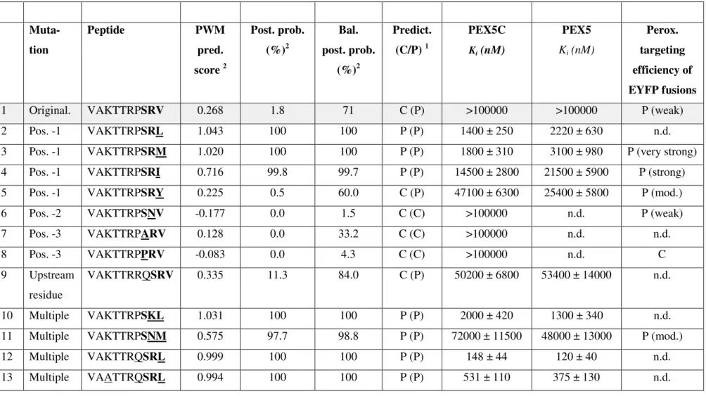
competition assay in which unlabelled peptides compete to displace the tracer from the PTS1 binding site on PEX5. The concentration of the peptide of interest required to displace 50% of the initially bound fluorophore from PEX5 (IC50) can be mathematically combined with the known affinity of the tracer for PEX5 to give the binding constant (expressed as Ki) for the sequence of interest. Example competition curves are shown in Fig. 4 for the peptide VAKTTRPSRV> and variants ending in M, I and Y binding to PEX5C. Affinity of both full length PEX5 and PEX5C for a total of 19 peptides was determined and are shown in Table 2 and Supplementary Figs 4 and 5. The peptides tested showed a range of Kivalues from 100
nM to undetectable (>100M). No significant differences in binding affinity of individual
peptides to PEX5 compared to PEX5C were observed (Table 2).
In accordance with the critical function of the most C-terminal residue in PTS1 tripeptides in peroxisome targetingin vivo(see above), initial studies focused on point mutations at
position -1. The affinity of the original model peptide of ZeACX4 (VAKTTRPSRV>) to PEX5 and PEX5C was below the detection limit with Ki>100 µM (Table 2, Entry 1; Fig. 4).
to PEX5C (Ki=1-3M) with Leu marginally better (Table 2, Entries 2 & 3). The mutation to
Ile in the -1 position resulted in an order of magnitude decrease in binding affinity (15-21
M), consistent with the PWM model prediction scores (Table 2, Entry 4; Fig. 4). As
predicted and consistent with thein vivodata, the mutation to Tyr caused a further 3-fold
decrease (25-47M). Contrary to the PWM model predictions but fully consistent with the semi-quantitativein vivo peroxisome targeting analyses, the SRY> peptide showed higher PEX5 binding affinity and moderate peroxisome targeting efficiency compared to the original model peptide terminating with SRV> (undetectable PEX5 binding, weakin vivoperoxisome
targeting, Figs. 1 and 4, Tables 1 and 2, Entry 5). The thermodynamic results demonstrate the preference of Arabidopsis PEX5 for long hydrophobic side chains at position -1 (L, M) since
both the branched Ile and especially Val significantly reduced PEX5 binding. Consistent with this conclusion, V-to- L/M mutations increased PEX5 binding affinity also for
VAKTTRPSN(V-to-M) and the shorter peptides YQSK(V-to-L) (Table 2, Entries 6 & 11; 17 & 18).
To investigate the effect of position -2 mutations on PEX5 binding affinity, multiple mutations were introduced into the original model peptide because the affinities of both the SRV> and the SNV> decapeptide for PEX5 were below detection limit (Table 2, Entries 1 & 6). Fully consistent with the PWM predictions, the mutation of Arg at position -2 to Lys combined with the V-to-L mutation at position -1 in the first model peptide maintained the high binding affinity of 1-2 µM (VAKTTRPSRL>, PWM score: 1.043; VAKTTRPSKL>, PWM score: 1.031), showing equivalence of Arg and Lys (position -2) in terms of PEX5 binding (Table 2, Entries 2 & 10). Asn in position -2, however, greatly decreased the binding affinity when combined with the favourable Met in the -1 position about 20-fold
(VAKTTRPSRM>, Ki=1.8-3.1 µM; VAKTTRPSNM>, Ki=48-72 µM) (Table 2 , Entries 3 & 11). This result is also consistent with the reduction in PWM score (from 1.02 to 0.58) and thein vivodata from strong to moderate peroxisome targeting (Fig. 1, Table 1, 2). SNM (in context with different upstream residues) had been previously characterized as a functional non-canonical PTS1 in plants.12
Upstream residues are known to be able to enhance the function of weak PTS1 tripeptides
and Pro is found reasonably frequently in positions -4 and -5 of natural plant PTS1 proteins12. Therefore the effect of substituting the Pro at position -4 with Gln was examined.
2, Entries 12 & 2)), showing that, at least in this specific context of the strong PTS1 tripeptide SRL, Pro did not show an additional targeting enhancing effect. Substitution of Pro with Gln in the peptide VAKTTRQSRV> (Table 2, Entries 1 & 9), however, significantly increased binding from undetectable to Ki=50-53 µM, similar to VAKTTRPSNM> (Ki=48-72 µM) (Table 2, Entry 11). Substitution of the two basic residues in the peptide VAKTTRQSRL> with neutral Ala residues, singly and in combination, resulted in a decrease in affinity of binding that was additive (Table 2, Entries 13,14 & 15), confirming the importance of upstream basic residues.
Discussion
Despite molecular details on binding of peptides and cargo proteins to PEX521; 22; 23; 24,
mutational studies of PTSs11; 14; 15and the availability of an increasing catalogue of
peroxisomal proteins from proteomic studies2; 3; 4; 5; 34, it remains difficult to predict reliably the identity of non-canonical PTS1 domains. Therefore, improved informatic tools that can
accurately predict the peroxisomal complement of organisms from sequenced genomes would be very useful. In addition, it is desirable to predict the strength of peroxisome targeting for PTS1 proteins of interest to infer, for instance, quantitative peroxisome targeting or dual protein localization in different subcellular compartments for proteins with multiple targeting signals. Also, understanding potential variations in PTS targeting strength can give insight into regulation of the composition of the peroxisome proteome and the evolution of PTSs to endow peroxisomes with new capabilities.
Comparison ofin vivoandin vitroexperimental data within silicopredictions
Overall the three methods deployed in this study agreed remarkably well, even at highest resolution of the targeting/affinity strength of position-specific single aa residues of the PTS1 tripeptide. Fig. 5 shows a graphical representation of the relationship between the peroxisome targeting prediction by the PWM model, the measured binding affinity by fluorescence anisotropy and the strength of targeting as determined semi-quantitatively byin vivoassay.
Sequences that behaved as strong PTSsin vivo(giving rise to fluorescent peroxisomes within 24 h) such as VAKTTRPSRM> and VAKTTRPSRI> had high PWM scores and balanced post posterior probabilities and bound both PEX5 and PEX5C with micromolar affinity. Both
terminal (-1) position L>M>I>Y>V,and this matched well to the individual scores for these
residues in the same position (Supp. Table 1), and is in good agreement with the aa residue frequency of naturally occurring Arabidopsis PTS1 proteins12.
The targeting strength of Tyr at pos. -1 might have been underestimated due to the complete lack of Tyr at this position in any of the 2600 positive example sequences of plant PTS1 tripeptides used for model training. The first plant PTS1 protein carrying Tyr at pos. -1 was only identified relatively recently.35The experimental data suggest that the PWM score for this sequence should be 0.2 to 0.4 units higher to bring the result in line with sequences with similar affinities and biological import properties. This Tyr example stresses the importance of identifying novel Arabidopsis proteins carrying novel residues in their non-canonical PTS1 tripeptides since these residues are often conserved in orthologs of diverse plant species and
altogether significantly improve residue representation in the large dataset of positive
example sequences against predominance of canonical PTS1 triptide residues. A more precise evaluation of the effect of Tyr at pos – 1 will require further investigation of this residue in a wider range of sequence contexts.
The peptide terminating in SRV> was below the binding detection limit for thein vitroassay. The higher sensitivity of thein vivosystem in detecting (weak) peroxisome targeting (Fig. 1, 2) compared to the thermodynamic assays, is remarkable and might indicate that additional components such as binding partners and/or posttranslational mechanisms enhance the affinity of non-canonical PTS1 tripeptides for PEX5in vivo. Conversely, thein vitrobinding assays were able to discriminate between strong targeting peptides that were not able to be resolved by thein vivoassays (Figs. 1 and 5, Table 2), revealing complementary information and an important advantage of thermodynamic binding studies.
Similarly, the STV> peptide was non-peroxisomal in this study, still consistent with the fact that Thr has been characterized as a plant PTS1 tripeptide residue for STL>12.
Clear evidence was also obtained for the importance of aa residues upstream of the PTS1 tripeptide in modulating PEX5 affinity and peroxisome import efficiency. Pro occurs at position -4 with reasonable frequency in natural PTS1s12; 31. In thein vitroexperiments the most significant effect was changing the Pro at position -4 to Gln which increased the affinity by a factor of 10 (Table 2). For shorter pentapeptides, however, only a very small effect on the binding affinities was observed (compare YQSKL> and YPSKL>, Table 2). It is possible that the cis-trans isomerisation of Pro could result in a conformation of the backbone within the longer decapeptide which does not favour receptor binding whereas the structural change does not affect binding in the shorter sequence context. Replacement of the two basic
residues at position -5 and -8 reduced binding affinity in an additive fashion.In vivo, replacement of these residues with neutral ones had no detectable effect but acidic residues were clearly deleterious. Taken together, the results of upstream residue mutations confirmed the targeting enhancing role basic and Pro residues compared to the generally inhibitory role of acidic residues upstream of PTS1 tripeptides, as reported previously.32
The experimentally determined threshold for peroxisome targeting appears to be near 0.15-0.05 since both weakly peroxisomal and cytosolic constructs are located in this prediction grey-zone, which is now much better defined. Except for one apparent outliner (SNY>), four mutated model sequences with PTS1 scores below 0.05 were cytosolic, strongly suggesting that this is a realistic threshold to delineate experimentally the lower limit of the prediction grey-zone.
Implications for cargo binding to PEX5
It has recently been proposed that PEX5 from Pichia undergoes redox regulated disulfide bond formation at the conserved N terminal Cys which alters the affinity of the receptor for its cargo.36One surprising observation is that the binding affinity of all the peptides tested in the present study was, within experimental error, identical for both truncated and full length PEX5. Since the N terminally truncated PEX5C lacks this redox sensitive Cys such a
mechanism would appear not to be relevant in the context of the binding of short peptides in our experimental system. It should be noted that there are several known cases where
instance, human alanine-glyoxylate aminotransferase (AGT), which has a non-canonical PTS1 (KKL>), binds much more tightly to HsPEX5C than the equivalent peptide24. The X-ray crystal structure of AGT in complex with HsPEX5C revealed a folded and enzymatically active dimer with each subunit bound via its PTS1 to PEX5. In addition to the interaction of the PTS1 tripeptide with the central funnel formed from the TPRs, an extended interface between the C-terminal domain of AGT and the PEX5 surface was observed. While residues immediately upstream of the PTS1 contributed to binding, there were also contributions from more distant residues. Further, residues that affected AGT folding, even to a minor extent, disrupted the interaction and therefore the import24. The other structure where a complex between a full length cargo protein (mSCP2 which has a canonical PTS1) and PEX5C is known23shows a complete lack of conservation of interactions outside the PTS124. This, together with the reports that certain proteins, such as catalase, with non-canonical PTS1
make additional contacts to the region of PEX5 outside of the TPR domain37may make the prediction of ‘weak’ PTS1s by only bioinformatic or experimental analysis of the C-terminal region very challenging. The more extensive use of biophysical tools to measure quantitative binding constants for a range of recombinant peroxisomal proteins and full length and
truncated PEX5 constructs may help to address these questions.
Towards mechanistic and quantitative models of import
A simple pre-equilibrium model, in which the concentration of cargo loaded PEX5 determines the likelihood of import, requires the concentrations of the cargo protein or its receptor in the cytosol to be close to the Kdfor binding. It has been suggested that,
consequently, proteins with lower expression levels may well have evolved stronger PEX5 binding sequences to offset their low abundance28. In contrast, the most abundant plant peroxisomal enzymes, generally carry canonical PTS1s of high peroxisome targeting strength12; 31. It is notable that both bioinformatics andin vivomeasurements show that protein import into the peroxisome can be observed using sequences that have an affinity for the receptor that exceeds 100 µMin vitro. Given that it is unlikely that either PEX5 or the cargo protein generally reach this level of expression, these detailed thermodynamic insights pose interesting questions about the underlying mechanistic details of the import process.
Previous work28; 29; 33suggested that the C-terminal peptide motifs tend to havein vitro
affinity were also observed. An affinity limit of ~500 nM for import competent sequences was proposed based on the measured affinities of model peptides for human PEX5 and two pathogenic mutants, and deduced binding energies of native PTS1 sequences29and a dataset of PTS1 sequences selected from a yeast 2 hybrid library using human PEX5 as bait10; 29. These observations are markedly different to those associated with import competent systems in this study, with import being observed for protein tagged with PTS-1 sequences that have significantly weaker affinitiesin vitrothan previously reported. While noting that the
experimental data in the earlier studies were obtained with the human PEX5 protein, the high degree of homology between the PTS-1 import apparatus of eukaryotes means that such different observations are hard to reconcile on this basis. However it is worthy of note that some natural human PTS1 peptides have higher Kds and correspondingly lower calculated binding energies28than the previously proposed threshold29. In the present study a range of
non-canonical PTS-1 sequences with predicted weaker targeting efficiency were
systematically tested for theirin vitrobinding and explicitly tested for theirin vivotargeting. This has enabled more light to be shed on the precise limits of the targeting peptide affinities that can actually drive import. Nevertheless the weakin vitrobinding of some of the PTS-1 sequences which are import competent still poses interesting questions about the precise mechanistic details of the import process.
One possibility is that other factors may influence the overall magnitude of the binding constants within the import system, although not the fundamental rank order for effects of individual residues. For instance, PEX5 interacts with PEX7 in the cytosol, and both PEX5 and many of its cargoes may exist as oligomers allowing for multivalent interactions to occur that might alter binding constants measured in a simplified system. In our hands (and
consistent with the data reported for the truncated human PEX520) at the low protein concentrations used in these assays 1:1 binding models provided good fits to the observed data, although it was noted that at much higher PEX5 concentrations deviations from idealised 1:1 binding curves started to be observed consistent with the presence of higher order oligomers affecting the equilibria being studied.
Thein vivodata also show that proteins with weaker PTS1s take longer to accumulate in the peroxisome. Thein vivolong-term expression studies resemble pulse chase experiments in the sense that protein synthesis primarily occurs within the first 24 h p.t. during cell
incubation and import seems to occur gradually over this time. In a model in which reversible binding to PEX5 is more rapid than import, the fraction of any given cargo bound to the receptor is determined by the ratio of the products of the individual Kds with the individual protein concentrations. The slower import of more weakly targeted proteins is hence consistent with two possible import mechanisms: either the weaker binding affinity of the non-canonical PTS1s results in only a small fraction of the cargo being imported at any time or (under these experimental conditions) the weaker PTS1s are only imported after
endogenous proteins with canonical PTS1s have been quantitatively imported and eliminated as competing cargo from the cytosol. However, in either case, the strength of the PTS1 determines the priority of the protein for import. In some situations slow import may be desirable, if a protein requires assembly and maturation steps in the cytosol as proposed for catalase.38Importantly, at all PTS1 tripeptide positions, single point mutations (SR(V-to-K),
S(R-to-T)V and (S-to-P)RV) completely abolished peroxisome targetingin vivo, demonstrating that peroxisome import is specific in the experimental system.
The wide range ofin vitrobinding affinities determined for the strong targeting sequences, all of which show exclusive peroxisomal localisation, may provide further evidence for the plant’s requirement to control the priority for import of peroxisomal proteins within the context of a complex and ever changing expression profile. In the physiological situation, changes in expression (for example due to circadian rhythm, tissue differentiation or stress situations) will alter the composition of the pool of proteins competing to be imported into peroxisomes. Understanding the processes governing competitive import will require detailed understanding of the kinetic parameters of the import system and measurement of the steady state pools of cargo and receptor, which will be determined by the rates of protein synthesis, cycling rate between cytosol and peroxisome, and turnover. In addition the extent to which these processes are operating at equilibrium would need to be determined to allow
Evolution of peroxisome targeting signals
Genome size expansion in multicellular complex organisms also increased the absolute number of nuclear-encoded proteins targeted to subcellular organelles. While the N-terminal targeting signals for mitochondria, plastids and the secretory pathway generally evolved by exon shuffling, the relatively short C-terminal PTS1 appears to be able to evolve by random point mutations of 3’coding regions, alternative splicing and ribosomal read-through of stop codons39; 40; 41. Indeed, phylogenetic analysis suggested, and experimental analyses validated, that cytosolic and mitochondrial proteins of green algae and mosses can slowly evolve non-canonical and subsequently non-canonical PTS1s in higher plants to facilitate peroxisome
targeting26; 42. Hence, cytosolic proteins that have entered this evolutionary track and initially possess extremely weak affinity to PEX5 must be given an opportunity of being successfully imported into peroxisomes, at least under some specific circumstances. Peroxisome import
then offers a selective advantage, thereby increasing organismal fitness and propagation which, in a positive feed-forward spiral, further advances and accelerates C-termini evolution into weak non-canonical and ultimately strong canonical PTS1s. This import capability of newly evolving peroxisomal cargo of lowest PEX5 affinity is difficult to envisage in a model where proteins with strong PTS1s are constantly synthesized and saturate the import
Experimental Procedure
Peptides were prepared using standard Fmoc based peptide synthesis strategies43using 2-chloro-trityl linked solid supports which were purchased with the C-terminal residue already loaded. Standard side chain protection was employed: Arg (Pbf), Asn & Gln (Trt), Glu & Asp (OtBu), Lys (Boc), Ser, Thr & Tyr (tBu). Coupling cycles were performed in
dimethylformamide, using 5 eq. of Fmoc protected aa activated with 5 eq. HCTU (O-(1H-6-chlorobenzotriazole-1-yl)-1,1,3,3-tetramethyluronium hexafluorophosphate) and 10 eq. of di-isopropylethylamine . Fmoc deprotection was performed with 20% piperidine in DMF. Following assembly of the sequence the peptide was cleaved from the resin using a cocktail
of CF3CO2H /H2O, triethylsilane (95:2.5:2.5). When sequences contained Met residues an additional 1% ethanedithiol was introduced and the solution degassed with nitrogen prior to use to prevent sulfoxide formation. The cleavage solutions were concentrated and crude
peptide was isolated by precipitation from diethyl ether and purified by preparative HPLC. Fluorescently labelled YQSKL was prepared by coupling the N-terminus of the peptide with lissamine sulfonyl chloride prior to cleavage. Detailed procedures and peptide
characterisation are reported in the Supplementary Information.
Recombinant PEX5 and PEX5C were prepared as described in Lanyon-Hogg et al.17
Fluoresence Anisotropy assays we performed in 384 well microtitre plates (Black Perkin Elmer Optiplates) as follows. Five solutions were prepared [A: FA Buffer (HEPES (20 mM), NaCl (150 mM), pH 7.5); B: Blocking solution: FA buffer containing 0.32 mg/ml of porcine gelatine; C: 12 point dilution series of test peptide in FA buffer (4 mM-20 nM); D:
channels each with 595(60) nm filters but with orthogonal polarisation (S and P polarisers). 30 flashes were used per measurement. The instrument response factor (g value) was set to 1 on the instrument. The data were blank corrected and processed to give a blank corrected anisotropy and the data processed as detailed in the Supplementary information Section 3 to give an IC50value for the competition experiment which was combined with the Kdof the Tracer to give the Ki, the binding constant for the unlabelled peptide.
Transient Import
Inin vivosubcellular targeting analyses, the C-terminal 10 residues of the wild-type model sequence from Zinnia ACX4 and of mutagenized variants thereof were fused to the C-terminus of EYFP by PCR using an extended reverse primer (see Suppl. Table 4) and
subcloned into the plant expression vector pCAT under control of a double 35S cauliflower mosaic virus promoter44and sequenced. For labeling of peroxisomes in double
transformants, DsRed-SKL was used45; 46. Onion epidermal cells were transformed
biolistically as described46. The onion slices were placed on wet paper in Petri dishes, stored at room temperature in the dark for approx. 16 h, and analyzed directly (referred to as 18-24 h p.t.) or after additional tissue incubation at 10°C in the dark for approx. 1 d (referred to as 48 h p.t.) to 6 d (referred to as 7 d p.t.). Fluorescence image acquisition was performed on a Nikon TE-2000U inverted fluorescence microscope equipped with an Exfo X-cite 120 fluorescence illumination system and single filters for YFP (exciter HQ500/20, emitter S535/30) and DsRed (exciter D560/40X, emitter D630/60M). The images were captured using a Hamamatsu Orca ER 1394 cooled CCD camera. Standard image acquisition and analysis was performed using Volocity II software (Improvision) and Photoshop.
Acknowledgements
This work was supported by a targeted priority studentship in chemical biology from the BBSRC (to NS), a YGGDRASIL IS-MOBIL fellowship from the Research Council of Norway (RCN, to GC), a FRIBIOMED grant by RCN (NFR 204822/F20 to SR) and
a Leonardo Da Vinci fellowship for technicians (to MD). We also thank Dr Thomas Lingner for critical reading of the manuscript.
1. Hu, J., Baker, A., Bartel, B., Linka, N., Mullen, R., Reumann, S. & Zolman, B. (2012). Plant Peroxisomes: Biogenesis and Function.Plant Cell24, 2279-2303.
2. Eubel, H., Meyer, E., Taylor, N., Bussell, J., O'Toole, N., Heazlewood, J., Castleden, I., Small, I., Smith, S. & Millar, A. (2008). Novel Proteins, Putative Membrane Transporters, and an Integrated Metabolic Network Are Revealed by Quantitative Proteomic Analysis of Arabidopsis Cell Culture Peroxisomes.Plant Physiology148, 1809-1829.
3. Quan, S., Yang, P., Cassin-Ross, G., Kaur, N., Switzenberg, R., Aung, K., Li, J. & Hu, J. (2013). Proteome Analysis of Peroxisomes from Etiolated Arabidopsis Seedlings Identifies a Peroxisomal Protease Involved in beta-Oxidation and Development.Plant Physiology163, 1518-1538.
4. Reumann, S., Babujee, L., Ma, C., Wienkoop, S., Siemsen, T., Antonicelli, G., Rasche, N., Luder, F., Weckwerth, W. & Jahn, O. (2007). Proteome analysis of Arabidopsis leaf peroxisomes reveals novel targeting peptides, metabolic pathways, and defense mechanisms.Plant Cell19, 3170-3193.
5. Reumann, S., Quan, S., Aung, K., Yang, P., Manandhar-Shrestha, K., Holbrook, D., Linka, N., Switzenberg, R., Wilkerson, C., Weber, A., Olsen, L. & Hu, J. (2009). In-Depth Proteome Analysis of Arabidopsis Leaf Peroxisomes Combined with in Vivo Subcellular Targeting Verification Indicates Novel Metabolic and Regulatory Functions of Peroxisomes.Plant Physiology150, 125-143.
6. Fan, J., Quan, S., Orth, T., Awai, C., Chory, J. & Hu, J. (2005). The Arabidopsis PEX12 gene is required for peroxisome biogenesis and is essential for development.Plant Physiology139, 231-239.
7. Sparkes, I., Brandizzi, F., Slocombe, S., El-Shami, M., Hawes, C. & Baker, A. (2003). An arabidopsispex10null mutant is embryo lethal, implicating peroxisomes in an essential role during plant embryogenesis.Plant Physiology133, 1809-1819.
8. Gould, S., Keller, G. & Subramani, S. (1987). Identification of a Peroxisomal Targeting Signal at the Carboxy Terminus of Firefly Luciferase.Journal of Cell Biology105, 2923-2931.
9. Gould, S., Keller, G., Hosken, N., Wilkinson, J. & Subramani, S. (1989). A Conserved Tripeptide Sorts Proteins to Peroxisomes.Journal of Cell Biology108, 1657-1664.
10. Lametschwandtner, G., Brocard, C., Fransen, M., Van Veldhoven, P., Berger, J. & Hartig, A. (1998). The difference in recognition of terminal tripeptides as peroxisomal targeting signal 1 between yeast and human is due to different affinities of their receptor Pex5p to the cognate signal and to residues adjacent to it.Journal of Biological Chemistry273, 33635-33643.
11. Lee, M., Mullen, R., Flynn, C. & Trelease, R. (1997). Characterization of the type 1 peroxisomal targeting signal (PTS1).Plant Physiology114, 1195-1195.
12. Lingner, T., Kataya, A., Antonicelli, G., Benichou, A., Nilssen, K., Chen, X., Siemsen, T., Morgenstern, B., Meinicke, P. & Reumann, S. (2011). Identification of Novel Plant
Peroxisomal Targeting Signals by a Combination of Machine Learning Methods andin Vivo
Subcellular Targeting Analyses.Plant Cell23, 1556-1572.
13. Neuberger, G., Maurer-Stroh, S., Eisenhaber, B., Hartig, A. & Eisenhaber, F. (2003). Motif Refinement of the Peroxisomal Targeting Signal 1 and Evaluation of Taxon-specific Differences.Journal of Molecular Biology328, 567-579.
14. Brocard, C. & Hartig, A. (2006). Peroxisome targeting signal 1: Is it really a simple tripeptide?
Biochimica Et Biophysica Acta-Molecular Cell Research1763, 1565-1573.
15. Mullen, R., Lee, M., Flynn, C. & Trelease, R. (1997). Diverse amino acid residues function within the type 1 peroxisomal targeting signal. Implications for the role of accessory residues upstream of the type 1 peroxisomal targeting signal.Plant Physiology115, 881-889.
17. Lanyon-Hogg, T., Hooper, J., Gunn, S., Warriner, S. L. & Baker, A. (2014). PEX14 binding to Arabidopsis PEX5 has differential effects on PTS1 and PTS2 cargo occupancy of the receptor.
FEBS letters588, 2223-2229.
18. Grou, C., Carvalho, A., Pinto, M., Alencastre, I., Rodrigues, T., Freitas, M., Francisco, T., Sa-Miranda, C. & Azevedo, J. (2009). The peroxisomal protein import machinery - a case report of transient ubiquitination with a new flavor.Cellular and Molecular Life Sciences66, 254-262.
19. Francisco, T., Rodrigues, T., Pinto, M., Carvalho, A., Azevedo, J. & Grou, C. (2014). Ubiquitin in the peroxisomal protein import pathway.Biochimie98C, 29-35.
20. Williams, C., van den Berg, M., Sprenger, R. R. & Distel, B. (2007). A conserved cysteine is essential for Pex4p-dependent ubiquitination of the peroxisomal import receptor Pex5p.J Biol Chem282, 22534-43.
21. Gatto, G., Geisbrecht, B., Gould, S. & Berg, J. (2000). Peroxisomal targeting signal-1 recognition by the TPR domains of human PEX5.Nature Structural Biology7, 1091-1095. 22. Sampathkumar, P., Roach, C., Michels, P. & Hol, W. (2008). Structural insights into the
recognition of peroxisomal targeting signal 1 by Trypanosoma brucei peroxin 5.Journal of Molecular Biology381, 867-880.
23. Stanley, W., Filipp, F., Kursula, P., Schuller, N., Erdmann, R., Schliebs, W., Sattler, M. & Wilmanns, M. (2006). Recognition of a functional peroxisome type 1 target by the dynamic import receptor Pex5p.Molecular Cell24, 653-663.
24. Fodor, K., Wolf, J., Erdmann, R., Schliebs, W. & Wilmanns, M. (2012). Molecular Requirements for Peroxisomal Targeting of Alanine-Glyoxylate Aminotransferase as an Essential Determinant in Primary Hyperoxaluria Type 1.Plos Biology10.
25. Reumann, S. (2011). Toward a definition of the complete proteome of plant peroxisomes: Where experimental proteomics must be complemented by bioinformatics.Proteomics11, 1764-1779.
26. Reumann, S., Buchwald, D. & Lingner, T. (2012). PredPlantPTS1: a web server for the prediction of plant peroxisomal proteins.Frontiers in Plant Science3, 194.
27. Gatto, G., Maynard, E., Guerrerio, A., Geisbrecht, B., Gould, S. & Berg, J. (2003). Correlating structure and affinity for PEX5 : PTS1 complexes.Biochemistry42, 1660-1666.
28. Ghosh, D. & Berg, J. (2010). A Proteome-Wide Perspective on Peroxisome Targeting Signal 1(PTS1)-Pex5p Affinities.Journal of the American Chemical Society132, 3973-3979. 29. Maynard, E. & Berg, J. (2007). Quantitative analysis of peroxisomal targeting signal type-1
binding to wild-type and pathogenic mutants of Pex5p supports an affinity threshold for peroxisomal protein targeting.Journal of Molecular Biology368, 1259-1266.
30. Chowdhary, G., Kataya, A., Lingner, T. & Reumann, S. (2012). Non-canonical peroxisome targeting signals: identification of novel PTS1 tripeptides and characterization of enhancer elements by computational permutation analysis.Bmc Plant Biology12, 142.
31. Reumann, S., Ma, C., Lemke, S. & Babujee, L. (2004). AraPerox. A database of putative Arabidopsis proteins from plant peroxisomes.Plant Physiology136, 2587-2608. 32. Ma, C. & Reumann, S. (2008). Improved prediction of peroxisomal PTS1 proteins from
genome sequences based on experimental subcellular targeting analyses as exemplified for protein kinases from Arabidopsis.Journal of Experimental Botany59, 3767-3779.
33. Maynard, E., Gatto, G. & Berg, J. (2004). Pex5p binding affinities for canonical and noncanonical PTS1 peptides.Proteins-Structure Function and Bioinformatics55, 856-861. 34. Arai, Y., Hayashi, M. & Nishimura, M. (2008). Proteomic analysis of highly purified
peroxisomes from etiolated soybean cotyledons.Plant and Cell Physiology49, 526-539. 35. Waller, J., Dhanoa, P., Schumann, U., Mullen, R. & Snedden, W. (2010). Subcellular and
36. Ma, C., Hagstrom, D., Polley, S. & Subramani, S. (2013). Redox-regulated Cargo Binding and Release by the Peroxisomal Targeting Signal Receptor, Pex5.Journal of Biological Chemistry
288, 27220-27231.
37. Freitas, M., Francisco, T., Rodrigues, T., Alencastre, I., Pinto, M., Grou, C., Carvalho, A., Fransen, M., Sa-Miranda, C. & Azevedo, J. (2011). PEX5 Protein Binds Monomeric Catalase Blocking Its Tetrarnerization and Releases It upon Binding the N-terminal Domain of PEX14.
Journal of Biological Chemistry286, 40509-40519.
38. Williams, C., Bener Aksam, E., Gunkel, K., Veenhuis, M. & van der Klei, I. J. (2012). The relevance of the non-canonical PTS1 of peroxisomal catalase.Biochimica et Biophysica Acta (BBA) - Molecular Cell Research1823, 1133-1141.
39. Freitag, J., Ast, J. & Bolker, M. (2012). Cryptic peroxisomal targeting via alternative splicing and stop codon read-through in fungi.Nature485, 522-U135.
40. Sorhagen, K., Laxa, M., Peterhansel, C. & Reumann, S. (2013). The emerging role of
photorespiration and non-photorespiratory peroxisomal metabolism in pathogen defence.
Plant Biology15, 723-736.
41. Ast, J., Stiebler, A. C., Freitag, J. & Boelker, M. (2013). Dual targeting of peroxisomal proteins.
Front Physiol.4, 297.
42. Xu, L., Law, S., Murcha, M., Whelan, J. & Carrie, C. (2013). The dual targeting ability of type II NAD(P)H dehydrogenases arose early in land plant evolution.Bmc Plant Biology13.
43. Chan, W. C. & White, P. D. (1999).Fmoc solid phase peptide synthesis : a practical approach, OUP, Oxford.
44. Fulda, M., Shockey, J., Werber, M., Wolter, F. & Heinz, E. (2002). Two long-chain acyl-CoA synthetases from Arabidopsis thaliana involved in peroxisomal fatty acid beta-oxidation.
Plant Journal32, 93-103.
45. Matre, P., Meyer, C. & Lillo, C. (2009). Diversity in subcellular targeting of the PP2A B'eta subfamily members.Planta230, 935-945.
46. Ma, C., Haslbeck, M., Babujee, L., Jahn, O. & Reumann, S. (2006). Identification and
Figure Legends
Figure 1: Analysis of the effect of point mutations in the PTS1 domain of a model sequence on semi-quantitativein vivoperoxisome targeting of the EYFP reporter fusion.
Onion epidermal cell were biolistically transformed with EYFP fusion constructs that were C-terminally extended by the decapeptide PTS1 domain of a model sequence, acyl-CoA
oxidase isoform 4 from Zinnia elegans (ZeACX4, VAKTTRP-SRV>) or various mutant
versions. The PTS1 tripeptide alterations included four point mutations at position -1, namely V-to-I (e1-e3), V-to-M (f1-f3), V-to-Y (g1-g3) and V-to-K (h1-h3), two mutations at pos. -2, namely R-to-N (i1-i3) and R-to-T (j1-j3), and one mutation at pos. -3 (S-to-P, k1-k3). The
multiple tripeptide mutations included SRV-to-SNM (l1-l3) and SRV-to-SNY (m1-m3). Furthermore, three mutations of upstream residues were investigated (VAKTTRP(SRV>)-to-VAGTTGG, n1-n3; VAKTTRP-to-VAETTDP, o1-o3, and VAKTTRP-to-VAKTTRD,
p1-p3). The mutated aa are underlined. If not otherwise defined, “7aa” represents the seven upstream residues of ZeACX4 (VAKTTRP). Subcellular targeting was analyzed by fluorescence microscopy 18-24 h (a1-p1 except for b1), 48 h (a2-p2 except for b2) and 7 d (a3-p3) post transformation (p.t.). To document the efficiency of peroxisome targeting, EYFP images were not modified for brightness or contrast in single transformants (a, c-p). For each experiment at least 10-15 fluorescent cells were analysed and the results reproduced in at least 3 independent experiments. All transformed cells showed similar stages of targeting efficiency. As positive and negative controls, EYFP extended C-terminally by a PTS1 decapeptide terminating with CKI> (c) and EYFP without any extensions (d), respectively, were used. Peroxisome targeting was verified by colocalization of EYFP fluorescence with the peroxisomal marker, DsRed-SKL, for the original model sequence (b) and the PTS1 tripeptide point mutations from SRV> to SRI>, SRM> and SRY> (Suppl. Fig. 1a-c).
semiquantitatively (see Fig. 1 and Table 1) and categorized as strong (peroxisomal after 18-24 h post transformation), moderate (peroxisomal only after 48 h) and weak (peroxisomal only after 7 days, i.e. 1 d RT and 6 d approx. 10°C).
Figure 3: Purification and binding activity of full length and truncated Arabidopsis PEX5. His6-tagged versions of (a) N-terminally truncated AtPEX5C (containing the entire
PTS1 binding TPR domain) and (b) full-length AtPEX5 were expressed in E. coli and
purified by immobilised metal affinity chromatography Proteins from the indicated fractions were separated by SDS PAGE and stained with Coomassie Blue. Concentrations in the labels above the lanes refer to the concentration of imidazole used in the wash buffer and E1,E2 etc refer to elution fractions. The arrow indicates AtPEX5 and AtPEX5C(c)Determination of the binding constant for binding of lissamine rhodamine-YQSKL for N-terminally truncated
AtPEX5C (~100 nM). The amount of fluorescent tracer bound was calculated from the
anisotropy measurements (see Supplementary Information). The curve represents a non-linear least squares fit to a single site binding equation and was generated in OrginPro.
Figure 4: Exemplar competitive binding assays between PTS1 peptides with mutations in position -1 and the tracer peptide, lissamine rhodamine-YQSKL, for binding to PEX5C.The total protein concentration was 200 nM and the tracer concentration was 30 nM. The amount of fluorescent tracer bound was calculated from the anisotropy measurements (see Supplementary Information). The curves are generated by non-linear least squares fitting to a single site competition model with the lower asymptote fixed at 0 in OrginPro. The midpoint of the transition is the IC50 value. The lack of inflection in the data for VAKTTRPSRV shows that the binding affinity is below the detection limit for this method.
Figure 5: Comparison ofin vitrobinding affinity of PTS1 peptides to PEX5 and PEX5C with bioinformatic predictions of targeting efficiency.
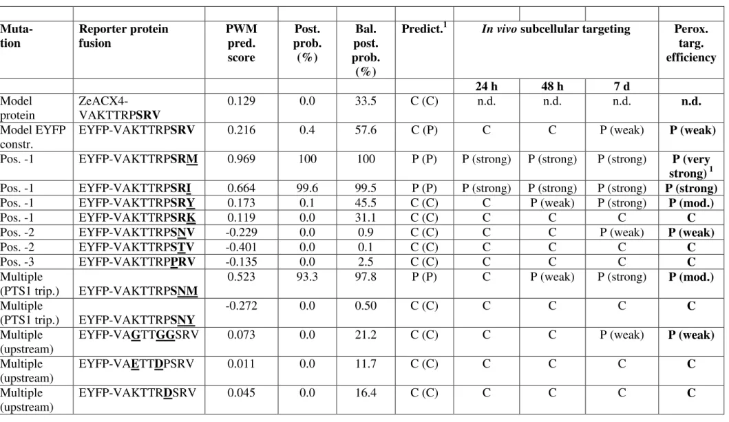
Table 1: Analysis of the effect of PTS1 domain mutations on the efficiency ofin vivo peroxisome targeting of reporter protein fusions.
As model PTS1 domain, the C-terminal decapeptide of acyl-CoA oxidase isoform 4 (ACX4)
homolog from Zinnia elegans (ZeACX4) was chosen. Single and multiple aa residue
mutations were introduced into the model sequence and the decapeptides were attached to the C-terminal end of EYFP. The effect on the efficiency of peroxisome targeting was analysed by fluorescence microscopy. The PWM prediction scores, which are based on the C-terminal 14 aa of proteins of interest, were determined for various mutagenized decapeptides fused to EYFP by extending them N-terminally by the four C-terminal aa residues of EYFP (ELYK). The PWM score of the original 14 C-terminal aa residues of ZeACX4 (SFQL-VAKTTRP-SRV>) and those of the EYFP fusion (ELYK-VAKTTRP-(SFQL-VAKTTRP-SRV>) were 0.129 and 0.216, respectively. PWM prediction scores for the EYFP fusions (with a threshold of 0.412) and the standard and balanced posterior probabilities for peroxisome targeting were determined as described previously12; 26. The peroxisome targeting efficiency was determined semi-quantitatively and categorized as strong (peroxisomal after 18-24 h post transformation), moderate (peroxisomal only after 48 h) and weak (peroxisomal only after 7 days, i.e. 1 d RT and 6 d approx. 100C). For each experiment at least 10-15 fluorescent cells were analysed and the results reproduced in at least 3 independent experiments. The subcellular targeting prediction is provided according to the posterior probability and the balanced posterior probability (in parenthesis). 1 The SRM> construct could be detected in peroxisomes at very
early time-points (i.e. 12 h post transformation, see Suppl. Fig. 1) and, hence, was referred to as conferring very strong peroxisome targeting to the reporter protein. C, cytosol; p,
peroxisome; n.d., not determined.
Table 2: Analysis of the effect of PTS1 domain mutations on the binding affinity to AtPEX5 andin vivoperoxisome targeting.
determined as described previously12; 26. The peroxisome targeting efficiency was determined semi quantitatively as described for reporter fusions of the given peptides with EYFP (Table 1). The C-terminal tripeptides are printed bold.1The subcellular targeting prediction
g2 20 µm g3 20 µm a3 20 µm 20 µm 20 µm 20 µm
e1 EYFP-7aa-SRI> e2 e3
20 µm 20 µm
f1 EYFP-7aa-SRM> f3 B1. EYFP-CKI> B2. EYFP-CKI>
40 µm 40 µm
a1 EYFP-7aa-SRV> a2
40 µm g1 EYFP-7aa-SRY>
20 µm 20 µm
h1 EYFP-7aa-SRK> h2
18-24 hr 48 hr 7 d
B3. EYFP-CKI>
20 µm f2
Pos. -1 mutations: EYFP-7aa-SR(V to I/M/Y/K)>
20 µm h3
i3
20 µm
40 µm 40 µm
i1 EYFP-7aa-SNV> i2
n3
20 µm 40 µm 40 µm
n1 EYFP-VAGTTGG-SRV> n2 20 µm 20 µm 20 µm
k1 EYFP-7aa-PRV> k2 k3
l2 20 µm l3 20 µm 40 µm l1 EYFP-7aa-SNM> m2 m3 m1 EYFP-7aa-SNY>
40 µm 40 µm 40 µm
20 µm 20 µm 20 µm p1 EYFP-VAKTTRD-SRV> p2 p3 20 µm 20 µm 20 µm
j1 EYFP-7aa-STV> j2 j3
20 µm b1 EYFP-7aa-SRV>
(7 d)
b2 DsRed-SKL> b3 Merge
20 µm 20 µm
40 µm 40 µm
40 µm
d1 EYFP (w/o PTS1) d2 d3
20 µm
Figure 1
c1 EYFP-PTS1> c2 c3
18-24 hr 48 hr 7 d
Pos. -2 mutations: EYFP-7aa-SNV> and –STV>
18-24 hr 48 hr 7 d
Pos. -3 mutation: EYFP-7aa-PRV>
Multipe tripeptide mutations: EYFP-7aa-SNM and –SNY>
Upstream residue mutations of EYFP-VAKTTRP-SRV> Model sequence EYFP-VAKTTRP-SRV> and controls
18-24 hr 48 hr 7 d
Table 1: Analysis of the effect of PTS1 domain mutations on the efficiency ofin vivoperoxisome targeting of reporter protein fusions. Muta-tion Reporter protein fusion PWM pred. score Post. prob. (%) Bal. post. prob. (%)
Predict.1 In vivosubcellular targeting Perox. targ. efficiency
24 h 48 h 7 d
Model protein
ZeACX4-VAKTTRPSRV
0.129 0.0 33.5 C (C) n.d. n.d. n.d. n.d.
Model EYFP constr.
EYFP-VAKTTRPSRV 0.216 0.4 57.6 C (P) C C P (weak) P (weak)
Pos. -1 EYFP-VAKTTRPSRM 0.969 100 100 P (P) P (strong) P (strong) P (strong) P (very
strong)1
Pos. -1 EYFP-VAKTTRPSRI 0.664 99.6 99.5 P (P) P (strong) P (strong) P (strong) P (strong)
Pos. -1 EYFP-VAKTTRPSRY 0.173 0.1 45.5 C (C) C P (weak) P (strong) P (mod.)
Pos. -1 EYFP-VAKTTRPSRK 0.119 0.0 31.1 C (C) C C C C
Pos. -2 EYFP-VAKTTRPSNV -0.229 0.0 0.9 C (C) C C P (weak) P (weak)
Pos. -2 EYFP-VAKTTRPSTV -0.401 0.0 0.1 C (C) C C C C
Pos. -3 EYFP-VAKTTRPPRV -0.135 0.0 2.5 C (C) C C C C
Multiple
(PTS1 trip.) EYFP-VAKTTRPSNM
0.523 93.3 97.8 P (P) C P (weak) P (strong) P (mod.)
Multiple
(PTS1 trip.) EYFP-VAKTTRPSNY
-0.272 0.0 0.50 C (C) C C C C
Multiple (upstream)
EYFP-VAGTTGGSRV 0.073 0.0 21.2 C (C) C C P (weak) P (weak)
Multiple (upstream)
EYFP-VAETTDPSRV 0.011 0.0 11.7 C (C) C C C C
Multiple (upstream)
Table 2: Analysis of the effect of PTS1 domain mutations on the binding affinity to AtPEX5 and peroxisome targeting efficiency of reporter fusions.
Muta-tion
Peptide PWM
pred.
score2
Post. prob.
(%)2
Bal.
post. prob.
(%)2
Predict.
(C/P)1
PEX5C
Ki(nM)
PEX5
Ki(nM)
Perox.
targeting
efficiency of
EYFP fusions
1 Original. VAKTTRPSRV 0.268 1.8 71 C (P) >100000 >100000 P (weak)
2 Pos. -1 VAKTTRPSRL 1.043 100 100 P (P) 1400 ± 250 2220 ± 630 n.d.
3 Pos. -1 VAKTTRPSRM 1.020 100 100 P (P) 1800 ± 310 3100 ± 980 P (very strong)
4 Pos. -1 VAKTTRPSRI 0.716 99.8 99.7 P (P) 14500 ± 2800 21500 ± 5900 P (strong)
5 Pos. -1 VAKTTRPSRY 0.225 0.5 60.0 C (P) 47100 ± 6300 25400 ± 5800 P (mod.)
6 Pos. -2 VAKTTRPSNV -0.177 0.0 1.5 C (C) >100000 n.d. P (weak)
7 Pos. -3 VAKTTRPARV 0.128 0.0 33.2 C (C) >100000 n.d. n.d.
8 Pos. -3 VAKTTRPPRV -0.083 0.0 4.3 C (C) >100000 n.d. C
9 Upstream
residue
VAKTTRRQSRV 0.335 11.3 84.0 C (P) 50200 ± 6800 53400 ± 14000 n.d.
10 Multiple VAKTTRPSKL 1.031 100 100 P (P) 2000 ± 420 1300 ± 340 n.d.
11 Multiple VAKTTRPSNM 0.575 97.7 98.8 P (P) 72000 ± 11500 48000 ± 13000 P (mod.)
12 Multiple VAKTTRQSRL 0.999 100 100 P (P) 148 ± 44 120 ± 40 n.d.
14 Multiple VAKTTAQSRL 1.028 100 100 P (P) 796 ± 240 824 ± 275 n.d.
15 Multiple VAATTAQSRL 1.023 100 100 P (P) 1360 ± 260 1560 ± 450 n.d.
16 Multiple VAKTTRPPRI 0.365 22.3 88 C (P) >100000 >100000 n.d.
17 Orig.
pentapept
ide
YQSKL 0.818 100 99.9 P (P) 166 ± 23 189 ± 52 n.d.
18 Pos. -1 YQSKV 0.043 0.0 16.0 C (P) 32400 ± 4800 24100 ± 6900 n.d.
19 Upstream
residue