transmembrane Δ6 desaturase from
Micromonas pusilla
A thesis submitted for the degree of Doctor of Philosophy of the Australian National University
Dongdi Li
June 2016
© Copyright by Dongdi Li 2016
Declaration
This is my original work carried out in the laboratory of Associate Professor Colin Jackson unless otherwise stated. The fatty acid desaturase characterization work
described in this dissertation was performed in the lipid metabolism laboratory at the Commonwealth Scientific and Industrial Research Organisation, under the supervision of
Dr. Thomas Vanhercke, Dr. Surinder Singh and Dr. James R. Petrie. The work presented here has not been submitted as part of any other degree in any university or tertiary institute.
Acknowledgements
I would like to thank Associate Professor Colin Jackson for giving me the
opportunity to work with him on this challenging and interesting project and for his encouragement and support during these past four years. The tremendous help and inspiration from him is the motivation that pushed me to achieve what I have achieved,
especially during the last few days of the critical write-up process. I am also grateful to the entire Jackson Group for their support and advice, both personal and professional, and
for proofreading my thesis chapters.
I also would like to thank our collaborators Dr. Thomas Vanhercke, Dr. James Petrie and Dr. Surinder Singh in the Metabolic Engineering Group at CSIRO, who provided me
with the facilities and resources to perform the yeast assays, for sharing ideas, and helping me with organizing my project. I would especially like to express my appreciation to Dr.
Vanhercke for his encouragement.
I would like to say a big thank you to my dear friends, especially Du, for the trips to Sydney and the occasional mouth-watering dining experiences after the long working
weeks. You made my PhD life a food adventure. I am grateful to Professor Susan Howitt for providing me with help even after I left her research group. I am also thankful for
David for proofreading my thesis multiple times, even if my work is out of the scope of his field of study.
Last but not least, I would like to express my biggest thank you to my parents for
Abstract
The increased awareness of the health benefits of ω3-long chain polyunsaturated
fatty acids (ω3-LCPUFAs) has led to a drastic increase in the consumption of fish-oil supplements. This has resulted in environmental concerns and the identification of key membrane-bound desaturases involved in the biosynthesis of ω3-LCPUFAs in order to
generate a sustainable source of ω3-LCPUFAs. The Micromonas pusilla Δ6 desaturase (MpΔ6des) is a membrane-bound desaturase that is specific for ω3-LCPUFA precursors
and acyl-Coenzyme A substrates (acyl-CoAs). The incorporation of MpΔ6des into the ω3-LCPUFA biosynthesis pathway allows efficient ω3-LCPUFA production in transgenic plants. However, little is known of the molecular basis underlying its
ω3-specificity, stability and acyl-CoAs specificity.
MpΔ6des is relatively challenging in terms of protein engineering targets in that there
is no molecular structure available, it cannot be expressed in easily manipulated prokaryotic systems such as Escherichia coli, and the activity cannot be rapidly screened viathe conventional techniques. Thus, computational, structure-based, protein design and
high-throughput directed evolution could not be used. To overcome the technical hurdles, we have applied bioinformatics-based techniques (consensus mutagenesis, ancestral
protein reconstruction and sequence similarity networks) to engineer MpΔ6des and to better define the sequence-structure-function relationship of proteins within the desaturase superfamily.
the substrate-binding cavity, but also by distal residues, possibly through intramolecular interaction networks.
An ancestral algal front-end Δ6 desaturase (ANC175) was inferred (Chapter 3), which resembles the properties of the progenitor of the algal Δ6 desaturases. The
comparison between ANC175 and contemporary desaturases indicated that the divergence of the ω3/ω6-specificity of algal Δ6 desaturases is associated with the environmental differences seen in the habitats of the different algal species.
Chapter 4 describes a bioinformatics analysis of the desaturase superfamily, showing that the four major desaturase subfamilies (the first desaturases, methyl-end desaturases, front-end desaturases and Δ4 sphingolipid desaturases) are structurally and functionally
distinct. Conserved motif analysis of the front-end desaturases suggested that two cytosolic regions (a loop between AH1 and H2, and the cytosolic side of TM3) play
crucial roles in determining the substrate head-group specificity of the front-end desaturase.
Altogether, this thesis promotes a more detailed structural and functional
understanding of the front-end desaturases, especially MpΔ6des. It validates the use of bioinformatics-based approaches such as consensus mutagenesis and ancestral protein
reconstruction, showing that small libraries that are relatively “rich” in valuable mutations can be produced, even in the absence of detailed structural information or a high-throughput screen. We have successfully created novel variants of MpΔ6des with
significantly enhanced ω3-specificity and with enhanced expression. These results also shed new light on the evolution of ω3/ω6-specificity in the front-end desaturase
Abbreviations
3-isopropylmalate dehydrogenase – ICDH
Alpha-linolenic acid – ALA
Acyl-carrier protein – ACP
First desaturase – FD
Arabidopsis thaliana Δ7/Δ9 desaturases – ACD1
Basic Local Alignment Search Tool – BLAST
Coenzyme A – CoA
Delta4 sphingolipid desatuarses – Δ4-SDs
Dihomo- γ-linolenic acid – DGLA
Docosahexaenoic acid – DHA Docosapentaenoic acid – DPA
Eicosapentaenoic acid – EPA
Eicosatetraenoic acid – ETA
Enhanced chemiluminescence – ECL
Fatty acid – FA
Fatty acid desaturases – FADs
Fatty acid elongases – FAEs
Fatty acid methyl esters – FAMEs
Front-end desaturase – FE Gas Chromatography – GC
Glycerolipid – GL
Human influenza virus hemagglutinin – HA
Linoleic acid – LA
Methyl-end desaturase – ME
Micromonas pusilla Δ6 desaturase – MpΔ6des Monogalactosyldiacylglycerol – MGDG
Molecular Evolutionary Genetics Analysis – MEGA
Multiple Expectation-maximum for Motif Elicitation – MEME
MUltiple Sequence Comparison by Log-Expectation – MUSCLE
Omega3 – ω3 Omega6 – ω6
Omega3 – Long chain polyunsaturated fatty acid – ω3-LCPUFA|
Ostreococcus tauri Δ6 desaturase – OtΔ6des
Phosphatidic acid – PA
Phosphatidylcholine – PC Phosphatidylethanolamine – PE
Phosphatidylserine – PS
Phosphatidylinositol – PI
Phospholipid – PL
Phylogenetic Analysis by Maximum Likelihood – PAML Polyunsaturated fatty acids – PUFAs
Sequence similarity networks – SSNs
Stearoyl-CoA desaturase – SCD1
Stearidonic acid – SDA
Transmembrane helix – TM Triacylglycerols – TAGs
Table of Contents
Declaration ... iii
Acknowledgements... v
Abstract ... vii
Abbreviations ... xi
Chapter 1 Introduction ... 1
1.6.1 The structural features of soluble desaturases ... 13
1.6.2 The functional mechanism of soluble desaturases ... 14
Chapter 2 Insight into the substrate specificity of the acyl-CoA Δ6 desaturase family from consensus mutagenesis... 29
2.1.1 The incorporation of consensus residues increases protein stability ... 32
2.1.2 Conserved residues are important for protein stability and/or function ... 32
2.1.3 The consensus mutagenesis of MpΔ6des ... 34
2.2.1 Phylogenetic analysis ... 35 2.2.2 Consensus mutagenesis using the broader algal desaturase members (Set I)36 2.2.3 Consensus mutagenesis using the small algal Δ6 acyl-CoA desaturases (Set II)
43
2.2.5 Topology guided mutagenesis ... 49
2.3.1 The possible role of cytochrome b5 in substrate binding ... 56
2.3.2 Functional and structural roles of the helix above substrate entry site ... 57
2.3.3 The amphiphilic helix near His box I is important for the expression of MpΔ6des ... 57
2.3.4 The consensus residues in the predicted helix bundle core exhibited different effects on stability ... 58
2.3.5 The trade-off between stability and specificity in algal front-end desaturase evolution ... 60
2.5.1 Molecular biology ... 61
2.5.2 HA tagging of wild-type MpΔ6des and its variants ... 62
2.5.3 Functional expression in yeast ... 63
2.5.4 Lipid analysis ... 64
2.5.5 Western blotting ... 65
2.5.6 Bioinformatics analysis ... 66
Chapter 3 Ancestral protein reconstruction of algal Δ6 desaturases ... 69
3.1.1 Ancestral resurrection as a method for generating thermophilic protein ... 70
3.1.2 The ancestor of FEs is a Δ6 desaturase ... 72
3.1.3 Chapter objectives ... 75
3.2.1 Phylogenetic analysis ... 76
3.2.2 Desaturation activity and expression of ANC175 at different temperatures . 80 3.2.3 Substrate specificity comparison ... 84
3.2.4 Sequence comparison ... 85
3.3.1 The divergence driven by habitat temperature difference ... 88
3.3.2 The residues that affect ω3/ω6-specificity ... 90
3.5.1 Bioinformatics analysis and structural modelling ... 91
3.5.2 Molecular biology ... 92
3.5.4 Western blotting for expression comparison ... 93
Chapter 4 The molecular basis underlying the diverse substrate head-group specificity of membrane-bound desaturases ... 97
4.3.1 The sequence-structure-function relationships of all known membrane bound desaturases ... 103
4.3.2 Differential distribution of charged residues in First desaturases reflect the absence of charged residues in the head-group binding cavity ... 106
4.3.3 Methyl-end desaturases present phospholipid specificity ... 110
4.3.4 The Δ4 sphingolipid desaturase subfamily. ... 111
4.3.5 Dissecting the diversity in FEs by SSNs ... 111
4.3.6 Conserved motifs distinguish the higher plant FE1-PCs and FE1-SDs ... 118
Chapter 5 General discussion ... 125
5.5.1 Future directions for the generation of highly ω3-specific and thermostable Δ6 desaturase ... 132
5.5.2 The crystallization and structural determination of FEs is needed for future protein engineering ... 133
References ... 135
Chapter 3 Supplementary Materials ... 165
Chapter 1
Introduction
Fatty acids and their lipid derivatives are one of the major components of biological
membranes [recently reviewed in (1,2)]. Lipid bilayers are the basis of cellular organelles that sequester cellular processes. This compartmentalization is the foundation of the evolution of complex organisms. The lipid composition of the bilayers is closely
associated with the function of membrane proteins and their related cellular functions. Highly unsaturated lipid bilayers ensure cell plasticity and help protect cells in cold
environments. In addition, the anti-inflammatory effects of the metabolites of the unsaturated lipids could protect cells from oxidation stress (3-5). As more clinical evidence is published (6,7), the physiological roles of unsaturated lipids and unsaturated
fatty acids (UFAs) in the lipid bilayer, especially the ω3-long chain polyunsaturated fatty acids (ω3-LCPUFAs), have become clearer in the cardiovascular and neurological
systems (8,9). The increasing production of ω3-LCPUFA supplements, using fish oil as the main source, led to the rapid depletion of marine fish sources. Consequently, the foreseeable environmental issues associated with the manufacturing of ω3-supplements
led to the generation of transgenic plant sources and the discovery and characterization of many membrane-bound desaturases (10). The membrane-bound desaturases have been
the focus of many studies into the metabolic engineering of lipid metabolism, not only for their roles in producing ω3-LCPUFAs, but also for their physiological roles in the lipid metabolism of their host organisms.
This chapter introduces the nomenclatures of lipids, the physiological roles of ω3-LCPUFAs, the current understanding of the structure and function of fatty acid desaturases, and most importantly, the driving force of this research project. The
appreciate the aims and the outcomes of this protein engineering research project on the membrane-bound Δ6 desaturase from Micromonas pusilla.
Fatty acids and lipids
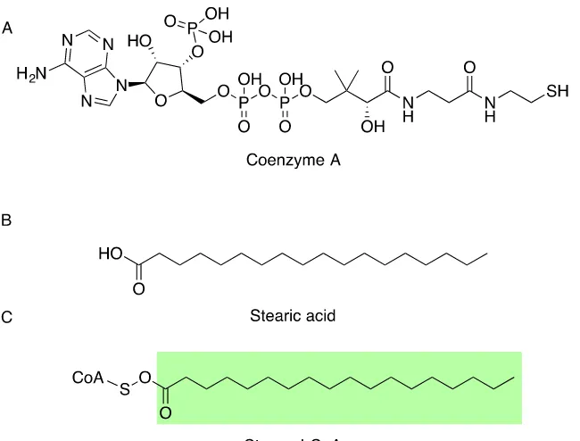
Fatty acids (FAs) are one of the major constituents of all organisms. Fatty acids
consist of a hydrocarbon chain with a carboxyl functional group at the end of the chain. These FAs can form a number of ester derivatives with different organic functional groups at the carboxylic ends (the head-group), in which the alkyl group and the carbon
oxygen double bond is then referred to as the acyl group. For example, a stearic acid forms an ester bond with coenzyme A (CoA), resulting in stearoyl-CoA (Figure 1.1). The chemical moiety of the head-groups will affect the physical properties of the fatty acid
ester and determine its biological role (Figure 1.2).
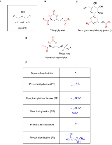
[image:20.595.120.445.409.656.2]One important class of lipids is the glycerolipids (GLs), which is based on the glycerol backbone (Figure 1.2A). The glycerophospholipids, being the main structural lipids found in cellular membranes are of particular importance, in which the hydrophobic tail is embedded within the cell membrane and the hydrophilic head is solvent accessible.
Glycerophospholipids have two acyl groups at the sn1 and sn2 positions on the glycerol backbone, but differ from other types of glycerolipids in that there is a variable chemical moiety at the sn3 position, connected through a phosphodiester bond (Figure 1.2D) (11). Due to their amphiphilic character, the hydrophobic acyl groups can aggregate and sequester themselves from water, while the hydrophilic heads remain in the aqueous phase, allowing for the formation of micelles or lipid bilayers (12). This characteristic
makes these lipids crucial for the formation of cellular organelles, intra/intercellular transport and metabolic processes.
The phosphorus-free glycerolipids are structurally more homogenous and are typically used for the purposes of energy storage. One example is the triacylglycerols (TAGs), a highly hydrophobic form of glycerolipid in which all three alcohol positions
on the glycerol backbone are esterified with acyl groups (Figure 1.2B) (13). They can aggregate into anhydrous fat storage structures (14), such as oil droplets. When nutritional
resources are limited, TAGs are broken down through lipolysis and the liberated acyl groups are re-introduced into lipid metabolism pathways (15-17). Another glycerolipid subclass is the galactolipids, which have a galactose substitution at the sn1 position or at
both sn1 and sn2 positions on the glycerol backbone (Figure 1.2C). They are more abundant in the membranes of photosynthetic organisms, such as cyanobacteria, green
Figure 1.2: The structure of representative glycerolipids. The acyl groups are in red.
Figure 1.3: The chemical structure of a sphingolipid. The acyl group in red is presented on the ceramide backbone.
The non-glycerol based lipids include the biologically active acyl-CoAs (Figure 1.1A). They have a negatively charged CoA region and can be readily utilised by various lipid-modifying enzymes such as desaturases, elongases and acyltransferases (20-25).
Therefore, they are important metabolites in lipid metabolism and cellular signalling (26,27). Sphingolipids are phospholipids, but instead of having a glycerol backbone, they
have a long hydrophobic base (e.g. ceramide) as the backbone (Figure 1.3). Due to the use of ceramide in multiple signalling pathways, sphingolipids are not only a class of essential structural lipids in lipid bilayers (28), but are also important in the regulation of
cell processes such as apoptosis, cell migration and cold tolerance (29-32).
Lipid nomenclature
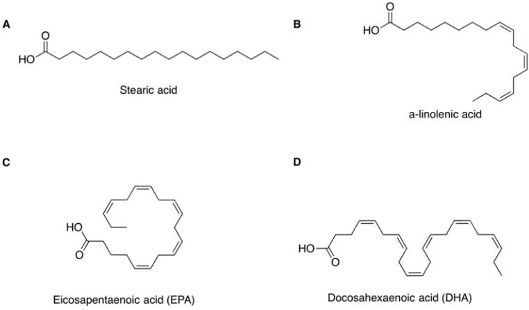
The level of unsaturation of a lipid molecule relates to the number of carbon-carbon double bonds (C=C) in the hydrocarbon chain of the acyl group. A lipid is saturated if the hydrocarbon chain is solely composed of carbon-carbon single bonds (C-C) i.e. an alkyl
chain, such as in stearic acid (C18:0) (Figure 1.4A). Polyunsaturated fatty acids (PUFAs) or lipids have at least two double bonds on the acyl chain. Unsaturated fatty acids could
A few nomenclature systems have been developed to describe the level of unsaturation and the locations of C=C bonds on acyl groups. For example, α-linolenic
acid (ALA) is the historical name for a common ω3-PUFA, the precursor of ω3-LCPUFAs (Figure 1.4B). It can also be expressed in lipid numbers and n terms as C18:3n3, where ‘C18’ indicates 18-carbon chain; ‘:3’ denotes there are three C=Cs on the carbon chain; and n3 is equivalent to ω3, indicating that the first C=C is located at the third carbon bond counting from the methyl end of the acyl group (33). In addition, the
locations of the C=Cs can also be specified by Δx, where x is the position of the first C=C counting from the carboxyl end of the acyl group. For instance, ALA can also be expressed as C18:3Δ9, 12, 15. When the first C=C is located at the third carbon bond from
the methyl end of the molecule, it is named as an omega3-LCPUFA (ω3-LCPUFA) (34). Two physiologically important examples of ω3-LCPUFAs are docosahexaenoic acid
(DHA, C22:5Δ4, 7, 10, 13, 16, 19) and eicosapentaenoic acid (EPA, C20:4 Δ5, 8, 11, 14, 17) (Figure 1.4C and D).
In the following chapters, both the “n terms” and “Δx” nomenclature systems were
utilised to clearly describe the level of unsaturation and positioning of the C=C bonds on the acyl groups of the different unsaturated lipids (for instance, EPA is C20:4n3 Δ5, 8, 11, 14, 17). The abbreviations of the historical names of different fatty acids were used
Figure 1.5: The structure of representative saturated and unsaturated fatty acids. A. A saturated fatty acid, stearic acid (C18:0). B. An ω3-PUFA ALA (C18:3n3 Δ9,12,15). C and D. The chemical structures of ω3-LCPUFAs EPA (C20:4n3 Δ5, 8, 11, 14, 17) and DHA (C22:5n3 Δ4, 7, 10, 13, 16, 19) respectively.
The physiological roles of ω3-PUFAs
In comparison to saturated lipids, the C=C bonds at the end of the hydrocarbon chain increase the level of disorder in the packing of the lipid bilayer, which increases
membrane permeability, reduces membrane thickness and can improve the flexibility of the membrane in response to changes in cell volume (35). This is because cis-C=C bonds
create “kinks”, steric hindrances to the hydrophobic interactions between membrane lipids (Figure 1.5). The improved lateral mobility of phospholipids and membrane proteins (36) can affect the function of mechanosensitive proteins such as ion channels
and transporter proteins (37-40), which can be physiologically significant. Moreover, the melting temperature of the lipid membrane that is rich in ω3-LCPUFAs is low, and this
Figure 1.6: A schematic diagram of the lipid bilayers.A. The compact assembly of
the lipid bilayer rich in saturated lipid. B. The loose assembly of the phospholipid in the lipid bilayer, rich in unsaturated lipid tails. This figure was generated based on a published figure (42)
Membrane fluidity is important for the correct function of transporters and pathways involved in photosynthesis, as well as cold and oxidation stress tolerance (43). Efficient enzyme turnover in photosynthetic systems is dependent on a number of protein synthesis
enzymes (44,45). Under environmental stress, protein synthesis and processing are further down regulated by the reduction of membrane fluidity as in many other membrane
proteins (45). The suppression of protein synthesis inhibits the recovery of protein damage in the photoinhibition of photosystems, which is one of the consequences of low temperature and salt stress in photosynthetic systems (44,45). In addition, the reduction
in membrane fluidity is essential for salt tolerance, because it affects the conformational changes of ion transporters (46).
In animals, abnormalities in neural signal transmission (47,48) and neural cell plasticity (the synaptic processes that are important for the processes of learning and memory formation), as well as a higher risk of cardiovascular diseases have a strong
correlation with reduction in ω3-LCPUFAs in the lipid composition of cell membranes (49-52). In addition, the anti-inflammatory and anti-oxidant properties of ω3-LCPUFAs
are evident in maintaining neural and cardiovascular functions (6,53-56). However,
animals such as humans and fish cannot synthesize a sufficient amount of EPA and DHA from the dietary intake of ALA and LA. They need to directly acquire the essential
ω3-LCPUFAs through diet from marine sources (57).
Natural sources of ω3-LCPUFAs
Animals, including humans, are unable to produce sufficient ω3-LCPUFAs after ingesting the precursor ALA from plants, even though functional animal fatty acid
desaturases and fatty acid elongases exist (58-61). The high level of ω3-LCPUFAs in marine fish oil is the result of nutrient enrichment in the fat tissue after fish feed on marine microorganisms such as marine algae. In addition, highly active fatty acid desaturases
and fatty acid elongases have been found in bacteria, diatom, yeast and moss, which can produce ω3-LCPUFAs using the endogenous ω3-LCPUFA precursor, ALA (62-64).
ALA is one of the most abundant unsaturated fats contained in most plants. However, because most plants lack the essential fatty acid desaturases and fatty acid elongases, very few of them are able to produce the essential oils (62,65). As a result, land animal fat and
most non-transgenic plant oils are not suitable natural sources of ω3-LCPUFAs.
The human diet has transformed from a vegetable-rich diet to a high animal product
diet and the ω3-lipid:ω6-lipid ratio has decreased significantly (66). Following this transition, there has been a significant increase in the occurrence of cardiovascular diseases in the adult population (67). As the public has become more aware of health
issues related to the modern diet, more ω3-LCPUFAs supplements have been sold worldwide. The food industry is reliant on fisheries to produce ω3 supplements. However,
marine fisheries (68), raising environmental concerns and eventually requiring the generation of sustainable ω3-LCPUFA sources. In the past few decades, a number of algal
FADs and FAEs in marine microbes have been studied in order to determine the underlying reasons for their high ω3 lipid content (69).
The recombinant synthesis of ω3-LCPUFAs
Some lower eukaryotes including fungi and algae are rich in DHA and EPA, and are
the primary source of ω3-LCPUFAs for other organisms. In order to produce a sustainable transgenic plant source for producing physiologically essential ω3-LCPUFAs, all of the enzymes needed to construct the biosynthesis pathway have been cloned and functionally
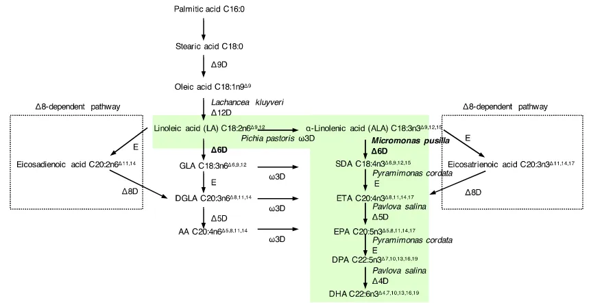
characterised (20). They included a range of fatty acid desaturases and fatty acid elongases (69) (Figure 1.7). Both ALA and linoleic acid (LA, the ω6-LCPUFAs precursor; 18:2n6Δ9, 12) are abundant in plants, especially in seeds and legumes (70). After the key algal and fungal enzymes were identified, they were used to reconstitute the ω3-LCPUFA synthesis pathway in model oil crop plants (63,64). The resulting transgenic
Figure 1.7: A schematic presentation of the marine algal ω3-LCPUFA synthesis Δ6
desaturase and Δ8 desaturase-dependent pathway. The ω3 specific Δ6 desaturase
pathway (shaded in green) has been reconstituted in model plant systems (the figure is drawn based on the published diagrams (20,71)). The species of origin of the introduced genes is given for the ω3 specific pathway. The subject of this study is written in bold letters. E, elongase; D, desaturase.
The recombinant DHA synthesis pathway in the transgenic oil crops consists of
FADs and FAEs from various organisms (Figure 1.7). The pathway starts from the Δ12 desaturation of oleoic acid C18:1n9Δ9 by Lachancea kluvveri Δ12 desaturase, then the ω3/Δ15 carbon double bond is introduced by Pichia pastoris ω3 desaturase. The
following Δ6 desaturation of ALA to stearidonic acid (SDA; C18:4n3Δ6,9,12,15) by Micromonas pusilla Δ6 desaturase (MpΔ6des) is the commitment step in the process,
because most plants have endogenous desaturases to produce ALA and LA, but not Δ6 desaturases and the enzymes of the steps following it. Δ6 elongation by Pyramimonas cordata Δ6 elongase produces eicosatetraenoic acid (ETA; C20:4n3Δ8,11,15,17), which is
followed by Δ4 desaturation to yield DHA. Δ6 desaturation is the bottleneck reaction of the pathway, because the substrate conversions of the following processes are close to
100% (20).
A Δ8 desaturase-dependent DHA and EPA synthesis pathway has been found in a
number of non-photosynthetic microorganisms and mammals as summarised previously (Figure 1.7) (72). It differs from the Δ6 dependent pathway by the initial elongation of ALA and LA to C20 followed by Δ8 desaturation to produce dihomo-γ-linolenic acid
(DGLA, C20:3n6Δ8,11,14) and EPA. However, the efficiency of this pathway is limited by the lipid substrate flow from the acyl-CoA pool (elongation product) to the acyl-lipid pool (substrate for Δ8 desaturation). This is known as the substrate dichotomy problem, which
explains the necessity for humans and animals to acquire essential ω3 lipid through dietary sources in addition to endogenous production.
Fatty acid desaturases
Fatty acid desaturases are one of the key enzymes in lipid modification, especially in
the synthesis of ω3-LCPUFAs. They form a very diverse family of enzymes sharing similar functionalities. The soluble desaturases are believed to form a smaller desaturase
family that is not related to the integral membrane desaturases. Soluble desaturases are the best understood desaturases in term of structure, substrate-binding and the structure-function relationship underlying their regiospecificity and substrate selectivity.
This section reviews the current understanding of soluble desaturases. This is followed by a discussion of the integral membrane desaturases and the most recent developments
1.6.1 The structural features of soluble desaturases
Soluble desaturases are only found in the plastids of higher plants (73,74). They are
responsible for introducing a C=C bond to a saturated fatty acid in plants. The lipid has to be esterified onto an acyl-carrier protein (ACP) for it to be available for the desaturation
reaction, so these desaturases are often referred to as acyl-ACP desaturases. After the publication of the crystal structures of the C18:0 acyl-ACP Δ4 desaturase from Hedera helix (Ivy) (PDB ID: 2UW1) and acyl-ACP Δ9 desaturase from Ricinus communis
(Castor) (PDB ID: 1AFR), structural comparisons in combination with mutagenesis studies have resulted in significant improvements to the molecular understanding of their stereoselectivity, substrate preference and functional mechanism.
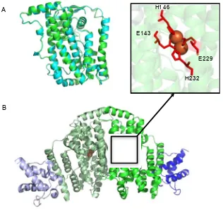
The structures of soluble desaturases consist of 11 α-helices, and they demonstrate high similarity to each other (Figure 1.8A). Four helices form a bundle that shapes the substrate-binding cavity (75). Two highly conserved E/DxxH motifs coordinate two iron ions to form the catalytic site (the di-iron centre) (76). The di-iron centre resides at the bend of the hydrophobic substrate tunnel coordinated by the two conserved E/DxxH
motifs (Figure 1.8B). Substrates bind in the textbook lock-and-key mode (77) with the target C-C bond positioned at the bend for the formation of the cis-product. The residues
at the end of the far end of the long hydrophobic substrate cavity determine the preferred length of the substrate acyl group, e.g. bulkier residues would shorten the preferred substrate length (74,78,79). The interaction between the desaturase and the acyl carrier
protein is predominantly electrostatic in that the positioning of the interaction site orients the acyl group in the substrate cavity in relation to the di-iron centre (80,81). Changing
of the acyl-ACP desaturases (80). Hence, the substrate specificities of FADs are determined by two factors: substrate-binding and the complementarity between the acyl
group and the substrate tunnel.
Figure 1.8: The crystal structures of soluble acyl-ACP desaturases. A. The
superimposed structures of Ricinus communis (Castor) Δ9 acyl-ACP desaturase (PDB ID: 2XZ0) in blue (82) and Δ4 desaturase from Hedera helix (Ivy) in green (PDB ID: 2UW1) (75). B. The structure of Castor Δ9 desaturase dimer with both active sites occupied by ACP through cross-linking. No acyl group is attached to the ACP. The desaturase monomers are in pale green and green. The ACPs are in dark blue and pale blue. The active site iron ions are shown as orange spheres. In the upper panel attached to B, the di-iron centre and the coordinating E/DxxH motifs are shown.
1.6.2 The functional mechanism of soluble desaturases
The functional mechanism of acyl-ACP desaturases is likely to resemble other non-heme di-iron enzymes, such as methane monooxygenases, owing to the similarity in
the catalytic residue orientation (78). While no functional mechanism has been proven
A
B
E143 H146
E229
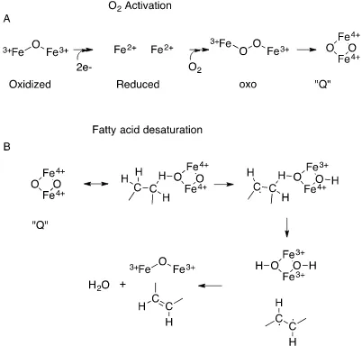
[image:32.595.115.431.188.486.2]biochemically, large-scale multi-reference ab initio calculations have proposed a possible likely reaction route from the energetic aspect of the desaturation reaction (83) (Figure 1.9). The first step is oxygen activation, which reduces the Fe(III) ions to Fe(II) ions. In turn, a diamond shaped Q species is formed. One of the activated oxygen atoms forms a
covalent bond with the hydrogen on the target C-C bond on the acyl group. The C-H bond breaks forming a radical C·. This is followed by the bonding of the other oxygen atom with the H atom on the other C of the target C-C. The next step is the process that is
Figure 1.9: A simplified schematic presentation of the mechanism of fatty acid
desaturation (83). There are two main processes: A. O2 activation and B.
dehydrogenation (fatty acid denaturation).
The oxygen activation of the di-iron centre requires electrons from an electron donor (84). All known acyl-ACP desaturases accept electrons from NADPH via ferredoxins
(85). However, the route through which electrons are transferred from ferredoxins to the di-iron centre in the core of the protein is unclear. A direct route can be ruled out, because
the di-iron centre in an acyl-ACP desaturase is not easily accessible from the solvent (75,78). The proposition based on the crystal structures is that a chain of aromatic or charged residues channels the electrons from the surface of the desaturase to the catalytic
[image:34.595.67.469.72.456.2]The crystal structures of the acyl-ACP desaturases have also shown that the soluble desaturases have a dimeric quaternary structure organisation (75). Conformational
changes of residues at the catalytic site are also observed during the enzymatic cycle of the enzyme as a result of the redox state of the metal ions in each subunit (84). The
reduction of the active site results in the formation of a complex of enzyme:substrate=2:2 (86) (Figure 1.8B). However, the dimers have demonstrated half-of-the-sites reactivity, meaning that only one of the two available active sites is catalytically active at one time,
with no inhibitory effect between the subunits (82).
The evolutionary origin of the soluble acyl-ACP desaturases is difficult to interpret due to the complex evolution of land plants (87). It is possible that after the initial
symbiogenesis, an ancestral protein with a different function in the engulfed prokaryote adapted for the fatty acid desaturation function to deliver the unsaturated fatty acid from
the only fatty acid synthesis organelle (plastid) to other organelles in plant cells (87). The possible common ancestors of land-plant plastids, cyanobacteria, do not have a homolog. While there are homologs found in the genomes of non-photosynthetic prokaryotes
including Mycobacterium and Streptomyces, and there is a crystal structure of a putative acyl-ACP desaturase from Mycobacterium tuberculosis, no direct experimental evidence
is available to suggest a functional homolog exists (88,89).
Membrane desaturases
The membrane-bound desaturase family is a much larger and diverse desaturase family that is responsible for generating unsaturated fatty acids (UFAs) in all organisms.
membranes (in photosynthesis organisms only), where the majority of unsaturated fatty acid production occurs (90,91). In prokaryotes, they are found on the plasma membrane,
with the catalytic domain in the cytosolic side of the membrane (92). For their capability to introduce a C=C on already unsaturated lipid substrates, the membrane-bound
desaturases are regarded as the key components in engineering a plant ω3-LCPUFA platform.
Based on their regiospecificity (the positioning of the newly formed C=C bond in
relation to the termini and the pre-existing C=C bond of the lipid molecule, if applicable), membrane desaturases have been further classified into three groups (87,93): First desaturases, Front-end desaturases and Methyl-end desaturases. The First desaturases
(FDs) mediate the formation of the first C=C bond at the Δ9 position of an acyl group. It has been postulated that the Δ9 desaturases are the most ancestral desaturases among the
three groups for their universal distribution in organisms (93). In addition, their desaturation products often serve as the substrates for the other two membrane desaturase groups (93). The Front-end desaturases (FEs) catalyse the formation of a C=C between
an existing C=C and the carboxyl end of the substrate (the “Front-end”), and they include the Δ4, Δ5, Δ6 and sphingolipid Δ8 desaturases. Δ12/ω6 desaturases and Δ15/ω3
desaturases form the third group, which is also known as the Methyl-end desaturases (MEs). They are responsible for introducing a C=C between a pre-existing C=C and the methyl end of the acyl group. The most important ones of all are the FEs, as they function
at the Δ6, Δ5 and Δ4 desaturation steps of the ω3-LCPUFAs as shown in Figure 1.7.
A four-transmembrane helix (4-TM) structure is believed to be the model topology
supported by hydrophobicity plots of some characterised desaturases (94-97) and the biochemistry characterization of the topology of the mouse stearoyl-CoA Δ9 desaturase
(98). Recently solved structures of human and mouse stearoyl-CoA Δ9 desaturases (PDB: 4ZYO and 4YMK respectively) are consistent with the 4-TM model structure (Figure 1.10A) (99,100). Eleven helical segments form a cytosolic domain (CAP domain) on the helix bundle in which the catalytic di-iron centre resides, and these form the substrate-binding cavity. The di-iron centre is located at the kink of the tunnel as in the soluble
desaturases (Figure 1.10B), but the kink is not as buried in the helix bundle (99,100). The shape and complementarity of the substrate-binding tunnel determines the acyl group preference of the desaturases, in line with the findings in the soluble proteins. However,
the di-metal centre in the membrane-bound desaturase is coordinated by three conserved histidine-rich motifs and a previously unknown NxxxH His motif located at the cytosolic
Figure 1.10: The structures of human stearoyl-CoA desaturases SCD1 (PDB ID:
4ZY0) (100). A. The structure of SCD1 in wheat with the substrate bound in the
substrate-binding pocket. The zinc ions in the di-metal centre are the red spheres. B. The di-metal centre is coordinated by nine conserved histidine residues. An asparagine (N265) coordinates one of the zinc ions through a water molecule. C. The topology of SCD1 presenting the locations of the coordinating His boxes, including the newly identified NxxxH at the cytosolic end of TM4.
Despite the differences in the metal coordinating motifs, in terms of functional mechanism the architecture of the active site agrees with the current understanding of the
fatty acid desaturation mechanism as discussed in section 1.6.1 (99). While some
H2 TM 1 TM 2 TM 4 TM 3 AH1 H3 H4 H5 H6 AH7 H8 AH9 Cyb5
C
N
Cytosol ER membrane ER lumen H11A
C
B
H302 H301 H125 H157 H120 H298 H269 N265 H160 H161H2O
[image:38.595.67.474.77.528.2]prokaryotic members utilise ferredoxins as electron donors like their soluble counterparts (101,102), cytochrome b5 is the most common electron donor for the membrane-bound
desaturases to obtain electrons from NADH via cytochrome b5 reductases (103-106). Since the catalytic sites of membrane-bound desaturases are more accessible, a direct
electron transfer from the cytochrome b5 domain to the catalytic site is possible (99,100). However, better experimental evidence is required to confirm this hypothesis. Moreover, some membrane-bound desaturases have a functionally crucial cytochrome b5 domain
fused at either the N-terminus or C-terminus (107). A tight association has been proposed for the two domains that should maximise the electron transfer efficiency (106).
Front-end desaturases (FEs)
The first Front-end desaturase (FE) was identified from a cyanobacterium, Synechocystis pcc6803, and can be seen as the representative of cyanobacterial FEs (102,108). The cyanobacterial FEs do not have a fused cytochrome b5 domain, instead
using monogalactosyl diacylglycerol (MGDG, a glycerolipid) as their substrate, and they can receive electrons from either ferredoxin or cytochrome b5 (109,110). Subsequently,
more FEs have been discovered from eukaryotes including marine algae, fungi, animals and plants. All these FEs carry a fused cytochrome b5 domain, and they present a great range of substrate head-group preferences (111). Some FEs from higher animals, some
algae and parasites are believed to use acyl-CoAs as a substrate (Figure 1.1) (59,112,113). The other FEs in plants, fungi, algae and lower animals utilise complex lipids, such as
suggests this family carries the structural information for understanding the different binding mode of the different head groups.
FEs have been the focus for metabolic engineering of plant ω3-LCPUFA sources, because of their unique capability to incorporate C=C at the Δ4, Δ5 and Δ6 positions of
long-chain acyl groups, as shown in the ω3-LCPUFA biosynthesis pathway (Figure 1.7). The most active Δ6 acyl-CoA desaturase from the marine alga Micromonas pusilla (MpΔ6des) preferentially uses the ω3-LCPUFA precursor, ALA as its substrate (20,63).
Mutagenesis studies and domain-swapping studies have suggested that the residues close to the histidine-rich motifs and the residues in the putative TM1 and TM2 are likely to be important for the regiospecificity of FEs (95). The residues in the helical core can
determine the preferred substrate length of the desaturases (114). However, the underlying basis of the substrate preferences and head group binding is still largely
unknown, owing to the absence of a crystal structure for a close FE homolog.
From an evolutionary perspective, the Δ6 regiospecificity is likely to be the ancient function of all FEs, as this function is ubiquitous for FEs in all kingdoms, despite their
varied substrate preferences. Δ8 Sphingolipid desaturases are only characterised in plant and fungi and they share high sequence identity with plant FE Δ6 acyl-PC desaturases
(96,115), while Δ5/Δ6 bifunctional acyl-CoA desaturases are found in the animal kingdom. It is believed that the emergence of animal Δ5 desaturases from the ancestral Δ6 desaturase occurred prior to gnathostome radiation (diversification of jawed
vertebrates), because Δ6 desaturases are ubiquitous to all marine teleost species (58,59,116). This scenario is further supported by the identification of a Δ6 FE desaturase
Interaction between the cytochrome b5 domain and the desaturase domain has not been elucidated due to the lack of an empirical structure for the fusion protein. The fusion
event makes sense from an evolutionary perspective as it provides a higher chance of optimising the electron transfer process, which is crucial for the efficiency of the catalytic
function. Moreover, the pairing between the two domains appears to be stringent, as the recombinant expression of an endoplasmic reticulum cytochrome b5 could not complement the deletion of the fused cytochrome b5 domain of a mammalian FE (104).
A phylogenetic analysis of both fusion proteins suggests that the interaction is the combination of hydrophobic interaction and electrostatic force, and the loss of electrostatic interactions was accompanied by the gain of hydrophobic interaction (106).
Research objectives
The focus of this work is to provide a more detailed structural-functional understanding of FEs to facilitate better metabolic engineering of transgenic plants for
ω3-LCPUFA production or biofuel production. Specifically, this project aims at investigating the possibility of using bio-informatics based methods to modulate the
substrate specificity or protein expression of the commercially valuable fatty acid desaturases, one of the Front-end desaturases. The detailed aims of each chapter are provided as the following. Using sequence alignment-based approaches, consensus
mutations were designed to determine the roles of conserved residues in the algal FE family (Chapter 2). Following that, an ancestral sequence was inferred for the algal Δ6
availability of the first crystal structures of mammalian Δ9 desaturases allowed for an in-depth structure-function analysis of the FE desaturase family using the data gathered in
each chapter. For the purpose of this dissertation, each objective is presented as its own chapter with an overview of the relevant research methodology.
Chapter 2 illustrates how consensus mutagenesis engineering has been used to determine the role of conserved residues in the algal desaturase family and to improve the ω3 specificity and ω6 specificity of MpΔ6des.
Chapter 3 explores the most likely ancestral characteristics of the algal FE desaturase family in terms of substrate specificity and stability by ancestral protein reconstruction.
Chapter 4 illustrates the structural basis underlying the diverse head group
preferences by conducting a broad informatics analysis of the membrane-bound desaturase superfamily and a more detailed analysis of the FE desaturase family.
Chapter 5 is a general discussion of the outcomes of research presented in the thesis, and highlights the main contributions to the understanding of the sequence-structure-function relationship of the membrane-bound desaturases.
The publications in preparation from this thesis
Li, D., Vanhercke, T., Singh, S. P., Petrie, J. R. and Jackson, C. J. Insight into the substrate specificity of the acyl-CoA Δ6 desaturase family from consensus mutagenesis.
Publication generated from this thesis
Li, D., Moorman, R., Vanhercke, T., Singh, S. P., Petrie, J. R. and Jackson, C. J. Classification and substrate head-group specificity of membrane fatty acid desaturases.
Comput. Struct. Biotechnol. J.14, 341-349
Declaration of author contribution.
The experimental work included in the manuscripts in preparation were completed
primarily by the author under the supervision of Associate Professor Colin J. Jackson. Dr. Thomas Vanhercke, Dr. Surinder P. Singh and Dr. James R. Petrie from the Oil Engineering Group at the Commonwealth Scientific and Industrial Research Organisation
CHAPTER 2
Insight into the substrate specificity of the
acyl-CoA Δ6 desaturase family from consensus
Chapter 2
Insight into the substrate specificity of the acyl-CoA Δ6
desaturase family from consensus mutagenesis
2.1 Introduction
As discussed previously in Chapter 1 section 1.4, the production of ω3-LCPUFAs
requires multiple steps of fatty acid desaturation and elongation (Figure 1.7). Previous attempts at recombinant biosynthesis of ω3-LCPUFAs in plants have showed that the product flow from the Δ6 desaturation step to the following elongation process restricted
the overall ω3-LCPUFA production yield (119). As the lipid preference of more desaturases was characterised, it was identified that an acyltransferase would be required
to convert the desaturation products (acyl-PC molecules) to acyl-CoA thioesters (the substrate dichotomy bottleneck). Indeed, when a phospholipid-specific Δ6 desaturase was replaced by an acyl-CoA-specific Δ6 desaturase, the overall ω3-LCPUFAs production
yield was found to increase substantially (120). In addition, it has also been identified that inclusion of a ω3-specific Δ6 desaturase (MpΔ6des) is essential for a high
ω3/ω6-LCPUFA ratio in transgenic plant oil, as the following processes are not ω3-specific (20).
The MpΔ6des-catalysed Δ6 desaturation efficiency within the ω3-LCPUFA biosynthetic pathway is consistently lower than that of the other proteins in the pathway
(20). A stronger promoter, or the incorporation of multiple copies of MpΔ6des in the transgenic plant system improved the ω3-substrate conversion rate from 50% to 60%
catalytic efficiency or expression of MpΔ6des through protein engineering could further improve transgenic ω3-LCPUFAs production.
Previous work has suggested that MpΔ6des related Front-end desaturases adopt a four-transmembrane helix (4-TM) topology (111) as is seen in the crystal structures of
mammalian membrane-bound desaturases (Figure 1.10) (99,100). The substrate-binding cavity is formed by the transmembrane helices (TMs) and the eleven short helices in the cytosolic loops of the desaturase (the cap domain) (Figure 1.8), with the di-iron active site being located in the cap domain, which determines the regioselectivity of the desaturase. Previous mutagenesis studies have established the role of the transmembrane helices (TMs) in determining the substrate length preference
(95,97,114,121). The cytosolic loops of the desaturase, as well as TM1, are also suggested to interact with the thioester substrate head group (Figure 1.10), thereby affecting the substrate head-group preference (e.g. acyl-PC vs acyl-CoAs) as well as the regioselectivity of the enzyme (80,100,122).
Algal Δ6 desaturases differ in terms of their ω3-specificity, meaning that the shapes
of substrate-binding cavity are able to differentiate between ω3/ω6-substrates with the same fatty acid chain length (C18). Because an ω3-substrate only differs from an
ω6-substrate by the C=C at the ω3 position of the acyl chain, we could expect a very subtle difference in the substrate cavity between an ω3-specific desaturase and an ω6-specific desaturase. A recent study successfully modulated the substrate preference of
ω3- and ω6-specific of MpΔ6des by generating protein chimeras (123). However, pinpointing the exact residues that were responsible for the changes is challenging,
ω3/ω6-substrate specificity shifts are genuine, as there are often overall reductions in the substrate desaturation efficiency of both substrates.
One of the most effective means to probe structure-function relationships within proteins is through mutagenesis. Random mutagenesis and screening is able to identify
functionally important mutations that affect activity, although it requires an ability to test the activity of a large numbers of variants. This is not applicable for the study of membrane-bound desaturases as the assays available to us are labour intensive. Rational
design is also frequently used to generate a more manageable number of variants for testing, but generally relies on sound structural understanding, which is not available for the algal Δ6 desaturases. An alternative approach involves the use of consensus
mutagenesis guided by phylogenetic analysis (124-127). Consensus mutagenesis reverts the residues that have diverged from highly conserved regions back to the conserved
sequence. There are five important advantages of this technique that make it suitable for this study: it does not require the availability of crystal structures, there is no bias from our prior assumptions regarding which regions might be important, the number of variants
that are generated is typically fewer than 100 (rather than several thousand in the case of random mutagenesis), this method focuses on highly conserved positions in the protein
sequence that are likely to be of structural or functional relevance, and it generally avoids introducing deleterious mutations because the fitness of the substitution at the location has been tested by natural evolution (128).
In order to identify potentially important residues in MpΔ6des, we implemented consensus mutagenesis. In the absence of a known structure of a membrane-bound
generated and characterised in this chapter to explore their roles in the algal Δ6 desaturases.
2.1.1 The incorporation of consensus residues increases protein stability
Consensus mutagenesis is based on the hypothesis that, during natural selection,
destabilising and deleterious mutations are eliminated, while stabilising residues (possibly in the folding network) are more persistent in protein evolution (128). Replacing a residue with the most frequent (consensus) amino acid by targeted mutagenesis has been
demonstrated to increase protein thermostability in a number of soluble protein families (124,128). As this method can be solely based on alignments of homologous sequences, it is ideal for studying protein families with no crystal structure.
2.1.2 Conserved residues are important for protein stability and/or function
In order to understand the reasoning underlying the functional or stability change as
a result of an incorporated consensus mutation, it is important to be aware of the possible molecular roles of the conserved residues in a protein family.
Conserved networks of molecular interactions maintain protein structures and these
networks are based on the formation of energetically favourable connections such as salt-bridges and hydrogen bonds between amino acid side chains and the peptide
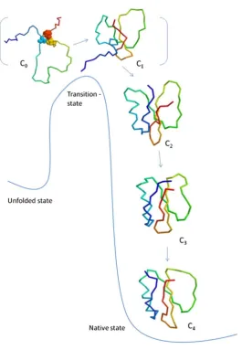
backbone (125). Computational simulations suggest that the process of folding follows the level-of-separation (LOS) hypothesis, which states that the formation of the network occurs in an orderly manner from the conserved core of the protein to the distal positions
(130,131) (Figure 2.1). Conserved residues often participate in the formation of the protein core and they are associated with optimum protein folding efficiency and
network and reduce protein expression (132,133). Therefore, by understanding the roles of the conserved residues, it may be possible to discover the residues that form the basis
[image:51.595.206.472.157.541.2]of the intramolecular network.
Figure 2.1: A schematic diagram of the level-of-separation (LOS) hypothesis. The
formation of the core network is the rate-limiting and determinant process in protein folding. This is followed by the energetically favourable formation of connections towards the distal locations. This picture is adapted from a previous publication (130).
Conserved residues form an evolutionarily conserved network that can also control the allosteric communication within a protein. For example, in G-protein coupled
cytoplasmic domain (134). Disruption of intermolecular connections between the regulatory domain and the catalytic domain in the 11R-lipoxygenase not only influences
its stability, but the effect also impacts the hydrophobic substrate-binding cavity and the catalytic site (135). Thus, if a substitution leads to a subtle substrate preference change of
an enzyme, it may indicate that this conserved residue plays a role in the allosteric control of the substrate cavity.
Whether conserved networks are important for the stability of membrane proteins is
less well studied, because the folding of membrane proteins is more complex than that of soluble proteins (136). In general, the folding process is composed of three steps: the integration of the transmembrane helix (TM) into a lipid bilayer by a translocon complex,
the packing of the TMs into its native tertiary structure and the oligomerisation into functional units (137). The stability of a membrane protein requires tight packing of the
helices, which is often mediated by small hydrophobic residues, such as alanine, glycine and the GxxG motif, in the helix-helix interface (138,139). For instance, incorporating small and hydrophobic residues such as alanine in the hydrophobic helical core of
G-protein coupled receptors could stabilise the membrane proteins and improve their expression [reviewed in (140)], but the effects are often not additive, because different
stabilising mutations may stabilise different conformations of membrane proteins (141).
2.1.3 The consensus mutagenesis of MpΔ6des
In this work, consensus mutagenesis was conducted to further investigate the
structure and function of the integral membrane desaturase family, and what underpins the ω3/ω6-specificity of these enzymes. Through the introduction of consensus
Furthermore, we demonstrate that the ω3/ω6-substrate specificity can be modulated through targeted mutagenesis.
2.2 Results
2.2.1 Phylogenetic analysis
Previous phylogenetic studies of membrane-bound desaturases revealed the
evolutionary separation of acyl-CoA desaturases and acyl-PC desaturases (4). For the purpose of consensus mutation design in this study, we conducted a focused phylogenetic
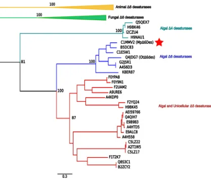
analysis of MpΔ6des homologs, obtained by a BLASTP search. The phylogenetic analysis of the above members using a maximum likelihood algorithm showed the clear separation between Δ5 and Δ6 desaturases from higher animals and other organisms
(Figure 2.2). In both groups, there were distinct Δ5 desaturase and Δ6 desaturase clades. Algal Δ6 desaturases formed one clade, whereas the proximal Δ5 clade contained Δ5 desaturases from algae and a number of other simple organisms (41). Homologous Δ4
desaturases were only present in algae and form a distinct clade from the other algal desaturases. Considering that Δ4 desaturation is the last step of
ω3-LCPUFA biosynthesis that converts DPA to DHA (Figure 1.7), the clustering of algal Δ4 desaturases may indicate an early gene duplication event as part of cold-adaptation before the divergence of algal Δ5 and Δ6 desaturases, since the increased
Figure 2.2: Phylogenetic analysis of the closely related homologous of MpΔ6des.
The tree was constructed using MEGA v.6. The analysis demonstrated the clear separation of algae, yeast and animal desaturases. A list of the included desaturase proteins is in supplementary materials. The bootstrap values of major nodes are denoted as percentages. For clarity, the animal Δ6 desaturase clade and yeast Δ6 desaturase clades are collapsed. The UniProt IDs of members are denoted in the tree. A red filled star is drawn next to MpΔ6des.
2.2.2 Consensus mutagenesis using the broader algal desaturase members (Set I)
Based on the sequence alignment of algal Δ5 and Δ6 desaturases (including the Δ5 desaturases from other unicellular organisms), we identified ten residues (consensus mutation set I) that are highly conserved throughout the alignment, but differ in MpΔ6des
tested in a yeast competition assay with both ALA and LA (the ω3-substrate and ω6-substrate respectively) added to the growth medium (at the same concentration, 0.25
mM each). The conversion of each substrate to its corresponding Δ6 desaturation product was measured and compared against the wild type activity in order to determine if the
mutation results in any change in terms of ω3/ω6 substrate preference and overall activity. In order to explain the observed effects of the mutations, the homology model structures of the desaturase domain and the cytochrome b5 domain of MpΔ6des were made using
the crystal structure of human stearoyl-CoA Δ9 desaturase (SCD1, PDB ID: 4ZYO) and the crystal structure of the outer mitochondrial membrane cytochrome b5 domain (PDB ID: 1ICC) respectively as the templates, as described in section 2.5.7. The modelled structures of the desaturase domain and the cytochrome b5 domain of MpΔ6des were used to locate the approximate location of the substituted residues. The mutations that
resulted in higher ω3-specificity were combined in different combinations to determine if the effects were additive, and to determine their effects on the expression of MpΔ6des.
The V57I, D138E and L285V substitutions were effectively neutral and exhibited
close to WT MpΔ6des activity on the ω3-substrate and ω6-substrate (Figure 2.4A, Table 2.2). In contrast, V74I, A184G, Q190M, G221A and D380N all showed significantly enhanced ω3-specificity, as a result of the reduced ω6-substrate desaturation (40% to 80% of WT MpΔ6des ω6-substrate conversion). The models of the desaturase domain and the cytochrome b5 domain of MpΔ6des were used to explain the possible effects of the
Figure 2.3: The segments of the alignment of algal and unicellular Δ5/Δ6
desaturases. The ten consensus substitutions shaded in pink. The locations of the
segments in relation to the sequence of MpΔ6des were labelled above the segments using the numbering of MpΔ6des.
55-59 68-77 135-141 182-196
[image:56.595.66.474.66.648.2]Three of them, A184G, Q190M, and G221A, were found to be in close proximity to the predicted substrate pocket, when mapped onto the model structure of MpΔ6des
(Figure 2.5A), suggesting they could affect the geometry of the substrate-binding cavity. D380N is located on the surface of the cap domain of MpΔ6des, where the cytochrome
b5 may interact with the desaturase domain. Therefore, D380N might affect the interaction between the two domains, thereby influencing the substrate recognition of MpΔ6des. V74I also exhibited higher ω3-specificity, but it is located in the cytochrome
b5 domain (Figure 2.5C). M223W and L348I exhibited a reduction in desaturation activity with both substrates, although more so with the ω6-substrate. M223W and L348I are located at the cytosolic side of the substrate cavity. In particular, L348I is likely to be
Figure 2.5: Homology models of the desaturase domain (A and B) and the
cytochrome b5 domain (C) of MpΔ6des. A. V74, A184, Q190, G221, M223 and D380
are labelled yellow on the modeled desaturase domain. M223 and L348 are labelled green. The conserved histidine residues in the three His boxes are coloured red. The stearoyl-CoA molecule (blue) was docked into the homology model. B. A close up view of the L348 residue (green) and its location in relation to the stearoyl-CoA (blue) molecule. C. V74I was found in the cytochrome b5 domain outside the heme-binding site.A prostheticheme molecule was docked into the homology model of the cytochrome b5 domain.
Since the V74I, A184G, Q190M, G221A and D380N single mutation variants retained more than 50% of the WT MpΔ6des ω3-substrate conversion activity, in an attempt to further increase the ω3-specificity, these mutations were combined (IGMAN).
This combination of algal desaturase consensus mutations generated a highly ω3-specific variant that exhibited undetectable ω6-substrate conversion (Figure 2.4A, Table 2.2). However, the expression of IGMAN was significantly lower than that of the WT
A
D380G221 Q190
A184 L348
V74I
B
Cytosol
ER membrane
ER lumen
M223
C
[image:59.595.119.535.76.349.2]MpΔ6des (Figure 2.4B, Table 2.2). Hence, there appeared to be a trade-off between the activity and expression.
Since the A184G and Q190M mutations are both located close to the first His box (Figure 2.5A), it is possible that they interact epistatically and the presence of both mutations is destabilizing. Therefore, two additional combined variants were generated to further explore the effects of A184G and Q190M: IGAN (V74I/A184G/G221A/D380N) and IMAN (V74I/Q190M/G221A/D380N). Both IGAN
and IMAN exhibited higher ω3-preference, as seen in IGMAN, with some increase in their expression levels (Figure 2.5B, Table 2.2), suggesting that the combination of the A184G and Q190M in the substrate-binding cavity had a negative influence on the
stability of MpΔ6des.
D380N is the only mutation that is located on the CAP domain of the desaturase. The
quadruple mutant IGMA (V74I/A184G/Q190M/G221A) was prepared to determine whether the presence of D380N could also affect protein expression and ω3-substrate recognition. The results showed that although the expression of IGMA was higher than
the other combined variants (IGMAN, IGAN and IMAN), the apparent activity was lower than IGAN and IMAN. Hence, the incorporation of D380N had a negative effect on
2.2.3 Consensus mutagenesis using the small algal Δ6 acyl-CoA desaturases (Set II)
Previous studies have characterised four of the seven identified algal Δ6 desaturases as displaying ω3-substrate specificity at various levels (20,112,120,142), from 63:5
(ω3:ω6 substrate desaturation efficiency) in MpΔ6des to 82:59 in OtΔ6des (20,112). The reported overall activity of Δ6 desaturase from Ostreococcus tauri (OtΔ6des) was the highest amongst the four, with a slight bias towards ω3-substrates when assayed
simultaneously (112).
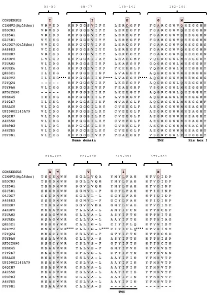
To investigate the roles of the residues conserved in the seven Δ6 desaturases, all seven of the identified algal Δ6 desaturases were aligned (Figure 2.6). The alignment revealed 25 residues that were highly conserved, but are not present in MpΔ6des (consensus mutations Set II). V57I and D138E were already characterised, and shown to
Figure 2.6: The alignment of contemporary algal Δ6 desaturases. The positions of the consensus mutations Set II of the algal Δ6 desaturases are shaded in grey. The consensus residue substitutions are highlighted in pink.
49-58 63-82
92-97 102-108 134-149
155-161 173-179 199-214
218-227 264-273 309-320
Most of the set II mutations to MpΔ6des were relatively neutral, exhibiting WT activity and specificity (N66D, A80S, R105A, I145P, T158M, V176I, S201N, V202I,
Y203W, V204W, L208I, M211F, A268L, L312F, V317I), while the T52R, M95Y, W335G, N372R, N385N and N465H mutations resulted in a loss of activity for both
substrates, and a generally small increase in ω3-specificity (Figure 2.7A, Table 2.2). Interestingly, the E222D and A270L (and potentially S201N) mutations increased the ω6 activity, with no effect on the ω3 activity. T52R and M95Y are located in the cytochrome
b5 domain, which is in agreement with the effects of the V74I mutation in Section 2.2, and highlights the role that this accessory domain plays in substrate binding and specificity. The E222D mutation, which increases conversion of the ω6 substrate, is
located near the substrate entry cavity (Figure 2.8A and B) on the opposite side to the S201N mutation, which could explain the subtle effects of these mutations on the
ω3/ω6-substrate preference. The A270L mutation is in a region of the protein that cannot be modelled (a long loop sequence in the CAP domain that is not present in SCD1), although we can conclude that it likely contributes to the formation of the
substrate-binding site (143).
The V176I, V202I and Y203W mutations appeared to improve the expression level
two- to three-fold. This suggests that these residues most likely play important roles in the folding and stability of the protein (Figure 2.7B). None of these mutations significantly affected the substrate conversion efficiency (although, if balanced against
the increased expression, the activity per molecule of protein must be less). V202I and Y203W are part of a unique motif 201-SVYV-204 in MpΔ6des that is situated in the
the seven algal Δ6 desaturases, a ~3-fold decrease in expression was observed along with an overall reduction in desaturation activity and a slight increase in ω3-specificity
Figure 2.8: Homology models of the desaturase domain and the cytochrome b5
domain MpΔ6des. A. and B. The modelled structure of the desaturase domain with the
substrate-binding pocket facing out (A) and facing the right side of the page (B). The residues that demonstrated effects on expression are coloured cyan (V176, V202, Y203 and W335). The residues that presented higher ω6-specificity are coloured magenta (S201 and E222).The conserved histidine residues that coordinate the di-iron center are coloured red.The stearoyl-CoA molecule was docked into the homology model. C. The modeled structure of the cytochrome b5 domain with the ω3-favouring residues coloured blue. A prosthetic heme group was docked into the model structure.
Anticlockwise Cytosol
ER membrane
ER lumen E222
B
S201
T52
V74
M95
C
V176 V202 Y203
A
W335 S201
V202 Y203 90 °
V176