JOURNALOFVIROLOGY,Oct.1967,P.968-979
Copyright © 1967 AmericanSocietyforMicrobiology Printed in U.S.A.Vol.1,No.5
Virogenic
Properties
of
Bromodeoxyuridine-sensitive
and
Bromodeoxyuridine-resistant
Simian
Virus
40-transformed
Mouse Kidney Cells
DEL R. DUBBS, SAUL KIT, RAMON A. DE
TORRES,
AND MILTON ANKENDivision of Biochemical Virology, Baylor University College of Medicine, Houston, Texas 77025
Receivedfor
publication
3July 1967When
simian virus
40(SV40)
-transformed
mousekidney
cells
(mKS)
were grown
in
the presenceof
susceptible indicator
cells,
SV40
wasreadily recovered from: (i)
15transformed cell
lines, (ii) transformed
cellssubcultured
45times
over a7-month
period in
medium
containing
antiviral
serumand
bromodeoxyuridine
(dBU),
(iii)
45of
46 clonallines
isolated in the
presenceof
antiviral serum,
(iv) 19 of 19
second-aryclones isolated from
twoclonal
lines,
and
(v) dBU-resistant transformed
cell
lines. dBU-resistant
SV40-transformed mouse
kidney
celllines
wereselected and
shown tocontain the
Tantigen
and
tohave normal levels of
thymidylate
kinase and
deoxyribonucleic
acid
(DNA) polymerase,
but
tobe
deficient in thymidine (dT)
kinase.
Radioautographic
and
biochemical
experiments
demonstrated
that very
little 3H-dT
wasincorporated
into
DNAof
dBU-resistant cells
during
a6-hr
labeling
period.
After infection of
dTkinase-deficient
mKS
cellswith vaccinia
virus, high
levels
of dT
kinase
wereinduced.
Theproperties
of
SV40 recovered from
dBU-sensi-tive
anddBU-resistant
cells werestudied. SV40 recovered from
transformed
cells wasshown
toexpressin
CV-1
cells
atleast six functions
characteristic
of parental
virus:
synthesis
of
capsid
antigen, synthesis
of
Tantigen, synthesis
of
viral
DNA,
induction of
dTkinase,
induction of
DNApolymerase,
and
induction
of host cell
DNA
synthesis.
Inaddition,
SV40
recovered from the
transformed
cellsinduced
T
antigen,
dT
kinase,
deoxycytidylate
deaminase, thymidylate
kinase,
and DNA
polym-erasein
abortively
infected
mousekidney cultures,
and the
virus
wasalso
capable
of
transforming primary
cultures of
mousekidney
cells.
The
fact that cells
transformed
by simian virus
40
(SV40)
contain
virus-induced
antigens
(3, 11,
28,
29, 32) has
provided
evidence
that atleast
partof
the SV40
genomepersists
in the
majority
of
transformed cells.
Recovery
of SV40 from
transformed
cells, however,
wasoften difficult
and
sporadic.
In1962,
Gerber and
Kirchstein
reported
that SV40recovery
couldbe enhanced
by seeding
SV40 tumorcells
directly
ontosen-sitive indicator cells
(10).
Studies
by
Sabin and
Koch, which showed
thatminute
amounts of viruscould be recovered fromtumorsinduced
by
inoculation
of 2 to 10 tumorcells into
adulthamsters,
provided
strong
evidence
thatSV40
tumor cells werecarrying
theentire
SV40 genome(30). More conclusive evidence
that themajority
oftransformed
cells
carry
the viral genome wasobtained
by
thestudies
of Black and co-workers(1,
2), who isolated
single-cell
clones
ofSV40-transformed
cells in the presence ofantiserum
and showed that those clonesyielded
infectious
SV40. Studies by Tournier
et al. (33)showed
by
neutralization andimmunofluorescence with
antiviral
sera thatthe SV40
recovered from a clonal strain ofSV40-transformed hamster
tu-mor cells was
similar
tothe parental SV40.The
virusrecovered from
transformed cells
wasalso
oncogenic
forSyrian hamsters
andtransformed
hamster
cells invitro.
The present
study provides evidence that, in
SV40-transformed
mousekidney lines, nearly
everycell
carries inanoninfectious state theSV40
genome.Cloning experiments,
as well as experi-ments inwhich
thecells
weresubcultured in
antiviral
sera andbromodeoxyuridine (dBU),
have ruled
out ahost
cell-virusrelationship in
which extracellular virus plays an essential role inmaintaining
the latent infection.Moreover,
astudy
of theproperties
ofSV40recovered
fromtransformed cells
strongly suggests
thatessentially
the entire
SV40 genome ismaintained
in thetransformed
cell.968
on November 11, 2019 by guest
http://jvi.asm.org/
VIROGENIC SV40-TRANSFORMED MOUSE CELLS
MATERIALS AND METHODS
Cell cultures. CV-1
cells,
an established strain of greenmonkeykidney cells(12), were grownin mono-layer cultures as previously described (17).Transformedmouse kidney lines (mKS) were
ob-tained by inoculating primary cultures (6 to 7 days old) ofmousekidneywith SV40 clone 307L at aninput multiplicity of approximately 150 plaque-forming units (PFU) percell. The mKS-A and mKS-B lines
wereobtained from cultures which displayed colonies oftransformed cells 22 and 30days, respectively, after inoculationwithSV40.Lines mKS-i, mKS-4, mKS-8, mKS-11, mKS-14, mKS-17,
imKS-21,
mKS-24, and mKS-28 were obtained by subculturing 1, 4, 8,11, 14, 17, 21,24,and 28days, respectively,afterinfectionwith SV40.Inthisseries,colonies of transformedcells
were not evident until 21 days postinoculation. The
secondpassage of each linewasmade whenthe cells
reachedadensityof about 10millioncells per 55 cm2.
Subsequently, the lines weresubcultured at 3- or
4-dayintervals.
mKS- B Regular Medium
mKS-BU 1 1[ig/ml dBU 17
mKS-BU5
23 5 1tg/ml d BU mKS
d BU- 6 2 6 10
1Ll,
dBU-l
Frozen Frozen Fr
102
ThemKS-BU cell lineswereobtained frommKS-B
by growing the cells in medium containing dBU as shownin Fig. 1.Afterthree passagesin25
,ug
of dBU per ml (passage 32, dBU 15), a critical point was reached, and cultures had to be fed for 2 weeks withmedium lacking dBU. At passage 36, dBU 19, the concentration of dBU was increased to 50
,ug
perml(mKS-BU 50). After two passages in 50
ug
of dBU per ml, a second critical stage was reached, duringwhich thecultureswerefedfor2weekswith medium lackingdBUandweresubculturedonceinthe absence
of dBU.Againat passage 49,dBU 30,itwasessential
to subculturethe lines four consecutivetimes in the absenceofdBU. Finally,atpassage 52, dBU35, the
concentration of dBU was increased to 100 ug of dBU perml, andthecellsweregrownin this concen-tration of dBU for 21 passages.
mKS cells were clonedwithout a feederlayer by thePuck technique (27) in medium
containing
1%SV40 antisera in 60-mm plastic petri dishes. Clones
were picked from plates containing less than five
dBU
81 dBU-64
mKS-BU 50 0gg-'mldBU
mKS- BU10 0 00
[ig'ml
dBUFIG. 1. Lineage of bromodeoxyuridine (dBU)-resistant lines of transformedmousekidney (mKS) cells. Lines
ofmKS cellsweredesignatedwith theconcentration ofdBUin which they weregrown. Hence,mKS-BU Iwas
growninmedium containingI,ugof dBU perml,and mKS-BU 5wasgrowninmediumcontaining5,ugof dBU per ml. The numbersattheleft indicate passage levelsatwhich theconcentrationof dBUwasincreased.
VOL. I, 1967
969
on November 11, 2019 by guest
http://jvi.asm.org/
[image:2.462.30.420.285.621.2]DUBBS ET AL.
colonies. Plating efficiencies of mKS-A and mKS-B cells were 45 to 55%, and clones were picked from plates receiving 5 or 10 cells. Plating efficiencies of mKS-BU and mKS-BU antisera-treated (mKS-BUAs) cells were 2.5 and 5.0%, respectively, and clones were picked from plates which initially
re-ceived10and 100 cells.
Virus. SV40 clone 307L was propagated and as-sayed in monolayer cultures of CV-1 as previously
described (17). SV40 clone 307L was used to
trans-form mouse kidney cultures and is referred to as "parental" virus. SV40 strains recovered from trans-formed mouse kidney cell strains are designated SV40 (mKS-A), SV40 (mKS-BU 25), and SV40 (mKS-BU100). Vaccinia (IHD) was grown in mono-layer cultures of LMorLM(TK-) cells (5) andtitrated byplaqueassayonCV-1monolayers.
Antisera. SV40 antiviral sera prepared in horses and SV40 antitumorserafrom hamstersbearing SV40
transplant (virus-free) tumors were obtained from
Flow Laboratories, Inc., Rockville, Md. The horse
antiviral serum had a log neutralization of 3.67 at 1:10dilutionagainst SV40 clone 307L and atiter of 1:640 against50TCD;,O SV40 antiviralserawerealso prepared inNewZealand whiterabbitsby
immuniza-tion with partially purified SV40 clone 307L. For
neutralization tests, serial dilutions of virus were made inmediumlackingcalfserum.Equal volumes of
virus dilutionsweremixed withequalvolumes of anti-sera orcontrol sera, and the mixtureswereincubated
at 37 Cfor 1hr.Samplesof each dilution(0.1ml) were plated on CV-1 monolayers in 60-mm petri dishes, andunneutralized virus waspermittedtoadsorb 1 hr
at 37Cbeforeoverlayingwithagarmedium.
IsolationzofSV40frommKS cells.Fordirect assay,
4- or 5-day-old cultures containing 10 million to 30 million cells were used. Cells were scraped into the growth medium with a rubber policeman. The cell suspension was treated by sonic oscillation at 10 kc for 1 min andwasthenassayedonCV-1 monolayers
for plaque formation. mKS-BU cells were grown for
atleastonepassage in medium lackingdBUpriorto
assay for SV40.
In a mixed-culture method, 1 million freshly trypsinized mKS cells were mixed with 1 million freshly trypsinized CV-1 cells in 20 ml of growth
mediumandwere
permitted
togrowtogetherfor3 to6 days. Then thecells were scraped into the super-natant fluid, and the suspension was treated with sonic oscillation at 10 kc for 1 min and assayed on CV-1 monolayers.
The capacity of mKS cells to produce infectious
centers when plated on CV-1 cells under agar was
determined as previously described (17).
Three-day-old cultures of mKS cells were trypsinized, and the cells were incubated in 2.5 ml of SV40 antiserum (Flow Laboratories, Inc., Rockville, Md.) prior to
diluting andplating.
Complemett-fixationi (CF) tests. SV40 tumor anti-genwas demonstrated by CFwith ascitic fluid from
hamstersbearing SV40transplant (virus-free) tumors
andwith 2full units ofcomplementas
previously
de-scribed (24). Cell extracts were prepared byresus-pendingwashed,centrifugedcells in4or9volumes of
modified barbital buffer (4), treating them at 4 C with a Raytheon sonic oscillator at 10 kc for 1 min, and centrifuging them for 1 hr at 34,000 X g or, occa-sionally, at 100,000 X g. Antigen titers were deter-mined as the highest dilution of antigen giving 3+ or
4+fixation in the presenceof 4units ofantibody (CF titer). For comparative purposes, the antigen titer values were divided by the protein content of the 50-,uliter samples used in the CF assay and are ex-pressed as CF units per milligram of protein.
Incorporation of tritium-labeled
deoxythyrnidine
(3H-dT)inlto
deoxyribonucleic acid (DNA). Cultures wereseeded with 2.5 million cells; 48 and 72 hr after planting,3H-dT (1 uc and 0.5,ug/ml) was added to the cultures and they were incubated at 37 C for 6 hr. Then, they were washed with saline-glucose solution and were trypsinized(0.05%o
trypsin and 0.05% ethylenediaminetetraacetic acid). A portion of the resulting cell suspension was used for radioautog-raphy, and the specific activity of the DNA was de-termined on the remainder of the cells (18). Grains were counted with a phase-contrast microscope equipped with an oil immersion objective, and at least 500cells were scored for each sample.Enzyme assays. Tritium-labeled deoxyuridine was used as nucleoside substrate in the thymidine kinase assay (14). The assays for thymidylate (dTMP) kinase, DNA polymerase, and deoxycytidylate (dCMP) deaminase have been described (16, 20, 22). The protein content of the centrifuged extracts was determined by the method of Lowry et al. (25).
Isolation of cellular and SV40 DNA from
SV40-in2fected
cellcultutres.
CV-1 cellcultures, infectedwith 75 to 270 PFU per cell of SV40 clone 307L, SV40 (mKS-BU 25), and SV40 (mKS-BU 100), wereincu-bated from 34 to 44 hr after infection with 3H-dT (0.5 ,ucand 2 ,ug/ml). Tritium-labeled DNA was
iso-lated from the infected cultures as described
previ-ously (24). Nitrocellulose chromatography and band centrifugation in CsCl density gradients were then carried out to determine the amounts of3H-labeled
SV40 DNA and3H-labeled cellular DNAsynthesized (24).
RESULTS
Transformation of primary mouse kidney cells by SV40. Dense
multilayered colonies
of cells becameevident
15 to 21 days after infection of primary mouse kidney cultures with SV40, clone 307L. These colonies increased insize to 7 to 10 mm in diameter. When culturescontaining
the transformedcolonies weretrypsinized
and seeded into new bottles, the transformed cells formedconfluent
monolayers veryquickly,
reaching
cell densities of 20million to 30 million cells per 55cm2, as
compared
to 5 million to 8 million cells per 55 cm2 forprimary
mousekidney
cells. Allof the transformed lines tested exhibited
high
levels oftumor
antigen
but no detectablecapsid
antigen or virusparticles
(20).Moreover, they
displayed
elevated levels of four enzymes whichparticipate
in DNAbiosynthesis:
dTkinase,
970 J. VIROL.
on November 11, 2019 by guest
http://jvi.asm.org/
VIROGENIC SV40-TRANSFORMED MOUSE CELLS
TABLE 1. Recovery ofSV40
from
transformed mouse kidney cell linesRecovery ofSV40 (PFU per culture) Cell line Cells required to produceone infectious center
Direct assay Mixed culture mKS-A >5 X 10⁵ (8)a 2 X 10²b (7) 4.0 X 10⁴ (31)
0 (15)
mKS-B >5 X 10⁵ (7) 0 (6) 9.1 X 10⁴ (17)
mKS-1 >5 X 10⁵ (6) 0 (5) 7.4 X 10³ (12)
mKS-4 >5 X 10⁵ (5) 2 X 10² (2) 1.6 X 10³ (15) 0
(4)
mKS-8 >5 X 10⁵ (5) 1.0 X 10² (1) 3.4 X 10³ (13) 0 (2, 4)
mKS-il >5 X 10⁵ (5) 0 (2,4) 2.2 X 10³ (14)
mKS-14 5 X 10⁵ (14)c 0 (2, 4) 3.3 X 10⁴ (12)
mKS-17 5 X 10⁵ (17)c 0(2, 4) 2.0 X 10² (15)
mKS-21 0 (2, 4) 1.2 X 10³ (4)
mKS-24 5.0 X 10³ (10)
mKS-28 2.6 X 10³ (12)
mKS-BU 10 1.6 X 10⁴ (39,BU 19)
mKS-BU 15 1.3 X 10⁴ (37, BU20)
mKS-BU 25 0 (74,BU 57) 1.3 X 10⁴ (74, BU 57)
mKS-BU 100 0 (71, BU49) 4.8 X 10⁴ (71, BU 49)
aNumbers in parenthesesrefer to passagelevels tested.
bOneor two plaques were
produced
onplates inoculated with undiluted cell extracts.cOne or twoplaques were
produced
on oneof fiveplates
receiving
105cells.dCMP
deaminase,
dTMPkinase,
and DNApolymerase
(13, 20).
Recovery
of
SV40from
mKS cells.Attempts
toisolate infectious SV40
from cell extracts and supernatant fluid from mKS cultures wererarely
successful (Table 1). Only mKS-A at passage 7,mKS-4
at passage2,
and mKS-8 at passage 1yielded infectious
SV40.Only
one or twoplaques
wereproduced
when CV-1monolayers
wereinoculated with undiluted cell
extractsfromthese
lines.
Subsequent
attempts to isolateSV40
fromcell
extracts and supernatantfluid
ofthese lines werecompletely
unsuccessful.
The mKS
cell
lines
were alsotested
fortheir
capacity
toproduce infectious
centerswhenplated
on CV-1monolayers and overlaid
with
agarmedium. With this
method,
two ofeight lines,
mKS-14 andmKS-17,
eachyielded
oneplaque
onone
of five plates
inoculated with 105 cells. Theother
sixlines tested failed
toproduce
anyplaques
when105
cells wereplated
oneach of
five
plates.In contrast to the
foregoing experiments,
SV40 was readily recovered from mKS cellsby
the mixed-culturetechnique.
When mKS cells werepermitted
togrow incontactwithgrowing
CV-1cells,
SV40 was recovered in substantialamounts(200 to 100, 000 PFU per
culture) from
15lines. Lines of mKS cells adapted to grow in mediumcontaining
dBU alsoreadily yielded
SV40 when grown in mixed culture with CV-1 cells.Repeated
attempts toisolate
virus from CV-1cells used
inpreparing
the mixed cultures wereunsuccessful.
Recovery
of
SV40from
mKS cells serially passaged inSV40
antiviral sera. Previous experi-mentsdemonstrated
that less than 1 % ofcells of
primary
mousekidney
cultures areproductively
infected with SV40,
while the
majority
ofcells
areabortively infected (20).
Tolearnwhether
a host-virusrelationship
existed inthe transformed
mousekidney
systemin
which
a very small percentageof
cellscontinually released virus,
producing multiple lytic cycles,
we cultured the mKScells
in the
presenceof
SV40antisera
(As) toeliminate extracellular
virus. mKS-BU 25 cells(passage
41,
dBU24)
werepassaged
at 4- to7-day intervals
inmedium
containing
25 ,ugof
dBU permland
1%
anti-SV40
serum.At
each passage, 1 million washed andtrypsinized
mKS-BU 25cells
weremixed
with
1million CV-1
cells in 20 mlof medium
lacking both
anti-SV40
sera anddBU; 4 to 6 days later, these mixed cultures were harvested, and thesonic-treated
cellsus-pension
wasassayed
forinfectious virus.
The mKS-BUAscells
have nowbeen cultured for
at least 7months and 45 passages in antiviral sera (Table 2), and SV40 has been recovered from every passage.Cloning of
mKS cells. To further rule out a carrier state inwhich virus
wascontinually
released,
the cells were cloned inthe presence of971
VOL. 1, 1967
on November 11, 2019 by guest
http://jvi.asm.org/
DUBBS ET AL.
TABLE2. Recovery ofSV40from mKS-BU 25 cells afterserialpassage in mediacontaining 25,g
of dBU per ml and 1% antiviral serum No. ofpassages
RecoveryofSV40
(PFUpermixed Total indBU antiserum cture)
41 24 0 8.8 X 10³
42 25 1 1.6 X 10³
46 29 5 1.6 X 10³
51 34 10 3.0 X 10³
56 39 15 5.4 X 10³
61 44 20 9.8 X 10³
66 49 25 3.8 X 10³
71 54 30 6.6 X 10³
76 59 35 8.4 X 10⁴
81 64 40 6.6 X 10³
86 69 45 8.0 X 10²
SV40antiviral serum (Tables 3and4). Although all of the clonal isolates (excepttwo which were
not tested) displayed complement-fixing T
TABLE 3. Recovery of SV40 from clonesa of
trans-formed mouse kidney (mKS-A and mKS-B) cells
Tantgn Recovery ofSV40b
Clone mgiof (avgno.of PFU
per
mteifn
permixedculture)mKS-A Cl 1 470 1.4 X 10³
2 310 6.6 X 10²
4 40 Negative
5 40 3.4X 10²
6 94 8.2 X 10²
7 240 9.0 X 10³
mKS-BCl 1 + 1.2X10³
2 420 8.0 X 10²
3 330 1.2 X 10³
4 140 2.8 X 10³
5 220 2.8 X 10²
6 80 3.4X 10²
7 180 4.5 X10³
8 170 7.4 X 101
9 NDd 3.4 X 10³
10 250 3.2 X 10³
11 120 1.4 X 10³
12 290 1.4 X 10³
15 260 1.3 X 10³
16 380 1.0X10³
mKS-A:clonedatpassage49;testedpassages, 51-63. mKS-B: cloned at passage 62; tested pas-sages,64-76.
bViruscould not be recovered fromanyclonal strains by the direct method.
cTantigenpresent butnot quantitated.
dNot done.
TABLE 4. Recovery ofSV40 from mKS-BU 25 and mKS-BUAs clones'
T antigen Recovery of Clone (CF unitsper mgof
SV4Ob
ofPFU(avgperno.protein) mixedculture) mKS-BU 25 Cl 1 83 7.2 X 10³
2 ND 1.1 X 10⁴ 3 140 2.5 X 10⁴ 4 180 6.7 X 10⁴ 5 130 3.6 X 10³ 6 150 4.1 X 10³
mKS-BU As Cl 1 200 1.0 X 10³
2 570 7.3 X 10⁴ 3 320 4.2 X 10³ 4 210 1.1 X 10³
5 130 2.1 X 10⁴
6 280 1.8 X 10' 7 280 2.8 X 10³ 8 750 4.4 X 10' 9 170 9.5 X 10² 10 270 6.0 X 10⁴ 11 140 4.3 X 10⁴ 12 230 1.3 X 10⁴ 13 190 5.3 X 10³ 14 140 6.3 X 10³
15 240 8.2X 10³ 16 100 2.9 X 10⁴ 17 260 1.8 X 10³ 18 240 2.8 X 10³ 19 360 1.8 X 10³ 20 190 1.5 X 10⁴
amKS-BU 25: cloned at passage 53, BU 36;
testedpassages, 55-65. mKS-BUAsclones: cloned
at passage 58, BU41, As-17; testedpassages, 62-69.
bVirus couldnot be recovered from anyclonal
strainsby thedirect method.
antigen,
none ofthem
yielded infectious virus by
the direct assay of cell extracts and supernatantfluid;
5of
6mKS-A clones and
16of
16mKS-B clonesyielded SV40 when grown in mixedculturewith
CV-1 cells(Table 3).
Each of these clonal lines was tested several times, and64%
of the trialsyielded
positive
results. mKS-A clone 4 wastestedseventimes withoutyielding
infectious SV40.Clones
(mKS-BU 25)
were also isolated from mKS linesserially passaged
inmedium
contain-ing
dBUand from cells grown for 17consecutive passagesin the presence ofanti-viral
sera(mKS-BUAs). Infectious
SV40wasrecoveredfrom 6of6mKS-BU25and 20 of 20 mKS-BUAs clones
by
themixed-culture
method(Table 4); 86%
of the attempts to recover SV40by
the mixed-culturetechnique
weresuccessful.
Twoof the mKS-BUAs clones
(2
and16)
were972 J. VIROL.
on November 11, 2019 by guest
http://jvi.asm.org/
[image:5.462.40.235.82.272.2] [image:5.462.40.233.325.604.2]VIROGENIC SV40-TRANSFORMED MOUSE CELLS
TABLE 5.RecoveryofSV40 fromtransformedmouse
kidney lines cloneda twice in the presence of anti-SV40 serum
Recovery ofSV40b Source Clone (PFU per mixed
culture)
miKS-BU As Cl 2 2-1 5.6 X 10⁴
2-2 4.4 X 10⁴ 2-3 9.4 X 10⁴ 2-4 4.4 X 10⁴ 2-5 8.6 X 10⁴ 2-6 1.1 X 10⁶ 2-7 7.2 X 10³ 2-8 1.7 X 10⁵ 2-9 1.3 X 10⁵ 2-10 9.2 X 101
mKS-BU As Cl 16 16-2 3.2 X 10⁴ 16-3 >1.0 X 10⁵ 16-4 1.4 X 10⁵ 16-5 4.8 X 10⁴ 16-6 3.0X 10⁴ 16-7 5.4 X 10⁴ 16-8 3.6 X 10⁴ 16-9 3.4 X 10⁴ 16-10 8.2 X 10⁴
amKS-BUAs
Cl
2: subcloned at passage 73, BU-41, As-17. mKS-BUAs Cl 16: subcloned at passage74,BU-41,
As-17.bVirus could not be recovered from any sub-clonesby the direct method.
selected for
recloning
in the presenceof
anti-serum.Secondary
cloneswere isolatedfrom
eachof
the twoclones,
and allyielded
SV40
when grownin
mixed culture with CV-1 cells
(Table 5).
These
results
rule
outthe
possibility
of
extra-cellular
virus
as a source ofinfection and
suggestthat,
at least in theSV40-transformed
mousekidney lines which
we studied, nearly everytransformed cell carried the potentialtoproduce
infectious
SV40and, therefore,musthavecarried theentire SV40 genomeinanoninfectious state.Properties of dBU-resistant mKS cell strains.
Previous studies with LM mouse
fibroblasts
andHeLa cells have shown that
development
ofresistance
to dBU was associated witha loss ofthe capacity of the cell to
phosphorylate
dBU (15, 19). Therefore, studies were initiated todetermine the levels of threeenzymes: dTkinase, which catalyzes the first step intheutilization of thymidine and dBU; dTMP
kinase,
whichcat-alyzes the second step; and DNA
polymerase.
Although the dTMP kinase and DNApolymerase
levels of the dBU-resistant lineswerenormal, thedT
kinase
levelswereonly about 1% ofthelevelof the parental mKS-B line (Table 6). It was
interesting
to note that, although the mKS-BU 25 and mKS-BU 100 cells weredeficient
in dTkinase activity, they displayed expected levels of T antigen (Table 6) and yielded SV40 when
grown in mixed culture with CV-1 cells (Tables
1 and 3).
Radioautographic
experiments were alsoper-formedtolearnwhether 3H-dTwas
incorporated
into DNA by dBU-resistant cells; 2- and 3-day-old cultures of cells were incubated for 6 hr
with 3H-dT.
Radioautographic
slides werepre-pared, and the percentage of cells with labeled nucleiwasdetermined. About80% of the mKS-A
and mKS-B cells displayed labeled nuclei 48 hr after
subculturing,
whereasonly 32 % of mKS-BU 25 and 15% ofmKS-BU
100 cellsincorporated
3H-dT into DNA (Table 7). GraincountsshowedTABLE 6.EnzymeactivitiesandTantigen titers ofmKS-B,mKS-BU25,and
mKS-BU100
cellsEnzymeactivitya
Cellsj)e~ ~ ~ ~ ________Tantigen(CF
Cell line Passage level Cellsper
-ons)
bottl-_________ unitsper mg of dTKinase dTMPKinase DNA Poly- protein)mersse
miKS-B
94 22.1 4.0 75097 24.3 39.4 0.12 210
mKS-BU25 74, BU 56 10.3 0.12 400
76,
BU 58 12.4 62.3 0.13 11077,BU59 11.3 0.02 0.18 130
mKS-BU 100 71,BU 48 12.2 0.04 400
73,BU50 4.8 42.1 0.11 41
83,BU54 14.4 0.05 260
adTkinase:,u,umoles ofdeoxyuridinemonophosphate (dUMP) formed per
pg
ofprotein in 10min at38 C; dTMPkinase:,u,moles ofdeoxythymidine diphosphate (dTDP) + deoxythymidine triphosphate
(dTTP) formed per ,ug ofprotein in 10 min at 38C; DNA polymerase:
pA,umoles
ofdTTP
incorporatedintoDNA per
plg
ofprotein in 30 min at 38 C.VOL.
1)1967
973
on November 11, 2019 by guest
http://jvi.asm.org/
[image:6.462.30.429.469.621.2]DUBBS ET AL.
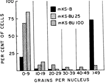
that 73% of the mKS-B cells were heavily
labeled (more than 49 grains per nucleus),
while 20% were unlabeled (less than 10 grains pernucleus) and7%werelightlylabeled (Fig. 2).
In contrast, only about 7% of the mKS-BU 25 cells and no mKS-BU 100 cells were heavily
labeled. Most of the labeled mKS-BU 100 cells had only 10to30 grainspernucleus. Thespecific
activity of the DNA was also determined. The
mKS-BU 25 and mKS-BU 100 cells incorporated only 6 and 1%, respectively, as much 3H-dT per
,ug of DNA as did the parental cells (Table 7).
Previous studies in ourlaboratory have shown
[image:7.462.42.234.255.443.2]that dT kinase can be induced in two dresistant cell strains, LM(TK-) and HeLa BU-100(15, 19), by infection with vaccinia orherpes simplex viruses. Experiments were performed
TABLE 7. Incorporation of3H-thymidine intoDNA of mKS-A, mKS-B, mKS-BU 25, andmKS-BU
100 cellsa
Cellline
mKS-A mKS-B mKS-BU25
mKS-BU 100
I Time
i after
sub-
cultur-ing ZH-dT added hr 48 72 48 72 48 72 48
72
Cellsper
bottle (millions)
10.9 18.7 11.0 22.6 11.6 20.5 8.7 15.4
Incor-pora
tion
(counts
permin perfg
iofDNA)
6, 270 3,134 5,920 2,489 376 161 51 16
aCultures were seeded with 2.5 X 10⁶ cells in
20 ml of medium. At indicated times, 0.1 ml of
3H-dT(20,uc and10lOg)wasadded to eachculture;
thecultureswerethenincubated at 37 C for 6 hr. 100
-rmKS-BU25
:
75EmKS-BU100
LL.
50
z w
25
0-9 10-19 20-29 30-39 40-49 >49 GRAINS PER NUCLEUS
FIG. 2. Incorporation of3H-dT into DNAofparental anddBU-resistant mKS cells. Cells wereincubatedfor
6 hr in thepresenceof3H-dT (20 /Acan1d10,ug).
to learn whether dT kinase and DNA polym-erase were induced
following
infection of mKS-BU 25 andmKS-BU
100 cells withvac-cinia
and SV40.Althoughuninfected mKS-BU 25 and mKS-BU 100 cells haveextremely low levels of dT
kinase,
both dTkinase and
DNA
polymerase arerapidly inducedafter infection
withvaccinia
(Table 8). Attempts to induce dT kinase in mKS-BU 25 cells by superinfecting these cells with SV40 (input multiplicity, 240 PFU per cell) were unsuccessful.Properties
of
SV40 recovered fromn inKS and dBU-resistant mKS cells. Further evidence thattransformed
mousekidney
cells carry the entire SV40 genome wasobtained by comparing
the properties of viruses isolated from mKS cells with the propertiesof
parental SV40. The virus isolates, as well as parental SV40, induced thesynthesis of
Tantigen
in both CV-1 cells and primary mousekidney cells, and were neutralizedby
SV40 antiviral sera. Moreover, the virusesisolated
from mKS cells transformed primary mousekidney
at anefficiency
approximately equaltothat ofparental
SV40.Productiveinfection of CV-1 cells with parental SV40 has been shown to
induce
several enzymes which function in DNAbiosynthesis: dT kinase,thymidylate
synthetase,dihydrofolate
reductase,and
DNApolymerase
(7,17, 22).
When CV-1 cells wereinfected
with virusrecovered from
mKS-A,
mKS-BU25,
or mKS-BU100,
dT [image:7.462.247.441.455.590.2]kinase was induced (Table 9). SV40 (mKS-BU 100) also
induced
DNA polymeraseactivity
in CV-1 cells. When virusstrains
SV40(mKS-A),
SV40(mKS-BU 25),
and SV40(mKS-BU 100)
TABLE 8. Induction of dT kinase and DNA
poly-merase in mKS-BU 25 and mKS-BU 100 cells infected with vaccinia viruSa
Time dT kinase DNA polymerase Cell line inocuafter
laio Con- In- Con- In-trol fected trol fected ltr
mKS-BU 25 1 0.00 0.00 0.12 0.16
3 0.01 0.50 0.17 0.17 7 0.00 4.36 0.13 0.56 mKS-BU 100 7 0.01 2.08 0.23 0.83
aThree-day-old cultures of mKS-BU 25 cells
containing 11.3 X 10⁶cells per bottlewere inocu-lated with vaccinia virus at an inputmultiplicity
of5.3 PFU per cell; 4-day-old cultures of
mKS-BU100cellscontaining13.7 X 106cellswere inoc-ulated with virus at an input multiplicity of 6 PFU per cell.
974
J.on November 11, 2019 by guest
http://jvi.asm.org/
[image:7.462.53.223.492.626.2]VIROGENIC SV40-TRANSFORMED MOUSE CELLS
TABLE 9. Induction of enzyme activity in CV-1 cultures by SV40 recovered from sensitive and dBU-resistant mKSlines
Virus
None
Parental SV40
SV40 (mKS-A)
SV40 (mKS-BU 25)
None
SV40 (mKS-BU 100)
None
Parental SV40
SV40 (mKS-BU 25)
SV40 (mKS-BU 100)
Recovered from mlKS passage
31
36,BU 19
71, BU49
36,BU 19 71, BU 49
Input Timeafter Tantigen (CF multiplicity SV40 units per mg (PFU percell) infection ofprotein)
138 85 61
198
127 75 270
hr 40 40 40
30 46 30 46
50 50 50 50
0 2,100 2,000 1,900
0 0 430 340
0
1,200 2,000 1,900
Enzyme activitya dT kinase
1.2
22.4 19.8 17.1
1.8
20.0 22.3 22.7
DNA polymerase
0.80 0.16 1.49 1.47
a dTkinase:,u,molesofdUMPformed per ,ug ofproteinin 10 minat38C;DNApolymerase:
M,uimoles
of3H-dTTPincorporated into DNA perMgofproteinin 30min at38 C.TABLE10. Induction of enzyme activity inprimarymousekidney culturesa bySV40 recoveredfrom
dBU-sensitiveand dBU-resistant mKS lines
Input Time T antigen Enzyme
activity,
Recoverd from multi- after___________________
Virus inKSerepassag plicity SV40 (CF units
mKSpssage (PFU infec- ofpermg
dTMPkinaseDdC
percell) tion of proten) dTkinas minase kinase polymerasehr
None 0.46 4.7
ParentalSV40 360 52 3.31 20.8
SV40 (mKS-A) 31 237 52 2.44 12.6
SV40 (mKS-BU 25) 36, BU 19 330 52 3.51 16.2
None 30 0 0.07 16.3 0.18
50 0 12.6
SV40 (mKS-BU 100) 71,BU49 417 30 140 0.34 20.4 0.45
50 42 32.7
None 0 11.8
Parental SV40 222 39 NDc 22.7
SV40 (mKS-A) 31 181 39 240 16.3
SV40 (mKS-BU25) 36,BU 19 508 39 500 20.8
a
Seven-day-old
primary mouse kidney cultures contained 4 X 10⁶ cells per culture.bdT kinase and dCMP deaminase:
Mumoles
ofdUMP formed perMug
ofprotein in 10 min at 38 C;dTMP kinase:
,u,umoles
ofdTDP+ dTTPformed per ,ug ofproteinin 10minat38 C;DNA polymerase:.uiumoles
of3H-dTTPincorporatedinto DNA per ,ug of protein in 30 min at 38 C.cNotdone.
were used to
infect
primary mouse kidney cells kidney cells induced host cell DNA synthesis,abortively,
dTkinase,
dCMPdeaminase,
dTMP CV-1 cells were infected with parental SV40,kinase,
and DNA polymerase were all induced SV40 (mKS-BU 25), and SV40 (mKS-BU 100),(Table
10).
and were pulse-labeled with 3H-dT at 34 to 44Induction of host cellDNA synthesis. To learn hr after infection. In cultures infected with each
whether SV40recovered from
transformed
mouse ofthe viruses, the total 3H-dTincorporated into975
VOL. l, 1 967
on November 11, 2019 by guest
http://jvi.asm.org/
[image:8.462.29.426.346.543.2]DUBBS ET AL.
91 8C
51
w 41
3C
)Q DNA FROM:
PURIFIED PARENTAL SV40 00_ PARTICLES
CV-I CELLS INFECTED WITHSTRAIN SV40 00 (mKS-BU25)
CV-I CELLS INFECTED
WITHSTRAIN SV40 I' (mKS-BU100)
1007
00
_
.1/
00
_
J
DO _ /
00
'
,'X
0 4 8 12 16 20 24 28 32 36 40
BOTTOM FRACTION NUMBER TOP
FIG. 3. Sedimentationvelocity (band centrifugation) of3H-dT-labeledDNAfrom CV-1 cells infectedwith
SV40 (mKS-BU 25) and SV40 (mKS-BU 100). A marker SV40 DNA (1,500 counts/min), isolated
from 3H-dT-labeled andpurified parental SV40
par-ticles, was also studied. The DNA preparations were
centrifuged for2.5 hrat 35,000 rev/minand20 C in the SW 39rotorofaSpincomodel L-2ultracentrifuge.
Bulk solution: 3 mlof CsCI; density, 1.503 g/cm-3.
Lamella: 50 plitersofDNA [2.6pigand4,760
countsl
minfrom SV40 (mKS-BU 25) and3.2 jigand5,400
counts/minfrom SV40 (mKS-BU 100)]. The solution wasoverlayedwith 1.8 mlofparaffin oil(petrolatum). Eight-drop fractionswere collectedon 2.5-cm squares of Whatman number 4 paper, washed with 5%
tri-chloroacetic acid andethyl alcohol, driedfor I hr at
70 C,andcountedinaPackardTri-Carb liquid scintil-lationspectrometer.
DNA was increased 10- to 13-fold, and the net
DNA synthesized (estimated by colorimetric
methods) was 50% greater than in uninfected
cultures. After heat denaturation and nitro-cellulose chromatography (24), 37 to
52%,
of the radioactive DNA from the infected culturesexhibited the properties of single-stranded cellular DNA. In contrast, 97% of the labeled
DNAfrom uninfectedcultures had theproperties ofsingle-stranded cellular DNA.
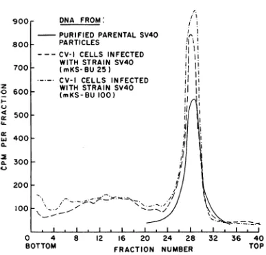
Figure 3 illustratesbandcentrifugation
experi-ments in CsC1 density gradients of the tritium-labeled DNA from CV-1 cell cultures infected with SV40 (mKS-BU 25) and SV40 (mKS-BU 100). A marker DNA isolated from purified SV40 parental virus particles was also centri-fuged. From35 to42% of the radioactive DNA from infected cultures (fractions 1-23) and over
90% of the labeled DNA from uninfected cultures sedimented more rapidly than marker
SV40 DNA. From the amount of radioactive
DNA sedimenting more rapidly than marker
SV40 DNA, an independent estimate canbe made of the relative amount of labeled cellular DNA synthesized in the infected cultures. This estimate
is in
good agreement with that obtained from the nitrocellulose chromatography experiments (24). Since the incorporation of 3H-dT into DNA was increased over 10-fold in the infected cul-tures, it can be concluded that a substantialincrease
in the labeling of cellular DNA occurred 34to 44 hr after SV40 (mKS-BU 25) andSV40
(mKS-BU
100) infections.About 48 to 65 % of the radioactive DNA from infected cultures was resistant to heat
denaturation
and banded at a position identical with that of radioactive DNA isolated from parental SV40 particles (Fig. 3). It is, therefore,likely
that the DNA fromSV40
(mKS-BU 25) and SV40 (mKS-BU 100) had approximately the same molecular weight and conformation asthatfrom parental
SV40.
The results summarized in Table 11 indicate that
SV40
strains recovered frommKS
cells express atleast six
functions characteristic of parental SV40in
CV-1cells
and also sixchar-acteristic
SV40 functions in abortively infected mousekidney
cells. The observations concerningthe
sedimentation
rates of DNA fromSV40
TABLE11. Summary of functions expressed inCV-1
and primary mousekidney cells by SV40 strains recoveredfrom mKS cells
Paren- SV40 SV40 SV40 Viralfunction tal (mKS- (mKS-
(mKS-SV40 A) BU 25) BU 100)
Synthesis
ofT antigen(CF) in CV-1 and
mouse
kidney...
+ + + +Synthesis of V antigen (neutralization) in
CV-1 ... + + + +
SynthesisofviralDNA
inCV-1. + + + +
Induction inCV-1
cul-turesof
dTkinase... + + + +
DNApolymerase. + NDa ND + Induction in mouse
kidneycultures of
dT
...+
kinase.. + + +dCMPdeaminase. + + + ND
dTMPkinase... + + + +
DNApolymerase. + ND ND +
Induction of host cell DNA synthesis in
CV-1 cells... + ND + +
Transformation of
mouse
kidney...
+ + + NDa Notdone.
z 0
4)
976
J. VIROL.7( 6(
I(c
on November 11, 2019 by guest
http://jvi.asm.org/
[image:9.462.46.240.74.260.2] [image:9.462.245.446.369.648.2]VIROGENIC SV40-TRANSFORMED MOUSE CELLS
strains and the phenotypic
characteristics
of these strains provide strongadditional
evidence that essentially the entire complement ofSV40
genetic information is maintained in the trans-formed cell.DIscussIoN
The induction of
synthesis
of SV40 intrans-formed cells by association with susceptible cells has previously been reported (10, 31, 33). Utilizing this technique, it has been possible to
show that nearly every cell in the transformed
mouse kidney lines which we studied carried
thepotentialto
synthesize
infectious virus. Theseresults are in contrast to those reported for
certain
SV40-transformed
hamsterkidneystrains(1, 8, 26, 32, 33), which have not yielded
in-fectious virus by this technique. At this time, SV40 has not been recovered from mKS-A, clone 4.Failure torecoverSV40fromthatclonal
line, as well as from other strains cited above, suggeststhatin these instances either a defective
SV40 genome is maintained in the transformed cell or that conditions for inducing virus
syn-thesis havenotyetbeen achieved.
The mechanism by which
copropagation
oftransformed cells with susceptible cells induces
SV40 synthesis has not been completely
eluci-dated. Experiments indicate that viability of the
transformed cell and direct contact with the
susceptible cell are essential (9). Perhaps,
inter-specific
hybridization
(6) of mKS and CV-1 cells occurs, thereby establishing an intracellular environment favorable for the expression ofthose SV40 functions blocked in mKS cells.
Some evidence supporting this
possibility
hasbeen obtained by Gerber (9), who has shown
that there is an earlier appearance of SV40 when mixed cultures of transformed and sensi-tive cells are treated with ultraviolet-irradiated Sendai virus, a treatment known to enhancecell
fusion.
In general, virus has been easier to recover
from mKS lines which have been passaged in medium containing dBU. Larger yields of virus
wereusually obtainedandahigherpercentageof
trials were successful. The reason for this
em-pirical finding hasnotbeen established.
Initially, mKS cellswerepropagatedinmedium containing dBU to inhibit possible replication of SV40 in occasional cells (17). However,
even after 69 passages in the presence ofdBU, SV40 could readily be recovered from mKS
cells. Moreover, cloning experiments demon-strated that virtually every cell in the mKS-BUAs lines carried the SV40 genome. It has
also been shown that SV40-induced hamster
tumor
cells serially
passaged in and
resistant
to10
,ugof
arabinofuranosylcytosine
(ara-C)
perml
arevirogenic, although
the same concentra-tionof
ara-Ccompletely
inhibitsSV40
replication
in
anestablished
strain ofrhesusmonkey kidney
cells
(9).
After
prolonged passage in the presenceof
increasing concentrations
of the analogue, the mKScells gradually
became resistant
todBU,
and
dTkinase activity
was lost.Some
of
the mKS(BU) cell
lines exhibited
lessthan 1%
of
the
dT kinase
activity of parental mKS
cells.
Nevertheless, all
dBU-resistant mKS cell lines
had normal
levels of
SV40T
antigen,
thusproviding further evidence
thatdT kinase
and
Tantigen
aredissimilar (21).
The fact that
dBU-resistant mKS
cells weredeficient in
dTkinase
activity
was notunex-pected.
It isinteresting, however,
that SV40strains recovered from either dBU-resistant
ordBU-sensitive
mKScell
lines
retained their
capacity
toinduce dT
kinase
in CV-1and in
primary
mousekidney
cultures. Onemotivation
for
propagating
mKS cells in
the
presenceof
dBU
wasthe
expectation
that mutantSV40
strains might be obtained,
someof
which would
lack dT kinase-inducing activity. Nevertheless,
all
of
theSV40 strains studied
to date retained anundamaged
genefor inducing this
enzyme. Thehypothesis
canbe advanced
that theSV40
function under
discussion is
aregulatory
genewhich
normally
derepresses
hostcell
dTkinase.
This
hypothesis would be consistent with
thefinding
that
dT kinase
activity is
enhanced in
mousekidney and in
CV-1cell
cultures bySV40
infection and that dT kinase activity is elevated
in mKS
cell lines.
This
function would be
in-operative
in mKS-BU 100
cells,
since these latter
cells
presumably
are mutantsselected
bypro-longed growth in dBU for defective cellular
dT
kinase cistrons (15, 19).
Thus,derepression
of the
defective dT kinase cistron would
notbe
expected
torestoredT
kinase
activity to mKS-BU 100 cells. However,infection
byvaccinia
virus,which
replicates in murine
cells andinduces
a newvirus-specific
enzyme(14), would
restore dTkinase activity
todBU-resistant mKS
cells.
The
postulate
thatSV40 derepresses
a hostcell
dT kinase does not account for the finding that theMichaelis
constant of the dT kinasepartially
purified from SV40-infected
CV-1 cells isaltered,
although the Michaelis constant of the enzyme fromSV40-infected
mouse kidney cultures or fromSV40-transformed mKS
cells
is similar to that of the enzyme fromuninfected
mouse or CV-1cells.
These latter findings mightsignify
the existence of a secondSV40
functioncontrolling
theformation
of a newSV40-specific
977
VOL.
1,
1967on November 11, 2019 by guest
http://jvi.asm.org/
DUBBS ET AL.
dT
kinase in CV-1 cells (13, 17, 21).
Toexplain
the
failure to express the second dT kinase
function in
mKS-BU 100 cells, it may be
as-sumed that expression of an SV40 gene
control-ling the
formation of a new dT
kinase
is
in-hibited in
primary
mouse
kidney
and in mKS
cell
lines.
It has
been
shown that
twisted-circular
SV40 DNA and SV40
capsid
proteins
are notmade in
primary
mousekidney
ormKS
cells,
although
Tantigen
is
formed in
both
of these
cell types and cellular
DNAsynthesis
is
induced
by
SV40
in mouse
kidney
cultures. The
sugges-tion
made
here is that the
block
in
expression
of SV40 genes in mKS cells is
prior
to astructural
gene for an
SV40-specific
dT
kinase.
In
contrast,
all
the
SV40 genes under discussion are expressed
in CV-1 cells, in which SV40 replicates.
An
implication
of our
findings
is
that
viral
genomes
persisting
intransformed
cells are
notnecessarily defective.
On
the
contrary,
genes for
capsid
protein
formation
orother
viral functions
may
be
intact
but
notexpressed
in
the
trans-formed cell. Further evidence that mKS cells
carry the
entire SV40 genome
wasobtained
by
investigating the properties of the virus recovered
from mKS lines. Not only
wasthe
SV40 from
mKS cells
able
toreplicate
in CV-1
cells,
but
italso
expressed several other viral functions
characteristic of the parental
virus,
such
asin-duction of
Tantigen,
dTkinase,
DNApolym-erase, and host cell DNA
synthesis. Moreover,
the virus
wasneutralized
by anti-SV40
sera,
was
capable
of
inducing
dT
kinase,
dCMP
deaminase,
dTMP
kinase,
and DNA
polymerase
in mouse
kidney cultures,
and could transform
the
mouse
kidney
cells.
Finally,
band
centrifuga-tion and
nitrocellulose
chromatography
experi-ments
have shown
thatthe
molecular size and
conformation
of the DNAobtained from
theSV40 strains recovered from mKS cells
wereindistinguishable
from that of
parental
SV40
DNA.
At this
time,
tumorinduction in newborn
hamsters
has notbeen studied
with the virus
recovered from mKS
cells,
nor have
transplanta-tion
antigens
beenstudied.
However, the
factsalready established suggest
that theSV40 genome
ismaintained in
toto inthe
transformed mouse
kidney
cells.
AcKNOWLEDGMENTS
This investigation was supported by grants from
the American Cancer
Society
(E 291), theNational Science Foundation (GB 5917),the Robert A.WelchFoundation (Q-163), and by Public Health Service grantsCA-06656,
1-K6-AI-2352,
and 5-K3-Ca25,797. We thank JudithRotbein, Carolyn Smith,
Sun OckYim,
andMarjorie
Johnson for able technicalassistance.
LrrERATuRECITED
1. BLACK,P. H. 1966. AnanalysisofSV40-induced transformation of hamster kidney tissue in
vitro.III. Persistenceof SV40viralgenomein clones oftransformed hamster cells. J. Natl.
Cancer Inst. 37:487-493.
2.
BLACK,
P.H.)
W. P. ROWE,ANDH. L. COOPER.1963. An analysis of SV40-induced trans-formation of hamster kidney tissue in vitro.
II. Studies ofthreeclones derivedfrom a
con-tinuous line oftransformed cells. Proc. Natl.
Acad. Sci. U.S. 50:847-854.
3. BLACK,P. H., W. P. RowE,H. C. TURNER, AND R. J. HUEBNER. 1963. A specific
complement-fixing antigen present in SV40 tumor and trans-formed cells. Proc. Natl. Acad. Sci. U.S. 50: 1148-1156.
4. CAMPBELL, D. H., J. S. GARVEY, N. E.CREMER, AND D. H. DussDoRF. 1963. Methods in im-munology,p. 246-247. W. A. Benjamin, Inc., NewYork.
5. DUBBS, D. R., AND S. KIT. 1964. Isolation and properties of vaccinia mutants deficient in
thymidine kinase-inducing activity. Virology
22:214-225.
6. EPHRUSSI, B.,ANDM. C.WEISS. 1965.Interspecific
hybridization of somatic cells. Proc. Natl.
Acad. Sci. U.S. 53:1040-1042.
7. FREARSON, P. M., S. KIT, AND D. R. DUBBS.
1966. Induction of dihydrofolate reductase
activity by SV40 and polyoma virus. Cancer
Res.26:1653-1660.
8. GERBER,P. 1964. Virogenic hamstertumorcells: induction of virus synthesis. Science145:833.
9.
GERBER,
P. 1966. Studies on thetransfer ofsub-viral infectivity from SV40-induced hamster
tumorcells to indicatorcells.Virology 28:501-509.
10. GERBER, P., AND R. L. KIRCHSTEIN. 1962. SV40-induced ependymomas in newborn hamsters. I.Virus-tumor relationships. Virology 18:582-588.
11. HOGGAN,M.D.,W. P.RowE,P.H.BLACK,AND R. J. HUEBNER. 1965. Production of
"tumor-specific" antigens by oncogenic viruses during
acute cytolytic infection. Proc. Natl. Acad. Sci. U.S. 53:12-19.
12. JENSEN,F.C.,A. J. GIRARDI,R. V.GILDEN,AND
H. KoPROWSKI. 1964. Infectionof human and simian tissue cultures with Rous sarcomavirus. Proc. Natl. Acad. Sci.U.S. 52:53-59. 13. KIT, S. 1967. Induction of enzymes of DNA
metabolismbysimian virus 40. In Y. Ito [ed.],
Subviralcarcinogenesis.
14. KIT, S., AND D. R. DUBBS. 1965. Properties of
deoxythymidine kinase partially purified from noninfected and virus-infected mouse fibro-blast cells. Virology 26:16-27.
15. KIT, S.,D. R.DUBBS,ANDP.M.FREARSON. 1966. HeLacells resistanttobromodeoxyuridineand deficient in thymidine kinsase activity. Intern. J. Cancer1:19-30.
16. KIT, S.,D.R.DUBBS,ANDP. M.FREARSON.1966. Enzymes of nucleic acid metabolism in cells
978
J. VIROL.on November 11, 2019 by guest
http://jvi.asm.org/
VIROGENIC SV40-TRANSFORMED MOUSE CELLS
infected with polyoma virus. Cancer Res.
26:638-646.
17. KIT,S., D. R. DUBBS, P. M. FREARSON,ANDJ.L. MELNICK. 1966. Enzyme induction in SV40-infected green monkeykidney cultures. Virology 29:69-83.
18. KIT, S., D. R. DUBBS, AND T. C. Hsu. 1963. Biochemistry of vaccinia-infected mouse fibro-blasts (strain L-M). III. Radioautographicand biochemical studies of thymidine-3H uptake into DNA of L-M cells and rabbit cells in primary culture.Virology19:13-22.
19. KIT, S., D. R.DUBBS,L. J.PEEKARSKI,ANDT. C.
Hsu. 1963. Deletion of thymidine kinase
ac-tivity from L cells resistant to
bromodeoxy-uridine. Exptl. Cell Res. 31:297-312. 20. KIT, S.,D. R.DUBBS, L. J. PIEKARSKI,R. A.DE
TORRES,ANDJ. L. MELNICK. 1966. Acquisition
of enzyme function by mouse kidney cells abortively infected with papovavirus SV40. Proc. Natl. Acad. Sci. U.S. 56:463-470. 21. KIT, S., J. L. MELNICK, M.ANKEN,D.R. DUBBS,
R. A. DE TORRES, AND T. KITAHARA. 1967. Nonidentity ofsome simian virus 40-induced
enzymes with tumor antigen. J. Virol. 1:684-692.
22. KIT, S., L. J. PEEKARSKI, ANDD. R. DUBBS. 1967. DNA polymerase induced by simian virus 40. J. Gen. Virol. 1:163-175.
23. KIT, S., R. A. DE TORRES, AND D. R. DUBBS. 1967. Induction of
deoxycytidylate
(dCMP) deaminase activity in SV40-infected mouse kidney cultures. Proc. Am. Assoc. Cancer Res.8:37.24. KIT, S., R. A.DETORRES, D. R.DUBBS,ANDM.L. SALvI. 1967. Induction of cellular deoxyribo-nucleic acid synthesis by simian virus 40. J.
Virol.1:738-746.
25. LOWRY, 0. H., N. J. ROSEBROUGH, A. L. FARR,
AND R. H. RANDALL. 1951. Protein
measure-ment with the Folin phenol reagent. J. Biol. Chem.193:265-275.
26. MELNICK, J.L., K. S.KHERA,ANDF. RAPP. 1964. Papovavirus SV40: failure to isolate infectious virus from transformed hamster cells synthe-sizingSV40-inducedantigens. Virology
23:430-432.
27. PUCK, T. T., P. I. MARCUS, ANDS. J. CIECIURA. 1956. Clonal growth of mammalian cells in vitro. Growth characteristics of colonies from
singleHeLacells with andwithout a "feeder" layer. J. Exptl. Med. 103:273-284.
28. RAPP, F.,J. S.BUTEL,ANDJ. L. MELNICK. 1964. Virus-induced intra-nuclear antigen in cells transformed by papovavirus SV40. Proc. Soc.
Exptl.Biol. Med. 116:1131-1135.
29. RAPP, F., T. KITAHARA, J. S. BUTEL, AND J. L. MELNICK. 1964. Synthesis of SV40 tumor antigen during replication of simian
papova-virus (SV40). Proc. Natl. Acad. Sci. U.S. 52:1138-1142.
30. SABIN, A. B.,ANDM. A.KOCH. 1963.Evidence of continuous transmission ofnoninfectious SV40 viral genome in most or all SV40 hamster tumorcells. Proc. Natl. Acad. Sci. U.S. 49:304-311.
31. SABIN, A. B.,ANDM.A.KOCH. 1963.Behavior of
noninfectious SV40 viral genome in hamster tumorcells: inductionofsynthesis of infectious virus. Proc. Natl. Acad. Sci. U.S. 50:407-417. 32. SABIN, A. B.,AND M. A. KOCH. 1964. Sourceof genetic information for specific complement fixing antigens in SV40 virus-induced tumors. Proc. Natl. Acad. Sci. U.S.52:1131-1138. 33. TOURNIER, P., R. CASSINGENA, R. WICKER, J.
COPPEY, AND H. SUAREZ. 1967. Etude du mechanismedel'induction chez des cellules de hamstersyrientransformeespar levirus SV40. I. Properties d'une lignee cellulaire clonale.
Intern. J. Cancer 2:117-132.
VOL. 1, 1 967