Drug delivery; lessons to be learnt from Leishmania studies. C. D. Shaw and K.C. Carter.
Strathclyde Institute for Pharmacy and Biomedical Sciences, 161 Cathedral Street, Glasgow. *Corresponding author:
Dr K.C. Carter,
Strathclyde Institute of Pharmacy and Biomedical Sciences University of Strathclyde
161 Cathedral Street Glasgow,
G4 0RE Scotland
Summary
Leishmaniasis is a disease caused by infection with the protozoan parasite Leishmania which is responsible for three main types of disease; cutaneous leishmaniasis, visceral leishmaniasis and mucocutaneous leishmaniasis which is related to the tissue tropism of the infecting species. This presents a major challenge to successful drug treatment, as a drug must not only reach antileishmanial concentrations in infected macrophages, the parasites’ host cell, but also reach infected cells in locations specific to the type of disease. In this paper we discuss how studies using Leishmania have contributed to our knowledge on how drug delivery systems can be used to improve drug efficacy and delivery.
Keywords: Leishmaniasis, drug delivery systems, intravenous, non-invasive
Introduction
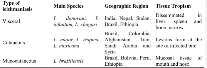
[image:2.595.50.403.547.663.2]Leishmaniasis is a disease caused by infection with the protozoan parasite Leishmania, which is transmitted by female sandflies. The type of disease caused by the parasite depends on the infecting species and the host’s immune response [1] but three main forms occur; cutaneous leishmaniasis (CL), mucocutaneous leishmaniasis (MCL) and visceral leishmaniasis (VL, Table 1). The World Health Organisation estimates that 350 million people, living in 98 countries, are at risk of contracting leishmaniasis, and each year approximately 1.5 million new cases of cutaneous and 500,000 of VL are reported. In terms of disease burden, leishmaniasis is responsible for 2,357,000 DALYs (Disability-Adjusted Life Years) lost due to ill effects caused by the disease.
Table 1 The main species responsible for leishmaniasis, their geographical distribution and site of the parasites within the body.
Type of
leishmaniasis Main Species Geographic Region Tissue Tropism
Visceral L. infantum, L. chagasi donovani, L. India, Nepal, Sudan, Brazil, Ethiopia Disseminated liver, spleen and in bone marrow
Cutaneous L. major, L. tropica, L. mexicana
Brazil, Colombia, Afghanistan, Iran, Saudi Arabia and Syria
Lesions form at the site of infected bite
The Leishmania parasite has two distinct morphological forms in its life cycle, the intracellular amastigote in the mammalian host and the extracellular promastigote, which is transmitted by a sand fly vector. Infective promastigotes are deposited into the skin when an infected sand fly feeds. The promastigotes are taken up by phagocytes in the vicinity and transform into amastigotes within the parasitophorous vacuole. Over a period of 4-6 days, the amastigotes multiply inside the macrophage until its maximum capacity is reached and then the macrophage ruptures, releasing amastigotes which can infect new macrophages. The ability to control the infection depends on stimulating a protective immune response in the host. This ultimately results in activation of the cell’s cytotoxic mechanisms, usually involving the production of reactive oxygen and nitrogen intermediates. Details on the specific responses involved, which vary between species, can be found in recent reviews [2, 3, 4]. The parasite’s life cycle is completed when an uninfected sand fly takes a blood meal from the infected host.
Currently there is no vaccine to prevent leishmaniasis in people therefore disease control depends on treating infected individuals or programmes which target the vector e.g. use of insecticide impregnated bed nets), vector control or the reservoir host e.g. the dog for VL. At present there are a limited number of drugs used in the treatment of leishmaniasis (Table 2) and many of the drugs are only suitable for use in certain geographical areas. For example, antimonials were the first line treatment for the majority of Leishmania infections for over 50 years but their use in treating VL is now limited due to the increasing incidence of drug resistance and relapse in endemic regions such as India and Nepal [5,6]. The introduction of miltefosine (MILT) for the treatment of VL was a major step forward as it was the first orally active drug. However there are already problems with a reduction in treatment efficacy, which could indicate that drug resistance is being introduced into the parasite population, possibly due to incorrect dosing by individuals [7]. Amphotericin B (AMB) is now the first line treatment for VL but it requires administration via the parenteral route and its use is limited by its inherent toxicity and high cost [2]. Although AMB resistance has been induced in laboratory strains [8], there is no evidence that it is present in field strains (9). Leishmaniasis is mainly a disease of the poor and so development of new drugs does not elicit the same interest for drug companies as other diseases. There is however a willingness to co-operate in providing drugs for leishmaniasis as the recent "London Declaration on
Neglected Tropical Diseases” recently showed
(http://www.who.int/neglected_diseases/London_Declaration_NTDs.pdf). One way to
improve drug treatment would be to use a drug delivery system to increase the efficacy of novel or existing drug. In this review we will discuss the variety of drug delivery systems that have been tested and demonstrate how studies on leishmaniasis have added to our knowledge on drug delivery.
Table 2 Drugs used in the treatment of leishmaniasis.
Drug Route Dose Adverse side effects reported
Pentavalent
antimonials Intravenous/ Intramuscular 20 mgSb
V/kg for 28
daysa
Vomiting and
nausea. Widespread resistance
Pentamidine Intravenous/ Intramuscular 4 mg/kg for 15 days alternative daysb Diabetes side effects
Paromomycin Intramuscular 15 or 20 mg/kg for 21 daysc
Renal Toxicity, Ototoxicity
Miltefosine Oral 2.5 mg/kg for 28 daysd Poor Teratogenic compliance.
Amphotericin B Intravenous 15 mg/kg for 30 days on alternative days, or 20 mg/kg/day for 20 dayse
High costs.
Nephrotoxicity
Sitamaquine Oral
1.75, 2, 2.5 or 3 mg/kg/day for 28 daysf
Abdominal pain, potential renal toxicity
The specific treatment regimen can vary for different Leishmania spp and their geographical location. Dosing regimens are shown from specific studies as an indication of treatment protocols used [a10; b11; c12; d13; e14, f15]
Drug delivery systems
amastigote (Fig. 1) as the parasite is located within the parasitophorous vacuole within the macrophage. Imaging studies have shown the dynamic nature of these vacuoles and information on their biogenesis and during infection (16). These technical developments will aid in characterising delivery to the parasites within the parasitophorous vacuole but drug delivery to the Leishmania parasite still presents technical difficulties. Most analytical methods for drugs are based on high performance liquid chromatography and assessment of delivery to the parasite would require isolation of amastigotes, which may cause drug loss, and an assay method of a suitable sensitivity level to detect the drug present. Therefore most studies use reduction in parasite burdens as a measure of successful drug delivery. In most in vivo studies traditional pharmacokinetic parameters (e.g. distribution phase half-life; elimination phase half-life; area under the plasma concentration-time curve; volume of distribution, total body clearance) and drug levels at the targeted site are used to assess drug delivery. For example, antimonial drugs given by the intravenous route are only present in the blood for a short period of time as they have a short half life (absorption phase mean half-life of 0.85 h) and a rapid clearance (elimination mean half-life of 2.02 h, 17), which would limit their uptake by the host cells and explain why multiple dosing is required for parasiticidal levels to reach the Leishmania parasites.Recent reviews discuss the problems associated with delivery of drugs using different routes of administration (18, 19, 20), the problems associated with drug delivery to specific sites or organelles within the body (20, 21) and drug deposition and uptake at these sites (21, 22). A drug delivery system can help achieve this aim as it directs more of the drug dose to tissues and away from the systemic circulation. Once the drug formulation has accessed cells at the site of uptake then the inherent pharmacokinetic properties of the drug within the formulation will influence its release into surrounding tissues (23, 24). Most drug delivery systems act as drug depots that decrease the release rate of the incorporated drug and therefore give more time for the drug to concentrate within the targeted cells. Macrophages, which are found in high concentrations in a number of locations in the body e.g. liver, lungs, spleen, play an important role in enhancing tissue uptake of particulate nanoformulations. Macrophages phagocytose particles from their immediate vicinity as part of their innate immune response and as a consequence act as a local drug depot. This means that the drug is directed directly to the Leishmania parasite in infected macrophages (25, 26, 27). Borborema et al., 2011, demonstrated the advantages of this type of approach using a liposomal formulation of meglumine antimoniate They showed that using the carrier system reduced the 50% inhibitory concentration (IC50 value) value
against the intracellular amastigote stage of L. major compared to the drug solution, from 93
µM to 10.5 µM. Moreover, they also showed and that infected macrophages were more efficient than uninfected macrophages at taking up the liposomes (28). A drug delivery system can facilitate a reduction in the total drug dose and/or number of doses required, which is particularly important for a potentially toxic drug. For example, amphotericin B (AMB) that is associated with nephrotoxicity. This can be important for a drug that causes nephrotoxicity such as amphotericin B (AMB). This beneficial feature for drug delivery systems has been clearly demonstrated by the higher efficacy and lower toxicity of lipid formulations of AMB compared to AMB solution [29]. However these lipid formulations are prohibitively expensive for widespread use in endemic countries. This problem is being addressed by a World Health Organisation (WHO) initiative, which facilitated the donation of 445,000 vials of AMBisome for the treatment of VL.
Repurposing drugs originally designed for other clinical conditions gave new antileishmanial treatments. Thus AMB was originally developed for the treatment of fungal infections and MILT was originally in development for the treatment of cancer. Repurposing clinically approved drugs for treatment of leishmaniasis is an attractive approach as the majority of the required toxicity testing has already been completed, although additional testing would be required if a different mode of administration is used. Endemic countries often have traditional medicines that have been used for the treatment of leishmianiasis, and development of novel drugs from plant products has been investigated (30).
Intravenous delivery
Liposomes are synthetic vesicles, prepared using phospholipids that form a natural bilayer. The exact composition of liposomes can vary and often includes phosphatidylcholine (PC) and cholesterol. By assembling the liposomes in conjunction with an aqueous solution, the compound is encapsulated in the inner core of the lipid bilayer. When the liposome comes in to contact with a cell membrane it may fuse with itor be taken up by phagocytosis, thus delivering the drug solution inside the cell. Vesicle characteristics such as composition, size, surface charge and drug loading all have important influences on drug delivery (31, 32) Roychoudhury et al. (34) prepared liposomes containing sodium stibogluconate (SSG) from PC and stearlyamine (PC-SA) or PC and cholesterol (PC-Chol) using sonication followed by centrifugation at 60,000 x g to remove unentrapped drug, a method that would not adapt well to large-scale manufacture and has been shown to damage the vesicle bilayer (35). Single-dose treatment of mice infected with L. donovani strains that had different inherent susceptibilities to SSG with PC-SA liposomes containing SSG (PC-SA-SSG, 12 mg Sbv/kg)
resulted in a significant reduction in liver, splenic and bone marrow parasite burdens (>84%) whereas similar treatment with PC-Chol liposomes containing SSG (PC-Chol-SSG) only caused a significant reduction in liver parasite burdens. The advantage of using a carrier system was clearly demonstrated in this study as similar treatment with SSG solution at a dose of 300 mg Sbv/kg only significantly affected liver parasite burdens in mice infected with
a SSG susceptible strain. Drug uptake studies in parasites showed that significantly higher levels of Sb were present in amastigotes recovered from infected macrophages treated with PC-SA-SSG compared with PC-Chol-SSG or SSG solution. The entrapment efficiencies quoted for the two formulations were similar, indicating that both type of carriers would deliver a similar amount of drug to cells, therefore different in drug delivery may reflect differences in the rate uptake for the two type of liposomes (36). PC can interact with phosphatidylserine (PS) residues present on the cell membranes and this could improve uptake of PC-SA liposomes [37].
the liver and spleen (39). Similar treatment with SSG-NIV had no adverse effect on lesion development in L. major infected mice (unpublished data), probably because the formulation did not target the drug to the skin parasites. More recent studies clearly demonstrated this effect. Iintravenous treatment with a NIV formulation containing luciferin did not enhance delivery of luciferin to luciferase-expressing L. major parasites within the footpad of a mouse, but did enhance delivery of the substrate to luciferase-expressing L. donovani parasites located within the liver of a co-infected mouse (Fig. 2).
Kansal et al. [42] showed that a nanocapsule emulsion (NC) containing doxorubicin (NC-DOX) had a 1.75 fold higher uptake if PS was anchored on to its surface compared to non-PS containing NC-DOX. The high drug levels within amastigotes after PC-SA-SSG compared to PC-Chol-SSG treatment could be related to lower inhibition of macrophage function, a factor that could be explored by infecting macrophages using different parasite: macrophage ratios and determining the effect on SSG uptake.
Modifying the surface of the delivery vehicle to increase the time it remains in the systemic circulation or by incorporating molecules, which target surface receptors on the target cell, can increase the efficacy of a drug formulation. This has been exploited by incorporation of polyethylene glycol (PEG) into liposomes to produce ‘long circulating’ or ‘stealth’ liposomes that are more likely to be taken up by tissues [43]. Integrating dendrimers, which interact with MHC class II molecules, into amphotericin B liposomes increased their uptake by phagocytic cells and intravenous treatment of L. major infected mice with this formulation was more effective at treating skin lesions compared to liposomal AMB alone. Interaction with MHC class II molecules was confirmed to be important as incorporation of a dendrimer that targeted a random peptide did not increase the efficacy of liposomal AMB [44] An unexpected side effect of this formulation was its ability to boost host immune responses, leading to enhanced interferon gamma production by L. major specific splenocytes. Infection with Leishmania suppresses immune responses in susceptible individuals both at the local (i.e. infected macrophage) and whole body level, so production of an immunotherapeutic drug formulation would be ideal for leishmaniasis [45].
mix, rotary evaporation was then used to remove the solvents and drug loaded liposomes were formed by hydration with water. Freeze-thaw cycles were used, presumably to improve drug entrapment as a reduction in size was achieved by passing the liposome suspension through an extruder fitted with an 80 nm polycarbonate membrane. Unentrapped drug was removed from the resulting suspension by passing down a PD-10 column and the liposomes were lyophilised in the presence of sucrose as the cryoprotector. On reconstitution, liposomes with a mean vesicle size of 75 ± 31 nm and an entrapment efficiency of 7% was obtained. Intravenous treatment with a single dose of resiquimod liposomes (0.38 mg/kg, assuming a 20 g mouse weight) caused a significant reduction in splenic, liver and bone marrow L. donovani parasites compared to treatment with the carrier alone but the drug formulation was not as effective as single dose treatment with SSG (500 Sbv/kg).
Determination of host immune responses showed that resiquimod liposome treatment was associated with enhanced interferon gamma (IFN-) and interleukin 10 (IL-10) production by splenocytes activated with specific antigen compared to control or carrier alone groups. These two cytokines act antagonistically as IFN-production stimulates macrophages to kill Leishmania and is associated with protection against L. dovovani infection whereas IL-10 suppresses macrophage activation and its production is associated with susceptibility [47]. However the relative local concentration of each cytokine at the site of infection will be important in determining whether the immunostimulatory effects aid parasite clearance or not.
related to the way AMB is incorporated into the ND. AMB causes ND bilayer interdigitation and in treated Leishmania cells there would be a reduction in bilayer thickness of the host/parasite membrane bilayer, so that only 1 AMB molecule spans the membrane bilayer, and as AMB molecules self-associate, a pore formed from 8-12 AMB would form. This pore results in leakage in the target cell membrane resulting in death of the host cell/parasite cell [51].
Conjugating drugs to polymeric nanoparticles is another strategy used to deliver drugs to particular target sites. Polymeric nanoparticles have an advantage over lipid formulations as their production costs are usually lower and the shelf life at room temperature is extended [52]. Gaspar et al. [53] and Paul et al. [54] showed that polyalkylcyanoacrylate(PACA) primaquine or polymethacrylate (PMMA) pentamidine nanoparticles respectively could be used to enhance drug delivery. Poly(lactide-co-glycolide) (PLGA) nanoparticles have been used to improve the delivery of AMB in a number of studies. For example, Nahar and Jain [55] produced PLGA nanoparticles conjugated to AMB (AMB-NP) which had a size of less than 200 nm and a polydispersity index (PDI) below 0.16. Additionally, inclusion of PEG to couple PGLA to mannose improved their uptake by macrophages and efficacy against L. donovani ex vivo amastigotes. An in vitro study showed that saponin loaded PLGA-nanoparticles were active against axenic and ex vivo amastigotes of L. infantum and confocal microscopy allowed visualisation of saponin-loaded nanoparticles uptake by L. donovani infected macrophages [56]. The same group recently characterised the delivery of AMB- PLGA nanoparticles against L. infantum parasites and several fungal species a showed that the formulation was was either equivalent or more efficacious than AMBisome or Fungizone against promastigotes and amastigotes [57].
Non-invasive drug delivery
Ideally, any new drug formulation developed for the treatment of leishmaniaisis should be administered by a non-invasive route as it removes the requirement for hospitalisation, improves patient compliance and removes other risks e.g. occupational risk of infection or environmental hazards associated with disposal of contaminated sharps. There are a number of routes that can be used [58] but the three that are probably most relevant for leishmaniasis are oral, pulmonary and topical.
Oral delivery is the preferred method for patients and clinicians, as it does not require hospitalization of the patient. Developing oral formulations of current chemotherapy options
Commented [CDS3]: For reviewer comments 3 and 4… I don’t think we’ve critically analysed the IV route like they asked. We could put a sentence or two of comment/opinion on the success of IV. We have listed the IV treatments but not evaluated them.
is attractive as it can help reduce the effective dose required, helping to reduce both side effects and the cost of treatment. However this route offers its own challenges as the gastrointestinal tract provides harsh physicochemical conditions. In addition any drug taken up in the gut has to undergo first pass metabolism in the liver, where it is exposed to enzymes [59]. Although patient compliance is higher for oral formulations, a lack of treatment supervision can lead to patients not completing the full course of treatment. This is a major issue as it may facilitate the development of drug resistance in Leishmania. MILT is the only oral drug used in the clinical treatment of leishmaniasis and it was originally developed as an anticancer agent. It is highly effective against VL, giving cure rates of 94%. But recent evidence in India indicates that relapse rates are now higher, even though there was no apparent increase in MILT resistance in parasites isolated from VL patients before, and after, treatment. Parasites isolated from post dermal kala patients do exhibit increased resistance to MILT [60] and in Nepal the number of VL patients are not responding to MILT treatment is increasing [61].
Imipramine, clinically used to treat treat depression by the oral route, has recently been shown to be effective against L. donovani. Repurposing drugs for other clinical indications has been suggested for a some time [62] but it is likely to require public funding for leishmaniasis.
formulation. Studies using Caco-2 cell monolayers have shown that f-CNT can cause reversible modulate tight junction formation, which would be required for transport across the gut epithelial barrier. In addition f-CNT had the ability to down regulate the activity of P glycoprotein efflux activity, which would help to increase the bioavailability of any drug incorporated into the carrier system [66]. CNT have also been shown to activate reactive oxygen species but this activity was associated with cytotoxicity in treated keratinocytes [67], so may not be important in the improved f-CNT formulations.
Nanoemulsions are mixtures of two normally immiscible liquids that are usually stabilised by using a surfactant, which can form a polymer shell around the outside of the emulsion to prevent coalescence. Formulating the emulsion as oil-in-water allows the solubilisation of poorly aqueous soluble drugs, such as AMB, and protects them from degradation. The first example of this formulation for treatment of VL was reported in 2004 [68]. IC0-010, a lipid emulsion formulation of AMB currently being developed by iCo-therapeutics, was granted orphan drug status by the US Food and Drug Administration [69]. The formulation contains a mono- and di-glycerides and d-α-tocopheryl polyethylene glycol succinate, which form a self-emulsifying structure that forms droplets in intestinal conditions at 37°C. The formulation has been shown to be highly effective in a murine model of VL and is stable at temperatures expected in tropical environments. Less developed formulations of AMB have been produced by other researchers. For example, a nanoparticle formulation of AMB was shown to be more effective that Fungizone in a murine model of VL but further work is required to improve its in vivo activity [70].
applied dose). Four weeks treatment using liposomal PMM at 50mg/dose twice a day cured mice [80]. However it is possible that mice ingested some of the drug formulation as we have found that mice clean away any formulation applied to the skin, even if it contains denatonium benzoate, a compound used to deter ingestion [81].
Future perspectives
The ability to successful treat Leishmania depends on not only identifying an effective drug, it is also important to deliver that drug at parasiticidal levels into infected macrophages, which can reside at multiple sites within the body. Advances in non-invasive imaging techniques [82] and the production of novel Leishmania strains expressing reporter genes e.g. green fluorescent protein (GFP), cherry red fluorescent protein or luciferase [83], have allowed detailed studies on disease burdens and pharmacokinetic studies at both the cellular level and in individual animals. Selection of the most appropriate label for experimental studies is essential. For example, GFP-labelled parasites allow direct visualisation of parasites without requiring a substrate (luciferin), which could be more beneficial for in vitro assays, however, for in vivo studies where parasites are located in deeper tissues rich in blood (e.g. the liver), then luciferase-expressing parasites may be more appropriate. Tissues can absorb the light emitted by fluorescent dyes and light scattering can mean that a lot of the light does not reach the detector or does not indicate the correct localization of parasites. Bioluminescence gives a stronger signal but it is not without its problems. Luminescence depends on delivery of luciferin to luciferase-expressing cells. Luciferin has a large volume of distribution after the recommended intraperitoneal injection but a short in vivo half-life, meaning that imaging is usually carried out within 30 min of dosing. Bioluminescence is also dependent on the presence of oxygen, therefore there will be a lo/no signal if if hypoxia is present at the site of the luciferase-expressing cells. The development of better gene reporter systems and continued development of imaging systems that allow integration of information from different types of imaging systems (e.g. fluorescence, bioluminescence, radioisotope imaging and X-ray-based computed tomography) means that a better understanding of the disease processes and treatment outcome is possible. It is now possible to have high throughput screening of compounds against the more clinically relevant intracellular amastigote stage of Leishmania rather than the promastigote stage [84] and it is possible to determine the effect of drug treatment on parasite burdens in multiple sites within the same animal at different times post-treatment, to get a better indication of how altering treatment regimens effects parasite numbers. It is also possible to determine how effective a drug
delivery system is at delivering a drug to different sites in the body, allowing rational design a drug delivery system so its effect on its delivery capabilities can be assessed. We have shown that it is possible to monitor drug delivery to multiple sites within the same animal using this type of imaging technology (Fig. 2). Eventually these types of studies will provide the data to mathematically model drug delivery so that the knowledge gained from other specialties e.g. fluid mechanics can be integrated so that it may eventually be possible to study in vivo drug delivery using very few animals and systems specific to the species being treated. Researchers need to keep in mind scale up parameters of their formulations at the early stage of development and try and use constituents of pharmaceutical grade where possible. Quite often experimental methods contain steps that are not practical for large-scale manufacture and considerations like these early on can save time further down the line. All our formulation studies have suggested that drug loading is one of the most important parameters in formulation efficacy, a factor strongly influenced by drug solubility. Therefore, chemical modification of active compounds to increase their aqueous solubility is an important area of research. In addition any drug development programme must include screening against recently isolated field strains in their studies as it is possible that by the time the drug formulations have gone through clinical trials it may be inappropriate for use in endemic countries. We developed a NIV formulation of sodium stibogluconate, which was effective a single intravenous dose in rodent VL models, but funding for this project stopped once it became apparent that antimony resistance was widespread in India. There are number of studies determining the molecular basis for resistance to antileishmanial drugs as knowing how the mechanism may allow the development of novel therapies that can block the mechanism(s) and turn a clinically ineffective drug back into an effective treatment. For example, resistance to PMM in L. donovani was related to increased membrane fluidity accompanied with decreased intracellular drug accumulation and was associated with increased expression of ATP-binding cassette (ABC) transporters (MDR1 & MRPA) [85] and the ABC transporter gene MRPA was amplified in antimony resistant field isolates of L. donovani [86]. ABC transporters also mediate drug resistance in cancer cells [87], therefore an understanding of how they operate in Leishmania is relevant to other clinical conditions.
Executive summary
Leishmaniasis: current drugs used and their limitations
Drug delivery systems: the role of drug delivery systems in modifying drug delivery to the Leishmania parasite are discussed: including vesicles, nanocapsule, nanodiscs, nanoparticles, nanotubes, nanoemulsions
Intravenous delivery: novel drug formulations given by the intravenous route are discussed
non-invasive drug delivery: novel drug formulations given by the oral, pulmonary and cutaneous routes are discussed
Future perspectives: co-ordinating data obtained using new imaging technologies along with engineering disciplines such as fluid dynamics may aid in the design of better drug delivery system
References
1. Heyneman D. Immunology of leishmaniasis Bull World Health Organ. 44(4), 499-514 (1971).
2. Moore EM, Lockwood DN. Treatment of visceral leishmaniasis. J Glob Infect Dis. 2(2), 151-8 (2010).
3. Okwor I, Mou Z, Liu D, Uzonna J. Protective immunity and vaccination against cutaneous leishmaniasis. Front Immunol. 29(3), 128 (2012).
4. de Oliveira CI, Brodskyn CI. The immunobiology of Leishmania braziliensis infection. Front Immunol. 8(3), 145 (2012).
5 Rijal S, Chappuis F, Singh R et al. Treatment of visceral leishmaniasis in south-eastern Nepal: decreasing efficacy of sodium stibogluconate and need for a policy to limit further decline. Trans R Soc Trop Med Hyg. 97(3), 350-354 (2003).
6. Musa A, Khalil E, Hailu A et al. (SSG) & paromomycin combination compared to SSG for visceral leishmaniasis in East Africa: a randomised controlled trial. PLoS Negl Trop Dis. 6(6), (2012).
7. Sundar S, Singh A, Rai M et al.Efficacy of miltefosine in the treatment of visceral leishmaniasis in India after a decade of use.Clin Infect Dis. 55(4):543-50 (2012
9. Prajapati VK, Mehrotra S, Gautam S, Rai M, Sundar S. In vitro antileishmanial drug susceptibility of clinical isolates from patients with Indian visceral leishmaniasis--status of newly introduced drugs. Am J Trop Med Hyg. 87(4), 655-657 (2012).
10. Ritmeijer K, Dejenie A, Assefa Y et al. A comparison of miltefosine and sodium stibogluconate for treatment of visceral leishmaniasis in an Ethiopian population with high prevalence of HIV infection. Clin Infect Dis 43(3), 357–364 (2006).
11. Das VN, Siddiqui NA, Pandey K,et al. A controlled, randomized nonblinded clinical trial to assess the efficacy of amphotericin B deoxycholate as compared to pentamidine for the treatment of antimony unresponsive visceral leishmaniasis cases in Bihar, India. Ther Clin Risk Manag. 5(1), 117-124 (2009).
12. Musa AM, Younis B, Fadlalla A, et al. Paromomycin for the treatment of visceral leishmaniasis in Sudan: a randomized, open-label, dose-finding study. PLoS Negl Trop Dis. 4(10), e855 (2010).
13. Sundar S, Chakravarty J, Rai VK, Agrawal N, Singh SP, Chauhan V, Murray HW. Amphotericin B treatment for Indian visceral leishmaniasis: response to 15 daily versus alternate-day infusions. Clin Infect Dis. 45(5), 556-561 (2007).
14 Rahman M, Ahmed BN, Faiz MA et al. Phase IV trial of miltefosine in adults and children for treatment of visceral leishmaniasis (kala-azar) in Bangladesh. Am J Trop Med Hyg. 85(1), 66-69 (2011).
15 Wasunna MK, Rashid JR, Mbui J et al. A phase II dose-increasing study of sitamaquine for the treatment of visceral leishmaniasis in Kenya. Am J Trop Med Hyg. 73(5), 871-876 (2005).
16. Real F, Mortara RA. The diverse and dynamic nature of Leishmania parasitophorous vacuoles studied by multidimensional imaging. PLoS Negl Trop Dis. 6(2), e1518 (2012) 17. Chulay JD, Fleckenstein L, Smith DH. Pharmacokinetics of antimony during treatment of visceral leishmaniasis with sodium stibogluconate or meglumine antimoniate. Trans. R. Soc Trop Med Hyg 82(1), 69-72 (1988).
18. Nino M, Calabrò G, Santoianni P.Topical delivery of active principles: the field of dermatological research. Dermatol Online J. 16(1):4. (2010).
19. Zarogoulidis P, Chatzaki E, Porpodis K et al. Inhaled chemotherapy in lung cancer: future concept of nanomedicine. Int J Nanomedicine. 7:15, 51-72, (2012).
20. Diab R, Jaafar-Maalej C, Fessi H, Maincent P. Engineered nanoparticulate drug delivery systems: the next frontier for oral administration? AAPS J. 14(4), 688-702 (2012).
22. Sakhrani NM, Padh H.Organelle targeting: third level of drug targeting. Drug Des Devel
Ther. 17;7, 585-99 (2013)
23. Benet LZ. Predicting Drug Disposition via Application of a Biopharmaceutics Drug Disposition Classification System. Basic Clin Pharmacol Toxicol. 106(3), 162–167 (2010). 24. Burton PS, Goodwin, JT, Vidmar TJ, Amore BM. Predicting Drug Absorption: How Nature Made It a Difficult Problem J Pharmacol Exp Ther. 303, 889-895 (2002).
25. Date AA, Joshi MD, Patravale VB. Parasitic diseases: Liposomes and polymeric nanoparticles versus lipid nanoparticles. Advanced Drug Delivery Reviews 59(6), 505–521 (2007).
26. Romero EL, Morilla MJ. Drug delivery systems against leishmaniasis? Still an open question. Expert Opin. Drug Deliv. 5(7), 805–823 (2008).
27. Espuelas S. Delivery systems for the treatment and prevention of Leishmaniasis. Gaz. Med. Bahia 79 (supl. 3), 134–146 (2009).
28 ????????? Borborema et al., 2011???????????????????????
29. Balasegaram M, Ritmeijer K, Lima MA et al. Liposomal amphotericin B as a treatment for human leishmaniasis. Expert Opin Emerg Drugs. 17(4), 493-510 (2012).
30. Brito AM, Dos Santos D, Rodrigues SA, Brito RG, Xavier-Filho L. Plants with anti-Leishmania activity: Integrative review from 2000 to 2011. Pharmacogn Rev. 7(13), 34-41 (2013).
31. Abra RM, Hunt CA. Liposome disposition in vivo. III. Dose and vesicle-size effects. Biochim Biophys Acta. 23;666(3), 493-503 (1981).
32. Abra RM, Bosworth ME, Hunt CA. Liposome disposition in vivo: effects of pre-dosing with lipsomes. Res Commun Chem Pathol Pharmacol. 29(2), 349-60 (1980).
33. Abra RM, Hunt CA. Liposome disposition in vivo IV: the interaction of sequential doses of liposomes having different diameters. Res Commun Chem Pathol Pharmacol. 36(1), 17-31 (1982).
34. Roychoudhury J, Sinha R, Ali N. Therapy with sodium stibogluconate in stearylamine-bearing liposomes confers cure against SSG-resistant Leishmania donovani in BALB/c mice. PLoS One. 6(3), e17376. (2011).
35. Heeremans JL, Gerritsen HR, Meusen SP et al.. The preparation of tissue-type Plasminogen Activator (t-PA) containing liposomes: entrapment efficiency and ultracentrifugation damage. J Drug Target. 3(4), 301-310 (1995).
37. Fadok VA, Voelker DR, Campbell PA, Cohen JJ, Bratton DL, Henson PM Exposure of phosphatidylserine on the surface of apoptotic lymphocytes triggers specific recognition and removal by macrophages. J Immunol. 148(7), 2207-2216 (1992).
38. Carter KC, Dolan TF, Alexander J, Baillie AJ, McColgan C. Visceral leishmaniasis: drug carrier system characteristics and the ability to clear parasites from the liver, spleen and bone marrow in Leishmania donovani infected BALB/c mice. J Pharm Pharmacol. 41(2), 87-91 (1989).
39. Collins M, Carter KC, Baillie AJ, O'Grady J. The distribution of free and non-ionic vesicular sodium stibogluconate in the dog. J Drug Target. 1(2), 133-142 (1993).
40. Mullen AB, Baillie AJ, Carter KC. Visceral leishmaniasis in the BALB/c mouse: a comparison of the efficacy of a nonionic surfactant formulation of sodium stibogluconate with those of three proprietary formulations of amphotericin B. Antimicrob Agents Chemother. 42(10), 2722-2725 (1998).
41. Williams D, Mullen AB, Baillie AJ, Carter KC.Comparison of the efficacy of free and non-ionic-surfactant vesicular formulations of paromomycin in a murine model of visceral leishmaniasis. J Pharm Pharmacol. 50(12), 1351-1356 (1998).
42. Kansal S, Tandon R, Dwivedi P et al. Development of nanocapsules bearing doxorubicin for macrophage targeting through the phosphatidylserine ligand: a system for intervention in visceral leishmaniasis. J Antimicrob Chemother. 67(11), 2650-2660 (2012).
43. Immordino ML, Dosio F, Cattel L. Stealth liposomes: review of the basic science, rationale, and clinical applications, existing and potential. Int J Nanomedicine. 1(3), 297-315 (2006).
44. Daftarian PM, Stone GW, Kovalski L et al. A targeted and adjuvanted nanocarrier lowers the effective dose of liposomal amphotericin B and enhances adaptive immunity in murine cutaneous leishmaniasis. J Infect Dis. 208(11), 1914-1922 (2013).
45. Khan N, Gowthaman U, Pahari S, Agrewala JN. Manipulation of costimulatory molecules by intracellular pathogens: veni, vidi, vici!! PLoS Pathog. 8(6), e1002676 (2012). 46. Peine KJ, Gupta G, Brackman DJ, Papenfuss TL, Ainslie KM, Satoskar AR, Bachelder EM. Liposomal resiquimod for the treatment of Leishmania donovani infection. J Antimicrob Chemother. (2013)
47. Das A, Ali N.Vaccine Development Against Leishmania donovani. Front Immunol. 15 (99), (2012).
49. Tufteland M, Ren G, Ryan RO. Nanodisks derived from amphotericin B lipid complex. J Pharm Sci. 97(10), 4425-4432 (2008).
50. Nelson KG, Bishop JV, Ryan RO, Titus R. Nanodisk-associated amphotericin B clears Leishmania major cutaneous infection in susceptible BALB/c mice. Antimicrob Agents Chemother. 50(4), 1238-1244 (2006).
51. Nguyen TS, Weers PMM, Raussens V et al. Amphotericin B induces interdigitation of apolipoprotein stabilized nanodisk bilayers. Biochim Biophys Acta. 1778(1), 303–312 (2008).
52. Lemke A, Kiderlen AF, Kayser O. Amphotericin B. Appl. Microbiol. Biotechnol. 68, 151–162 (2005).
53. Gaspar R, Opperdoes FR, Préat V, Roland M. Drug targeting with polyalkylcyanoacrylate nanoparticles: in vitro activity of primaquine-loaded nanoparticles against intracellular Leishmania donovani. Ann Trop Med Parasitol. 86(1), 41-49 (1992).
54. Paul M, Durand R, Boulard Y et al. Physicochemical characteristics of pentamidine-loaded polymethacrylate nanoparticles: implication in the intracellular drug release in Leishmania major infected mice. J. Drug Target. 5, 481–490 (1998).
55. Nahar M, Jain NK. Preparation, characterization and evaluation of targeting potential of amphotericin B-loaded engineered PLGA nanoparticles. Pharm Res. 26(12), 2588-2598 (2009)
56. Van de Ven H, Vermeersch M, Vandenbroucke RE et al. Intracellular drug delivery in Leishmania-infected macrophages: Evaluation of saponin-loaded PLGA nanoparticles. J Drug Target. 20(2), 142-154 (2011).
57. Van de Ven H, Paulussen C, Feijens PB et al. PLGA nanoparticles and nanosuspensions with amphotericin B: Potent in vitro and in vivo alternatives to Fungizone and AmBisome. J Control Release. 161(3), 795-803 (2012).
58. Li H, Yu Y, Faraji Dana S, Li B, Lee CY, Kang L. Novel engineered systems for oral, mucosal and transdermal drug delivery. J Drug Target. 21(7), 611-629 (2013).
59. Diab R, Jaafar-Maalej C, Fessi H, Maincent P. Engineered nanoparticulate drug delivery systems: the next frontier for oral administration? AAPS J. 14(4), 688-702 (2012).
61. Rijal S, Ostyn B, Uranw S. Increasing failure of miltefosine in the treatment of kala-azar in Nepal and the potential role of parasite drug resistance, reinfection, or noncompliance. Clin Infect Dis. 56(11), 1530-1538 (2013).
62. Cavalla D. Predictive methods in drug repurposing: gold mine or just a bigger haystack? Drug Discov Today. 18(11-12), 523-532 (2013).
63. Mazzaferro S, Bouchemal K, Ponchel G. Oral delivery of anticancer drugs III: formulation using drug delivery systems. Drug Discov Today. 18(1-2), 99-104 (2013). 64. Prajapati VK, Awasthi K, Yadav TP, Rai M, Srivastava ON, Sundar S. An oral formulation of amphotericin B attached to functionalized carbon nanotubes is an effective treatment for experimental visceral leishmaniasis. J Infect Dis. 205(2), 333-336 (2012). 65. Prajapati VK, Awasthi K, Gautam S, Yadav TP, Rai M, Srivastava ON, Sundar S. Targeted killing of Leishmania donovani in vivo and in vitro with amphotericin B attached to functionalized carbon nanotubes. J Antimicrob Chemother. 66(4), 874-879 (2011).
66. Coyuco JC, Liu Y, Tan BJ, Chiu GNC. Functionalized carbon nanomaterials: exploring the interactions with Caco-2 cells for potential oral drug delivery. Int J Nanomedicine (6), 2253-2263 (2011).
67. Shvedova A.A., Castranova V, Kisin E.R., Schwegler-Berry D, Murray A.R., Gandelsman, V.Z. Maynard A., Baron P. Exposure to carbon nanotube material: assessment of nanotube cytotoxicity using human keratinocyte cells. J. Toxicol. Environ. Health. 66: 1909–1926 (2003).
68. Veerareddy PR Vobalaboina V, Nahid A. Formulation and evaluation of oil-in-water emulsions of piperine in visceral leishmaniasis. Pharmazie. 59(3), 194-197 (2004).
69. Wasan EK, Gershkovich P, Zhao J et al. A novel tropically stable oral amphotericin B formulation (iCo-010) exhibits efficacy against visceral Leishmaniasis in a murine model. PLoS Negl Trop Dis. 4(12), e913 (2010).
70. Italia JL, Kumar MN, Carter KC. Evaluating the potential of polyester nanoparticles for per oral delivery of amphotericin B in treating visceral leishmaniasis. J Biomed Nanotechnol. 8(4), 695-702 (2012).
71. Rubin BK. Air and soul: the science and application of aerosol therapy. Respir Care. 55(7), 911-21 (2010).
72. Darquenne C. Aerosol deposition in health and disease. J Aerosol Med Pulm Drug Deliv. 25(3), 140-147 (2012).
74. Westphalen K, Gusarova GA, Islam MN, Subramanian M, Cohen TS, Prince AS, Bhattacharya J.Sessile alveolar macrophages communicate with alveolar epithelium to modulate immunity. Nature. 506(7489), 503-506 (2014).
75. GeurtsvanKessel1 CH, B N Lambrecht B.N. Division of labor between dendritic cell subsets of the lung. Mucosal Immunology. 1, 442–450 (2008).
76. Alsaadi M, Italia JL, Mullen AB et al. The efficacy of aerosol treatment with non-ionic surfactant vesicles containing amphotericin B in rodent models of leishmaniasis and pulmonary aspergillosis infection. J Control Release. 160(3), 685-691 (2012).
77. Elsabahy M, Wooley KL. Design of polymeric nanoparticles for biomedical delivery applications. Chem Soc Rev. 41(7), 2545-2561 (2012).
78. El-On J, Jacobs GP, Witztum E, Greenblatt CL. Development of topical treatment for cutaneous leishmaniasis caused by Leishmania major in experimental animals. Antimicrob Agents Chemother. 26(5), 745-51 (1984).
79. Carneiro G, Santos DC, Oliveira MC et al. Topical delivery and in vivo antileishmanial activity of paromomycin-loaded liposomes for treatment of cutaneous leishmaniasis. J Liposome Res. 20(1), 16-23 (2010).
80. Jaafari MR, Bavarsad N, Bazzaz BS et al. Effect of topical liposomes containing paromomycin sulfate in the course of Leishmania major infection in susceptible BALB/c mice. Antimicrob Agents Chemother. 53(6), 2259-2265 (2009)
81. Berning CK, Griffith JF, Wild JE. Research on the effectiveness of denatonium benzoate as a deterrent to liquid detergent ingestion by children. Fundam Appl Toxicol. 2(1), 44-48 (1982).
82. Thalhofer CJ, Graff JW, Love-Homan L et al. In vivo imaging of transgenic Leishmania parasites in a live host. J Vis Exp. 27;(41), (2010).
83. Brogan J, Li F, Li W, He Z, Huang Q, Li CY. Imaging molecular pathways: reporter genes. Radiat Res. 177(4), 508-13 (2012).
84. Bringmann G, Thomale K, Bischof S et al. A novel Leishmania major amastigote assay in 96-well format for rapid drug screening and its use for discovery and evaluation of a new class of leishmanicidal quinolinium salts. Antimicrob Agents Chemother. 57(7), 3003-3011 (2013).
85. Bhandari V, Sundar S, Dujardin JC, Salotra P. Elucidation of cellular mechanisms involved in experimental paromomycin resistance in Leishmania donovani. Antimicrob Agents Chemother. 2014 (in press).
87. Zinzi L, Capparelli E, Cantore M, Contino M, Leopoldo M, Colabufo NA. Small and Innovative Molecules as New Strategy to Revert MDR. Front Oncol. 4:2, (2014).
Fig legends
Fig. 1 The route a drug must take to access intracellular Leishmania amastigotes within macrophages. A drug enters the body by the route administered and has to reach the sites where infected macrophages reside and as a consequence have to cross multiple membranes to enter the parasite (N is the nucleus of the cell).
Fig. 2. Imaging of mice and organs samples using the IVIS® inaging system. Mice (A and
B) were inoculated intravenously with 5 x 10⁵ B16 F0 luc cells and 30 minutes later treated
intravenously with luciferin solution (free, 3.38 mg/ml; 30 mg/kg) ) or luciferin-NIV (NIV, prepared using luciferin solution at 18.69 mg/ml and diluted 1:5 just before use; 30 mg/kg). Animals were imaged at 2 min intervals from 10 minutes after dosing. The results for 14 (A) and 28 (B) minutes after dosing clearly show that using NIV improves delivery of luciferin to the cancer cells. BALB/c mice were inoculated with 2 x 10⁷ luciferase expressing L.