City, University of London Institutional Repository
Citation
:
Hickey, M., Samuels, N., Randive, N., Langford, R. M. and Kyriacou, P. A.
(2011). An in vivo investigation of photoplethysmographic signals and preliminary pulse
oximetry estimation from the bowel using a new fiberoptic sensor. Anesthesia and Analgesia,
112(5), pp. 1104-1109. doi: 10.1213/ANE.0b013e31820f8df3
This is the draft version of the paper.
This version of the publication may differ from the final published
version.
Permanent repository link:
http://openaccess.city.ac.uk/13327/
Link to published version
:
http://dx.doi.org/10.1213/ANE.0b013e31820f8df3
Copyright and reuse:
City Research Online aims to make research
outputs of City, University of London available to a wider audience.
Copyright and Moral Rights remain with the author(s) and/or copyright
holders. URLs from City Research Online may be freely distributed and
linked to.
City Research Online:
http://openaccess.city.ac.uk/
publications@city.ac.uk
An In Vivo Investigation of Photoplethysmographic
Signals and Preliminary Pulse Oximetry
Estimation from the Bowel Using a New
Fiberoptic Sensor
Michelle Hickey, PhD,* Neal Samuels, BSc(Pharm), MBBS, FRCA,† Nilesh Randive, MBBS, MD, FRCA,†
Richard M. Langford, MMBS, FRCA,† and Panayiotis A. Kyriacou, PhD*
S
planchnic ischemia can ultimately lead to cellular hyp-oxia and necrosis, and may contribute to the develop-ment of multiple organ failure and increased mortality.1–3Many techniques, such as laser Doppler and gastric tonom-etry, have been investigated as a means of monitoring the perfusion of splanchnic organs.4 –7
However, none of these methods has been widely accepted as a routine method of assessing splanchnic perfusion.3
Pulse oximetry has been
widely accepted as a reliable method for monitoring oxy-gen saturation of arterial blood (Spo2).
8 –10
However, the reliable estimation of Spo2depends on the presence of good
quality photoplethysmographic (PPG) signals. Restricted arterial flow may result in diminished PPG signals, and, therefore, it has been suggested that unmeasurable Spo2
due to the loss of pulsatility, or the amplitude and mor-phology of PPG signals, may reflect intestinal ischemia.11,12
Animal studies have shown that surface pulse oximetry was rapid, reproducible, and a highly sensitive and specific technique for detecting small bowel ischemia.12–15
How-ever, in smaller-animal models, such as rabbits, whose intestinal diameters approach that of infants, it has been found that pulse oximetry results in a high number of false-negative results.16
The feasibility of estimating splanchnic blood oxygen saturation in humans has also been demonstrated in a study of 30 patients using commer-cial transmission pulse oximeters on the intestine11
and in a single case study of a 12-month-old girl.17However, there
are difficulties in applying commercial pulse oximeters in abdominal human organs because available probes are unsuitable and are not easily sterilizable.18
A preliminary
From the *School of Engineering and Mathematical Sciences, City University London; and †Anaesthetic Laboratory, St. Bartholomew’s Hospital, Bart’s and The London NHS Trust, London, United Kingdom.
BACKGROUND:The continuous monitoring of splanchnic organ oxygen saturation could make the early detection of inadequate tissue oxygenation feasible, reducing the risk of hypoperfusion, severe ischemia, multiple organ failure, and, ultimately, death. Current methods for assessing splanchnic perfusion have not been widely accepted for use in the clinical care environment. In an attempt to overcome the limitations of the current techniques, a new fiberoptic photoplethys-mographic (PPG)/pulse oximetry sensor was developed as a means of assessing splanchnic organ perfusion during surgery in humans.
METHODS:A new fiberoptic splanchnic pulse oximeter and an optically identical fiberoptic finger pulse oximeter have been developed. Simultaneous PPG signals and preliminary estimates of arterial oxygen saturation from the bowel (small and large) and finger were obtained in 17 patients (3 men and 14 women) undergoing open laparotomy.
RESULTS:Good quality PPG signals were obtained from the small and large bowel and from the finger in all patients (lower 95% confidence limit for the proportion was 0.64). Comparisons of blood oxygen saturation values acquired when using the splanchnic and the finger fiberoptic sensors and a commercial finger pulse oximeter indicated that there was no statistically significant difference between them (allP⬎0.454). A Bland and Altman plot of the difference between blood oxygen saturation values from the bowel fiberoptic pulse oximeter and the fiberoptic finger pulse oximeter against their mean showed that the limits of agreement between the 2 pulse oximeters were⫺3.8% and 4.2% for small bowel measurements, and⫺3.4% and 4.3% for large bowel measurements. The 95% prediction interval for the difference between the 2 devices was between⫺4.2% and 4.7%.
study using a prototype electrooptical system showed that measurable PPG signals with large amplitudes and good signal-to-noise ratio at 2 wavelengths (red and infrared) could be detected from various abdominal organs such as the liver and bowel.19
The limitation of this device was that it was difficult to be held on the surface of the investigated organ.
In an attempt to overcome the limitations of previous attempts, a new sensor technology to facilitate direct mea-surements of splanchnic blood oxygen saturation, using fiberoptic technology, is suggested. The main advantages of fiberoptic technologies for such applications are character-ized mainly by their flexibility in size, their electrical and thermal safety when placed in the body (compared with optoelectronic components attached on the body or organ), and their ease in sterilization using conventional medical sterilization techniques.18
A new handheld fiberoptic reflectance pulse oximetry sensor and processing system have been developed with the main aim of investigating PPG signals from the bowel. The fiberoptic sensor and its associated instrumentation and software are described before presenting results from a clinical investigation.
METHODS
Fiberoptic Pulse Oximetry Sensors
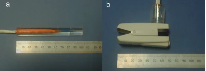
A new reflectance fiberoptic splanchnic pulse oximetry sensor was developed that comprises 2 emitters (660 and 850 nm, FFT 2000 BHR and H22E4020IR; the Optoelectronic Manufac-turing Corporation, Cornwall, UK), a 1 mm2
photodiode, a custom-made Y piece (Ocean Optics, Dunedin, FL), and 600-m optical fiber cables (Fig. 1a).18
The sensor was de-signed in a handheld configuration (pencil probe) to enable the ease of application of the sensor on the investigated organs by the clinical expert during the pilot clinical trials. This was achieved by inserting the transmitting and light-receiving fibers into a precision-drilled Perspex rod (diameter, 13 mm; height, 100 mm). To facilitate comparisons of the acquired Spo2values from splanchnic organs with those from
the finger, an optically identical fiberoptic finger pulse oxim-etry sensor was also developed. This sensor was inserted into a modified pulse oximetry finger clip (Fig. 1b).
Instrumentation and Software
An isolated 3-channel battery-powered instrumentation sys-tem was designed to drive the optical components of both the splanchnic and finger sensors and to preprocess all AC and DC PPG signals at both wavelengths.18
All PPG signals were digitized by a 12-bit data acquisition card (DAQCard-6024E; National Instruments, Austin, TX) in the laptop computer and
displayed and analyzed by a Virtual Instrument running in LabVIEW (National Instruments). All signals were electroni-cally archived for further offline analysis.
Patients and Measurements
After obtaining local research ethics committee approval, informed written consent was obtained from ASA physical status I or II, male or female (nonpregnant) adult patients (aged between 18 and 65 years) who were due for elective surgery involving laparotomy. Patients who were under-going acute emergency operations or had abnormal labo-ratory results that were considered clinically significant to this study, or patients who were unwilling or unable to conform to the protocol, were excluded from the study.
Seventeen patients (3 men and 14 women; mean age⫾ SD: 54⫾ 9.7 years), undergoing general anesthesia, were studied. None of the patients had a history of peripheral vascular disease or diabetes. One of the patients had a medical history of hypertension, and another had previ-ously undergone heart failure surgery. None of the patients was receiving medication that would affect the objectives of this preliminary investigation. The study was observational and patients’ surgical, anesthetic, and monitoring manage-ment were as per routine. All patients were induced with propofol 2 mg/kg IV, and after placement of an endotracheal tube, anesthesia was maintained with either 1% to 3% isoflu-rane or sevofluisoflu-rane in a 50% to 70% air/oxygen or N2O/oxygen mixture. The inspired concentration of the
vola-tile anesthetic was varied to maintain hemodynamic stability. All patients were tracheally intubated and their lungs venti-lated using volume-controlled intermittent positive pressure ventilation set at 12 breaths per minute (Datex-Ohmeda Aestiva/5; GE Healthcare, Hertfordshire, UK). Patient tem-perature was measured using temtem-perature probes (Tempre-cise; Arizant, Wakefield, UK) sited in the esophagus. Arterial blood pressure was measured noninvasively from the arm every 5 minutes. The blood pressure cuff was placed on the opposite arm to the finger pulse oximetry sensor.
[image:3.594.207.539.56.171.2]After the attachment of all operating room monitoring devices (electrocardiographic leads, commercial pulse oxime-ter, etc.), the custom-made fiberoptic finger pulse oximetry sensor was placed on the index finger of the patient. Before any splanchnic PPG measurements were taken, the fiberoptic splanchnic pulse oximetry sensor was inserted into a sterile medical ultrasound probe cover (Cory Medical, Taunton, UK) and was secured in place using 2 sterile fixation bands. At an appropriate time during the surgery (decided by the surgeon), the sensor was passed to the surgeon and it was applied gently to the surface of accessible bowel (small or large) so
that the emitted light was reflected from its surface. Simulta-neous AC and DC splanchnic PPG signals, and AC and DC peripheral PPG signals were recorded for approxi-mately 2 minutes for each site under investigation. During the splanchnic PPG measurements, values of Spo2 from a commercial finger transmission pulse
oxi-meter (OxyTip⫹; GE Healthcare, UK) were also manu-ally recorded in a notebook. The researcher recording all PPG signals and Spo2 values was not blinded to the
results from different measurement sites (splanchnic or peripheral) or different measurement devices (fiberoptic sensors or commercial finger pulse oximeter).
Data Analysis and Statistics
Although this is an uncalibrated system, preliminary Spo2 values were estimated for each splanchnic site
under investigation to provide an indication of the system’s ability to estimate Spo2. The Spo2 values were
estimated by using a typical linear equation used in pulse oximetry18
:
Spo2ⴝ110ⴚ25ⴛR
R is known as the ratio of ratios and is given by:
Rⴝ rac兾rdc irac兾irdc
where racand iracare the amplitudes of the AC red and
infrared signals, respectively, and rdc and irdc are the
amplitudes of the DC red and infrared signals, respectively. Mean splanchnic and finger Spo2values were estimated
by averaging the Spo2values over the 2-minute monitoring
period. The values recorded from the commercial device during the same monitoring period were also averaged to obtain a mean commercial Spo2value.
Pairedttests were performed using SigmaStat software (Systat Software, Inc., San Jose, CA) on all Spo2datasets to
determine whether there was a statistically significant difference between Spo2estimation from the finger and the
bowel (small and large). The limits of agreement between
the finger fiberoptic Spo2 values and those from the
com-mercial pulse oximeter were calculated using the between-method differences analysis outlined by Bland and Altman.20
The Bland and Altman method was also used to determine the limits of agreement between the finger and splanchnic fiberoptic Spo2datasets.
RESULTS
Good quality, easily recognizable PPG signals with large amplitudes were recorded in all attempts from the small bowel (n ⫽ 17) and large bowel (n ⫽ 14) (lower 95% confidence limit for the proportion was 0.64). In 3 cases, the large bowel was not accessible by the surgeon and as a result only PPG signals from the small bowel were ob-tained. During the clinical measurements, the heart rate, core temperature, mean arterial blood pressure, end-tidal CO2, and arterial blood pressure were recorded from all
patients. The mean (range) of these variables were as follows: heart rate 75.94 bpm (60 –104 bpm), temperature 36.38°C (35.9°C–36.9°C), mean arterial blood pressure 78.24 mm Hg (55–98 mm Hg), end-tidal CO24.46 kPa (3.9–5 kPa),
and arterial blood pressure 108/64 mm Hg (90/50–144/85 mm Hg).
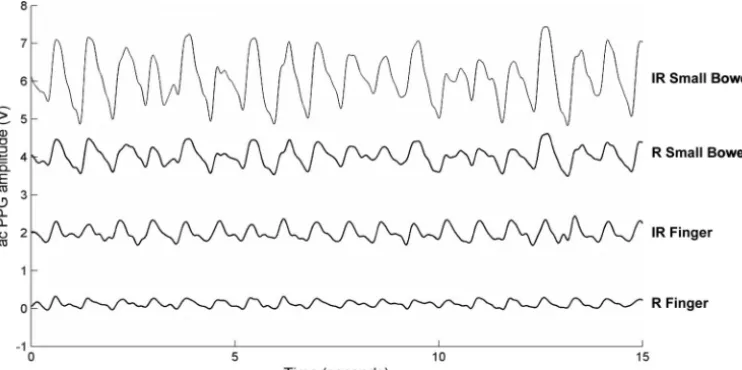
Figure 2 depicts typical AC PPG traces from the small bowel and the corresponding peripheral signals. The recorded PPG signals from the bowel displayed larger amplitudes than the corresponding peripheral signals obtained from the iden-tical fiberoptic sensor. It is believed that this is attributable to the underlying difference in tissue and vasculature between the peripheral and splanchnic sites. The low-frequency artifact present on the PPG traces from the small bowel (Fig. 2) was caused by the mechanical ventilator and also, perhaps, move-ment of the handheld sensor.
The mean of the mean Spo2 value estimated from the
[image:4.594.110.481.56.241.2]large bowel (97.41%⫾3.14% and 97.14%⫾3.10%, respec-tively) were almost identical and are also in good agree-ment with the Spo2values from the finger (both fiberoptic
and commercial sensors).
Paired t tests (P ⬍ 0.05) were performed on the Spo2
data from the small bowel, large bowel, and periphery (Table 1). The mean absolute pairwise differences were
⬍0.4%, and no significant difference was found between different sites (allP⬎0.454).
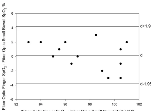
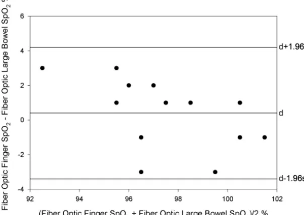
The Bland and Altman method was used to compare Spo2
values obtained from the small bowel and large bowel with their corresponding Spo2 values from the finger using the
fiberoptic sensor. Figures 3 and 4 show the plots of the difference between finger and splanchnic Spo2values against
the mean for the small bowel and large bowel data, respec-tively. Because no obvious relation was observed between the difference and the mean in both cases, the limits of agreement were calculated as described above. The limits of agreement were found to be⫺3.8% and 4.2% for the small bowel data,
and⫺3.4% and 4.3% for the large bowel. The 95% prediction intervals for the small and large bowel data were calculated to be⫺4.2% to 4.6% and⫺3.8% to 4.7%, respectively.
DISCUSSION
A new handheld fiberoptic pulse oximeter system recorded good quality PPG signals and provided Spo2 estimates
with⫾4% accuracy from the small bowel (n⫽17) or large bowel (n ⫽ 14) in all of the 17 patients studied. The fiberoptic pulse oximetry system in its current handheld design could be a useful tool for surgeons, enabling the assessment of some surgical outcomes early, such as the quality of surgical anastomosis.
However, the main limitation of the study was that it did not allow for the assessment of the device on patients with compromised bowel perfusion. As a result, all Spo2
[image:5.594.57.537.80.246.2]values were within the normal range. The data range of the results presented may be too narrow for Bland and Altman analysis to be helpful in assessing interchangeability of
Figure 3.Difference against mean for arterial blood pressure saturation (SpO2) data obtained from the
[image:5.594.237.538.272.493.2]small bowel using the splanchnic pulse oximetry sensor and the corresponding finger values ob-tained using the fiberoptic pulse oximetry sensor. d⫽mean; s⫽standard deviation.
Table 1. Results of PairedtTests on the Arterial Blood Pressure Saturation (SpO2) Data for the Small Bowel, Large Bowel, and Finger
Treatment name Mean (%)
Standard deviation (%)
Standard error of the mean (%)
Small bowel SpO2(n⫽17) 97.472 3.231 0.784
Large bowel SpO2(n⫽14) 97.14 3.091 0.826
Difference ⫺0.19 1.861 0.497
95% confidence interval for difference of means:⫺1.264% to 0.884% No significant difference,P⫽0.708
Small bowel SpO2(n⫽17) 97.472 3.231 0.784
Finger SpO2(n⫽17) 97.681 2.355 0.571
Difference ⫺0.21 2.15 0.521
95% confidence interval for difference of means:⫺1.315% to 0.896% No significant difference,P⫽0.693
Large bowel SpO2(n⫽14) 97.14 3.091 0.826
Finger SpO2(n⫽14) 97.573 2.168 0.579
Difference ⫺0.433 2.1 0.561
methods. Therefore, the next step in evaluating the system as a potential method for detecting splanchnic ischemia would be to conduct more rigorous clinical investigations on a group of patients in which splanchnic perfusion is compromised. Also, it would be desirable to develop an experimental model (perhaps an animal study) to induce abnormally low splanch-nic (bowel) Spo2values.
To provide a better understanding of the perfusion of the bowel, future work should include the comparison of the splanchnic Spo2 results against other indicators of
microvasculature perfusion (Doppler techniques). This may provide valuable information regarding the sensitivity of the fiberoptic sensor to changes in perfusion states.
In its current state, the developed technology is only capable of assessing PPG signals from the bowel during open laparotomy. Further probe miniaturization may en-able its use in a postoperative environment. Further work needs to be performed to validate this hypothesis.
CONCLUSION
Good quality PPG signals have been obtained from the small and large bowel using a new splanchnic fiberoptic sensor. Preliminary clinical results are positive and suggest that such a sensor may prove a useful tool for the intraop-erative assessment of splanchnic perfusion. Future work will assess the performance of the sensor in cases of poor bowel perfusion.
REFERENCES
1. Rittoo D, Gosling P, Bonnici C, Burnley S, Millns P, Simms MH, Smith SRG, Vohra RK. Splanchnic oxygenation in patients undergoing abdominal aortic aneurysm repair and volume expansion with eloHAES. Cardiovasc Surg 2002;10:128 –33
2. Koch T, Geiger S, Ragaller MJ. Monitoring of organ dysfunc-tion in sepsis/systemic inflammatory response syndrome: novel strategies. J Am Soc Nephrol 2001;12:S53–9
3. Richard C. Tissue hypoxia: how to detect, how to correct, how to prevent? Intensive Care Med 1996;22:1250 –7
4. Bulkley GB, Zuidema GD, Hamilton SR, O’Mara CS, Klacsmann PG, Horn SD. Intraoperative determination of small intestinal viability following ischemic injury: a prospec-tive, controlled trial of two adjuvant methods (doppler and fluorescein) compared with standard clinical judgment. Ann Surg 1981;193:628 –37
5. Kram HB, Shoemaker WC. Method for intraoperative assess-ment of organ perfusion and viability using a miniature oxygen sensor. Am J Surg 1984;148:404 –7
6. Hatherill M, Tibby SM, Evans R, Murdoch IA. Gastric tonom-etry in septic shock. Arch Dis Child 1988;78:155– 8
7. Corbett EJ, Barry BN, Pollard SG, Lodge JP, Bellamy MC. Laser doppler flowmetry is useful in the clinical management of small bowel transplantation. The Liver Transplant Group. Gut 2000;47:580 –3
8. Tremper KK, Barker SJ. Pulse oximetry. Anesthesiology 1989;70:98 –108
9. Mendelson Y. Pulse oximetry: theory and applications for noninvasive monitoring. Clin Chem 1992;38:1601–7
10. Jubran A. Pulse oximetry. Intensive Care Med 2004;30:2017–20 11. Ouriel K, Fiore WM, Geary JE. Detection of occult colonic ischemia during aortic procedures: use of an intraoperative photoplethysmographic technique. J Vasc Surg 1988;7:5–9 12. Pearce WH, Jones DN, Warren GH, Bartle EJ, Whitehill TA,
Rutherford RB. The use of infrared photoplethysmography in identifying early intestinal ischemia. Arch Surg 1987;122:308 –10
13. DeNobile J, Guzzetta P, Patterson K. Pulse oximetry as a means of assessing bowel viability. J Surg Res 1990;48:21–3
14. MacDonald PH, Dinda PK, Beck IT, Mercer CD. The use of oximetry in determining intestinal blood flow. Surg Gynecol Obstet 1993;176:451– 8
[image:6.594.47.349.62.276.2]15. Ferrara JJ, Dyess DL, Lasecki M, Kinsey S, Donnell C, Jurkov-ich GJ. Surface oximetry: a new method to evaluate intestinal perfusion. Am Surg 1988;54:10 – 4
Figure 4.Difference against mean for arterial blood pressure saturation (SpO2) data obtained from the
16. Dyess DL, Bruner BW, Donnel CA, Ferrara JJ, Powell RW. Intraoperative evaluation of intestinal ischemia: a comparison of methods. South Med J 1991;84:966 –9
17. Hei ERL, Shun A. Intra-operative pulse oximetry can help determine intestinal viability. Pediatr Surg Int 2001;17:2–3 18. Hickey M, Kyriacou PA. Development of a new splanchnic
perfusion sensor. Conf Proc IEEE Eng Med Biol Soc 2007;2007:2952–5
19. Crerar-Gilbert AJ, Kyriacou PA, Jones DP, Langford RM. Assessment of photoplethysmographic signals for the determi-nation of splanchnic oxygen saturation in humans. Anaesthe-sia 2002;57:442–5