An electrooptical muscle contraction sensor
Alessio Chianura and Mario E Giardini
Medical and Biological Engineering and Computing
48 (2010) 731-734
This is an author-generated version
The final publication is available at www.springerlink.com
An electrooptical muscle contraction sensor
Alessio Chianura* and Mario E. Giardini#
*Università degli Studi di Pavia – Dipartimento di Elettronica – Pavia – Italy #Istituto Nazionale per la Fisica della Materia – INFM – Genova – Italy
#Corresponding author:
current affiliation and present address:
Mario E. Giardini University of St Andrews
SUPA - School of Physics and Astronomy J.F. Allen Physics Research Laboratories North Haugh
St Andrews KY16 9SS United Kingdom
E-mail: [email protected] Tel. +44 1334 46.3439
Fax +44 1334 46.3104
The total number of words of the manuscript, including entire text from title page to figure legends: 1522 The number of words of the abstract: 88
ABSTRACT
An electrooptical sensor for the detection of muscle contraction is described. Infrared light is injected into the muscle, the backscattering is observed, and the contraction is detected by measuring the change, that occurs during muscle contraction, between the light scattered in the direction parallel and perpendicular to the muscle cells. With respect to electromyography and to optical absorption-based sensors, our device has the advantage of lower invasiveness, of lower sensitivity to electromagnetic noise and to movement artifacts, and of being able to distinguish between isometric and isotonic contractions.
KEYWORDS
1. Introduction
In the design of an active prosthetic device, the detection of a signal suitable as a trigger or as a proportional controller for the prosthesis actuation is among the key issues. In current clinical
applications, surface electromyography (EMG), that detects the electric signals that underlie muscle contraction, is in widespread use1,2,3. The patient is trained to contract appropriate muscles when he
needs to actuate a specific movement in the prosthesis.
However EMG, based on the detection of low voltage signals at high impedance through
electrodes applied on the surface of the skin, it is highly sensitive to electromagnetic interference4.
Moreover, in order to reduce the impedance and to minimize movement artifacts, electrodes are put in
direct contact with the tissue, often with a significant pressure, and the consequent tissue reaction and eventual reduction in tissue blood flow may cause malfunction and/or tissue damage5,6.
For these reasons, in order to detect muscle contraction, optical techniques are particularly
appealing, as they are intrinsically free from electromagnetic noise and, in principle, are not invasive on the tissues. As the near-infrared optical absorption of muscle is dominated by blood7 and as, during
contraction, the muscle undergoes blood depletion, it has been demonstrated that the contraction can be detected as a decrease in the optical absorption of the muscle8,9. Though this technique can indeed
be used to generate a signal for prosthesis actuation, the sensitivity is low and, so far, viable sensor configurations have shown to be prone to artifacts due to patient movement10.
2. Materials and methods
Our solution for the optical detection of muscle contraction is based on the observation that
muscle cells are shaped as elongated fibers, aligned along the main muscle axis. In muscle tissue, we can therefore expect light to be scattered anisotropically. Namely, if we inject light into the muscle using a suitable point light source applied to the overlaying skin, the backscattered light, collected
through the skin a few centimeters away from the source, depends on the collection point position with respect to the muscle fiber direction11. In particular, the light scattered in a direction perpendicular to
the muscle fibers differs from the light scattered parallel to the fibers. As the muscle contracts, the fiber aspect ratio changes, and the scattering anisotropy varies accordingly. We have therefore
designed our sensor to detect such variation, responding to contraction with intrinsic rejection to non-anisotropic signals, such as those resulting from patient movement.
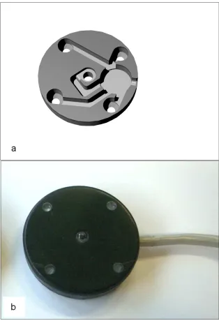
The sensor head is constituted of a round PVC housing, 50 mm in diameter and 8 mm thick, with five 5 mm PMMA windows arranged on the vertices and in the center of an ideal square with 40
Using such configuration, the sensor collects light coming from a depth on the order of 2 cm under the skin surface7.
A 1 m multicore cable connects the photodiodes and the LED to an appropriate photodiode preamplifier and a continuous-wave LED driver.
Inside the head, the cable is separated in its individual cores, that run in channels, held in place by an optically absorbing potting compound. This minimizes the optical crosstalk between each
component. Figure 1 shows the lower part of the housing, with the channels, rendered from its CAD design (a), and a photograph of the assembled sensor head (b).
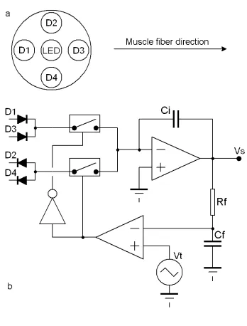
The sensor is placed directly on the skin, over a muscle. The LED emits light through the skin and into the muscle. It is oriented so that two photodiodes (1 and 3) collect the light scattered in the
direction of the muscle fibers, while the other two photodiodes (2 and 4) collect the light scattered
perpendicularly to such direction (photocurrents I// and I⊥ respectively).
I// and I⊥ are time-multiplexed, with opposite signs, on the input node of a current integrator. A
feedback loop controls the duty cycle of the multiplexer balancing the output of the integrator for zero
signal at steady state. Any imbalance between I// and I⊥ faster than the loop time constant (22 s)
appears as a non-zero signal on the integrator output. A simplified preamplifier schematic diagram is
reported in Figure 2b.
3. Results and discussion
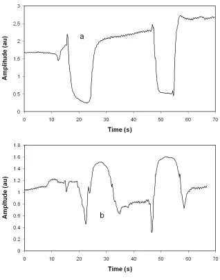
The sensor has been tested on a 28 year old male volunteer, placed over his biceps muscle, held in place by an elastic dark cloth bandage, taking care not to occlude blood circulation. The
volunteer has then performed series of isotonic contractions (i.e., at constant muscle force), lifting a 8 kg dumbbell, and series of isometric contractions (i.e., at constant muscle length) against a fixed
obstacle. Typical signals detected at the integrator output are reported in Figure 3.
Stable and consistent individual contraction signals can be easily distinguished against the steady-state baseline, and the shape of the signals corresponding to isotonic and isometric contractions
is clearly different. In particular, in a first phase of the isotonic contractions, the muscle is rapidly depleted of blood, and then the differential optical scattering contribution due to the shortening of the
muscular fibers prevails. Conversely, in isometric contractions, the muscle fibers are constrained to a quasi-constant length. Therefore, differential scattering contributes to a first phase of the signal only,
and then haematic depletion dominates. As, in our configuration, differential scattering and blood depletion contribute to the signal with opposing signs, the isometric and isotonic signals present
4. Conclusions
For the quality of the signals obtained, the device appears promising as an alternative to surface EMG for monitoring muscle contraction. As described, it enables a clear non-invasive
detection of the contraction signal, with the substantial advantages on electromagnetic noise that optical methods present over low-signal electric detection, and with the additional capability to
distinguish between isometric and isotonic contractions.
References
1 Merletti R, Parker PA (eds) (2004), Electromyography: Physiology, Engineering and Noninvasive
Applications, Wiley-IEEE, USA
2 Reaz MBI, Hussain MS, Mohd-Yasin F (2006) Techniques of EMG signal analysis: detection, processing,
classification and applications. Biol. Proced. Online 8: 11-35
3 Parker P, Englehart K, Hudgins B (2006) Myoelectric signal processing for control of powered limb
prostheses. J Electromyogr. Kinesiol. 16: 541-548
4 Andreasen LNS, Struijk UJJ (2003) Artefact reduction with alternative cuff configurations. IEEE Trans.
Biomed. Eng 50: 1160-1166
5 Lai JCK, Schoen MP, Gracia AP, Naidu DS, Leung SW (2007) Prosthetic devices: challenges and implications
of robotic implants and biological interfaces. Proc. Institut. Mech. Eng. H – J. Eng. Med. 221: 173-183
6 Sood A, Taylor JS, Billock JN (2003) Contact Dermatitis to a limb prosthesis. Am. J. Contact Dermatitis 14:
169-171
7 Tuchin V (2000) Tissue Optics, SPIE Publishing, USA
8 Gelmetti A, Giardini ME, Lago P, Pavesi R, Zambarbieri D, Maestri R, Felicetti G (1997) Preliminary Study of
Muscle Contraction Assessment by NIR Spectroscopy. SPIE Proc. 3199: 61-67
9 Bianchi T, Zambarbieri D, Beltrami G, Verni G (1999) NIRS monitoring of muscle contraction to control a
10 Lopez NM, di Sciascio F, Soria CM, Valentinuzzi ME (2009) Robust EMG sensing system based on data
fusion for myoelectric control of a robotic arm. Biomed Eng Online 8: Art. No. 5
11 Fomenko VN, Shwarts FM, Shwarts MA (1998) Exact description of the photon migration in anisotropically
Figure legends
FIG. 1. CAD rendering of the sensor head housing (a) and photograph of the assembled
sensor head (b)
FIG. 2. Simplified schematic diagram of the sensor amplifier. (a): photodiodes D1 and
D3 collect the light scattered parallel to the muscle fibers, D2 and D4 the light
perpendicular to the fibers. (b): the photocurrents are integrated on Ci to yield the output
signal Vs. The background, filtered with time constant RfCf is compared with a triangular
signal Vt to balance the photocurrents.