Autographa californica Nucleopolyhedrovirus AC141 (Exon0), a
Potential E3 Ubiquitin Ligase, Interacts with Viral Ubiquitin
and AC66 To Facilitate Nucleocapsid Egress
Siddhartha Biswas,aLeslie G. Willis,bMinggang Fang,bYingchao Nie,bDavid A. Theilmanna,b
aPlant Science, Faculty of Land Food Systems, University of British Columbia, Vancouver, BC, Canada
bSummerland Research and Development Center, Agriculture and Agri-Food Canada, Summerland, BC, Canada
ABSTRACT During the infection cycle of Autographa californica multiple nucleopo-lyhedrovirus (AcMNPV), two forms of virions are produced, budded virus (BV) and occlusion-derived virus (ODV). Nucleocapsids that form BV have to egress from the nucleus, whereas nucleocapsids that form ODV remain inside the nucleus. The mo-lecular mechanism that determines whether nucleocapsids remain inside or egress from the nucleus is unknown. AC141 (a predicted E3 ubiquitin ligase) and viral ubiq-uitin (vUbi) have both been shown to be required for efficient BV production. In this study, it was hypothesized that vUbi interacts with AC141, and in addition, that this
interaction was required for BV production. Deletion of both ac141 and vubi
re-stricted viral infection to a single cell, and BV production was completely eliminated. AC141 was ubiquitinated by either vUbi or cellular Ubi, and this interaction was re-quired for optimal BV production. Nucleocapsids in BV, but not ODV, were shown to be specifically ubiquitinated by vUbi, including a 100-kDa protein, as well as high-molecular-weight conjugates. The viral ubiquitinated 100-kDa BV-specific nucleocap-sid protein was identified as AC66, which is known to be required for BV production and was shown by coimmunoprecipitation and mass spectrometry to interact with AC141. Confocal microscopy also showed that AC141, AC66, and vUbi interact at the nuclear periphery. These results suggest that ubiquitination of nucleocapsid proteins by vUbi functions as a signal to determine if a nucleocapsid will egress from the nu-cleus and form BV or remain in the nunu-cleus to form ODV.
IMPORTANCE Baculoviruses produce two types of virions called occlusion-derived virus (ODV) and budded virus (BV). ODVs are required for oral infection, whereas BV enables the systemic spread of virus to all host tissues, which is critical for killing in-sects. One of the important steps for BV production is the export of nucleocapsids out of the nucleus. This study investigated the molecular mechanisms that enable the selection of nucleocapsids for nuclear export instead of being retained within the nucleus, where they would become ODV. Our data show that ubiquitination, a universal cellular process, specifically tags nucleocapsids of BV, but not those found in ODV, using a virus-encoded ubiquitin (vUbi). Therefore, ubiquitination may be the molecular signal that determines if a nucleocapsid is destined to form a BV, thus en-suring lethal infection of the host.
KEYWORDS AcMNPV, AC141, RING motif, viral ubiquitin, cellular ubiquitin, budded virus (BV), occlusion-derived virus (ODV), nucleocapsids, AC66, VP80, ubiquitination
A
utographa californica multiple nucleopolyhedrovirus (AcMNPV) has a largedouble-stranded DNA genome that is packaged into rod-shaped nucleocapsids, which are utilized to form two types of virions, budded virus (BV) and occlusion-derived virus (ODV). During the late stage of infection, new nucleocapsids are synthesized in the
Received6 October 2017Accepted8 November 2017
Accepted manuscript posted online15 November 2017
CitationBiswas S, Willis LG, Fang M, Nie Y, Theilmann DA. 2018. Autographa californica nucleopolyhedrovirus AC141 (Exon0), a potential E3 ubiquitin ligase, interacts with viral ubiquitin and AC66 to facilitate nucleocapsid egress. J Virol 92:e01713-17.https://doi.org/10 .1128/JVI.01713-17.
EditorRozanne M. Sandri-Goldin, University of California, Irvine
© Crown copyright 2018. The government of Australia, Canada, or the UK (“the Crown”) owns the copyright interests of authors who are government employees. TheCrown Copyright is not transferable.
Address correspondence to David A. Theilmann, david.theilmann@agr.gc.ca.
crossm
on November 6, 2019 by guest
http://jvi.asm.org/
nuclear virogenic stroma, followed by transport to the ring zone at the nuclear periphery. Nucleocapsids destined to become BV egress from the nucleus and traverse through the cytoplasm to the plasma membrane, from where they bud to form BV. During the late and very late phases of infection, nucleocapsids that are retained inside the nucleus form ODV. BV obtains its envelope by budding from the plasma membrane, whereas the ODV envelope is derived from the inner nuclear membrane (1). The function of BV is to mediate the systemic spread of viral infection between insect tissues, whereas ODVs are utilized for interhost transmission and are specifically re-quired for initiating the infection of the insect by binding to the midgut epithelial cells. The mechanism by which nucleocapsids are designated to become BV or ODV is unknown, but some form of signaling to enable the differential trafficking is assumed to occur.
A wide variety of cellular processes, including trafficking, are regulated by post-translational modifications of proteins by ubiquitin (Ubi) (2–4). The ubiquitination pathway initiates with a thiol-ester linkage between ubiquitin-activating E1 enzymes and the C-terminal glycine of ubiquitin. The activated ubiquitin is transferred to a ubiquitin-conjugating E2 enzyme. The transfer of ubiquitin from E2 to a substrate is catalyzed by E3 ubiquitin ligases (2, 5, 6). Substrate specificity for ubiquitination is determined by the E3 ubiquitin ligases, which is why mammals have hundreds of E3 proteins but only approximately 35 E2s and two E1s (5, 7, 8). E3 enzymes are classified into three families according to their structures and functions: (i) the homologous to E6AP C-terminus (HECT) domain family, (ii) the really interesting new gene (RING) domain family, and (iii) the RING-between-RING (RBR) domain family (7, 8). Many studies have shown that viruses utilize the host ubiquitination system at different stages of the infection cycle, but they are also known to either encode their own E3 ubiquitin ligases or encode adapter proteins that recruit the cellular E3 ligases (9, 10). AC141 (also known as Exon0) is a nucleocapsid-associated protein expressed at late times postinfection (p.i.) and is specifically required for BV production (11). Deletion of
ac141reduces BV production by more than 99.99%, and nucleocapsids are not able to egress from the nucleus (11, 12). AC141 contains a C-terminal RING domain that is homologous to those found in E3 ubiquitin ligases, and deletion analysis has shown it is required for optimal BV production (13). The predicted RING motif of AC141 is conserved among sequenced genomes of alphabaculoviruses that infect lepidoptera
(11). The consensus sequence of the AC141 RING motif, C3C(Y/F)C4, is different from the
C3HC4motif of most other cellular or AcMNPV RING domain proteins (11). A tyrosine or
phenylalanine replaces histidine, and there is an additional conserved cysteine residue adjacent to the third cysteine. The RING motif binds two zinc atoms in a cross-brace finger configuration and is required for interaction with E2 enzymes (14).
Ubiquitin is a small, 76-amino-acid (aa) protein that is present in cells either in a free form or covalently attached to other proteins. Ubiquitin can be linked to one lysine or to the multiple lysine residues of a substrate, resulting in mono- or multiubiquitination, respectively. Ubiquitin also has intrinsic lysine residues that can be used to form polyubiquitin chains (3, 15). Polyubiquitin chains, linked to the various ubiquitin lysines, are required for different cellular processes. For example, attachment of K48 polyubiq-uitin chains to substrate proteins is a signal for degradation via the 26S proteasome-mediated pathway (3, 15, 16). K63-linked polyubiquitination of proteins is a signal for protein trafficking or for a DNA damage response (15, 16). Baculoviruses are a unique
group of viruses, as they encode their own ubiquitin (vubi). AcMNPV viral ubiquitin
(vUbi) is 77 amino acids long and has 76% amino acid identity with cellular ubiquitin (cUbi) (17). All the cUbi lysine residues are conserved, but vUbi also has an extra lysine
residue at position 54. Analysis ofvubihas shown that it is expressed as a late gene, and
disruption by a frameshift mutation did not have any impact on virus replication; however, BV production was reduced 5- to 10-fold (18, 19). Biochemical analysis has shown that both cUbi and vUbi are attached to the inner side of the BV envelope via
a phospholipid anchor (20). Inin vitroubiquitination assays using mammalian E1, E2,
and E3 enzymes, vUbi was found to be less efficient than cUbi in supporting
on November 6, 2019 by guest
http://jvi.asm.org/
dependent proteolysis. vUbi and cUbi were functionally indistinguishable in the E1 and E2 steps of ubiquitin conjugation. However, the rate of transfer by the mammalian E3 ubiquitin ligase of vUbi to a substrate was significantly lower than for cUbi (21). Thus, the transfer of vUbi to a substrate might require a different E3 ligase or a different mechanism of interaction between E3 ligase and an E2 conjugated with vUbi.
In addition to AC141, the AcMNPV regulatory proteins IE2, PE38, IAP1, and IAP2 all contain classic cellular C3HC4 RING motifs and have been shown, or predicted, to be E3 ligases (22–25). The AC141 RING motif, as indicated above, has conserved differences compared to cellular E3 proteins. As both AC141 and vUbi are required for BV production, we hypothesized that AC141 is a potential ubiquitin E3 ligase that specif-ically interacts with vUbi, as opposed to cUbi, and that this activity is required for optimal BV production. The results described in this study show that AC141 and vUbi
interact and the deletion of bothac141andvubieliminates BV production. In addition,
BV nucleocapsids were found to be highly ubiquitinated by vUbi compared to ODV nucleocapsids. We also demonstrate that the nucleocapsid protein AC66 associates with vUbi and AC141 and that this interaction suggests a mechanism by which nucleocapsids are designated for nuclear egress and subsequent BV formation.
RESULTS
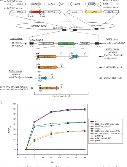
BV production ofac141andvubisingle- and double-gene-knockout viruses.To study the interaction between AC141 and vUbi, a series of single- and
double-gene-knockout (KO) viruses were constructed in which ac141, vubi, or both genes were
deleted, followed by repair with either one or both genes. Theac141andvubirepair
genes included sequences for either C-terminal hemagglutinin (HA) or Myc epitope
tags, respectively. These viruses were named ac141KO, vubiKO, vubiKO-Myc-vubi,
ac141⫹vubi2⫻KO, 2⫻KO-HA-ac141⫹Myc-vubi, 2⫻KO-HA-ac141, and 2⫻KO-Myc-vubi
(Fig. 1A and Table 1). To determine and compare the impacts of deleting either one or
both of theac141andvubigenes, time course experiments were conducted to analyze
BV production. Cells were transfected with purified bacmids from the above-mentioned
viruses. In addition, a control transfection of agp64 deletion virus that is unable to
produce BV was also performed (26–28). Theac141KO repair virus,ac141KO-HA-ac141,
was not used in this experiment, as it has been previously analyzed and has been
shown to be equivalent to the wild type (WT) (11, 13, 29). BV production of 2⫻
KO-HA-ac141⫹Myc-vubiandvubiKO-Myc-vubiwere equivalent to that of the WT at 24, 48, 72,
and 96 h posttransfection (hpt) (Fig. 1B). The vubi knockout viruses, vubiKO and
2⫻KO-HA-ac141, had similar BV production but were equivalent to only 0.26% of WT
levels. Theac141knockout viruses,ac141KO and 2⫻KO-Myc-vubi, had BV production
reduced to only 0.005% of WT levels, which was similar to what has been previously
described (11, 12). The double-knockout virus,ac141⫹vubi2⫻KO, and the control virus,
gp64KO, did not produce any detectable BV. These results confirmed previous analyses
showing that both AC141 and vUbi are required for BV production. In addition, the
cumulative effect of knocking out bothvubiandac141results in the complete
elimi-nation of even the low level of virus observed with the single-gene KO viruses.
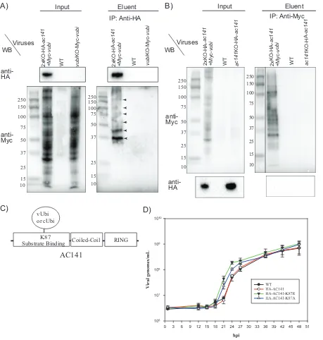
Coimmunoprecipitation of AC141 and vUbi. The elimination of BV production
through simultaneous deletion of bothac141andvubisuggests these two proteins may
interact. To study the possible association between AC141 and vUbi,
coimmunopre-cipitation experiments were performed. Spodoptera frugiperda IPLB-Sf21-AE clonal
isolate 9 (Sf9) cells were infected with 2⫻KO-HA-ac141⫹Myc-vubi expressing
N-terminal HA-tagged AC141 (HA-AC141) and N-terminally Myc-tagged vUbi
(Myc-vUbi). As a control, Sf9 cells were infected with WT or vubiKO-Myc-vUbi, which
ex-presses only Myc-vUbi and untagged AC141. Protein complexes were immunoprecipi-tated with anti-HA antibodies, and eluted material was analyzed by Western blotting. The input lanes showed that there was extensive ubiquitination of proteins with vUbi (Fig. 2A). HA-AC141 specifically coimmunoprecipitated Myc-vUbi-ubiquitinated
pro-teins from 2⫻KO-HA-ac141⫹Myc-vubi virus-infected cells and not from the control
samples (Fig. 2A). The Western blot identified two prominent proteins of approximately
on November 6, 2019 by guest
http://jvi.asm.org/
ac34 CmR
Tn7L Tn7R
GenR egfp
polyhedrin ac142
ac141 5’ end
me53 ZeoR
ac141 3’ end
ac141
HA
vubi
Myc
pA pA
ac141
HA
vubi
Myc
ac141 locus vubi locus polh locus
1xKO virus 2xKO virus
bMON14272
ac141KO or vubiKO
ac141+vubi-2xKO
2xKO-HA-ac141 + Myc-vubi
2xKO-HA-ac141
2xKO-Myc-vubi vubiKO-Myc-vubi
ac142
me53 ac141 ac34 vubi ac36
ac141 K87R/A ac141KO-HA-ac141K87R
ac36
hpt
0 12 24 36 48 60 72 84 96
TC
ID
50
0 1
101
102
103
104
105
106
107
108
W T
2xKO-HA-ac141+Myc-vubi
2xKO-HA-vubiKO (=ac141KO) ac141KO
2xKO-HA-ac141 (=vubiKO) vubiKO
vubiKO-Myc-vubi
ac141+vubi-2xKO gp64KO
A)
B)
pA
pA
pA
ac141 WT locus vubi WT locus
ac141KO-HA-ac141K87A HA
Repair constructs
pFAcT-GFP
2xKO repair viruses
1xKO repair viruses
ac141 KO locus vubi KO locus
FIG 1Construction ofac141andvubisingle-knockout (1⫻KO) and double-knockout (2⫻KO) viruses and time course analysis of BV production. (A) Using the bacmid bMON14272, theac141KO virus was generated by deleting theac141ORF and replacing it with the zeocin resistance (ZeoR) gene. Similarly thevubiKO virus was generated by replacing thevubiORF with the chloramphenicol resistance (CmR) gene. A double-knockout virus was generated by knocking out bothac141andvubiwith zeocin and chloramphenicol resistance genes, respectively. The three KO viruses were repaired using pFAcT-GFP or pFAcT-GFP containing HA-ac141, Myc-vubi, or both genes. pFAcT-GFP contains theegfpmarker gene, as well aspolyhedrin. The resulting viruses wereac141⫹vubi2⫻KO, 2⫻KO-HA-ac141⫹Myc-vubi, 2⫻KO-HA-ac141, 2⫻KO-Myc-vubi,ac141KO,vubiKO, andvubiKO-Myc-vubi. The ac141KO virus was also repaired with twoac141mutants,ac141K87Randac141K87A, generating the virusesac141KO-HA-ac141K87Rand ac141KO-HA-ac141K87A. (B) Analysis of BV production at 12, 24, 48, 72, and 96 hpt. Sf9 cells were transfected with 2g of bacmid DNA of each virus. The media containing the BV were collected at different time points and assayed for BV production by 50% endpoint dilution assay (TCID50). Each data point represents a set of four biological repeats, and the error bars represent standard errors.
on November 6, 2019 by guest
http://jvi.asm.org/
[image:4.585.44.472.77.637.2]37 and 45 kDa, along with other minor higher-molecular-mass proteins. These results showed that HA-AC141 associates either directly or in a complex with viral ubiquiti-nated proteins. Reciprocal coimmunoprecipitation was also done to confirm the asso-ciation of AC141 and Myc-vUbi (Fig. 2B). As shown in Fig. 2B, the input lane showed numerous proteins that were ubiquitinated with Myc-vUbi. As expected, the majority of the viral ubiquitinated proteins were specifically pulled down with anti-Myc. The 26-kDa HA-AC141 was not detectable in the coimmunoprecipitations with Myc-vUbi. This suggests that only a small fraction of the extensive number of Myc-tagged viral ubiquitinated proteins interact with HA-AC141.
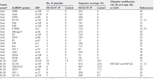
Mass spectrometric analysis of proteins coimmunoprecipitated with HA-AC141 to identify potential substrates and viral ubiquitination sites. Mass spectrometry (MS) analysis of the coimmunoprecipitated material with HA-AC141 was performed to identify the viral proteins that potentially interact with AC141 and are associated with BV development and egress. The MS analysis was applied to total pulldown eluent and material gel purified from the region containing the prominent Myc-Ubi-tagged 37- and 45-kDa proteins (Fig. 2A and Table 2). In addition to AC141, a number of the major BV structural proteins were detected, including VP80, VP39, GP41, P78/83, vUbi, GP64, and AC66. VP39 is the most abundant nucleocapsid protein. VP80 interacts with the F-actin cytoskeleton and is required for the movement of nucleocapsids from the nuclear virogenic stroma to the nuclear periphery regions (30). GP41 is a tegument protein required for nucleocapsid egress from the nucleus and efficient BV production (31, 32). P78/83 is required for actin nucleation during entry and is located at the base of the nucleocapsid (33, 34). GP64 is the major BV envelope protein, and AC66 is a nucleocapsid protein required for
BV production, as deletion ofac66results in nucleocapsids that do not egress from
the nucleus (35, 36). The primary proteins specific to the 35- to 45-kDa region (Table 2) were identified as ChiA, AC141, ODV-EC27, GP37, VLF-1, and AC114, but of these, only AC141 is essential for BV production.
[image:5.585.40.551.83.223.2]MS analysis of AC141-coimmunprecipited proteins was also utilized to identify potential ubiquitination sites on the coimmunoprecipitated proteins. Ubiquitinated peptides increase in mass by the terminal GG from ubiquitin. The C terminus of vUbi, however, is GGY, but it is believed that any C-terminally modified ubiquitin-like molecules are trimmed by isopeptidases prior to any ubiquitin ligase event. It is known that cellular ubiquitins, even with one extra amino acid at the C terminus, are cleaved to make ubiquitin functional (37, 38). In case the vUbi C-terminal tyrosine was not cleaved, peptides were also analyzed for a GGY modification. Ubiquitinated peptides were detected only in the gel-purified 30- to 50-kDa region, and they were found to originate from AC141 at K87 (Fig. 2C and Table 2). Interestingly, two different mass-shifted peptides from the same AC141 sequence were identified, corresponding to a GGY and a GG modification at K87. The K87 ubiquitination site is located in the
TABLE 1Summary of viruses used in this study
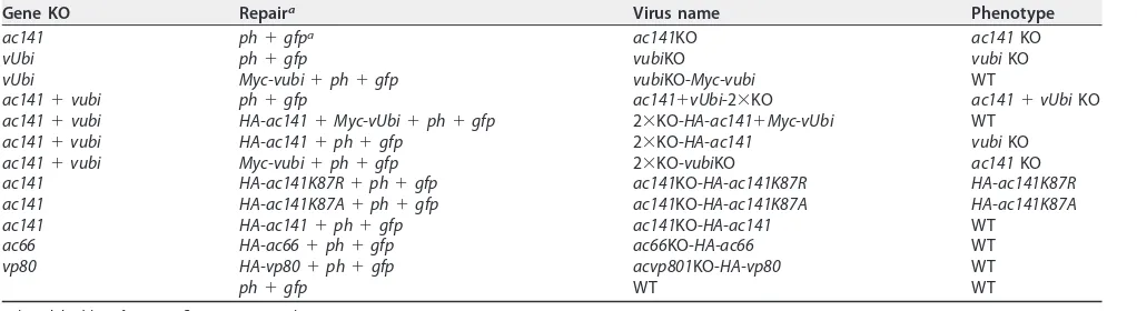
Gene KO Repaira Virus name Phenotype
ac141 ph⫹gfpa ac141KO ac141KO
vUbi ph⫹gfp vubiKO vubiKO
vUbi Myc-vubi⫹ph⫹gfp vubiKO-Myc-vubi WT
ac141⫹vubi ph⫹gfp ac141⫹vUbi-2⫻KO ac141⫹vUbiKO ac141⫹vubi HA-ac141⫹Myc-vUbi⫹ph⫹gfp 2⫻KO-HA-ac141⫹Myc-vUbi WT
ac141⫹vubi HA-ac141⫹ph⫹gfp 2⫻KO-HA-ac141 vubiKO ac141⫹vubi Myc-vubi⫹ph⫹gfp 2⫻KO-vubiKO ac141KO ac141 HA-ac141K87R⫹ph⫹gfp ac141KO-HA-ac141K87R HA-ac141K87R ac141 HA-ac141K87A⫹ph⫹gfp ac141KO-HA-ac141K87A HA-ac141K87A ac141 HA-ac141⫹ph⫹gfp ac141KO-HA-ac141 WT
ac66 HA-ac66⫹ph⫹gfp ac66KO-HA-ac66 WT vp80 HA-vp80⫹ph⫹gfp acvp801KO-HA-vp80 WT
ph⫹gfp WT WT
aph, polyhedrin;gfp, green fluorescent protein.
on November 6, 2019 by guest
http://jvi.asm.org/
potential substrate binding region of AC141 (Fig. 2C). This result, therefore, suggests that vUbi might become attached to AC141 with a novel isopeptide linkage of tyrosine to lysine, as well as the normal glycine-lysine linkage. Overall, the MS results show that AC141 and vUbi interact during viral infection.
250
75 100 150 250
25
10 15 37 75
50 100 150
15 37
10 50
25
WT WT
2xKO-HA
-ac141
+Myc
-vubi
2xKO-HA-ac141
+Myc-vubi
ac141
KO-HA
-ac141
vubi
KO-Myc-vubi
Input Eluent
anti-Myc anti-HA
Viruses
A)
B)
RING Coiled-Coil
Substrate Binding vUbi or cUbi
K87
WB
WT 2xKO-HA- WT
ac141
+Myc-vubi
Input Eluent
Viruses WB
75 100 150 250
25
15 37
10 50
75 100 150 250
25
15 37
10 50
vubi
KO-Myc-vubi
2xKO-HA-ac141
+Myc-vubi
ac141
KO-HA
-ac141
anti-HA anti-Myc
C)
D)
IP: Anti-HA IP: Anti-Myc
AC141
FIG 2Coimmunoprecipitation of HA-AC141 and Myc-vUbi and impact of AC141 K87 mutations on BV production. (A) Sf9 cells were infected with 2⫻KO-HA-ac141⫹Myc-vubi, the WT, orvubiKO-Myc-vubi. Infected cells were harvested at 24 hpi, and total cell lysates were pulled down with HA beads and subjected to Western blot (WB) analysis. The input lanes were loaded with 0.25% of the total input, and the eluent lanes were loaded with 20% of the total eluent. The blots were probed with the antibodies indicated on the left. IP, immunoprecipitation. (B) Sf9 cells were infected with 2⫻KO-HA-ac141⫹Myc-vubi, the WT, andac141KO-HA-ac141. The virusac141KO-HA-ac141has been previously described (29). Infected cells were harvested, and total cell lysates were pulled down with anti-Myc antibody bound to protein G beads. The input lanes were loaded with 0.25% of the total input, and eluent lanes were loaded with 20% of the total eluent. The blots were probed with the antibodies indicated on the left. The marker lane was from the same gel and was moved to be adjacent to the sample lanes. The numbers on the left of the gels are kilodaltons. (C) Schematic diagram showing the RING, coiled-coil, and putative substrate binding domains of AC141. Also indicated is the location of the viral ubiquitination site of the substrate binding site of AC141 at lysine residue 87 (K87). (D) Sf9 cells were infected with the WT, HA-AC141, and mutant virusesac141KO-HA-ac141K87R andac141KO-HA-ac141K87Aat an MOI of 5, and BV production was determined at 1, 12, 15, 18, 21, 24, 27, 36, 42, and 48 hpi. The media containing the extracellular virus or BV were collected at each time point, and the titer was determined by droplet digital PCR. Each data point shown represents a set of two biological repeats and two technical repeats. The error bars represent standard errors.
on November 6, 2019 by guest
http://jvi.asm.org/
[image:6.585.41.500.70.552.2]Mutational analysis of the AC141 K87 vUbi ubiquitination site to determine the effect on BV production. AC141 coimmunoprecipitated viral ubiquinated proteins, and in addition, using MS, it was found that AC141 was ubiquitinated at K87. Auto-ubiquitination of E3 ubiquitin ligases can act as a regulatory mechanism and can result in either enhancing substrate ubiquitination or signaling its own degradation (39–43). To investigate the significance of the AC141 ubiquitination, viruses were generated that mutated the K87 of HA-AC141 to arginine (K87R) or alanine (K87A) (Fig. 1A). A time course of virus production was performed and showed that mutation of AC141 K87 to arginine or alanine altered temporal aspects of BV production (Fig. 2D). In WT-infected cells, BV production initiates between 15 and 18 h postinfection (hpi) with an expo-nential increase between 21 and 27 hpi. In comparison, the K87R and K87A viruses both initiated BV production at the same time as the WT, with an exponential increase between 18 and 24 hpi, 3 h earlier than the WT. Compared to the WT, both mutant viruses produced nearly a log-unit-higher level of BV by 21 hpi, but maximum BV levels were similar at later times postinfection. These results indicated that the mutation of K87, to prevent AC141 ubiquitination at the site, results in more rapid production of BV.
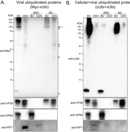
Western blot analysis of purified BV and ODV for viral and cellular ubiquiti-nated proteins.If, as was hypothesized, vUbi is required to differentiate nucleocapsids destined for BV or ODV, possibly catalyzed by AC141, then the two forms of virions could potentially be differentially ubiquitinated. To address this, BV and ODV were
purified from vubiKO-Myc-vUbi-infected Sf9 cells by sucrose density gradient and
[image:7.585.43.547.93.347.2]separated into envelope and nucleocapsid fractions. Total and fractionated protein samples were analyzed by Western blotting. The results showed that BVs contained approximately 4-fold-higher levels of vUbi than ODV (Fig. 3A). Fractionated samples showed that the vast majority of viral ubiquitinated proteins were in the nucleocapsid fraction. In addition, there was a specific viral ubiquitinated major band of approxi-mately 100 kDa in the nucleocapsid fraction of BV, but not in the ODV (Fig. 3A, arrow). There were also other minor bands (indicated by asterisks), which were also present in ODV nucleocapsids, of approximately 86 kDa, 45 kDa, 43 kDa, 30 kDa, and 8 kDa
TABLE 2Most prominent AcMNPV proteins known to be associated with BV or BV synthesis and transport, coimmunoprecipitated with HA-AC141 and identified by mass spectrometry
Protein
sourcea AcMNPV protein ORF
No. of peptides Sequence coverage (%) Ubiquitin modification site (K) and type (GG
or GGY) Reference(s)
HA-AC141 IP Control HA-AC141 IP Control
Total VP80 ac104 11 0 20.3 0 30
Total ChiA Ac126 9 0 19.2 0 72
Total VP39 ac89 8 0 28.8 0 73
Total AC141 ac141 8 0 29.1 0 11, 12
Total P40 ac103 6 0 14.7 0 72
Total P49 ac142 6 0 14.9 0 74
Total P78/83 ac9 5 0 11.8 0 33, 75
Total 39k/pp31 ac36 5 0 27.3 0 35
Total IE1 ac147 5 0 13.9 0 76
Total GP41 ac80 5 0 19.3 0 32
Total FP ac61 3 0 15 0 35
Total PTP ac1 3 0 19.6 0 35
Total bro ac2 3 0 11.2 0 77
Total VLF-1 ac77 3 0 8.7 0 35
Total vUbi ac35 2 0 23.4 0 18
Total GP64 ac128 2 0 5.1 0 28
Total AC66 ac66 2 0 2.8 0 36
30–50 ChiA Ac126 22 14 47.7 24.7 72
30–50 AC141 ac141 19 7 61.3 22.6 K87-GGY and K87-GG 11, 12
30–50 ODV-EC27 ac144 6 3 49.7 27.6 35
30–50 GP37 ac64 8 3 45 17.2 78
30–50 VLF-1 ac77 8 5 20.8 9.7 35
30–50 AC114 ac114 7 3 20.8 9.7 35
aTotal, proteins identified from the total eluent of the AC141 immunoprecipitation (IP); 30 –50, proteins isolated from the 30- to 50-kDa gel fragment of the AC141
pulldown eluent.
on November 6, 2019 by guest
http://jvi.asm.org/
(Fig. 3A). A very long exposure revealed that trace amounts of vUbi could also be detected in the envelope of BV, but not ODV. To examine total ubiquitination levels of both vUbi and cUbi in purified BV and ODV, a duplicate Western blot was performed and probed with anti-cUbi, which detects both cellular and viral ubiquitin (Fig. 3B). The results showed that BV was 80-fold more ubiquitinated than ODV. Most of the cellular and viral ubiquitin detected with anti-cUbi antibody is in the high-molecular-weight proteins of the nucleocapsid fractions of both BV and ODV. A low level of ubiquitin was also detected in the envelope fraction of BV, but not ODV. The higher levels of ubiquitin detected in the BV envelope with anti-cUbi antibody suggest it must be predominantly cellular in origin (Fig. 3A). To ensure correct fractionation of BV and ODV, blots were probed with antibodies to the nucleocapsid protein VP39, the BV envelope protein GP64, and the ODV envelope protein PIF-1 (Fig. 3A and B).
FIG 3Western blot analyses of the isolated BV and ODV for viral ubiquitinated proteins. BV and ODV were purified from Sf9 cells that were infected withvubiKO-Myc-vbi. Purified BV and ODV were fractionated into envelope (ENV) and nucleocapsid (NC) fractions, separated by SDS-PAGE, and analyzed by Western blotting. The blots were probed with anti-Myc antibody to identify proteins ubiquitinated by vUbi (A) or with anti-cellular ubiquitin antibody to identify proteins ubiquitinated by cUbi and vUbi (B). Control blots using antibodies against the nucleocapsid protein VP39, the BV envelope protein GP64, and the ODV envelope protein PIF-1 to confirm the efficiency of fractionation are shown below. The arrow in panel A indicates a specific viral ubiquitinated major band of approximately 100 kDa in the nucleocapsid fraction of BV, but not in the ODV, while the asterisks mark other minor bands of approximately 86 kDa, 45 kDa, 43 kDa, 30 kDa, and 8 kDa that were also present in BV or ODV nucleocapsids upon long exposures.
on November 6, 2019 by guest
http://jvi.asm.org/
[image:8.585.42.459.72.496.2]MS analysis of purified BV and ODV for potential viral ubiquitinated peptides.
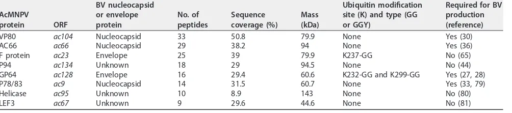
Western blot analyses showed that an approximately 100-kDa protein was specifically ubiquitinated in BV, but not in ODV. To identify the proteins in the 100-kDa band, the region from 85 to 110 kDa of an SDS-PAGE gel was isolated and analyzed by MS (Table 3). Similar to previously reported BV MS results (35), three known BV structural or associated proteins in the 100-kDa size range were identified: VP80 (80 kDa), AC66 (94 kDa), and F protein (80 kDa). F protein is an envelope protein, whereas AC66 and VP80 are both nucleocapsid proteins. It is therefore possible that either AC66 or VP80 is the viral ubiquitinated 100-kDa protein detected in BV (Fig. 3A). AC66 is required for nucleocapsid egress from the nucleus, and VP80 is required for movement of nucleo-capsids from the virogenic stroma to nuclear periphery regions. These results sug-gested that AC66 or VP80 could be the potential substrate for viral ubiquitination in BV nucleocapsids. Both AC66 and VP80 were also identified as proteins that interact with AC141 (Table 2). P94 was also isolated from the 100-kDa region, but it is known not to impact BV development (44).
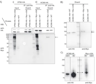
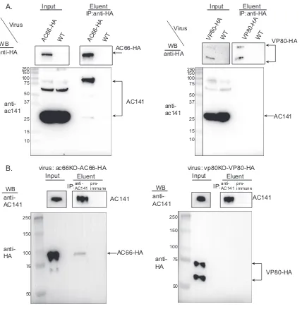
Coimmunoprecipitation analysis of Myc-vUbi with AC66 or VP80. If AC66 or VP80 is ubiquitinated by Myc-vUbi, then it should be possible to immunoprecipitate the 100-kDa viral ubiquitinated nucleocapsid protein with either VP80 or AC66. To enable
this analysis, two viruses were constructed that expressed HA-tagged AC66 (ac66
KO-ac66-HA) or VP80 (vp80KO-vp80-HA) (Fig. 4 and Table 1). Each virus (ac66KO-ac66-HA
andvp80KO-vp80-HA) was coinfected withvubiKO-Myc-vubior the WT (control virus
with no epitope tag). At 24 hpi, cells were harvested, the supernatants were immuno-precipitated, and the eluents were analyzed by Western blotting. The Western blots showed the expression of VP80-HA-, AC66-HA-, and Myc-vUbi-tagged proteins in the input lanes (Fig. 5A). Coimmunoprecipitation of proteins from VP80-HA- and Myc-vUbi-expressing cells did not detect any vUbi-conjugated proteins in the eluent. In contrast,
analysis ofac66KO-ac66-HA-infected cells showed that AC66-HA specifically
coimmu-noprecipitated a Myc-vUbi-conjugated protein of approximately 100 kDa (Fig. 5A). The coimmunoprecipitated band was of approximately the same size as the major vUbi-conjugated band from BV nucleocapsids (Fig. 3A). This result indicates that either AC66 is ubiquitinated by vUbi or it interacts with a 100-kDa vUbi-conjugated protein.
If AC66 is the target of vUbi ubiquitination, then HA-AC66-immunoreactive proteins should comigrate with the coimmunoprecipitated 100-kDa Myc-vUbi-conjugated
pro-tein. Therefore, a coinfection ofac66KO-ac66-HA and eithervubiKO-Myc-vubior WT was
[image:9.585.42.544.84.195.2]repeated and the cell lysates immunoprecipitated with anti-HA. Duplicate samples of the eluent material were separated on a 7.5% gel and probed with either anti-HA or anti-Myc to determine if the coimmunoprecipitated cellular AC66-HA and 100-kDa Myc-vUbi-conjugated protein comigrated (Fig. 5B). The Myc-vUbi band was observed to migrate approximately 7 to 10 kDa higher than the primary cellular AC66 band, which suggests that AC66 was monoubiquitinated. Longer exposures were used to determine if there were higher-molecular-mass species of AC66 that comigrated with the Myc-vUbi band; however, the signal from the primary HA-AC66 band occluded all higher-molecular-mass species (data not shown). This result suggested that if AC66 is ubiquitinated by vUbi,
TABLE 3Most prominent AcMNPV proteins in the region of 85 to 110 kDa of purified BV identified by mass spectrometry
AcMNPV
protein ORF
BV nucleocapsid or envelope protein
No. of peptides
Sequence coverage (%)
Mass (kDa)
Ubiquitin modification site (K) and type (GG or GGY)
Required for BV production (reference) VP80 ac104 Nucleocapsid 33 50.8 79.9 None Yes (30)
AC66 ac66 Nucleocapsid 29 38.2 94 None Yes (36)
F protein ac23 Envelope 25 39 79.9 K237-GG No (65)
P94 ac134 Unknown 18 29 94.5 None No (44)
GP64 ac128 Envelope 16 29.4 60.6 K232-GG and K299-GG Yes (27, 28)
P78/83 ac9 Nucleocapsid 14 31.5 60.7 None Yes (33, 79)
Helicase ac95 Unknown 10 8.9 143 None No (80)
LEF3 ac67 Unknown 9 29.6 44.6 None No (81)
on November 6, 2019 by guest
http://jvi.asm.org/
it represents only a small proportion of the total cellular AC66. Therefore, to increase the proportion of any Myc-vUbi-conjugated AC66, we compared the Myc-vUbi and AC66-HA
proteins in purified BV and ODV isolated from cells coinfected withac66KO-ac66-HA and
vubiKO-Myc-vubi(Fig. 5C). The purified BV and ODV samples were run side by side on a
high-resolution SDS-7.5% PAGE gel, and duplicate lanes were probed with either anti-HA or anti-Myc. The Western blot showed that AC66-HA bands of higher molecular mass than its native size were present in the BV samples, but not in the ODV samples (Fig. 5C). The higher-molecular-mass HA-AC66 bands comigrated with the primary Myc-vUbi band that was present in BV but not ODV. This result supports the conclusion that AC66 is the target for vUbi ubiquitination, but only in BV nucleocapsids. MS analyses were performed on AC66-HA-coimmunoprecipitated material to identify potential AC66 ubiquitination sites. Two potential ubiquitination sites were identified on AC66, a high-probability site at K451 and a low-probability site at K91.
Coimmunoprecipitation of AC141 with AC66 or VP80.We further analyzed the interaction between AC66 and AC141 by coimmunoprecipitation and Western blotting.
Sf9 cells were infected withac66KO-ac66-HA or as controls withvp80KO-vp80-HA or the
WT. The cell lysates were immunoprecipitated with anti-HA beads, and the eluents were analyzed by Western blotting and probed with a polyclonal antibody against AC141 or with anti-HA to detect AC66 or VP80. The input lanes showed expression of AC66-HA and VP80-HA, as expected (Fig. 6A). AC141 was expressed primarily as a 30-kDa protein,
DNA-polymerase ac66 lef-3
DNA-polymerase ZeoR lef-3
vp80 he65
p48
he65
p48 ZeoR
ac66 locus vp80 locus
polh locus
KO virus
WT (ac66 locus)
ac66KO
vp80KO WT (vp80 locus)
bMON 14272 ac66 L
egfp
polyhedrin GenR Tn7R
Tn7L
HA ac66
vp80 L
ac66 L
vp80 L
HA
ac66KO or vp80KO
ac66KO-ac66-HA
vp80KO-vp80-HA pA
pA vp80
pFAcT-GFP
Repair viruses
FIG 4Construction of ac66 and vp80 KO and repaired bacmids. The schematic diagram shows how theac66KO and vp80KO viruses were generated. Theac66orvp80ORF was deleted by recombination with the zeocin resistance gene. The ac66KO andvp80KO viruses were repaired using pFAcT-GFP containing eitherac66orvp80, respectively, with a C-terminal HA tag. The viruses were namedac66KO-AC66-HA andvp80KO-VP80-HA.
on November 6, 2019 by guest
http://jvi.asm.org/
[image:10.585.42.421.74.436.2]along with two minor high-molecular-mass species. The eluent results showed that AC66-HA coimmunoprecipitated with the 30-kDa AC141. Surprisingly, however, a protein of approximately 80 to 90 kDa that was immunoreactive to the AC141 poly-clonal antibody was also specifically coimmunoprecipitated. In contrast, no AC141 was coimmunoprecipitated with VP80-HA (Fig. 6A). Reciprocal coimmunoprecipitation was also done to confirm the association between AC66-HA and AC141. Sf9 cells were
infected withac66KO-ac66-HA andvp80KO-vp80-HA, and the cell lysates were pulled
down with either AC141 polyclonal antibody or preimmune serum as the control. The input lanes showed the expression of AC141, AC66-HA, and VP80 in the corresponding lanes (Fig. 6B). AC66-HA, but not VP80-HA, was specifically coimmunoprecipitated by AC141. These results showed that AC66-HA interacts with AC141 during the course of
i b U v-c y M + A H-6 6 C A T W + A H-6 6 C A i b U v-c y M + A H-6 6 C A T W + A H-6 6 C A i b U v-c y M + A H-0 8 P V T W + A H-0 8 P V i b U v-c y M + A H-0 8 P V T W + A H-0 8 P V Input
Eluent Eluent
t u p n I anti-Myc anti-HA Viruses anti-HA anti-Myc T W + A H-6 6 C A T W + A H- 6 6 C A i b U v-c y M + A H-6 6 C A i b U v-c y M + A H-6 6 C A 250 10 15 37 75 50 100 150 25 250 150 100 75 50 A) B) WB
VP80-HA AC66-HA Eluent
IP: anti-HA IP: anti-HA
250 75 100 150 anti-HA anti-Myc AC66-HA BV ODV Myc-vUbi BV ODV C) i) ii)
FIG 5Coimmunoprecipitation analysis of AC66-HA or VP80-HA with Myc-vUbi. (A) Sf9 cells were coinfected with vp80KO-VP80-HA andvubiKO-Myc-vUbi or withvp80KO-VP80-HA and the WT (i) and withac66KO-AC66-HA and vubiKO-Myc-vUbi or withac66KO-AC66-HA and the WT (ii). Infected cells were harvested at 24 hpi, and total cell lysates were pulled down with HA beads. Samples were separated on SDS-12% PAGE gels and analyzed by Western blotting. The input lanes were loaded with 0.25% of the total protein, and the eluent lanes were loaded with 15% of the total eluent. The blots were probed with the corresponding antibodies listed on the left. The arrow on the right indicates the location of the Myc-vUbi immunoprecipitated by AC66. (B) Theac66KO-AC66-HA- andvubiKO-Myc-vUbi-infected cell eluent materials from panel A were separated in duplicate on an SDS-7.5% PAGE gel and blotted, and the two halves of the blot were probed with anti-HA (left) or anti-Myc (right) to determine if the detectable cellular forms of AC66 (94 kDa) comigrated with the 100-kDa viral ubiquitinated bands. The arrows indicate the positions of the dominant anti-HA- and anti-Myc-reactive bands. Each half of the blot represents different exposure times for each antibody, and the images were aligned using Adobe Photoshop. (C) BV and ODV were purified from Sf9 cells infected withac66KO-AC66-HA andvubiKO-Myc-vUbi. Total virion protein was separated in an SDS-7.5% PAGE gel and analyzed by Western blotting. The membrane was cut in half at the ladder. The two halves of the blot were probed with anti-HA (left) or anti-Myc (right) to determine if the higher-molecular-mass forms of BV or ODV AC66 comigrated with the 100-kDa viral ubiquitinated bands. The double arrow shows the comigration of a 100-kDa AC66 band with the 100-kDa vUbi band. Each half of the blot probed with anti-HA and anti-Myc represents different exposure times; the images were aligned using Adobe Photoshop. The ODV and BV lanes of the anti-HA blot are from the same gel with the same exposure times, but intervening lanes were removed. The numbers on the left of the gels are kilodaltons.
on November 6, 2019 by guest
http://jvi.asm.org/
[image:11.585.44.432.71.411.2]infection. The higher-molecular-mass form of AC141 that is specifically coimmunopre-cipitated by AC66-HA could be polyubiquitinated or multiubiquitinated with 6 or 7 ubiquitin molecules. Neither the 30- or 90-kDa form of AC141 was coimmunoprecipi-tated by VP80-HA, which, like AC66, is a nucleocapsid protein. This specific interaction suggests that AC66 is a substrate for the predicted ubiquitin ligase activity of AC141.
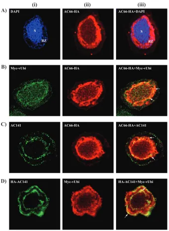
Localization of AC66-HA and colocalization of AC66-HA with Myc-vUbi and AC141. The Western blot analysis, mass spectrometry, and coimmunoprecipitation data indicated that AC66 is specifically ubiquitinated in BV nucleocapsids by vUbi and suggests this process is mediated by AC141. To further analyze these interactions, confocal microscopy was performed to determine the cellular localization of AC66-HA and also to determine if AC66 colocalizes with Myc-vUbi and AC141. Sf9 cells were
coinfected withac66KO-ac66-HA andvubiKO-Myc-vubi, fixed at 24 hpi, and stained for
AC66, vUbi, or AC141. As shown in Fig. 7A, AC66-HA was localized predominantly inside
AC66-HA WT AC66-HA VP80-HA VP80-HA
anti-ac141 anti-HA
Virus
Input Eluent Input Eluent
250
10 15 37 75 50 100 150
25 250
10 15 37 75 50 100 150
25
AC66-HA
VP80-HA
AC141
AC141
B.
WT WT
WT
Input Eluent
Input Eluent
anti-AC141
pre-immune
anti-AC141 pre-immune WB
virus: ac66KO-AC66-HA
anti-HA
anti-HA
anti-AC141
anti-AC141 AC141
AC66-HA
VP80-HA AC141 Virus
WB
A.
anti-HA
anti-ac141
250
75
50 100 150
250
75
50 100 150
IP:anti-HA IP:anti-HA
WB IP: WB IP:
virus: vp80KO-VP80-HA
FIG 6Coimmunoprecipitation analysis of AC66-HA or VP80-HA with AC141. To determine if AC141 interacts with either AC66 or VP80, Sf9 cells were infected with eitherac66KO-AC66-HA,vp80KO-VP80-HA, or the WT as a control. Cell lysates from 24 hpi were immuno-precipitated (IP) with anti-HA (A) or anti-AC141 (B). The eluents were analyzed by Western blotting and probed with the respective antibodies indicated on the left of each blot. For the VP80 blot in panel A, the marker lane was from the same gel and was moved to be adjacent to the sample lanes. The numbers on the left of the gels are kilodaltons.
on November 6, 2019 by guest
http://jvi.asm.org/
[image:12.585.45.473.68.506.2]the nucleus, with the majority of the signal in the ring zone, but lower levels were evenly distributed throughout the virogenic stroma. Small amounts of AC66 were also observed in the cytoplasm and at the plasma membrane. Myc-vUbi localizes through-out the cell in both the nucleus and cytoplasm (Fig. 7B and C). AC66-HA and Myc-vUbi exhibited colocalization that was primarily in the ring zone (Fig. 7B, iii). AC141 distri-bution was concentrated at both the nuclear and cytoplasmic peripheries, as shown previously (12). AC66-HA and AC141 showed colocalization, but only within the RING zone at the nuclear periphery in the regions outside the virogenic stroma (Fig. 7C, iii). Colocalization of HA-AC141 and Myc-vUbi was also analyzed in cells infected with
DAPI AC66-HA AC66-HA+DAPI
AC66-HA+AC141 AC66-HA
AC141
AC66-HA AC66-HA+Myc-vUbi
Myc-vUbi
A)
B)
C)
D)
HA-AC141 Myc-vUbi HA-AC141+Myc-vUbi(i)
(ii)
(iii)
S
RZ
S
RZ
FIG 7Colocalization analysis of AC66-HA, AC141, and Myc-vUbi. Sf9 cells were infected with ac66KO-AC66-HA (A),ac66KO-AC66-HA andvubiKO-Myc-vUbi (B),ac66KO-AC66-HA (C), orac141KO-HA-AC141 and vubiKO-Myc-vUbi (D). The cells were fixed at 24 hpi and stained for AC66-HA (A), AC66-HA and Myc-vUbi (B), AC66-HA and AC141 (C), or HA-AC141 and Myc-vUbi (D) using anti-HA, anti-Myc, or anti-AC141 antibodies. The nuclear regions in panel A were stained with DAPI. Single staining (i and ii) and merged images (iii) are shown. Regions of colocalization are shown in yellow (arrows). S, virogenic stroma; RZ, ring zone.
on November 6, 2019 by guest
http://jvi.asm.org/
[image:13.585.40.381.69.536.2]2⫻KO-HA-ac141⫹Myc-vubi. HA-AC141 and Myc-vUbi were observed to colocalize in both the nuclear and cytoplasmic periphery regions (Fig. 7D, iii). The colocalization analysis showed that the three proteins AC141, Myc-vUbi, and AC66-HA colocalize primarily in the ring zone of the nucleus outside the virogenic stroma. It is from this region that nucleocapsids either egress from the nucleus or are assembled into ODVs.
DISCUSSION
Baculoviruses are unique, as they produce two structurally different virions: BV, which is required for systemic transmission, and ODV, which is utilized for interhost transmission. Nucleocapsids for both virion types are synthesized in the infected cell nucleus in the virogenic stroma, from which they are transported to the nuclear periphery, called the ring zone. Nucleocapsids are either retained within the ring zone to form ODV or they egress from the nucleus, transit the cytoplasm, and bud from the plasma membrane to form BV. The focus of this study was to investigate the mecha-nism that determines how nucleocapsids are selected for nuclear egress and BV formation instead of remaining in the nucleus and forming ODV. AcMNPV encodes a predicted E3 RING domain ubiquitin ligase called AC141, as well as a viral ubiquitin that has 74% similarity to cellular ubiquitin. Both of these proteins had previously been shown to be specifically required for the production of BV. In this study, it was proposed that vUbi interacts with AC141 and that this interaction is required for the development of BV. The results have shown that AC141 and vUbi interact and that deletion of both the encoding genes eliminates BV production. In addition, nucleocapsids of BV, but not ODV, were shown to be extensively ubiquitinated by vUbi. Lastly, the nucleocapsid protein AC66 was identified as a potential substrate for AC141 and vUbi.
RING motifs of E3 ubiquitin ligases are required for binding E2 conjugating enzymes that are charged with ubiquitin (14). AC141 appears to be structurally distinct from the majority of E3 ubiquitin ligases, as it contains within the RING domain an extra cysteine residue adjacent to the third cysteine, and the histidine residue is replaced with tyrosine or phenylalanine (11). This suggests that AC141 may have a mechanistically different transfer of ubiquitin to a substrate. In cells infected with AcMNPV, there are both vUbi and cUbi in the cellular milieu. vUbi and cUbi are equally efficient in binding cellular E2s; however, vUbi is 40% less efficient than cUbi in transfer to a cellular E3 (21). This suggests a viral ubiquitin ligase is required for efficient recognition of the E2-vUbi complexes. The linkage of ubiquitin and E2 has been shown to be very flexible and to be able to exist in different structural orientations (45). However, upon binding of the E2-Ubi complex by the RING domain of an E3 ligase, the bound ubiquitin becomes locked into the active-site groove of E2 for catalysis (14). E3 ligases therefore have a close interaction with ubiquitin, and the structural differences of the AC141 RING motif may permit selective interaction with E2 enzymes conjugated with the structurally different vUbi as opposed to cUbi.
To study the interaction of AC141 and vUbi, single- and double-gene-knockout viruses were constructed and examined for their impacts on BV production. Deletion of
vubiorac141resulted in BV production being reduced to 0.26% or 0.005% that of the
WT, respectively (Fig. 1B). When bothac141andvubiwere deleted, BV production was
eliminated, suggesting cooperative interaction between AC141 and vUbi. Deletion of
vubi had a significant but smaller impact on BV production than deleting ac141. A
possible reason for this is that, as described above, cUbi can be interchanged with vUbi inin vitroubiquitination assays (21). It is therefore possible that in the absence of vUbi, cUbi may be utilized, albeit less efficiently, with a resulting decrease in BV production. E3 ubiquitin ligases are normally highly specific for their substrates (7), which suggests
that deletion ofac141would result in the loss of substrate recognition and
ubiquiti-nation and therefore a more dramatic drop in BV production. The very low levels of BV
that are observed in theac141KO viruses (estimated to be⬍1.0 50% tissue culture
infective dose [TCID50] per 500 transfected cells) (Fig. 1B) could be explained by
nonspecific stochastic tagging of nucleocapsids by cellular or viral E3 ligases from the
pool of vUbi present in an infected cell. Deletion of bothac141andvubiwould result
on November 6, 2019 by guest
http://jvi.asm.org/
in neither of the potential compensation mechanisms being available, and therefore, no BV would be produced, which is what was observed (Fig. 1).
AC141 was also found to be a substrate for ubiquitination, as either viral or cellular ubiquitin was found to be linked to residues in the AC141 substrate binding domain. Autoubiquitination has been shown to downregulate E3 ubiquitin ligases by polyubiq-uitination and proteasome degradation or to upregulate them by monoubiqpolyubiq-uitination, resulting in the enhanced ubiquitination of target substrates (39–43). Mutation of the AC141 ubiquitination sites results in accelerated BV production, suggesting ubiquiti-nation of AC141 may act as a switch mechanism to regulate its activity, which results in altered rates of BV production.
The C-terminal amino acids of cellular ubiquitins are normally diglycines, whereas vUbi has diglycine followed by a tyrosine. Inactive variants of cellular ubiquitin that have extra amino acids after the C-terminal diglycine exist, but they are normally cleaved by cellular isopeptidases or deubiquitinases (DUBs), resulting in functional ubiquitin (37, 38). Most ubiquitin binding proteins, including DUBs, bind cUbi at a hydrophobic surface or patch centered on I44 of cUbi (46). The vUbi I44 patch is conserved (38, 46); however, DUB binding domains have greater affinity for polyubiq-uitin or ubiqpolyubiq-uitin linked to a substrate than for free ubiqpolyubiq-uitin (47). AcMNPV vUbi is encoded as a monomer, whereas cUbi is usually expressed as a linear fusion of multiple ubiquitin molecules or with ribosomal proteins (37). As yet, no viral or cellular protein has been identified that has DUB activity and that can process vUbi to a diglycine C terminus. The C-terminal tyrosine of vUbi may play an important regulatory role, but this remains to be determined.
The envelope fraction from purified BV contained mainly monomeric cUbi and high-molecular-mass cUbi conjugates but also contained traces of vUbi (Fig. 3). Previ-ous biochemical analyses of BV envelopes by Guarino et al. (20) showed that they contained two forms of monomeric ubiquitin, nonmodified or covalently linked to envelope phospholipid, in the proportions 80% cUbi and 20% vUbi. Similarly, this study also showed that in BV envelopes, cUbi makes up the majority of monomeric and conjugated ubiquitin. In other viral systems, cUbi has been shown to become associ-ated with viral envelopes via the host endosomal sorting complex required for trans-port (ESCRT) pathway (10, 48). Viruses either encode E3 ubiquitin ligases or encode adapter proteins that recruit other components of ESCRT to support the process of budding from the plasma membrane. Interestingly, it has recently been shown that during baculovirus infection, ESCRT pathway components are required for BV produc-tion (49). If AcMNPV utilizes the host ESCRT components similarly to other viral systems, this could account for the specific cUbi and vUbi incorporation into the envelope or as conjugates attached to BV envelope proteins or lipids.
This study proposed that AcMNPV may utilize AC141 as a virus-specific E3 ubiquitin ligase to ubiquitinate nucleocapsids with vUbi to differentiate those that become BV or ODV. In support of this, a protein of approximately 100 kDa was found to be specifically ubiquitinated in the BV nucleocapsids but not in the ODV nucleocapsids (Fig. 3). Two of the potential nucleocapsid proteins in that region are AC66 and VP80 (30, 36). MS and coimmunoprecipitation analyses showed that AC141 associates specifically with AC66, which has also been shown to be required for nucleocapsid egress. Ke et al. (36) performed a detailed analysis of AC66 and proposed that AC66 and AC141 interact inside the nucleus to facilitate nucleocapsid egress. Two AC66 peptides were found by MS analysis to be modified by viral or cellular ubiquitin. Higher-molecular-mass forms of AC66 were identified in nucleocapsids, the dominant form of which was 7 to 10 kDa larger than its native size and comigrated with the nucleocapsid-specific 100-kDa viral ubiquitinated protein. The higher-molecular-mass form of AC66 is significantly enriched in BV compared to infected cells, where the vast majority of the AC66 exists in its native form and higher-molecular-mass species could not be resolved in total cell extracts. This was expected, because only approximately 2.3% of the viral genomes synthesized are utilized to form BV and the remaining 97.7% is retained inside the nucleus to form
on November 6, 2019 by guest
http://jvi.asm.org/
the ODV (50). Overall, these results suggest that only a minor fraction of the total AC66 produced becomes ubiquitinated and that it is associated only with BV nucleocapsids. Other viral systems use ubiquitination as a signal for the trafficking of nucleocapsids. For example, ubiquitination of the herpesvirus tegument protein pUL36 can act as a switch that determines capsid retrograde or anterograde cytoplasmic movement along microtubules, which can significantly impact neurovirulence (51–53). Nuclear egress of herpes simplex virus 1 (HSV-1) capsids has been extensively studied, and it is known that nucleocapsids egress from the nuclear envelope by utilizing a budding process. Several HSV-1-encoded proteins, including the pUL31-pUL34 nuclear egress complex, are associated with the inner and outer nuclear envelope membranes. These proteins assist in the budding of nucleocapsids from the nucleus and mediate the primary envelopment into the perinuclear space and de-envelopment from the outer nuclear envelope (54, 55). Recently, it was also shown that ubiquitination of the pUL34 homolog of Epstein-Barr virus (BFRF1) regulates the modulation of the nuclear enve-lope and nuclear egress of viral nucleocapsids (56). Although electron micrographs of AcMNPV-infected cells have suggested that nucleocapsids may also egress from the nucleus by budding through the nuclear envelope, definitive evidence is lacking (12, 57). Our coimmunoprecipitation and MS analyses suggested that, in addition to AC66, AC141 also interacts with GP41, which is known to be required for nuclear egress (32, 35).
Interestingly, GP41 was found to associate with the SNARE (soluble N
-ethylmaleimide-sensitive factor attachment protein receptor) proteins, which are required for fusion of transport vesicles with target membranes and for nuclear egress of AcMNPV nucleocapsids (58). It is possible that during egress of nucleocapsids from the nucleus, AC141, AC66, and GP41 form part of a nuclear-egress complex. The ubiquitination of nucleocapsid proteins, such as AC66, may therefore be part of the signaling process required for the recognition of nucleocapsids by a nuclear envelope complex composed of viral and cellular proteins. In support of this, confocal microscopy showed that AC141, AC66, and vUbi associate near the nuclear periphery regions (Fig. 7). AC66 may also play a role once nucleocapsids traverse the nuclear envelope, since the N terminus has a conserved desmoplankin domain. Desmoplankin domains can bind intermediate filaments and interact with microtubule components, which are known to be required for nucleocapsid egress (59–61). It is therefore possible that AC66 enables interaction with the cellular cytoskeleton during egress from the nucleus.
In summary, we have shown that the nucleocapsids that egress from the nucleus to form BV are differentially modified by extensive ubiquitination with vUbi compared to those used to form ODV. In addition, these data support the conclusion that AcMNPV AC141 is involved in the ubiquitination of the nucleocapsid protein AC66, or associated proteins, with vUbi to enable BV production. Ubiquitination with vUbi is therefore the potential mechanism by which a nucleocapsid is directed to become a BV, a critical event in the baculovirus replication cycle that enables systemic virus spread in an infected host.
MATERIALS AND METHODS
Cell lines and viruses.Sf9 cells were maintained in Grace’s insect medium supplemented with
L-glutamine, 3.33 g/liter lactalbumin hydrolysate, and 3.33 g/liter yeastolate (Gibco Life Technologies) and further supplemented with 10% fetal bovine serum at 27°C. The WT virus was AcMNPV-E2, and the AcMNPV-E2 bacmid used for construction of recombinant viruses was bMON14272 (62).
Construction ofvubiknockout,ac141plusvubidouble-knockout, and repaired viruses.The vubiopen reading frame (ORF) is close toac34, and the late promoter sequence ofac34is within thevubi ORF (63). Upon deletion ofvubi, the late promoter sequence forac34was reintroduced in the primers used to amplify the chloramphenicol resistance gene. The primer sequence to amplify thecatgene was based on previously published sequences (64, 65). Thecatgene was PCR amplified using the plasmid pKD3 as the template with primer pair 746-747 (Table 4; the core late promoter sequence forac34in the 746 primer sequence is underlined). The PCR product was gel purified and transformed into bMON14272 containingEscherichia coliBW25113/pKD46 electrocompetent cells (64). The recombinant cells were selected, and correct insertion of thecatgene in thevubilocus was confirmed with primer pairs 763-764 and 765-766 (Table 4), which confirmed recombination into the correct locus.
Theac141andvubidouble-knockout AcMNPV bacmid was generated by using a strategy similar to that described above (64). Theac141ORF was knocked out using a 2.9-kbp EcoRI-HindIII restriction
on November 6, 2019 by guest
http://jvi.asm.org/
fragment from the previously described plasmid vector pAc-exon0-KO (11). The restriction fragment contained the 483-bp zeocin resistance gene under the control of the EM7 promoter and 1,547 bp of 5=
and 543 bp of 3=ac141flanking sequences. The 5=flanking sequence contained 142 bp of the 5=ORF of ac141, which contains the splice site ofie0and the 3=flanking sequence of the promoter downstream ofac142(11). The restriction fragment was transformed intoE. coliBW25113/pKD46 competent cells containing thevubiKO bacmid. The recombinant cells were selected, and correct insertion of the zeocin resistance gene in theac141locus was confirmed with primer pairs, as previously described (11). The double-gene-knockout bacmid containing the desired recombination was selected, and the resulting virus was namedac141⫹vubi2⫻KO.
The transfer vector pFAcT-GFP, which contains polyhedrin and enhanced green fluorescent protein (GFP) was used to repair theac141,vubi, and ac141⫹vubiknockout bacmids. These plasmids were pFAcT-GFP-Myc-vubi, pFAcT-GFP-HA-ac141, and pFAcT-GFP-HA-ac141⫹Myc-vubi. To construct pFAcT-GFP-Myc-vubi, thevubisequence was amplified with the primer pair 1833-1834 containing AgeI and XbaI sites. The primers also inserted the Myc epitope tag (EQKLISEEDL) coding sequence after the second codon ofvubi(66) and thevubipoly(A) signal at the 3=end.
The amplified 320-bp fragment was digested with AgeI and XbaI and ligated to a similarly digested vector that contained theac141late promoter. The resulting plasmid, pMyc-vubi, was digested with XhoI and XbaI, which generated a 395-bp fragment [ac141late promoter plus Myc-vubiplusvubipoly(A)] that was inserted into pFact-GFP-digested with XhoI and XbaI, to generate GFP-Myc-vubi. The pFAcT-GFP-HA-ac141plasmid has been previously described (11). The pFAcT-GFP-HA-ac141⫹Myc-vubiplasmid was constructed as follows. Myc-vubi, along with theac141promoter and thevubipoly(A) signal, was amplified by PCR, using pFAcT-GFP-Myc-vubias a template, with the primer pair 1834 and 1835, which contained PstI and XhoI sites. The fragment was digested with PstI and XbaI and cloned into pFAcT-GFP-HA-ac141 to generate the plasmid pFAcT-GFP-HA-ac141⫹Myc-vubi. The plasmids pFAcT-GFP, pFAcT-GFP-Myc-vubi, and pFAcT-GFP-HA-ac141or the pFAcT-GFP-HA-ac141⫹Myc-vubibacmid was used to transform E. coli DH10B cells containing one of the knockout bacmids and the helper plasmid (pMON7124), which encodes the Tn7transposase, as described previously (62). The genotype was verified by PCR. Seven repaired bacmids were generated, which were called ac141KO, vubiKO, ac141⫹vub2⫻KO,vubiKO-Myc-vubi, 2⫻KO-HA-ac141⫹Myc, 2⫻KO-HA-ac141, and 2⫻KO-Myc-vubi.
Construction ofac66knockout,vp80knockout, and repaired viruses.ac66was deleted from the AcMNPV bacmid bMON14272, using recombination inE. colias described above. The 5=terminus of the ac66ORF contains the late promoter sequence of the adjacentdnapol, and the 3=terminus contains the poly(A) region of the adjacentlef3. Therefore, 270 bp of theac665=terminus and 880 bp of the 3=
[image:17.585.43.559.84.346.2]terminus were retained. The zeocin resistance gene was PCR amplified from p2ZeoKS with the primer pair 1266 and 1267 (Table 4), which also containac66homologous sequences. The PCR-amplified 625-bp fragment contains a zeocin resistance gene under theEM7promoter and theac66flanking sequence for recombination. The amplified PCR product was transformed into electrocompetent E. coliBW25113/ pKD46, and recombinants were selected as described above. The correctac66deletion and insertion of TABLE 4Primers used for construction of plasmid clones and viruses
Primer no. Primer sequence (5=to 3=)
2587 GCTTTTTTTATTACTATGATCAAT
2588 GATTCTCTATTCTATGCTTGTACA
2589 CCTTTTTTTATTACTATGATCAAT
2590 CATTCTCTATTCTATGCTTGTACA
746a CAAGGGCGCATTCACAGCAACCGTTGTCATTTATAAGTAAACTTATCTAAGTGTAGGCTGGAGCTGCTTC
747 CAAGATACAAATATGTCAGATTAAATAAAAAACTTTTATGTATATTTAATGATATGAATATCCTCCTTAG
763 TGTGAATAAAGGCCGGATAAAACT
764 CCCATTAGCGGCAGCAGGAAA
765 GTCGGCGTGCGTGTAACAAAGT
766 CAAGGCGACAAGGTGCTGATGC
1833 ACACCGGTAAAATGCAAGAACAAAAACTTATTTCTGAAGAAGATCTTATATTCATCAAAACAT 1834 GG TCTAGAAATAAAAAACTTTTATGTATATTTA
1835 CCCTGCAGATCAATTGTG
1313 GCAACTGTGACGCCATAG
1239 CTGACCGACGCCGACCAA
1629 GCGTCTAGATGCTTTGTTTCTTTCGTATT
1630 GCGGCGGCCGCTTAGGCGTAGTCGGGCACGTCGTAGGGGTATTCGACGTTTGGTTGAAC
1695 GCGCTGCAGGTACCTGTTTGATAAACTC
1696 GCGTCTAGATTAGGCGTAGTCGGGCACGTCGTAGGGGTATATAACATTGTAGTTTGCGTT
1266 CGGATTTCGCCGGCCGAGATATCAACACGTTGACGCACAACATCAACTACTTCGGATCTCTGCAGCAC3 1267 TTGTCGCGACTTGAGACAATTCATTTTTAGTTGCAGTTAATTCATTTACATCGAGGTCGACCCCCCTG
1268 CGCGCACTGTACACGATT
1014 CCGATATACTATGCCGATGATT
1310 GAAGAGTGTTATGTTAAAATTGATAGACTATTTAAAGAGAGCATTAAAAATTCGGATCTCTGCAGCAC 1311 TTATATAACATTGTAGTTTGCGTTCATCAACATTATTAGTCTTTGCAAATTCGAGGTCGACCCCCCTG
1312 GCCGCGGGTAACAT
aThe underlined region in the 746 primer sequence is the core late promoter sequence forac34.
on November 6, 2019 by guest
http://jvi.asm.org/
the zeocin resistance gene were confirmed by PCR with the primer pair 1268 and 1014 (Table 4). The deletion bacmid was namedac66KO.
Thevp80ORF was deleted using the same method. Due to regulatory sequences of upstream genes, 473 bp in the 5=terminus of thevp80ORF was retained, but the remaining ORF sequences were deleted. The zeocin resistance gene was PCR amplified from p2ZeoKS with primer pair 1310 and 1311, which also contain thevp80homologous sequences for recombination. Correct insertion of the zeocin resistance gene was confirmed with primer pairs 1312 and 1014, and also 1313 and 1239 (Table 4). The deletion bacmid was namedvp80KO.
To generate a C-terminal HA epitope-tagged clone ofac66, theac66late promoter and ORF were PCR amplified from the AcMNPV genome with primer pair 1629 and 1630, which contained a 5=XbaI and a 3=NotI site. The HA epitope nucleotide sequence was contained in the 3=primer 1630 (Table 4). The PCR product was digested with XbaI and NotI and inserted into pFAcT-GFP-Tnie1PA digested with the same restriction enzymes, resulting in the clone pFAcT-GFP-AC66-HA. Theac66KO bacmid was repaired with pFAcT-GFP-AC66-HA using Tn7-mediated transposition, as described above, to generate the ac66KO-ac66-HA repaired virus. Similarly, thevp80late promoter and ORF, with a C-terminal HA epitope tag, were PCR amplified from the AcMNPV genome with the primer pair 1695 and 1696, which contain PstI and XbaI sites, respectively. The HA epitope nucleotide sequence was contained within the 3=primer 1696. The PCR product was digested with PstI and XbaI and inserted into pFAcT-GFP-Tnie1PA digested with the same restriction enzymes. The resulting clone was named pFAcT-GFP-VP80-HA. Thevp80KO bacmid was repaired with pFAcT-GFP-VP80-HA by TnI7-mediated transposition to generate the vp80KO-vp80-HA repaired virus.
Mutation in the ubiquitination site of AC141 (K87).The potential ubiquitinated lysine 87 of AC141 was mutated to an arginine or an alanine by inverse PCR. The PCR template was a previously constructed plasmid, p2Zeo-HA-Acexon0. Site-directed mutagenesis was carried out by standard PCR procedures using the primer pair 2587 and 2588 for K87R (AAA¡CGC) and primer pair 2589 and 2590 for K87A (AAA¡GCC). A 1.4-kbp XhoI and XbaI fragment excised from mutagenized p2Zeo-HA-Acexon0 was cloned into XhoI- and XbaI-digested pFAcT-GFP to produce K87R and pFAcT-HA-ac141-K87A. These clones were used to repair the bacmidac141KO, using the method described above, to generate the virusesac141KO-HA-ac141-K87R andac141KO-HA-ac141-K87A.
Time course analysis of virus infection in bacmid-transfected or virus-infected cells for analysis of BV production and viral protein synthesis.Sf9 cells were seeded into 6-well plates (2.0⫻106cells per 35-mm-diameter well) and transfected with 1.0 g of each bacmid or infected with BV at a multiplicity of infection (MOI) of 5 or 10. To measure BV production, the culture medium was harvested at various times posttransfection and centrifuged at 3,000 ⫻g for 5 min to pellet cell debris. BV production was determined by the TCID50endpoint dilution assay or by droplet digital PCR (12, 67).
Coimmunoprecipitation of protein complexes. Sf9 cells (5.0 ⫻ 107) were infected with the respective viruses at an MOI of 10. Infected cells were harvested at 24 hpi and resuspended in 1.25 to 1.5 ml of either HEPES lysis buffer (15 mM HEPES, pH 7.6, 10 mM KCl, 0.1 mM EDTA, 0.5 mM EGTA, 1 mM dithiothreitol [DTT], and 1% protease inhibitor cocktail [Invitrogen]) or EBC lysis buffer (50 mM Tris-HCl, pH 8.0, 120 mM NaCl, 0.5% Nonidet P-40, 0.2 mM sodium orthovanadate, 1% sodium fluoride, and 1% protease inhibitor cocktail). Following cell lysis, the immunoprecipitation protocol was followed, as previously described (61). The eluent volume was vacuum concentrated to 60l and mixed with 20l of 4⫻protein sample buffer (PSB) (277.8 mM Tris-HCl, pH 6.8, 44.4% [vol/vol] glycerol, 0.02% bromophe-nol blue, 4%-mercaptoethanol, 1% protease inhibitor cocktail [Sigma-Aldrich]). The sample was boiled for 10 min, subjected to SDS-polyacrylamide gel electrophoresis (PAGE) using Mini-Protean TGX stain-free gels (Bio-Rad), and examined by Western blotting.
Confocal microscopy.Sf9 cells were plated on sterile coverslips and allowed to settle overnight as previously described (61). The cells were infected with viruses at an MOI of 10. At different times postinfection, cells were fixed (100 mM HEPES, 5 mM MgCl2, 5 mM EGTA, pH 6.9, and 1.5 or 4% formaldehyde) for 45 min, followed by permeabilization with 0.01% Triton X-100 solution in 1⫻PBS for 15 min. Incubation of primary and secondary antibodies was as previously described (61). The coverslips were mounted with Prolong Gold antifade reagent (Molecular Probes) with or without DAPI (4=,6= -diamidino-2-phenylindole). The primary antibodies included mouse monoclonal anti-c-Myc 9E10 (1:50; Santa Cruz Biotechnology), rabbit polyclonal anti-HA (1:200; Abcam), and rabbit polyclonal AC141 (1:400). The secondary antibodies used were goat anti-mouse Alexa-647 (1:500; Molecular Probes), goat anti rabbit Alexa-488 (1:500; Molecular Probes), goat anti rabbit Alexa-647 (1:500; Molecular Probes), goat anti mouse Alexa-405 (1:500; Molecular Probes), and goat anti rabbit Alexa-405 (1:500; Molecular Probes). All images were acquired using a Leica TCS SP8 confocal laser scanning microscope (CLSM) with a 63⫻oil immersion lens. Samples were sequentially excited at 488 nm for Alexa-488 or GFP, 633 nm for Alexa-647, and 405 nm for DAPI or Alexa-405.
Purification of BV and ODV and fractionation into envelope and nucleocapsid fractions.Sf9 cells (2.0⫻108) were infected withvubiKO-Myc-vubivirus at an MOI of 0.1, and cells were harvested at 7 days p.i. The infected cells were pelleted at 3,000 rpm to separate the medium containing BV and the cell pellet containing ODV. The BV-containing medium (90 ml) was centrifuged at 8,000⫻gin a Beckman JA12 rotor to pellet cell debris. The BV in the cell debris-free supernatant was pelleted by centrifugation at 100,000⫻g(21,000 rpm) in a Beckman SW28 rotor at 4°C. The pelleted BV was resuspended in 400
l of 0.1⫻Tris-EDTA (TE) buffer with 1% protease inhibitor cocktail as described above. The resuspended BV was loaded onto a continuous sucrose density gradient (25 to 60%) and centrifuged at 100,000⫻g (24,000 rpm) in a Beckman SW41 rotor at 4°C for 1.5 h. The BV band was collected and diluted with an equal volume of 0.1⫻TE. The diluted, purified BV was centrifuged at 100,000⫻g(24,000 rpm) in a
on November 6, 2019 by guest
http://jvi.asm.org/
Beckman SW41 rotor at 4°C for 30 min to pellet the BV. The purified BV protein concentration was determined by the Bradford assay (68).
The cell pellet containing occlusion bodies (OBs) was washed twice with 5 ml of sterile distilled water (dH2O) to remove any trace medium. The cell pellet was resuspended in 2 ml of 2% (vol/vol) Triton X-100 and incubated at 37°C for 1 h. Following Triton X-100 treatment, the cells were treated with 2% deoxycholate at 37°C for 1 h. The material was then treated with 0.1% SDS and vortexed for 10 min. OBs were pelleted at 3,000⫻gfor 1 min and resuspended in 100l dH2O. ODVs were released from OBs by incubation with alkaline dissolution buffer (0.002 M EDTA, 0.2 M Na2CO3, 0.34 M NaCl, pH 10.8) for 30 min. The dissolution of OBs was checked by microscopy. The reaction was neutralized with 20l 1.0 M Tris-HCl, pH 7.2. The remaining cell debris and insoluble material were pelleted for 5 min at 3,000⫻g. ODVs in the supernatant were loaded onto a step sucrose gradient (30 to 60%). The gradient was centrifuged at 100,000⫻g(24,000 rpm) in a Beckman SW41 rotor at 4°C for 90 min. The multiple ODV bands were collected and diluted with 2