Fever Virus to Vero Cells Leads to a Gradual Attenuation of Virulence
in Swine Corresponding to Major Modifications of the Viral Genome
Peter W. Krug,aLauren G. Holinka,aVivian O’Donnell,a,bBo Reese,cBrenton Sanford,aIgnacio Fernandez-Sainz,a,b Douglas P. Gladue,a,bJonathan Arzt,aLuis Rodriguez,aGuillermo R. Risatti,bManuel V. Borcaa
Agricultural Research Service, U.S. Department of Agriculture, Plum Island Animal Disease Center, Greenport, New York, USAa
; Department of Pathobiology and Veterinary Science, CANHR, University of Connecticut, Storrs, Connecticut, USAb
; Center for Applied Genetics and Technology, University of Connecticut, Storrs, Connecticut, USAc
ABSTRACT
African swine fever virus (ASFV) causes a contagious and often lethal disease of feral and domestic swine. Experimental vaccines derived from naturally occurring, genetically modified, or cell culture-adapted ASFV have been evaluated, but no commercial vaccine is available to control African swine fever (ASF). We report here the genotypic and phenotypic analysis of viruses obtained at different passages during the process of adaptation of a virulent ASFV field isolate from the Republic of Georgia (ASFV-G) to grow in cultured cell lines. ASFV-G was successively passaged 110 times in Vero cells. Viruses ob-tained at passages 30, 60, 80, and 110 were evaluatedin vitrofor the ability to replicate in Vero cells and primary swine macrophages cultures andin vivofor assessing virulence in swine. Replication of ASFV-G in Vero cells increased with suc-cessive passages, corresponding to a decreased replication in primary swine macrophages cultures.In vivo, progressive loss of virus virulence was observed with increased passages in Vero cells, and complete attenuation of ASFV-G was ob-served at passage 110. Infection of swine with the fully attenuated virus did not confer protection against challenge with virulent parental ASFV-G. Full-length sequence analysis of each of these viruses revealed significant deletions that gradu-ally accumulated in specific areas at the right and left variable ends of the genome. Mutations that result in amino acid sub-stitutions and frameshift mutations were also observed, though in a rather limited number of genes. The potential impor-tance of these genetic changes in virus adaptation/attenuation is discussed.
IMPORTANCE
The main problem in controlling ASF is the lack of vaccines. Attempts to produce vaccines by adaptation of ASFV to cul-tured cell lines have been made. These attempts led to the production of attenuated viruses that conferred only homolo-gous protection. Specifics regarding adaptation of these isolates to cell cultures have been insufficiently described. Details like the numbers of passages required to obtain attenuated viruses, genetic modifications introduced into the virus ge-nomes along passages, and the extent of attenuation and induced protective efficacy are not readily available. In this study, we assessed the changes that lead to decreased growth in swine macrophages and to attenuation in swine. Loss of virulence, probably associated with limited replicationin vivo, may lead to the lack of protective immunity in swine observed after challenge. This report provides valuable information that can be used to further the understanding of ASFV gene function, virus attenuation, and protection against infection.
A
frican swine fever (ASF) is a contagious disease of swine caused by ASF virus (ASFV), an enveloped virus containing a large (190-kbp) double-stranded DNA (dsDNA) genome. ASFV infection of domestic pigs can induce a spectrum of disease sever-ity, from highly lethal to subclinical, depending on host character-istics and the particular circulating virus strain (1).ASF is endemic in most sub-Saharan African countries, while in Europe it is also endemic on the island of Sardinia (Italy). Since 2007, outbreaks of the disease have been declared in the Caucasus region, including Georgia, Armenia, Azerbaijan, and Russia. Iso-lated outbreaks of the disease have recently been identified in Ukraine, Belarus, Lithuania, Latvia, and Poland. The epidemic virus ASFV Georgia 2007/1 is a highly virulent virus of genotype II (2).
Since no commercial vaccine is currently available, disease control measures consist of elimination of infected and suscepti-ble animals. Experimental live attenuated ASF viruses that have been engineered to contain deletions within virulence-associated
genes or have been produced by cell culture adaptation usually provide protection to immunized pigs when challenged with ho-mologous parental virus. (3,4,5,6,7,8,9).
Adaptation of several virulent field isolates to grow in
estab-Received7 November 2014Accepted1 December 2014
Accepted manuscript posted online10 December 2014
CitationKrug PW, Holinka LG, O’Donnell V, Reese B, Sanford B, Fernandez-Sainz I, Gladue DP, Arzt J, Rodriguez L, Risatti GR, Borca MV. 2015. The progressive adaptation of a Georgian isolate of African swine fever virus to Vero cells leads to a gradual attenuation of virulence in swine corresponding to major modifications of the viral genome. J Virol 89:2324 –2332.doi:10.1128/JVI.03250-14.
Editor:S. Perlman
Address correspondence to Manuel V. Borca, manuel.borca@ars.usda.gov. P.W.K., L.G.H., and V.O. contributed equally to this work.
Copyright © 2015, American Society for Microbiology. All Rights Reserved.
doi:10.1128/JVI.03250-14
on November 7, 2019 by guest
http://jvi.asm.org/
lished cell lines were usually accompanied by decreased virulence in swine and significant genomic modifications (3,5,7,10). De-tails regarding the process of adaptation, phenotypic changes, and precise genomic modifications of ASFV grown/adaptedin vitro
remain elusive, making it challenging to elucidate the genomic mechanisms governing phenotypic modifications.
Here we describe thein vivoandin vitrocharacteristics of a highly virulent ASFV strain isolated in the Republic of Georgia (ASFV-G) during propagation in Vero cells. It was observed that the adaptation of ASFV-G to replicate in Vero cells is associated with a progressively decreased ability of the virus both to replicate in primary cultures of swine macrophages and to produce disease in swine. Full-length genomic analyses of viruses obtained from Vero cells at passages 30, 60, 80, and 110 were performed to iden-tify genetic changes associated with the gradual adaptation of ASFV-G to growin vitro.
MATERIALS AND METHODS
Cell cultures and viruses.The ASFV Georgia (ASFV-G) isolate was de-rived from the spleen of a naturally infected pig and was kindly provided to Plum Island Animal Disease Center by Nino Vepkhvadze from the Laboratory of the Ministry of Agriculture (LMA) in Tbilisi, Georgia. The clinical material was macerated, suspended in Dulbecco’s minimal essen-tial medium (DMEM) (Gibco, Grand Island, NY) with 10% fetal calf serum (FCS) (Atlas Biologicals, Fort Collins, CO), clarified by centrifuga-tion (20 min at 10,000 rpm and 4°C), and suspended in DMEM with 10% FCS. Vero cells were obtained from the ATCC and subcultured in DMEM with 10% FCS. Growth kinetics were assessed either in Vero cells or in primary swine macrophage cell cultures, prepared as described before (9). Titration of ASFV from freeze-thawed samples was performed using pri-mary swine macrophage cell cultures in 96-well plates (Primaria, Cam-bridge, MA). Viral infectivity was detected, after 7 days in culture, by hemadsorption (HA). When Vero cells were used for virus titrations, the presence of virus-infected cells was detected by immunocytochemistry using an anti-ASFV p30 monoclonal antibody (kindly provided by Javier Dominguez, INIA, Spain). Titers were calculated using the method of Reed and Muench (11) and expressed as 50% hemadsorption doses (HAD50)/ml.
Next-generation sequencing (NGS) of ASFV genomes.ASFV DNA was obtained from the cytoplasms of infected swine macrophages using the TRIzol method (Life Technologies, Grand Island, NY, USA). ASFV DNA was extracted from infected Vero cells using a previously described method (12). DNA concentration was determined using the Qubit dsDNA high-sensitivity (HS) assay kit (Life Technologies) and read on a Qubit 2 fluorometer (Life Technologies). One microgram of virus DNA was enzymatically fragmented to obtain blunt-end fragments in a length range of 200 to 300 bp using the Ion Shear Plus reagent kit (Life Technol-ogies) and incubated at 37°C in a Peltier DNA Engine Tetrad 2 thermal cycler. After shearing, the fragmented DNA library was loaded onto a DNA chip (Agilent, Santa Clara, CA, USA) and analyzed using a Bioana-lyzer 2100 (Agilent) to assess DNA size distribution and range. Frag-mented DNA was ligated to Ion-compatible adapters and library bar-codes, followed by nick repair to complete the linkage between adapters and DNA inserts using the Ion Plus fragment library kit (Life Technolo-gies). The adapter-ligated library was size selected for optimum length on 2% agarose gel cassettes (Sage Science, Beverly, MA, USA) using the Pip-pin Prep instrument (Sage Science). Library concentration was normal-ized using the Ion library equalizer kit (Life Technologies). Next, the DNA library was clonally amplified onto Ion Sphere particles (ISPs), generating template-positive ISPs using the Ion PGM template OneTouch 2 200 kit (Life Technologies) with the Ion OneTouch 2 instrument (Life Technol-ogies). Before proceeding to enrichment, quality assessment of nonen-riched template-positive ISPs was performed using the Ion Sphere quality control (QC) assay kit (Life Technologies) and a Qubit 2 fluorometer. The
template-positive ISPs were then enriched using the Ion PGM template OneTouch 2 200 kit (Life Technologies) and Ion OneTouch enrichment system (ES) instrument (Life Technologies) to eliminate template-nega-tive ISPs and to denature DNA on template-positemplate-nega-tive ISPs. Using the Ion PGM 200 sequencing v2 kit (Life Technologies), enriched template ISPs were prepared for sequencing, loaded onto either an Ion 314 or Ion 316 chip, v2 (Life Technologies), and run on the Ion PGM sequencer (Life Technologies). The sequences obtained were then trimmed using Galaxy (https://usegalaxy.org/) NGS QC and manipulation tools. Sequences were aligned and analyzed using Sequencher 5.2.2 (Genecodes) and CLC Genomics Workbench (CLCBio) software. For comparative purposes, genes and putative open reading frames (ORFs) of ASFV-G described here follow the nomenclature as for the ASFV Georgia 2007/1 annotated ge-nome (GenBank no.FR682468.1) (2).
Animal experiments.Animal experiments were performed under biosafety level 3 conditions in the animal facilities at Plum Island Animal Disease Center following a protocol approved by the Institutional Animal Care and Use Committee.
Vero-adapted ASFV-G (ASFV-G/V) strains were assessed for their virulence phenotype relative to the virulent parental ASFV-G using 80- to 90-pound commercial-breed swine. Groups of five pigs were inoculated intramuscularly (IM) with either 102or 104HAD
50of ASFV-G/V strains or ASFV-G. Clinical signs (anorexia, depression, fever, purple skin discol-oration, staggering gait, diarrhea, and cough) and changes in body tem-perature were recorded daily throughout the experiment.
For protection studies, pigs surviving infection with attenuated viruses were IM challenged with 104HAD
50of parental ASFV-G at either 21 or 28 days postinfection. Clinical signs and body temperature were recorded daily throughout the experiment.
RESULTS
Development of the ASFV Georgia Vero-adapted strain (ASFV-G/V).A spleen homogenate from a pig naturally infected with the Georgia strain of ASFV (ASFV-G) was used to infect a subconflu-ent monolayer of Vero cells, which was then incubated for 4 days at 34°C. Neither cytopathic effect (CPE) nor hemadsorption was observed in these cells. The infected cells were then subcultured successively twice under the same conditions, and the presence of several foci of rounded cells was observed. The presence of ASFV-infected cells was detected in these cultures by immunohisto-chemistry using serum from an ASFV-infected pig, showing evi-dence of virus spread in the Vero cells (data not shown). Infected cells were subcultured two additional times until CPE became evident in the cultures. At this point, a virus stock was prepared (5⫻10650% tissue culture infective doses [TCID
50]/ml). There-after, virus was successively passed under similar conditions for a total of 110 passages, counting from the initial infection of Vero cells with the spleen lysate. Viruses harvested at passages 30, 60, 80, and 110 were used to produce stocks (named ASFV-G/VP30, -P60, -P80, and -P110) that were used in subsequent studies.
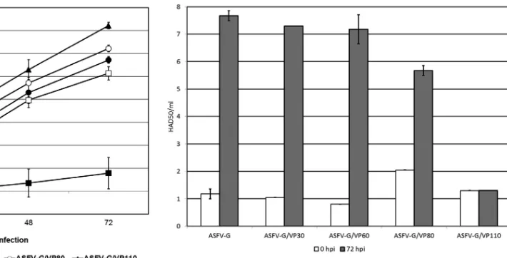
Ability of ASFV-G/V to grow in Vero cells.In vitro character-istics of the ASFV-G/V were evaluated relative to the parental ASFV-G isolate in multistep growth curves (Fig. 1). Vero cell cul-tures were infected at a multiplicity of infection (MOI) of 0.01. Virus was adsorbed for 1 h (time zero), and samples were collected at 0, 24, 48, and 72 h postinfection (hpi). Virus yields were assessed by titration using Vero cells. Results demonstrated that ASFV-G/V yields increased with increasing passages in Vero cells, while the virus yield of parental ASFV-G was almost undetectable in these cells. ASFV-G/VP30 virus already exhibited a significantly increased virus yield in Vero cells (between 100- and 10,000-fold, depending on the time point considered) compared to parental Adaptation of African Swine Fever Virus to Vero Cells
on November 7, 2019 by guest
http://jvi.asm.org/
ASFV-G. Additional cell passages resulted in virus yields 1,000- to 1,000,000-fold greater than the yield obtained with parental ASFV-G, depending on the time point considered. Results are indicative of an increased fitness of the virus to grow in Vero cells with an increased number of virus passages.
Replication of ASFV-G/V in primary swine macrophages.In vitrogrowth characteristics of ASFV-G/V were evaluated in pri-mary swine macrophage cell cultures, the pripri-mary cell targeted by ASFV during infection in swine, and compared to those of paren-tal ASFV-G in one-step growth curves (Fig. 2). Cell cultures were infected at an MOI of 0.01, and samples were collected at 96 hpi. Results demonstrated that although ASFV-G/VP30 and -P60 yields are similar to or slightly lower than those of parental ASFV-G, the ASFV-G/VP80 yield is approximately 10-fold less than that of parental ASFV-G, whereas ASFV-G/VP110 showed a drastically reduced ability to replicate in swine macrophages, with virus yields being 1,000,000- to 10,000,000-fold lower than those of parental ASFV-G. These results demonstrate that while cell-adapted ASFVs replicate up to high titers in Vero cells, they pro-gressively lose their ability to replicate in primary swine macro-phagesin vitro. These results suggest that Vero-adapted viruses lack specific gene functions that are associated with ASFV replica-tion and growth in swine macrophages.
Assessment of virulence of ASFV-G/V in swine.To assess how virus adaptation to replicate in Vero cells affects virulence of ASFV-Gin vivo, 80- to 90-pound pigs were intramuscularly (IM) inoculated with G, G/VP30, G/VP60, ASFV-G/VP80, or ASFV-G/VP110 (Table 1). Pigs that received an inoc-ulum of 104HAD50of parental ASFV-G displayed increased body temperature (⬎104°F) by 3 to 4 days postinfection, presenting typical clinical signs associated with ASF, including anorexia, de-pression, purple skin discoloration, staggering gait, and diarrhea. Signs of the disease grew more severe over time, and animals either died or were euthanizedin extremisby day 7 or 8 postinfection. Pigs inoculated IM with an inoculum containing 102HAD50of
parental ASFV-G exhibited a slightly delayed onset of clinical dis-ease relative to pigs inoculated with 104HAD
50of the same virus. Pigs presented a short period of fever starting by day 7 postinfec-tion, with animals dying or euthanizedin extremisaround 8 to 9 days postinfection.
Animals inoculated IM with 104 HAD
50 of ASFV-G/VP30, ASFV-G/VP60, or ASFV-G/VP80 developed clinical disease sim-ilar to that observed in animals inoculated IM with the same dose of parental ASFV-G. Animals displayed ASF-related signs by 3 to 4 days postinfection (p.i.), dying or being euthanized by 8 to 9 days p.i. (Table 1). It should be noted that animals inoculated with ASFV-G/VP80 presented a delayed onset of fever that was mani-fested at least 3 days later than that in pigs inoculated with ASFV-G/VP30 or ASFV-G/VP60. Interestingly, animals inoculated with ASFV-G/VP110 by using the same route and dose as described above did not present any signs of ASF during the entire observa-tional period (21 days).
IM inoculation of swine with 102 HAD
50 of ASFV-G/VP30 induced milder signs of ASF than inoculation with parental ASFV-G at a similar route and dose. Two out of five pigs presented clinical signs of ASF between 4 and 6 days p.i. and died or were euthanized by 11 days p.i., manifesting disease at least 4 days later than pigs that were inoculated with parental ASFV-G. Three out of five pigs showed a delayed onset of the disease (by 2 to 5 days) with milder signs, denoting a rather transient disease manifestation. These animals did not show signs of ASF by the end of the obser-vational period. Interestingly, animals inoculated IM with 102 HAD50of ASFV-G/VP60, ASFV-G/VP80, or ASFV-G/VP110 re-mained clinically normal throughout the observational period (Table 1).
In vitroandin vivoexperiments demonstrated that progressive adaptation of ASFV-G to replicate in Vero cells after successive pas-sages is accompanied with a gradually increased attenuation of the virus. ASFV-G passed 30 times (ASFV-G/V30) produced a significant attenuation at relatively low doses (102HAD50). Attenuation was clearly enhanced at passage 110, when high doses (104HAD
50) of ASFV-G/VP110 were unable to induce any signs of the ASF. FIG 1In vitrogrowth kinetics of ASFV-G and Vero-adapted viruses ASFV-G/
VP30, ASFV-G/VP60, ASFV-G/VP80, and ASFV-G/VP110 in Vero cell cul-tures. Cell cultures were infected (MOI⫽0.01) with either ASFV-G or any of the Vero-adapted viruses, and virus yields obtained at the indicated times postinfection were titrated in Vero cell cultures. Data are means and standard deviations from two independent experiments. The sensitivity of virus detec-tion wasⱖ1.8 TCID50/ml.
FIG 2In vitrogrowth kinetics of ASFV-G and the Vero-adapted viruses ASFV-G/VP30, ASFV-G/VP60, ASFV-G/VP80, and ASFV-G/VP110 in pri-mary cultures of swine macrophages. Data are means and standard deviations from two independent experiments. The sensitivity of virus detection was
ⱖ1.8 TCID50/ml.
on November 7, 2019 by guest
http://jvi.asm.org/
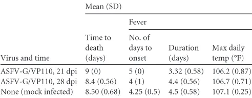
[image:3.585.177.532.64.245.2] [image:3.585.43.291.66.244.2]Protective efficacy of ASFV-G/VP110 virus against challenge with parental ASFV-G.Since pigs inoculated via IM with 104 HAD50of ASFV-G/VP110 survived the infection without signs of the disease, these animals were challenged IM with 104HAD
50of parental ASFV-G at either 21 or 28 days postinoculation (homol-ogous challenge). Five naive animals that were challenged using the same route and dose served as the control group. All ASFV-G/ VP110 inoculated/challenged animals presented severe signs of the disease and were euthanizedin extremisby approximately day 9 postchallenge regardless the time of the challenge (Table 2). All the animals in the control group developed disease with a clinical course similar to that observed in ASFV-G/VP110-inoculated an-imals challenged with 104 HAD50 of ASFV-G (Table 1). Thus, under the conditions tested here, inoculation with ASFV-G/ VP110 did not induce protection against challenge with the viru-lent parental virus. Perhaps the inability of ASFV-G/VP110 to replicate in swine macrophages relates to the lack of induction of a protective immune response in infected animals when challenged with virulent parental virus.
Animals surviving intranasal infection with 102 HAD 50 of ASFV-G/VP60, -VP80, and -VP110 were also challenged using similar conditions. All animals died or were euthanizedin extremis
with disease presentations resembling those observed in mock-infected animals and challenged pigs.
Genetic alterations acquired by the ASFV-G/V during the process of adaptation to grow in Vero cells. The complete genomic sequences of the ASFV-G/VP30, -P60, -P80, and -P110 were obtained and compared to the genome of the parental ASFV-G. In some cases, the existence of more than one virus se-quence variant at a particular cell passage of the ASFV-G/VPs was noticed. These populations may represent the presence of a pre-dominant virus variant that coexists with several other virus pop-ulations present at lower frequency. Unless explicitly indicated, mutations described in this section were all unequivocally ob-served.
ASFV-G/VP30 accumulated three nucleotide mutations at passage 30 in Vero cells relative to parental ASFV-G (Table 3). A G-to-A mutation in theEP424Rgene results in a C131Y substitu-tion in the protein encoded by the predicted ORF. TheEP424R
[image:4.585.40.546.78.267.2]gene encodes a putative 481-amino-acid protein that, according to reported sequence analysis, harbors an Fts-J-like methyl-trans-ferase domain between residues 105 and 313 (2). A G-to-A muta-tion in theCP530Rgene results in a G257S substitution in the TABLE 1Swine survival and fever response following infection with ASFV-G/V viruses and parental ASFV-G virus
Dose and virus
No. of survivors/total
Mean (SD)
Time to death (days)
Fever
No. of days
to onset Duration (days) Max daily temp (°F)
102HAD 50
ASFV-G 0/7 8.4 (0.56) 6.14 (1.57) 2.88 (0. 69) 107.1 (0.59)
ASFV-G/VP30 3/5 11.5 (0.7) 8.8 (1.3) 3. 4 (0.89) 105.42 (1.25)
ASFV-G/VP60 5/5 103.72 (0.12)
ASFV-G/VP80 5/5 103.18 (0.86)
ASFV-G/VP110 5/5 102.7 (0.22)
104HAD 50
ASFV-G 0/5 7.32 (1.03) 3.67 (0.52) 3.67 (0.82) 107.4 (0.52)
ASFV-G/VP30 0/5 8 (0.71) 3.6 (0.55) 4.4 (1.14) 106.5 (0.14)
ASFV-G/VP60 0/5 8.75 (0.5) 4 (0.0) 4.25 (0.5) 107.15 (0.34)
ASFV-G/VP80 0/5 9. 5 (0.5) 7 (0.25) 2.25 (0.5) 105.12 (1.06)
ASFV-G/VP110 5/5 102.4 (0.17)
TABLE 2Swine survival and fever response in ASFV-G/VP110 virus-inoculated animals challenged with parental ASFV-G virusa
Virus and time
Mean (SD)
Time to death (days)
Fever
No. of days to onset
Duration (days)
Max daily temp (°F)
ASFV-G/VP110, 21 dpi 9 (0) 5 (0) 3.32 (0.58) 106.2 (0.87) ASFV-G/VP110, 28 dpi 8.4 (0.56) 4 (1) 4.4 (0.56) 106.7 (0.71) None (mock infected) 8.50 (0.68) 4.25 (0.5) 4.5 (0.58) 107.1 (0.25)
aThe animals were infected IM with 104HAD
50of ASFV-G/VP110 and challenged
either 21 or 28 days later with 104
HAD50of ASFV-G virus. None of the animals
[image:4.585.298.543.554.696.2]survived (n⫽5 per group).
TABLE 3Summary of differences between the full-length genome sequence of ASFV-G/V viruses and the parental ASFV-G compared with ASFV Georgia 07/1
NPNa Gene Modification
Virus
P30 P60 P80 P110
41137 MGF5057R Asp 131 Asn ⫺ ⴙ ⫺ ⫺
45785 MGF50510R His 408 Tyr ⫺ ⴙ ⴙ ⴙ
45979 MGF50510R Ile 472 Met ⫺ ⫺ ⴙ ⴙ
71002 EP424R Cys 131 Tyr ⴙ ⴙ ⴙ ⴙ
78032 M1249L Arg 471 Gln ⫺ ⫺ ⫺ ⴙ
124092 CP2475L Glu 200 Gly ⫺ ⫺ ⫺ ⴙ
126174 CP530R Gly 257 Ser ⴙ ⴙ ⴙ ⴙ
166065 E199L Gly 127 Glu ⴙ ⴙ ⴙ ⫺
166551 E165R Frameshift ⫺ ⫺ ⫺ ⴙ
174137 I215L Gly98 Met99 ins Glyb ⫺ ⫺ ⴙ ⴙ
174548 I177L Pro 42 Ser ⫺ ⴙ ⴙ ⴙ
aNPN, nucleotide position number (based on the sequence of ASFV Georgia 2007/1
published by Chapman et al. [2]).
bA Gly residue was inserted at position 99 of the predicted ORF.
Adaptation of African Swine Fever Virus to Vero Cells
on November 7, 2019 by guest
http://jvi.asm.org/
[image:4.585.40.286.600.696.2]predicted protein. This ORF encodes a predicted 530-amino-acid protein that harbors an SMI1 domain (13). Proteins harboring SMI1 domains have been grouped into the SUKH superfamily (13) which, in some animal viruses, seems to be involved in the modulation of antiviral responses. Finally, a C-to-T mutation in theE199L gene results in a G127Q substitution in the protein encoded by this predicted ORF, which encodes a predicted protein of 199 amino acids of unknown function.
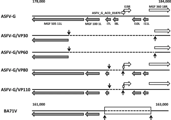
In addition to these point mutations, analysis of the ASFV-G/ VP30 sequence revealed an area of noticeably lower read coverage between nucleotide positions 178,630 and 183,476 of ASFV-G. Together with a population resembling the sequence of ASFV-G, there were several distinct variants containing large stretches of deleted nucleotides. The predominant variant population con-tained a deletion from nucleotide position 178,631 to 183,475, encompassing the region ofMGF505 11L(gene11Lof multigene family 505 [MGF505]) encoding the amino-terminal portion of the protein as well as the complete sequences of the genesMGF100 1L,I7L,I8L,I9R,I10L, andI11LandACD_01870and the region of theMGF360 18Rencoding the amino-terminal portion of the pro-tein (Fig. 3). Two other variant populations were also observed but at much lower frequencies, one containing a deletion from nucleotide position 179,341 to 183,476, encompassing the same genes as the major population described above. The other variant contained a deletion from nucleotide position 176,588 to 183,392 which expands into two additional genes, theMGF360 16Rand
DP36R. While MGFs have been associated with important host range functions (1), the ORFI10Lencodes a second copy of the gene for structural protein p22 (14,15,16), which is also expressed on the surfaces of ASFV-infected cells.
ASFV-G/VP60 harbors single mutations in six different genes relative to parental ASFV-G (Table 3). The mutations inEP424R,
CP530R, andE199Lwere identical to those observed in the ASFV-G/VP30 genome. Additionally, a G-to-A mutation in the ORF
MGF505 7Rresults in a D131N substitution in the predicted 527-amino-acid protein. Also, a C-to-T mutation was observed in the ORFMGF505 10R, leading to an H408Y substitution in the puta-tive 542-amino-acid protein encoded by this gene. Interestingly, ASFV MGF505 and -530 genes have been implicated in virus rep-lication in ticks and macrophages (17). Finally, a G-to-A mutation was observed in theI177Lgene, located in the right variable end of the ASFV genome, leading to a P42S substitution in the predicted protein of unknown function.
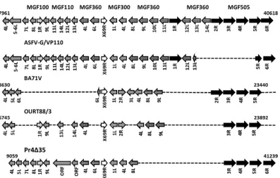
In addition to those mutations, the ASFV-G/VP60 genome harbors two major deletions (Fig. 3and4). The deletion located at the left variable end of the genome gives rise to two population variants. One variant harbors a deletion between nucleotide posi-tions 32,610 and 39,798 that results in the removal of the portion of the ORFMGF360 14Lcorresponding to the amino terminus, the complete deletion of the ORFs ASFV_G_ACD_00520 and
MGF505 2R,3R,4R, and5Rand a partial deletion of the portion of the ORFMGF505 6Rcorresponding to the amino terminus. The second variant instead is much less represented and contains a deletion spanning positions 30,783 to 38,026 of the ASFV-G ge-nome. This deletion affects almost the entire coding sequence of the ORFMGF360 13L, along with deletions of the ORFsMGF360 14LandMGF505 2R,3R, and4Rand the deletion of the portion of the ORFMGF505 5Rcorresponding to the amino-terminal half of the protein (Fig. 3). Interestingly, the latter population becomes the major population variant observed at passage 80 in ASFV-G/ VP80. The genes affected by the deletions that map to the left variable end of the ASFV-G genome have been implicated in virus replication in ticks and in swine macrophages (17), as well as in interferon response and virus attenuation in swine (18,19). The deletion observed at the right variable end of the ASFV-G/VP60 genome spans a similar area of low read coverage, as observed for ASFV-G/VP30 (suggesting that a discrete virus population that resembles the ASFV-G genome may still exist). None of the dele-FIG 3Schematic representation of the right variable region of the Vero-adapted ASFV-G. Vertical arrows indicate approximate boundaries of the observed deletions. Dotted lines indicate regions within the genomes of viruses that were deleted at different passages.
on November 7, 2019 by guest
http://jvi.asm.org/
[image:5.585.122.460.68.305.2]tion variants observed in ASFV-G/VP30 were observed at passage 60, with the exception of the prevalent ASFV-G/VP30 population, which was now present at a very low frequency (Fig. 3). At passage 60, a new variant emerged as the predominant population, har-boring a slightly different deletion (from positions 178,645 to 183,475). In addition, two additional population variants were also detected at passage 60, albeit at a low frequency (harboring deletions at positions 178,795 to 183,476 and 177,102 to 181,500). These two population variants were not observed at passage 30.
The ASFV-G/VP80 genome harbors single mutations in six different genes relative to the parental ASFV-G genome. The mutations inEP424R,CP530R, andE199Lwere similar to those observed in the ASFV-G/VP30 and ASFV-G/VP60 genomes.
Mutations observed in I177L and the gene MGF505 10R
(H408Y substitution) were similar to those observed in the ASFV-G/VP60 genome. At passage 80, there was an additional change in the geneMGF505 10Rthat results in an I472M sub-stitution in the ORF. Interestingly, it was observed that in the ASFV-G/VP80 genome, the G-to-A mutation previously ob-served in the ORFMGF505 7Rreverted back to adenine, as in the parental ASFV-G genome. In addition, a TCC insertion was observed in ORFI215Lof ASFV-G/VP80, which resulted in the insertion of a Gly residue at position 98 of the predicted poly-peptide. TheI215LORF harbors a region with homology to the gene for ubiquitin-conjugating enzyme E2, which is involved in the protein degradation pathway (2) (Table 3).
The left-end deletion (ASFV-G positions 30,783 to 38,026) that is a minor variant in VP60 is a major variant in VP80. This deletion spans nucleotides 30,783 to 38,026 of the ASFV-G genome. In addition, at passage 80 it is possible to observe sequence reads mapping between nucleotide positions 30,783 and 33,640 of the ASFV-G genome which most likely represent a discrete popula-tion variant that encompasses genes corresponding to the ORFs
MGF360 13Land14L(Fig. 4). The deletion at the right variable terminal end of the genome spans nucleotides 180,950 to 181,698. This 749-nucleotide deletion affects the portion of theI7Lgene corresponding to the amino terminus, along with the deletion of
theI8Lgene and the deletion of the portion of ORFASFV_G_ ACD_01870, corresponding to the amino-terminal end of the protein. None of the deletions previously observed in this area of the genome at previous passages were observed in ASFV-G/VP80. ASFV-G/VP110 harbors single mutations in nine different genes relative to the parental ASFV-G genome. The mutations in
EP424RandCP530Rwere similar to those observed in the ge-nomes of ASFV-G/VP30, ASFV-G/VP60, and ASFV-G/VP80. The mutations in the genesMGF505 10R(H408Y substitution) and
I177L(P42S substitution) were similar to those observed in the genomes of ASFV-G/VP60 and ASFV-G/VP80. Similarly, the modifications in ORFI215Land the ORFMGF505 10R(I472M substitution) observed in the ASFV-G/VP80 genome were also observed in the genome of ASFV-G/VP110 (Table 3). In addition, in ASFV-G/VP110 the C-to-T mutation in theE199L gene re-verted back to the parental cysteine residue. Three additional mu-tations were observed in ASFV-G/VP110: a C-to-T mutation in theM1249Lgene, leading to an N471Q substitution in the pre-dicted ORF; a T-to-C mutation in theCP2475Lgene, leading to an E200G substitution in the ORF; and a deletion in theE165Rgene resulting in a frameshift. TheM1249Lgene encodes a putative protein of 1,249 residues of unknown function. TheCP2475Lgene encodes a 220-kDa protein in ASFV that is further processed into smaller polypeptides (20). The predicted protein encoded by the
E165RORF (165 amino acids) harbors a domain between residues 25 and 158 present in trimeric dUTP-diphosphatase proteins (dUTPases) that catalyze the hydrolysis of the dUTP-Mg complex, preventing dUTP from being incorporated into DNA (2) (Table 3). The deletion observed in theE165RORF leads to the introduc-tion of a premature stop codon at residue 32 of the protein, result-ing in the truncation of the putative dUTPase domain. The dele-tion at the left variable end of the genome observed at passage 110 is similar to the deletion observed at passage 80. However, se-quence reads that map between nucleotides 30,783 and 33,640 of the ASFV-G genome at passage 110 were not observed in passage 80. Thus, at passage 110, ASFV-G/VP110 harbors a unique dele-tion at the left variable end of the genome. However, the deledele-tion at the right variable end of the genome observed in ASFV-G/ FIG 4Schematic representation of the left variable region of the Vero-adapted ASFV-G. Vertical arrows indicate approximate boundaries of the observed deletions (solid and dotted arrows indicate major and minor populations). Dotted lines indicate regions within the genomes of viruses that were deleted at different passages. Dotted arrows represent ORFs (white and gray for MGF505 and MGF360, respectively) in variant populations found at low frequency.
Adaptation of African Swine Fever Virus to Vero Cells
on November 7, 2019 by guest
http://jvi.asm.org/
[image:6.585.109.476.72.247.2]VP110 is not unique, as the population variants observed at pas-sage 80 were still observed at paspas-sage 110.
DISCUSSION
Live attenuated strains of ASFV obtained by cell culture adapta-tion or genetic recombinaadapta-tion and naturally attenuated field iso-lates have been shown to induce variable degrees of protection in pigs against challenge with homologous virulent isolates. How-ever, commercial vaccines against ASFV are not yet available.
Adaptation of virulent field isolates to replicate in established cell lines from different species has been attempted, and the pro-cess has led to decreased virulence of adapted viruses in swine (7, 21,22). The process of ASFV adaptation to be able to replicate in established cell lines is usually accompanied by attenuation of vir-ulence and significant modifications of the viral genome, generally affecting the variable genomic ends (21,22,23,24,25). Unfortu-nately, in most of these cases, detailed descriptions of the adapta-tion process and the associated genetic and phenotypic changes of ASFV are not readily available.
Here, we present a systematic analysis of the genotypic and phenotypic changes that occurred during the process of adapting a highly virulent ASFV isolate from Georgia to grow in Vero cells. Emphasis has been placed on comparing the changes in the ability of viruses from different cell passages to replicate in both Vero cells and primary swine macrophages, as well as the ability of these viruses to cause disease in swine.
Basically, successive passages of the parental virus ASFV-G in Vero cells were associated with an increased ability to replicate in this cell line. Adaptation to Vero cells corresponded with a de-creased ability to replicate in primary swine macrophages after successive passages. Virus virulence, as evaluated in this report, progressively decreased with successive passages, and complete attenuation of ASFV-G was observed by passage 110.
Genomic alterations of ASFV-G occurring throughout succes-sive passages in Vero cells can be grouped by the appearance of point mutations, leading to residue substitutions or frameshifts (Table 3) and by the appearance of relatively large deletions affect-ing a group of genes (Fig. 3,4, and5).
Here, it is difficult to assess the effects that point mutations observed during the adaptation of ASFV-G to Vero cells may have had on the observed phenotypic changes of the virus. The func-tions of most of ASFV genes are still unknown. Thus, prediction of functions to assign biological or biochemical roles to viral proteins encoded by putative ORFs of ASFV may provide important leads. For instance, all ASFV-G/VP genomes present point mutations in theCP530Rgene, which harbors a SMI1 like-domain (13). Pro-teins within the SMI1/KNR4 family have been grouped into the SUKH superfamily, and animal DNA viruses bearing SUKH do-mains, such as herpesviruses, poxviruses, iridoviruses, and adeno-viruses, have been shown to restrict antiviral responses by inter-acting with specific host proteins (13). So far, there is no evidence supporting similar functions forCP530Rduring ASFV infection. Point mutations observed in the genesMGF505 10RandI177L
may have contributed to the phenotypic changes observed with ASFV-G/VP60 and ASFV-G/VP80. TheI177Lgene seems to be involved in the process of protein degradation, although the role of this gene in ASFV virulence is unknown. The geneMGF505 10R
is a member of a gene family associated with replication of ASFV in swine macrophages, virus virulence, and blocking of interferon production (18, 19, 26). The point mutations observed in the ASFV-G/VP110 genome lead to a frameshift in the ORF corre-sponding to theE165Lgene. This gene encodes a putative protein which harbors a domain that is present in dUTPase proteins (that catalyze the hydrolysis of the dUTP-Mg complex into dUMP and pyrophosphate), preventing dUTP from being incorporated into DNA. Also at passage 110, a point mutation was observed in the FIG 5Schematic representation of the deletions observed at the left variable region of the ASFV genome in cell-adapted viruses (ASFV-G/VP110 and BA71V), an attenuated field isolate (OURT88/3), and a recombinant deletion mutant virus (Pr4⌬35). ASFV-G is depicted at the top.
on November 7, 2019 by guest
http://jvi.asm.org/
[image:7.585.96.493.61.314.2]CP2475Lgene, which encodes a 220-kDa polyprotein that is pro-cessed into four major structural proteins: p150, p37, p34, and p14 (20). Nonetheless, the roles that the mutations observed inE165L
orCP2475Lgenes may have in the phenotypic changes observed in ASFV-G/VP110in vitroandin vivoare unknown.
The process of adaptation of ASFV-G to grow in Vero cells also led to two major deletions that mapped to specific regions within the left and right variable ends of the viral genome. The deletion at the right end of the genome developed first and was observed at passage 30 in ASFV-G/VP30. The same deletion was observed at passage 60 in ASFV-G/VP60 (Fig. 3). This deletion, of approxi-mately 5 kbp, affects members of MGF100, MGF360, and MGF505. Although the role of members of MGF100 in the virus replication cycle is still unknown, the functions of MGF505 mem-bers have been associated with the ability of ASFV to replicate in macrophages and may account for the decreased virulence ob-served in swine when the virus is administered at low doses (18,19, 26). This deletion of approximately 5 kbp also includes theI10L
gene, which encodes a second copy of the ASFV structural protein p22 (14,15,16). This structural protein of unknown function is expressed in the membranes of cells infected with ASFV. At pas-sage 80, the deletion observed in the genome of ASFV-G/VP80 was significantly shorter than that observed in the genomes of ASFV-G/VP30 or ASFV-G/VP60, recovering the geneMGF505 11Land the genesMGF100 1LandI7L. The observation suggests that these three genes are not major contributors to the phenotypic differ-ences observed between ASFV-G/VP60 and ASFV-G/VP80/110, since the deletions observed in the genomes of ASFV-G/VP80 and ASFV-G/VP110 are comparable.
A deletion in the right variable end of the genome, encompass-ing theI7L,I8L,I10L, andI11Lgenes was also observed in Vero-adapted ASFV BA71V (Fig. 3), a virus that is considered attenu-ated in domestic swine (21). Similar deletions have not been described for any other cell-adapted ASFV isolate, although infor-mation regarding the full-genome sequences of those isolates is quite limited.
The deletion at the left variable end of the genome was first observed at passage 60 in ASFV-G/VP60. At this passage, two par-tially overlapping population variants of the virus were identified. The deletion observed in these variants encompassMGF360 14L,
ORF_G_ACD_00520, andMGF505 2R,3R,4R, and5R. The dele-tion observed in the genome of the most frequent variant also includes the geneMGF505 6R, whereas the deletion in the less frequent population variant includes the geneMGF360 13L. Most of the ASFV genes affected by this deletion have been linked to virus host range, innate immune response to ASFV, and virus virulence (18,19,26). Interestingly, the minor population variant that emerged by passage 60 becomes the major virus population observed at passage 80 (ASFV-G/VP80). However, at this passage, additional populations were also observed. Those variants harbor
MGF360 13Land14L. It may be possible that the increased atten-uation of ASFV observed at passage 110 is due to the absence of additional virus populations that harborMGF360 13Land14L.
Deletions encompassing regions within the left variable ends of the ASFV genome have been observed in the genomes of several attenuated isolates, including a naturally occurring attenuated isolate conferring protection against homologous viruses (OURT88/3), cell culture-adapted viruses (MS14 and MS44, de-rived from isolate E70; CV1, dede-rived from isolate E75; and the attenuated Vero cell-adapted virus BA71V, derived from isolate
Badajoz 60) (5,7,21,22), recombinant deletion mutant viruses (Pr4⌬35) derived from isolate Pretorisuskop/96/4 (26), and a de-rivative of a Malawi Lil-20/1 isolate (Mal⌬SVD) lacking the viru-lence-associated NL gene (19). In general, these deletions encom-pass members of MGF360 and MGF505, as observed here during the adaptation of ASFV-G to grow in Vero cells. At passage 110, the deletions observed in ASFV-G were significantly shorter and encompassed fewer genes than the deletions observed in OURT88/3, BA71V, and Pr4⌬35, suggesting that a limited num-ber of genes may be involved in virulence/attenuation (OURT88/3 and Pr4⌬35) or cell adaptation (BA71V). The inability of cell-adapted BA71V to grow in swine macrophages can be rescued by recombination using an ASFV genomic fragment containing six members of MGF360 and one member of MGF505 from the left variable region of the ASFV E70 isolate (26), whereas deletion of these MGF members from the genome of the virulent ASFV iso-late Pretorisuskop/96/4 severely affected the ability of this virus to replicate in swine macrophages (Fig. 5) (26). Similarly, deletions of several MGF360 and MGF505 genes (includingMGF360 13L
and14L) from the left variable region of the genome of the viru-lent recombinant virus Malawi Lil-20/1, which already lacks the
NLgene, results in complete attenuation of the virus (19). In summary, it is possible that the attenuation observed with ASFV-G/VP110 is due to the progressive deletion of genes of MGF360 and MGF505 in the left variable end of the ASFV ge-nome. However, the contribution to the observed phenotypes of the genes that are affected by the deletion at the right variable end of the genome is more difficult to assess. As mentioned, deletions at the right variable end of the genome have been observed only in cell-adapted BA71V. That deletion encompasses the same area as the deletion observed in the genomes of ASFV-G/VP30, -P60, -P80, and -P110. Although the extent and the number of genes affected by the deletion particularly in the genome of attenuated ASFV-G/VP110 and BA71V differ, data suggest that this area of the genome is also important for the process of adaptation to the new host (i.e., Vero cells). Unfortunately, the complete genome sequences of the cell-adapted viruses M14, M44, and CV1 are not available for further comparisons.
The use of a combination of deep sequencing andin vitroand
in vivostudies in swine to analyze viruses from different passages during adaptation to Vero cells has enabled, for the first time, a thorough and complete description of the genotypic and pheno-typic changes that take place during the adaptation of ASFV to grow in cultured cell lines. This report specifically estimated changes in ASFV, providing valuable information that can be ap-plied to the understanding of ASFV gene function, virus attenua-tion, and protection against infection.
ACKNOWLEDGMENTS
We thank the Plum Island Animal Disease Center Animal Care Unit staff for excellent technical assistance. We particularly thank Melanie V. Prarat for editing the manuscript.
This project was funded through an interagency agreement with the Science and Technology Directorate of the U.S. Department of Homeland Security under award numbers HSHQDC-11-X-00077 and HSHQPM-12-X-00005. We thank ARS/USDA-University of Connecticut SCA 58-1940-1-190 for partially supporting this work.
REFERENCES
1.Tulman ER, Delhon GA, Ku BK, Rock DL.2009. African swine fever virus. Curr Top Microbiol Immunol328:43– 87.
Adaptation of African Swine Fever Virus to Vero Cells
on November 7, 2019 by guest
http://jvi.asm.org/
2.Chapman DA, Darby AC, Da Silva M, Upton C, Radford AD, Dixon LK.2011. Genomic analysis of highly virulent Georgia 2007/1 isolate of African swine fever virus. Emerg Infect Dis17:599 – 605.http://dx.doi.org /10.3201/eid1704.101283.
3.Boinas FS, Hutchings GH, Dixon LK, Wilkinson PJ.2004. Character-ization of pathogenic and non-pathogenic African swine fever virus iso-lates from Ornithodoros erraticus inhabiting pig premises in Portugal. J Gen Virol85:2177–2187.http://dx.doi.org/10.1099/vir.0.80058-0. 4.Lewis T, Zsak L, Burrage TG, Lu Z, Kutish GF, Neilan JG, Rock DL.2000.
An African swine fever virus ERV1-ALR homologue, 9GL, affects virion mat-uration and viral growth in macrophages and viral virulence in swine. J Virol 74:1275–1285.http://dx.doi.org/10.1128/JVI.74.3.1275-1285.2000. 5.Martins CL, Lawman MJ, Scholl T, Mebus CA, Lunney JK. 1993.
African swine fever virus specific porcine cytotoxic T cell activity. Arch Virol129:211–225.http://dx.doi.org/10.1007/BF01316896.
6.Moore DM, Zsak L, Neilan JG, Lu Z, Rock DL.1998. The African swine fever virus thymidine kinase gene is required for efficient replication in swine macrophages and for virulence in swine. J Virol72:10310 –10315. 7.Ruiz Gonzalvo F, Caballero C, Martinez J, Carnero ME.1986.
Neutral-ization of African swine fever virus by sera from African swine fever-resistant pigs. Am J Vet Res47:1858 –1862.
8.Zsak L, Caler E, Lu Z, Kutish GF, Neilan JG, Rock DL.1998. A nonessential African swine fever virus gene UK is a significant virulence determinant in domestic swine. J Virol72:1028 –1035.
9.Zsak L, Lu Z, Kutish GF, Neilan JG, Rock DL.1996. An African swine fever virus virulence-associated gene NL-S with similarity to the herpes simplex virus ICP34.5 gene. J Virol70:8865– 8871.
10. Enjuanes L, Carrascosa AL, Moreno MA, Vinuela E.1976. Titration of African swine fever (ASF) virus. J Gen Virol32:471– 477.http://dx.doi.org /10.1099/0022-1317-32-3-471.
11. Reed LJ, Muench HA.1938. A simple method of estimating fifty per cent endpoints. Am J Hyg27:493– 497.
12. Poffenberger KL, Roizman B.1985. A noninverting genome of a viable herpes simplex virus 1: presence of head-to-tail linkages in packaged ge-nomes and requirements for circularization after infection. J Virol53: 587–595.
13. Zhang D, Iyer LM, Aravind L. 2011. A novel immunity system for bacterial nucleic acid degrading toxins and its recruitment in various eu-karyotic and DNA viral systems. Nucleic Acids Res39:4532– 4552.http: //dx.doi.org/10.1093/nar/gkr036.
14. Vydelingum S, Baylis SA, Bristow C, Smith GL, Dixon LK. 1993. Duplicated genes within the variable right end of the genome of a patho-genic isolate of African swine fever virus. J Gen Virol74:2125–2130.http: //dx.doi.org/10.1099/0022-1317-74-10-2125.
15. Camacho A, Vinuela E.1991. Protein p22 of African swine fever virus: an
early structural protein that is incorporated into the membrane of infected cells. Virology 181:251–257. http://dx.doi.org/10.1016/0042-6822(91) 90490-3.
16. Dixon LK, Chapman DA, Netherton CL, Upton C.2013. African swine fever virus replication and genomics. Virus Res173:3–14.http://dx.doi .org/10.1016/j.virusres.2012.10.020.
17. Burrage TG, Lu Z, Neilan JG, Rock DL, Zsak L.2004. African swine fever virus multigene family 360 genes affect virus replication and generaliza-tion of infecgeneraliza-tion in Ornithodoros porcinus ticks. J Virol78:2445–2453.
http://dx.doi.org/10.1128/JVI.78.5.2445-2453.2004.
18. Afonso CL, Piccone ME, Zaffuto KM, Neilan J, Kutish GF, Lu Z, Balinsky CA, Gibb TR, Bean TJ, Zsak L, Rock DL.2004. African swine fever virus multigene family 360 and 530 genes affect host interferon response. J Virol 78:1858 –1864.http://dx.doi.org/10.1128/JVI.78.4.1858-1864.2004. 19. Neilan JG, Zsak L, Lu Z, Kutish GF, Afonso CL, Rock DL.2002. Novel
swine virulence determinant in the left variable region of the African swine fever virus genome. J Virol76:3095–3104.http://dx.doi.org/10.1128/JVI .76.7.3095-3104.2002.
20. Simon-Mateo C, Andres G, Vinuela E.1993. Polyprotein processing in African swine fever virus: a novel gene expression strategy for a DNA virus. EMBO J12:2977–2987.
21. Yanez RJ, Rodriguez JM, Nogal ML, Yuste L, Enriquez C, Rodriguez JF, Vinuela E.1995. Analysis of the complete nucleotide sequence of African swine fever virus. Virology208:249 –278.http://dx.doi.org/10.1006/viro .1995.1149.
22. Tabares E, Olivares I, Santurde G, Garcia MJ, Martin E, Carnero ME. 1987. African swine fever virus DNA: deletions and additions during ad-aptation to growth in monkey kidney cells. Arch Virol97:333–346.http: //dx.doi.org/10.1007/BF01314431.
23. de la Vega I, Vinuela E, Blasco R.1990. Genetic variation and multigene families in African swine fever virus. Virology179:234 –246.http://dx.doi .org/10.1016/0042-6822(90)90293-Z.
24. Pires S, Ribeiro G, Costa JV.1997. Sequence and organization of the left multigene family 110 region of the Vero-adapted L60V strain of African swine fever virus. Virus Genes 15:271–274. http://dx.doi.org/10.1023 /A:1007992806818.
25. Chapman DA, Tcherepanov V, Upton C, Dixon LK.2008. Comparison of the genome sequences of non-pathogenic and pathogenic African swine fever virus isolates. J Gen Virol89:397– 408.http://dx.doi.org/10.1099/vir .0.83343-0.
26. Zsak L, Lu Z, Burrage TG, Neilan JG, Kutish GF, Moore DM, Rock DL. 2001. African swine fever virus multigene family 360 and 530 genes are novel macrophage host range determinants. J Virol75:3066 –3076.http: //dx.doi.org/10.1128/JVI.75.7.3066-3076.2001.


