Fatty Acid Synthase Promotes the Palmitoylation of
Chikungunya Virus nsP1
Na Zhang,
aHongjian Zhao,
aLeiliang Zhang
a,baMOH Key Laboratory of Systems Biology of Pathogens, Institute of Pathogen Biology, Chinese Academy of Medical Sciences & Peking Union Medical College,
Beijing, China
bKey Laboratory of Medicine and Biotechnology of Qingdao, Department of Microbiology, Medical College of Qingdao University, Qingdao, China
ABSTRACT
Chikungunya virus (CHIKV) is transmitted to people by mosquitoes, and
CHIKV infection causes fever and joint pain. Fatty acid synthase (FASN) has been
identified as a proviral factor for CHIKV. How FASN participates in CHIKV replication
remains to be elucidated. In this study, we demonstrated that palmitic acid (PA) can
restore the suppression of CHIKV replication by FASN inhibitors. The palmitoylation
and plasma membrane localization of CHIKV nsP1 were reduced by FASN inhibitors.
Triple mutation of Cys417, Cys418, and Cys419 in nsP1 blocked its palmitoylation
and severely disrupted CHIKV replication. Furthermore, two zinc finger DHHC
domain-containing palmitoyltransferases (ZDHHCs), ZDHHC2 and ZDHHC19, promoted nsP1
palmitoylation and CHIKV replication. Our results not only identified the key
en-zymes for the palmitoylation of nsP1 but also provided mechanistic insights into the
roles of FASN in CHIKV replication.
IMPORTANCE
S-palmitoylation is an important form of lipid posttranslational
modi-fication, which affects the function of proteins by regulating their transport, stability,
and localization. Previous studies have shown that FASN is critical for CHIKV
replica-tion; however, the mechanism for this function of FASN remains unknown. The key
zinc finger DHHC domain-containing palmitoyltransferases involved in the
palmitoyl-ation of nsP1 are not clear. We demonstrated that FASN promoted CHIKV replicpalmitoyl-ation
through nsP1 palmitoylation. ZDHHC2 and ZDHHC19 were identified as the major
enzymes for nsP1 palmitoylation. Since nsP1 proteins are conserved in alphaviruses,
our results highlight the mechanisms by which alphavirus nsP1 is palmitoylated.
KEYWORDS
CHIKV, FASN, ZDHHC, nsP1, palmitoylation
C
hikungunya virus (CHIKV) is transmitted to people by
Aedes aegypti
and
Aedes
albopictus
mosquitoes (1). The most common symptoms of CHIKV infection include
severe joint pain, fever, headache, muscle pain, and rash (1, 2). CHIKV is responsible for
a reemerging epidemic in countries in the Indian Ocean region and has been identified
in Europe and the United States. Currently, no vaccine exists to prevent CHIKV infection.
CHIKV is a member of the genus
Alphavirus
in the family
Togaviridae
(3). The genome
of CHIKV consists of two open reading frames (ORFs). One ORF encodes a nonstructural
polyprotein that is posttranslationally processed to generate four nonstructural
pro-teins (nsP1, nsP2, nsP3, and nsP4) that are involved in genome replication. The other
ORF encodes a structural polyprotein that is also processed and forms the viral particles,
which are composed of the capsid, E3, E2, 6K, and E1 proteins (3).
CHIKV genomic viral RNA and the 26S subgenomic RNA require a 5
=
cap structure
to direct efficient translation of the viral polyprotein and prevent degradation of the
viral RNA genome by host exonucleases. Capping of the CHIKV RNA is orchestrated by
nsP1 protein, which binds GTP and confers the N-7 methyltransferase and
guanylyl-transferase activities that are necessary for cap formation in a noncanonical manner (4).
CitationZhang N, Zhao H, Zhang L. 2019. Fatty acid synthase promotes the palmitoylation of chikungunya virus nsP1. J Virol 93:e01747-18.
https://doi.org/10.1128/JVI.01747-18.
EditorJulie K. Pfeiffer, University of Texas Southwestern Medical Center
Copyright© 2019 American Society for Microbiology.All Rights Reserved. Address correspondence to Leiliang Zhang, armzhang@hotmail.com.
Received3 October 2018
Accepted26 October 2018
Accepted manuscript posted online7 November 2018
Published
VIRUS-CELL INTERACTIONS
crossm
17 January 2019
on November 6, 2019 by guest
http://jvi.asm.org/
Addition of palmitic acid (PA), a 16-carbon saturated fatty acid, to the thiol group of
a cycteine residue of substrate is defined as S-palmitoylation, which was generalized as
“palmitoylation” (5). Palmitoylation is catalyzed by the zinc finger Asp-His-His-Cys
(DHHC) domain-containing (ZDHHC) palmitoyl acyltransferase (PAT) family (6). ZDHHC
proteins were paired with their specific substrates and were closely involved in the
substrates’ function. As a posttranslational lipid modification, palmitoylation increases
protein hydrophobicity and facilitates protein trafficking to cellular membranes. The
RNA capping enzyme nsP1 from Semliki Forest virus (SFV) and Sindbis virus (SINV)
anchors to the cellular membrane through an amphipathic peptide and a palmitoylated
cysteine in nsP1 (7, 8). Mutations that prevent nsP1 palmitoylation reduce the
replica-tion of SFV and SINV (9). Whether CHIKV nsP1 is also palmitoylated is unknown.
Moreover, which ZDHHC enzymes contribute nsP1 palmitoylation is not clear.
Fatty acid synthase (FASN) catalyzes the
de novo
synthesis of palmitic acid, a fatty
acid that is utilized for the posttranslational palmitoylation of host and viral proteins.
FASN has been demonstrated to be critical for the replication of many viruses (2, 8, 10,
11). How FASN participates in CHIKV replication remains to be elucidated.
In this study, we found that FASN inhibitor reduced the palmitoylation of nsP1.
ZDHHC2 and ZDHHC19 associated with nsP1 and mediated its palmitoylation. Silencing
of ZDHHC2 and ZDHHC19 reduced CHIKV replication. Taken together, our findings
identified the key enzymes for the palmitoylation of nsP1 and revealed the mechanism
by which FASN contributes to CHIKV replication.
RESULTS
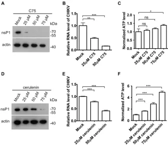
FASN inhibitors reduced CHIKV replication.
To determine whether the inhibition
of FASN enzyme activity disrupted CHIKV replication, HeLa cells were treated with the
compounds C75 and cerulenin, two known FASN inhibitors (12), and then infected with
CHIKV strain 181/25. Consistent with the results from a previous study (13), C75
inhibited CHIKV replication, as assessed by the protein level of CHIKV nsP1 and the
CHIKV RNA level (Fig. 1A and B). C75 did not reduce the cell viability as measured by
the intracellular ATP level (Fig. 1C). Similarly, cerulenin reduced the CHIKV viral nsP1
protein expression and viral RNA level (Fig. 1D and E) without reducing the cell viability
(Fig. 1F).
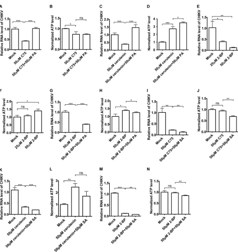
Palmitic acid restored the suppression of CHIKV replication caused by FASN
inhibitors or a palmitoylation inhibitor.
Since FASN inhibitors suppressed CHIKV
replication, we hypothesized that FASN might promote CHIKV replication through its
product palmitic acid. To test this hypothesis, we treated HeLa cells with palmitic acid
(PA) or left them untreated in the presence of a FASN inhibitor and infected the cells
with CHIKV strain 181/25. As shown in Fig. 2A and B, palmitic acid rescued the
suppression of CHIKV replication by the FASN inhibitors C75 without reducing the cell
viability. Similarly, palmitic acid rescued the suppression of CHIKV replication by
cerulenin without reducing the cell viability (Fig. 2C and D). The palmitate analog
2-bromopalmitate (2-BP) is commonly used to inhibit palmitoylation (14). We treated
the HeLa cells with 2-BP and then infected the cells with CHIKV strain 181/25. The
results indicated that 2-BP inhibited CHIKV replication in a dose-dependent manner
without reducing the cell viability (Fig. 2E and F). PA partially rescued the suppression
of CHIKV replication by 2-BP (Fig. 2G and H). However, stearic acid (SA), a downstream
lipid from palmitic acid, failed to rescue the suppression of CHIKV replication by C75
(Fig. 2I and J), cerulenin (Fig. 2K and L), or 2-BP (Fig. 2M and N), excluding the
downstream lipid role of palmitic acid complementation. Taken together, these data
suggested that palmitic acid could restore the suppression of CHIKV replication by
FASN inhibitors and a palmitoylation inhibitor.
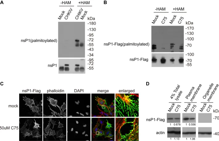
Palmitoylation of nsP1 was reduced by a FASN inhibitor.
The palmitoylation of
nsP1 from other alphaviruses, such as SINV, is critical for viral replication (9); thus, we
hypothesized that the palmitoylation of nsP1 from CHIKV is dependent on the synthesis
of palmitic acid by FASN. To investigate whether the palmitoylation of endogenous
nsP1 existed, Huh7.5.1 cells were infected with CHIKV strain 181/25 (multiplicity of
Zhang et al. Journal of Virology
on November 6, 2019 by guest
http://jvi.asm.org/
infection [MOI]
⫽
1) for 8 h and the palmitoylation of nsP1 was assayed by acyl-biotin
exchange (ABE) assay. The ABE assay contains three biochemical steps: blocking the
unmodified cystein thiol groups by
N
-ethylmaleimide (NEM), cleaving and unmasking
the palmitoylated cysteine’s thiol group by hydroxylamine (HAM), and labeling the
palmitoylated cysteine by a thiol-reactive biotinylation reagent, 1-biotinamido-4-[4
=
-(maleimidomethyl)cyclohexanecarboxamido]butane (biotin-BMCC). CHIKV nsP1 was
confirmed to be palmitoylated as measured by ABE assay (Fig. 3A). To investigate
whether the inhibition of FASN reduces the palmitoylation of nsP1, 293T cells were
transfected with FLAG-nsP1 for 24 h and treated with the FASN inhibitor C75 or
subjected to mock treatment for 12 h. As shown in Fig. 3B, the palmitoylation of CHIKV
nsP1 was reduced by the FASN inhibitor.
Palmitoylated alphavirus nsP1 is located in the plasma membrane and filopodial
extensions, while nonpalmitoylated nsP1 is not observed in filopodial extensions (7).
Indeed, immunofluorescence experiments showed that the filopodial localization of
nsP1 was reduced by the FASN inhibitor (Fig. 3C). The plasma membrane localization
of nsP1 was also reduced by the FASN inhibitor (Fig. 3D). We quantified the blots and
found that FASN inhibitor reduced the nsP1 protein level in the total lysate. The nsP1
without proper localization is degraded eventually, leading the reduction of the plasma
membrane level of nsP1. Taken together, these data suggested that FASN is important
for the palmitoylation and proper localization of nsP1.
FIG 1FASN activity was required for CHIKV replication. (A) HeLa cells were treated with difference dosages of C75 for 30 min and then infected with CHIKV strain 181/25 (multiplicity of infection [MOI]⫽1) for 8 h. The lysates were immunoblotted for nsP1 and actin. (B) HeLa cells were treated with difference dosages of C75 for 30 min and then infected with CHIKV strain 181/25 (MOI⫽1) for 5 h. RNA was assessed by qPCR. (C) HeLa cells were treated with difference dosages of C75 for 30 min and then infected with CHIKV strain 181/25 (MOI⫽1) for 5 h. ATP levels were measured by CellTiter-Glo luminescent cell viability assay. (D) HeLa cells were treated with difference dosages of cerulenin for 30 min and then infected with CHIKV strain 181/25 (MOI⫽1) for 5 h. The lysates were immunoblotted for nsP1 and actin. (E) HeLa cells were treated with difference dosages of cerulenin for 30 min and then infected with CHIKV strain 181/25 (MOI⫽1) for 5 h. RNA was assessed by qPCR. (F) HeLa cells were treated with difference dosages of cerulenin for 30 min and then infected with CHIKV strain 181/25 (MOI⫽1) for 5 h. ATP levels were measured by CellTiter-Glo luminescent cell viability assay.
FASN Promotes nsP1 Palmitoylation Journal of Virology
on November 6, 2019 by guest
http://jvi.asm.org/
[image:3.585.41.376.71.360.2]FIG 2Palmitic acid could rescue the inhibitory effects of 2-BP and FASN inhibitors on CHIKV replication. (A and B) HeLa cells were preinfected with CHIKV vaccine strain 181/25 (MOI⫽1) for 1 h and then treated with C75 or C75-palmitic acid for 5 h in the presence of CHIKV. Total RNA was isolated and reverse transcribed, and qPCR was then performed to measure CHIKV RNA levels (A). ATP levels were measured by CellTiter-Glo luminescent cell viability assay (B). (C and D) HeLa cells were preinfected with CHIKV vaccine strain 181/25 (MOI⫽1) for 1 h and then treated with cerulenin or cerulenin-palmitic acid for 5 h in the presence of CHIKV. Total RNA was isolated and reverse transcribed, and qPCR was then performed to measure CHIKV RNA levels (C). ATP levels were measured by CellTiter-Glo luminescent cell viability assay (D). (E and F) HeLa cells were preinfected with CHIKV vaccine strain 181/25 (MOI⫽1) for 1 h and then treated with 2-BP for 3 h in the presence of CHIKV. Total RNA was isolated and reverse transcribed, and qPCR was then performed to measure CHIKV RNA levels (E). ATP levels were measured by CellTiter-Glo luminescent cell viability assay (F). (G and H) HeLa cells were preinfected with CHIKV vaccine strain 181/25 (MOI⫽1) for 1 h and then treated with 2-BP or 2-BP–palmitic acid for 3 h in the presence of CHIKV. Total RNA was isolated and reverse transcribed, and qPCR was then performed to measure CHIKV RNA levels (G). ATP levels were measured by CellTiter-Glo luminescent cell viability assay (H). (I and J) HeLa cells were preinfected with CHIKV vaccine strain 181/25 (MOI⫽1) for 1 h and then treated with C75 or C75-SA for 5 h in the presence of CHIKV. Total RNA was isolated and reverse transcribed, and qPCR was then performed to measure CHIKV RNA levels (I). ATP levels were measured by CellTiter-Glo luminescent cell viability assay (J). (K and L) HeLa cells were preinfected with CHIKV vaccine strain 181/25 (MOI⫽1) for 1 h and then treated with cerulenin or cerulenin-SA for 5 h in the presence of CHIKV. Total RNA was isolated and reverse transcribed, and qPCR was then performed to measure CHIKV RNA levels (K). ATP levels were measured by CellTiter-Glo luminescent cell viability assay (L). (M and N) HeLa cells were preinfected with CHIKV vaccine strain 181/25 (MOI⫽1) for 1 h and then treated with 2-BP or 2-BP–palmitic acid for 3 h in the presence of CHIKV. Total RNA was isolated and reverse transcribed, and qPCR was then performed to measure CHIKV RNA levels (M). ATP levels were measured by CellTiter-Glo luminescent cell viability assay (N).
Zhang et al. Journal of Virology
on November 6, 2019 by guest
http://jvi.asm.org/
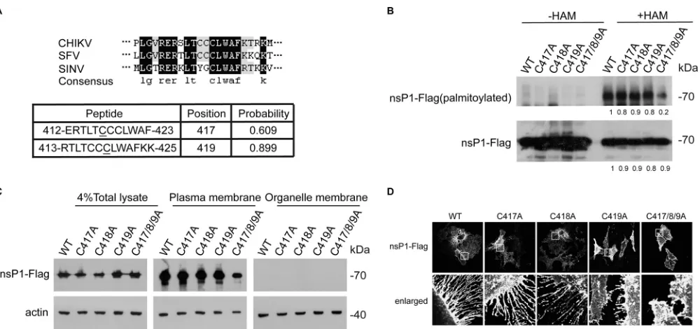
[image:4.585.43.518.64.567.2]The cysteines at sites 417 to 419 of CHIKV nsP1 are the key residues for nsP1
palmitoylation.
Based on predictions (15), the cysteines at sites 417 and 419 of CHIKV
nsP1 were found to be potential key residues for nsP1 palmitoylation (Fig. 4A). We
performed homology analysis to compare the sequence of CHIKV nsP1 with those of
SFV and SINV and found that C419 is the most conserved amino acid among C417 to
C419. We generated C417A, C418A, C419A, and C417/8/9A mutants of nsP1 and found
that the cysteines at sites 417 to 419 are the key residues for nsP1 palmitoylation (Fig.
4B). These cysteines are also critical for the plasma membrane targeting of nsP1 (Fig.
4C). The C419A and C417/8/9A mutants of nsP1 showed reduced localization to
filopodial extensions (Fig. 4D). Thus, we concluded that the cysteines at sites 417 to 419
of nsP1 are critical for the palmitoylation and proper localization of nsP1.
The cysteines at sites 417 to 419 of CHIKV nsP1 are critical for CHIKV
replica-tion.
To investigate whether the palmitoylation of nsP1 could affect CHIKV replication,
we generated the nsP1 mutant CHIKV-nsP1C/A, which is deficient in the palmitoylation
of nsP1, by mutating C417 to C419 to A417 to A419 in the context of CHIKV 181/25 (Fig.
5A). The infectivity of CHIKV-C3A-expressing virus was 4 orders of magnitude lower
than that of wild-type (WT) CHIKV, as measured by quantitative PCR (qPCR) (Fig. 5B),
and this finding is consistent with the result obtained with the nsP1
palmitoylation-deficient mutant of SFV (9).
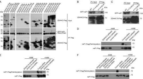
ZDHHC2 and ZDHHC19 promote the palmitoylation of nsP1.
Protein
palmitoyl-ation is mediated mainly by ZDHHCs (16). Next, we screened for ZDHHCs associated
with nsP1. 293T cells were transfected with 23 FLAG-tagged ZDHHCs and green
fluorescent protein (GFP)-tagged nsP1, and the lysates were incubated with an
anti-FLAG antibody to perform immunoprecipitation (IP) experiments. ZDHHC2 and
ZDHHC19 were identified as binding partners for nsP1 (Fig. 6A). Interestingly, nsP1C/A
FIG 3Palmitoylation of nsP1 was reduced by a FASN inhibitor. (A) Huh7.5.1 cells were infected with CHIKV strain 181/25 (MOI⫽1) for 8 h. The palmitoylation level of nsP1 was measured by ABE assay, and then cells were blotted with streptavidin-HRP and nsP1 antibody.⫺HAM, without HAM;⫹HAM, with HAM. (B) 293T cells were transfected with nsP1-FLAG plasmid overnight and treated with 50M C75 for 20 h. The palmitoylation level of nsP1 was measured by ABE assay, and then cells were blotted with streptavidin-HRP and FLAG antibody. The asterisk indicates the palmitoylated nsP1-FLAG. (C) HeLa cells were transfected with nsP1-FLAG plasmid overnight and then treated with 50M C75 for 8 h. The cells were fixed and immunolabeled with FLAG antibody (green). Phalloidin marks the actin (red). DAPI marks the nucleus. Scale bar: 10m. (D) HeLa cells were transfected with nsP1-FLAG plasmid overnight and then treated with 50M C75 for 8 h. The plasma membrane and other fractions of the cells were separated by a plasma membrane protein isolation and cell fractionation kit and then blotted with FLAG and actin antibodies. The blots were quantified by ImageJ.FASN Promotes nsP1 Palmitoylation Journal of Virology
on November 6, 2019 by guest
http://jvi.asm.org/
[image:5.585.43.398.73.302.2]mutants no longer interacted with ZDHHC2 or ZDHHC19 (Fig. 6B and C), further
confirming the specific association of ZDHHC2 and ZDHHC19 with nsP1.
We hypothesized that ZDHHC2 and ZDHHC19 participated in the palmitoylation of
nsP1. To test this hypothesis, ZDHHC2 or ZDHHC19 was knocked down in 293T cells by
small interfering RNAs (siRNAs) for 48 h, and the cells were then transfected with
nsP1-FLAG for 24 h. ABE assays showed that the palmitoylation of nsP1 was reduced
after silencing ZDHHC2 (Fig. 6D), ZDHHC19 (Fig. 6E) or ZDHHC2/ZDHHC19 (Fig. 6F).
These results suggested key roles for ZDHHC2 and ZDHHC19 in the palmitoylation
of nsP1.
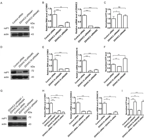
ZDHHC2 and ZDHHC19 promote CHIKV replication.
To assess whether ZDHHC2
and ZDHHC19 are crucial for CHIKV replication, we knocked down ZDHHC2 and
ZDHHC19 with siRNAs. Silencing ZDHHC2 inhibited CHIKV replication, as measured by
FIG 4The crucial sites of nsP1 were the three cysteines located at positions 417 to 419. (A) nsP1 from CHIKV nsP1 was compared with those from SFV and SINV. Protein palmitoylation software was used to predict the nsP1 palmitoylation sites. (B) 293T cells were transfected with the WT nsP1-FLAG plasmid and mutant plasmids for 24 h. The palmitoylation level of nsP1 was measured by an ABE assay, and the cells were then immunoblotted with streptavidin-HRP and an anti-FLAG antibody. The blots were quantified by ImageJ. (C) HeLa cells were transfected with a WT nsP1-FLAG plasmid and mutant plasmids for 24 h. Next the plasma membrane and other cell fractions were separated by a plasma membrane protein isolation and cell fractionation kit and then immunoblotted with antibodies against FLAG and actin. (D) HeLa cells were transfected with a WT nsP1-FLAG plasmid and mutant plasmids for 24 h and then immunolabeled with an anti-FLAG antibody. Scale bar: 10m.
FIG 5Mutation of nsP1 palmitoylation sites inhibited CHIKV replication. (A) Diagram of CHIKV-WT and CHIKV-nsP1 C/A. (B) Plasmids containing the CHIKV WT genome and the CHIKV-nsP1 C/A genome were linearized by NotI digestion and transcribed to generate viral RNA. In addition, Huh7.5.1 cells were transfected with viral RNA by electroporation, and the cells were cultured in 24-well plates. Sixteen hours later, the intracellular RNA was isolated and reverse transcribed. qPCR was performed to measure CHIKV RNA levels.
Zhang et al. Journal of Virology
on November 6, 2019 by guest
http://jvi.asm.org/
[image:6.585.45.545.68.303.2] [image:6.585.49.366.555.678.2]the levels of viral RNA and the viral protein nsP1, without reducing the cell viability (Fig.
7A to C). Similarly, knockdown of ZDHHC19 reduced the levels of CHIKV RNA replication
and the viral protein nsP1 (Fig. 7D to F). Combination of ZDHHC2 siRNA and ZDHHC19
siRNA treatment also reduced the CHIKV replication (Fig. 7G to I). Taken together, these
data indicate that both ZDHHC2 and ZDHHC19 are involved in CHIKV replication.
DISCUSSION
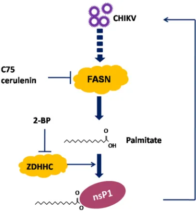
In this study, we demonstrated that FASN contributed to CHIKV replication by
producing palmitic acid for the palmitoylation of nsP1 (Fig. 8). Inhibitors of FASN and
ZDHHCs reduced the palmitoylation of nsP1 and suppressed CHIKV replication (Fig. 8).
The key residues in nsP1 for palmitoylation are C417 to C419. ZDHHC2 and ZDHHC19
interacted with nsP1, promoted nsP1 palmitoylation, and enhanced viral replication.
FASN catalyzes the synthesis of palmitic acid, which can be utilized for the
post-translational palmitoylation of proteins, including viral proteins. Alphavirus nsP1
pro-teins from SFV and SINV were reported to be palmitoylated (9, 17, 18). Our current
report raises the possibility that FASN is involved in viral replication through the
palmitoylation of CHIKV nsP1. Indeed, inhibition of FASN reduced CHIKV replication,
which was consistent with the findings of a previous study (13). Although we
estab-lished that FASN played a crucial role in CHIKV nsP1 palmitoyaltion, our data did not
rule out the possibility that FASN-mediated palmitoylation of other CHIKV proteins and
host proteins involved in replication. A previous study found that transframe (TF)
protein from SINV was palmitoylated (19). Thus, it is conceivable that inhibition of FASN
would also reduce the palmitoylation of CHIKV TF protein.
FIG 6ZDHHC2 and ZDHHC19 interacted with nsP1 and mediate nsP1 palmitoylation. (A) Lysates from 293T cells transfected with 23 ZDHHCs fused to FLAG and GFP-nsP1 of CHIKV were immunoprecipitated with an anti-FLAG antibody and then immunoblotted with antibodies against GFP and FLAG to screen for ZDHHCs that could interact with nsP1. Asterisks indicate the ZDHHCs. (B) Lysates from 293T cells transfected with ZDHHC2-FLAG and GFP-nsP1 or GFP-nsP1417-419C/A were immunoprecipitated with an anti-FLAG antibody and then immunoblotted with antibodies against GFP and FLAG. (C) Lysates from 293T cells transfected with ZDHHC2-FLAG and GFP-nsP1 or GFP-nsP1417-419C/A were immunoprecipitated with an anti-FLAG antibody and then immuno-blotted with antibodies against GFP and FLAG. (D) 293T cells were treated with siRNAs targeting ZDHHC2 or a control siRNA for 48 h and then transfected with an nsP1-FLAG plasmid for 24 h. The lysates were collected, and an ABE assay was performed to detect the nsP1 palmitoylation level. (E) 293T cells were treated with siRNAs targeting ZDHHC19 or a control siRNA for 48 h and then transfected with an nsP1-FLAG plasmid for 24 h. The lysates were collected, and an ABE assay was performed to detect the nsP1 palmitoylation level. (F) 293T cells were treated with siRNAs targeting ZDHHC2, ZDHHC19, or ZDHHC2/ZDHHC19 or a control siRNA for 48 h and then transfected with an nsP1-FLAG plasmid for 24 h. The lysates were collected, and an ABE assay was performed to detect the nsP1 palmitoylation level.
FASN Promotes nsP1 Palmitoylation Journal of Virology
on November 6, 2019 by guest
http://jvi.asm.org/
[image:7.585.44.543.71.347.2]Protein palmitoylation plays a key role in regulating the localization and function of
many proteins (20). Palmitoylated nsP1 from CHIKV localized in the plasma membrane
and filopodial extensions. CHIKV nsP1 was not observed in the plasma membrane or
filopodial extensions of cells treated with a FASN inhibitor (Fig. 3C), confirming that
FASN contributed to the proper distribution of nsP1 by producing palmitic acid.
The palmitoylation of nsP1 has been shown to be critical for alphavirus replication
(9). Since nsP1 proteins are conserved across alphaviruses, it is possible that the
palmitoylation of nsP1 from alphaviruses other than CHIKV is also reduced by FASN
FIG 7ZDHHC2 and ZDHHC19 were important for CHIKV replication. (A to C) HeLa cells were treated with siRNAs targeting ZDHHC2 or a control siRNA for 60 h and then infected with CHIKV vaccine strain 181/25 (MOI⫽1) for 6 h. The samples were analyzed by Western blotting (A). Total RNA was isolated and reverse transcribed. Subsequently, qPCR was performed (B). ATP levels were measured by CellTiter-Glo luminescent cell viability assay (C). (D to F) HeLa cells were treated with siRNAs targeting ZDHHC19 or a control siRNA for 60 h and then infected with CHIKV vaccine strain 181/25 (MOI⫽1) for 6 h. The samples were analyzed by Western blotting (D). Total RNA was isolated and reverse transcribed. Subsequently, qPCR was performed (E). ATP levels were measured by CellTiter-Glo luminescent cell viability assay (F). (G to I) HeLa cells were treated with siRNAs targeting ZDHHC2, ZDHHC19, or ZDHHC2/ZDHHC19 or a control siRNA for 60 h and then infected with CHIKV vaccine strain 181/25 (MOI⫽1) for 6 h. The samples were analyzed by Western blotting (G). Total RNA was isolated and reverse transcribed. Subsequently, qPCR was performed (H). ATP levels were measured by CellTiter-Glo luminescent cell viability assay (I).
Zhang et al. Journal of Virology
on November 6, 2019 by guest
http://jvi.asm.org/
[image:8.585.40.532.68.541.2]inhibitors, reflecting a general mechanism for the role of FASN in alphavirus replication
via the production of palmitic acid.
By prediction and mutagenesis experiments, we demonstrated that the cysteines at
sites 417 to 419 of CHIKV nsP1 are the key residues for nsP1 palmitoylation. In Fig. 4B,
mutations in individual cysteine residue showed no impact on nsp1 palmitoylation,
whereas mutations of three cysteine residues reduced nsp1 palmitoylation by about
80%. Mutation of the key cysteine for the palmitoylation could generate another new
palmitoylation site which did not exist in the endogenous protein. That is why we saw
the reduction of nsP1 palmitoylation only with the triple mutation. C417 to C419 of
CHIKV nsP1 affected more than just nsP1 palmitoylation. For instance, those residues
might affect other processes, such as nsP1 disulfide bond formation. Consistent with its
critical role in palmitoylation, CHIKV-nsP1 C/A triple mutation reduced viral replication
greatly. We did not isolate CHIKV-nsP1 C/A virus or a reversion mutant.
Protein palmitoylation is mediated by PATs and protein thioesterases (16). In our
study, ZDHHC2 and ZDHHC19 are the major PATs for nsP1. By associating with nsP1,
ZDHHC2 and ZDHHC19 promoted the palmitoylation of nsP1. ZDHHC2 has been
involved in the palmitoylation of postsynaptic density protein of 95 kDa (PSD-95) (21),
protein A kinase-anchoring protein 79 (AKAP79) (22), CD9 (23), and CD151 (23). For
instance, upon blocking synaptic activity, ZDHHC2 rapidly translocated to the
postsyn-aptic plasma membrane to induce the palmitoylation of PSD-95, which is beneficial for
synaptic recruitment of AMPA receptors (21). ZDHHC2 localized in plasma membrane
in some cells (24) and thus is a credible candidate to palmitoylate nsP1. ZDHHC19 is
able to promote the palmitoylation of R-Ras (25). R-Ras palmitoylation resulted in an
increased association with membranes enriched in cholesterol and sphingomyelin (25).
Given that nsP1 proteins are conserved across alphaviruses, ZDHHC2 and ZDHHC19
might also promote the palmitoylation of nsP1 from other alphaviruses. Since ZDHHC2
and ZDHHC19 palmitoylated more than one substrate, it is possible that silencing
ZDHHC2 or ZDHHC19 would inhibit CHIKV replication indirectly by affecting the
palmitoylation of other host proteins or CHIKV TF protein. Individual silencing of
ZDHHC2 or ZDHHC19 strongly inhibits nsP1 palmitoylation and viral replication. We
have a theory that ZDHHC2 and ZDHHC19 might participate in the palmitoylation of
FIG 8Schematic diagram of the proposed model depicting the mechanism of nsP1 palmitoylation. Palmitic acid synthesized by FASN was covalently attached to nsP1, and nsP1 palmitoylation was critical for CHIKV replication. Pharmacological inhibition of FASN by C75 and cerulenin inhibited nsP1 palmi-toylation and restricted CHIKV replication. The ZDHHC inhibitor 2-BP reduced nsP1 palmipalmi-toylation and suppressed CHIKV replication.FASN Promotes nsP1 Palmitoylation Journal of Virology
on November 6, 2019 by guest
http://jvi.asm.org/
[image:9.585.104.307.72.291.2]nsP1 in different steps of the life cycle. More work must be conducted to further dissect
the detailed mechanisms.
FASN is a host factor for several other viruses, such as hepatitis C virus (11, 26), HIV
(27), respiratory syncytial virus (RSV) (8), rotavirus (28), and dengue virus (10). Thus, it is
possible that FASN contributes to the replication of other viruses through the
palmi-toylation of some host and viral proteins.
How does nsP1 palmitoylation facilitate CHIKV replication? We think that nsP1
palmitoylation is not only crucial for CHIKV genome replication but also important for
postreplication steps. The nsP1 palmitoylation-deficient mutation reduced viral RNA
replication greatly, which suggested that nsP1 palmitoylation is critical for genome
replication. In order to fulfill the genome replication role, nsP1 required the
palmitoyl-ation modificpalmitoyl-ation. The palmitoylpalmitoyl-ation of nsP1 might affect the life cycle of CHIKV
during a process other than genome replication. The protrusion of nsP1 also required
palmitoylation (Fig. 4D). Previously, nsP1 was reported to reverse the anti-CHIKV role of
bone marrow stromal cell antigen 2 (BST2) to produce more viruses (29). It is possible
that the palmitoylation of nsP1 is important for this role.
In conclusion, we propose that FASN functions in CHIKV replication through nsP1
palmitoylation. ZDHHC2 and ZDHHC19 are the major PATs for nsP1. Our results provide
mechanistic insights into the role of FASN in CHIKV replication.
MATERIALS AND METHODS
Cells, virus, and reagents.HeLa, 293T, and Huh7.5.1 cells were maintained in Dulbecco’s modified Eagle’s medium (DMEM; Thermo Fisher Scientific, Waltham, MA) supplemented with 10% fetal bovine serum (FBS), glutamine, and gentamicin. CHIKV strain 181/25 plasmid pSinRep5-181/25ic was provided by Terence Dermody through Addgene (pSinRep5-181/25ic, plasmid 60078) (30). Plasmid pSinRep5-181/ 25ic was linearized by NotI digestion. RNA was synthesized from these cDNAs in vitro using the mMESSAGE mMACHINE SP6 transcription kit (Thermo Fisher Scientific). The cells were transfected with RNA by electroporation with a Bio-Rad Green Pulser II unit, as follows. The cells were resuspended in Opti-MEM and electroporated with one pulse at 270 V and 960F. Electroporations were performed in 4-mm cuvettes (Thermo Fisher Scientific). Cerulenin, C75, palmitic acid (PA), stearic acid (SA), and 2-bromopalmitate (2-BP) were purchased from Sigma-Aldrich.
Antibodies.The mouse antibodies used in this study have been described previously and included anti-FLAG (Sigma-Aldrich; A2220) and anti-actin (Sigma-Aldrich; A1978) antibodies. The rabbit antibodies used in this study included anti-nsP1 (provided by Andres Merits) and anti-GFP (XHY, XHY026L) antibodies. Horseradish peroxidase (HRP)-conjugated donkey antibodies against mouse IgG, rabbit IgG, and goat IgG were also used (Jackson ImmunoResearch, West Grove, PA).
Plasmids.Plasmid transfection into 293T cells was performed using FuGENE HD (Promega, Madison, WI) according to the manufacturer’s instructions. A mammalian expression construct for nsP1 was generated by using the HindIII and EcoRI sites of 3⫻FLAG–CMV-14. The forward and reverse primers for PCR were as follows: 5=-CCCAAGCTTATGGATCCTGTGTA-3= and 5=-CGGAATTCGCTGCGCCCGCTCT-3=. Constructs expressing nsP1C417A, nsP1C418A, nsP1C419A, and nsP1C417/8/9A were generated by a QuikChange site-directed mutagenesis kit (Thermo Fisher Scientific, Waltham, MA). CHIKV-nsP1C/A was generated by mutating C417 to C419 to A417 to A419. The plasmids expressing FLAG-tagged ZDHHC1 (MG5A3618-CF), ZDHHC2 (MG5A1611-CF), ZDHHC3 (MG5A0430-CF), ZDHHC4 (HG16190-NF), ZDHHC5 (MG5A2307-CF), ZDHHC6 (MG53691-NF), ZDHHC7 (HG19166-CF), ZDHHC8 (MG5A0792-CF), ZDHHC9 (HG21413-NF), ZDHHC11 (MG55410-CF), ZDHHC12 (MG5A7057-CF), ZDHHC13 (MG5A1809-CF), ZDHHC14 (MG54007-CF), ZDHHC15 (MG5A4866-CF), ZDHHC16 (MG5A5172-CF), ZDHHC17 (MG5A0647-CF), ZDHHC18 (MG5A2533-CF), ZDHHC19 (MG54666-CF), ZDHHC20 (MG5A6992-CF), ZDHHC21 (MG5A1530-CF), ZDHHC22 (MG54364-(MG5A1530-CF), ZDHHC23 (MG5A6405-(MG5A1530-CF), and ZDHHC24 (MG5A6597-CF) were from Sino Biological (Beijing, China). All ZDHHC plasmids contained a cytomegalovirus (CMV) promoter and a kanamycin resistance cassette.
Immunoprecipitation.Cells were washed three times with ice-cold phosphate-buffered saline (PBS) and lysed in lysis buffer (20 mM Tris [pH 7.4], 2 mM EDTA, 150 mM NaCl, and 1% Triton X-100) supplemented with a protease inhibitor cocktail. The cell lysates were incubated with protein A/G agarose beads slurry at 4°C for 30 min and centrifuged at 12,000⫻gat 4°C for 15 min. Then the cell lysate was incubated with protein A/G agarose beads bound to the antibody for 1.5 h and washed three times with PBS at 4°C at 500⫻g. The proteins bound to the beads were boiled in 4⫻loading buffer and then subjected to sodium dodecyl sulfate-polyacrylamide gel electrophoresis (SDS-PAGE).
Prediction of palmitoylation sites. Palmitoylation sites of nsP1 from CHIKV were predicted in NBA-Palm (nbapalm.biocuckoo.org) (15).
ABE assay.The acyl-biotin exchange (ABE) assay contains three biochemical steps: blocking the unmodified cysteine thiol groups byN-ethylmaleimide (NEM), cleaving and unmasking the palmitoylated cysteine’s thiol group by hydroxylamine (HAM), and labeling the palmitoylated cysteine by a thiol-reactive biotinylation reagent, 1-biotinamido-4-[4=-(maleimidomethyl)cyclohexanecarboxamido]butane
Zhang et al. Journal of Virology
on November 6, 2019 by guest
http://jvi.asm.org/
(biotin-BMCC). Detection of nsP1 palmitoylation by an ABE assay was performed based on a previous study (31).
Plasma membrane isolation.The plasma membrane was isolated using a Minute plasma membrane protein isolation and cell fractionation kit (Invent Biotechnologies; SM-005), and the assay was performed according to the manufacturer’s protocol.
Western blot analysis.The protein samples were separated by SDS-PAGE, transferred to a polyvi-nylidene difluoride (PVDF) membrane, and probed with appropriate primary and secondary antibodies. Immunofluorescence microscopy.Cells seeded onto glass coverslips were washed with PBS three times and fixed with 4% formaldehyde in PBS for 10 min at room temperature. The fixed cells were incubated with blocking solution (PBS containing 10% normal donkey serum) for 5 min at room temperature. For intracellular staining, the coverslips were incubated with primary antibodies diluted in permeabilization buffer (0.1% Triton X-100 in blocking solution) for 1 h. The coverslips were then washed three times with blocking solution, followed by incubation with an Alexa Fluor 488-conjugated second-ary antibody, Alexa Fluor 555 phalloidin (Molecular Probes; A34055), and 4=,6-diamidino-2-phenylindole (DAPI) for 1 h at room temperature. The cells were washed three times with blocking solution and one time with PBS. The cells were imaged using a 40⫻oil immersion lens with a Leica TCS microscope (Germany).
qPCR.Total cellular and viral RNA was isolated using RNeasy minicolumns (Qiagen) and reverse transcribed by random priming with a high-capacity cDNA reverse transcription kit (Applied Biosystems; Foster City, CA). Then the cDNA was quantitated by quantitative PCR (qPCR) using a DyNAmo HS SYBR green qPCR kit (Finnzyme, Espoo, Finland). The sequences of the primers used in qPCR were as follows. The forward and reverse primers for CHIKV were 5=-CATCTGCACCCAAGTGTACCA-3=and 5=-GCGCATTT TGCCTTCGTAATG-3=, the forward and reverse primers for glyceraldehyde-3-phosphate dehydrogenase (GAPDH) were 5=-GGAGCGAGATCCCTCCAAAAT-3=and 5=-GGCTGTTGTCATACTTCTCATGG-3=, the forward and reverse primers for ZDHHC2 were 5=-TCTTAGGCGAGCAGCCAAGGAT-3=and 5=-CAGTGATGGACGCG ATCTGGTT-3=, and the forward and reverse primers for ZDHHC19 were 5=-TTGCTGCCTTCAATGCGGTG-3= and 5=-CGGAGCCTTGATGTAAGATGC-3=.
siRNA and transfection.siRNAs were transfected into cells using Lipofectamine RNAiMAX transfec-tion reagent (Invitrogen, Carlsbad, CA). The siRNAs were from GenePharma (Shanghai, China) and had the following sequences: GCUACUGCUAGAAGUCUUATT (ZDHHC2#1), CCAAGGAUCUUCCCAUCUAUU (ZDHHC2#2), GCAUCGGUCACCGCAACUUTT (ZDHHC19#4), GCACAACCCACCUGCCCUUTT (ZDHHC19#5), and UUCUCCGAACGUGUCACGUTT (negative control).
Cell viability assays.Cell viability was assessed using a CellTiter-Glo luminescent cell viability assay kit (Promega, Madison, WI) according to the manufacturer’s instructions. Briefly, the CellTiter-Glo reagent was added to the cells in 96-well plates at room temperature. The plate was shaken for 2 min and incubated for 10 min. Luminescence was detected using an EnVision multilabel plate reader (Perkin Elmer).
Statistics. Statistically significant differences were assessed using a paired Student’s ttest in GraphPad Prism 5 (GraphPad Software Inc., La Jolla, CA). Data represent the averages from at least three independent experiments⫾the standard deviations (SD), unless stated otherwise. In figures, significant differences are represented as follows:*,P⬍0.05;**,P⬍0.001;***,P⬍0.0001; and ns, no significant difference.
ACKNOWLEDGMENTS
This work was supported by grants from the National Key Plan for Research and
Development of China (2016YFD0500304), the National Natural Science Foundation of
China (81871663, 81471955, and 81672035), and the CAMS Innovation Fund for Medical
Sciences (2016-I2M-3-020).
We thank Andres Merits and Terence Dermody for reagents.
We declare that there are no conflicts of interest.
REFERENCES
1. Burt FJ, Rolph MS, Rulli NE, Mahalingam S, Heise MT. 2012. Chikungunya: a re-emerging virus. Lancet 379:662– 671.https://doi.org/10.1016/S0140 -6736(11)60281-X.
2. Couderc T, Lecuit M. 2015. Chikungunya virus pathogenesis: from bed-side to bench. Antiviral Res 121:120 –131.https://doi.org/10.1016/j .antiviral.2015.07.002.
3. Schwartz O, Albert ML. 2010. Biology and pathogenesis of chikungunya virus. Nat Rev Microbiol 8:491–500.https://doi.org/10.1038/nrmicro2368. 4. Delang L, Li C, Tas A, Querat G, Albulescu IC, De Burghgraeve T, Guerrero
NA, Gigante A, Piorkowski G, Decroly E, Jochmans D, Canard B, Snijder EJ, Perez-Perez MJ, van Hemert MJ, Coutard B, Leyssen P, Neyts J. 2016. The viral capping enzyme nsP1: a novel target for the inhibition of chikungunya virus infection. Sci Rep 6:31819.https://doi.org/10.1038/srep31819. 5. Salaun C, Greaves J, Chamberlain LH. 2010. The intracellular dynamic of
protein palmitoylation. J Cell Biol 191:1229 –1238. https://doi.org/10 .1083/jcb.201008160.
6. Gottlieb CD, Linder ME. 2017. Structure and function of DHHC protein S-acyltransferases. Biochem Soc Trans 45:923–928. https://doi.org/10 .1042/BST20160304.
7. Laakkonen P, Ahola T, Kaariainen L. 1996. The effects of palmitoylation on membrane association of Semliki forest virus RNA capping enzyme. J Biol Chem 271:28567–28571.https://doi.org/10.1074/jbc.271.45.28567. 8. Ohol YM, Wang Z, Kemble G, Duke G. 2015. Direct inhibition of cellular fatty acid synthase impairs replication of respiratory syncytial virus and other respiratory viruses. PLoS One 10:e0144648.https://doi.org/10 .1371/journal.pone.0144648.
9. Ahola T, Kujala P, Tuittila M, Blom T, Laakkonen P, Hinkkanen A, Auvinen P. 2000. Effects of palmitoylation of replicase protein nsP1 on alphavirus
FASN Promotes nsP1 Palmitoylation Journal of Virology
on November 6, 2019 by guest
http://jvi.asm.org/
infection. J Virol 74:6725– 6733.https://doi.org/10.1128/JVI.74.15.6725 -6733.2000.
10. Heaton NS, Perera R, Berger KL, Khadka S, Lacount DJ, Kuhn RJ, Randall G. 2010. Dengue virus nonstructural protein 3 redistributes fatty acid synthase to sites of viral replication and increases cellular fatty acid synthesis. Proc Natl Acad Sci U S A 107:17345–17350.https://doi.org/10 .1073/pnas.1010811107.
11. Yang W, Hood BL, Chadwick SL, Liu S, Watkins SC, Luo G, Conrads TP, Wang T. 2008. Fatty acid synthase is up-regulated during hepatitis C virus infection and regulates hepatitis C virus entry and production. Hepatology 48:1396 –1403.https://doi.org/10.1002/hep.22508. 12. Flavin R, Peluso S, Nguyen PL, Loda M. 2010. Fatty acid synthase as a
potential therapeutic target in cancer. Future Oncol 6:551–562.https:// doi.org/10.2217/fon.10.11.
13. Karlas A, Berre S, Couderc T, Varjak M, Braun P, Meyer M, Gangneux N, Karo-Astover L, Weege F, Raftery M, Schonrich G, Klemm U, Wurzlbauer A, Bracher F, Merits A, Meyer TF, Lecuit M. 2016. A human genome-wide loss-of-function screen identifies effective chikungunya antiviral drugs. Nat Commun 7:11320.https://doi.org/10.1038/ncomms11320. 14. Webb Y, Hermida-Matsumoto L, Resh MD. 2000. Inhibition of protein
pal-mitoylation, raft localization, and T cell signaling by 2-bromopalmitate and polyunsaturated fatty acids. J Biol Chem 275:261–270.https://doi.org/10 .1074/jbc.275.1.261.
15. Xue Y, Chen H, Jin C, Sun Z, Yao X. 2006. NBA-Palm: prediction of palmitoylation site implemented in Naive Bayes algorithm. BMC Bioin-formatics 7:458.https://doi.org/10.1186/1471-2105-7-458.
16. Lemonidis K, Werno MW, Greaves J, Diez-Ardanuy C, Sanchez-Perez MC, Salaun C, Thomson DM, Chamberlain LH. 2015. The zDHHC family of S-acyltransferases. Biochem Soc Trans 43:217–221. https://doi.org/10 .1042/BST20140270.
17. Kiiver K, Tagen I, Zusinaite E, Tamberg N, Fazakerley JK, Merits A. 2008. Properties of non-structural protein 1 of Semliki Forest virus and its interference with virus replication. J Gen Virol 89:1457–1466.https://doi .org/10.1099/vir.0.2008/000299-0.
18. Zusinaite E, Tints K, Kiiver K, Spuul P, Karo-Astover L, Merits A, Sarand I. 2007. Mutations at the palmitoylation site of non-structural protein nsP1 of Semliki Forest virus attenuate virus replication and cause accumula-tion of compensatory mutaaccumula-tions. J Gen Virol 88:1977–1985.https://doi .org/10.1099/vir.0.82865-0.
19. Ramsey J, Renzi EC, Arnold RJ, Trinidad JC, Mukhopadhyay S. 2017. Palmitoylation of Sindbis virus TF protein regulates its plasma mem-brane localization and subsequent incorporation into virions. J Virol 91:e02000-16.https://doi.org/10.1128/JVI.02000-16.
20. Daniotti JL, Pedro MP, Valdez Taubas J. 2017. The role of S-acylation in protein trafficking. Traffic 18:699 –710.https://doi.org/10.1111/tra.12510. 21. Noritake J, Fukata Y, Iwanaga T, Hosomi N, Tsutsumi R, Matsuda N, Tani H, Iwanari H, Mochizuki Y, Kodama T, Matsuura Y, Bredt DS, Hamakubo T, Fukata M. 2009. Mobile DHHC palmitoylating enzyme mediates activity-sensitive synaptic targeting of PSD-95. J Cell Biol 186:147–160. https://doi.org/10.1083/jcb.200903101.
22. Woolfrey KM, Sanderson JL, Dell’Acqua ML. 2015. The palmitoyl acyl-transferase DHHC2 regulates recycling endosome exocytosis and syn-aptic potentiation through palmitoylation of AKAP79/150. J Neurosci 35:442– 456.https://doi.org/10.1523/JNEUROSCI.2243-14.2015. 23. Sharma C, Yang XH, Hemler ME. 2008. DHHC2 affects palmitoylation,
stability, and functions of tetraspanins CD9 and CD151. Mol Biol Cell 19:3415–3425.https://doi.org/10.1091/mbc.e07-11-1164.
24. Greaves J, Carmichael JA, Chamberlain LH. 2011. The palmitoyl trans-ferase DHHC2 targets a dynamic membrane cycling pathway: regulation by a C-terminal domain. Mol Biol Cell 22:1887–1895.https://doi.org/10 .1091/mbc.E10-11-0924.
25. Baumgart F, Corral-Escariz M, Pérez-Gil J, Rodríguez-Crespo I. 2010. Palmitoylation of R-Ras by human DHHC19, a palmitoyl transferase with a CaaX box. Biochim Biophys Acta 1798:592– 604. https://doi.org/10 .1016/j.bbamem.2010.01.002.
26. Huang JT, Tseng CP, Liao MH, Lu SC, Yeh WZ, Sakamoto N, Chen CM, Cheng JC. 2013. Hepatitis C virus replication is modulated by the inter-action of nonstructural protein NS5B and fatty acid synthase. J Virol 87:4994 –5004.https://doi.org/10.1128/JVI.02526-12.
27. Kulkarni MM, Ratcliff AN, Bhat M, Alwarawrah Y, Hughes P, Arcos J, Loiselle D, Torrelles JB, Funderburg NT, Haystead TA, Kwiek JJ. 2017. Cellular fatty acid synthase is required for late stages of HIV-1 replication. Retrovirology 14:45.https://doi.org/10.1186/s12977-017-0368-z. 28. Gaunt ER, Cheung W, Richards JE, Lever A, Desselberger U. 2013.
Inhi-bition of rotavirus replication by downregulation of fatty acid synthesis. J Gen Virol 94:1310 –1317.https://doi.org/10.1099/vir.0.050146-0. 29. Jones PH, Maric M, Madison MN, Maury W, Roller RJ, Okeoma CM. 2013.
BST-2/tetherin-mediated restriction of chikungunya (CHIKV) VLP bud-ding is counteracted by CHIKV non-structural protein 1 (nsP1). Virology 438:37– 49.https://doi.org/10.1016/j.virol.2013.01.010.
30. Mainou BA, Zamora PF, Ashbrook AW, Dorset DC, Kim KS, Dermody TS. 2013. Reovirus cell entry requires functional microtubules. mBio 4:e00405-13.https://doi.org/10.1128/mBio.00405-13.
31. Brigidi GS, Bamji SX. 2013. Detection of protein palmitoylation in cul-tured hippocampal neurons by immunoprecipitation and acyl-biotin exchange (ABE). J Vis Exp 2013(72):50031. https://doi.org/10.3791/ 50031.
Zhang et al. Journal of Virology
on November 6, 2019 by guest
http://jvi.asm.org/