Large Intestine, Play a Major Role in Oral Prion Disease Pathogenesis
David S. Donaldson,aKathryn J. Else,b Neil A. Mabbotta
The Roslin Institute & Royal (Dick) School of Veterinary Sciences, University of Edinburgh, Easter Bush, United Kingdoma, Faculty of Life Sciences, University of Manchester, Manchester, United Kingdomb
ABSTRACT
Prion diseases are infectious neurodegenerative disorders characterized by accumulations of abnormally folded cellular prion
protein in affected tissues. Many natural prion diseases are acquired orally, and following exposure, the early replication of some
prion isolates upon follicular dendritic cells (FDC) within gut-associated lymphoid tissues (GALT) is important for the efficient
spread of disease to the brain (neuroinvasion). Prion detection within large intestinal GALT biopsy specimens has been used to
estimate human and animal disease prevalence. However, the relative contributions of the small and large intestinal GALT to
oral prion pathogenesis were unknown. To address this issue, we created mice that specifically lacked FDC-containing GALT
only in the small intestine. Our data show that oral prion disease susceptibility was dramatically reduced in mice lacking small
intestinal GALT. Although these mice had FDC-containing GALT throughout their large intestines, these tissues were not early
sites of prion accumulation or neuroinvasion. We also determined whether pathology specifically within the large intestine
might influence prion pathogenesis. Congruent infection with the nematode parasite
Trichuris muris
in the large intestine
around the time of oral prion exposure did not affect disease pathogenesis. Together, these data demonstrate that the small
in-testinal GALT are the major early sites of prion accumulation and neuroinvasion after oral exposure. This has important
impli-cations for our understanding of the factors that influence the risk of infection and the preclinical diagnosis of disease.
IMPORTANCE
Many natural prion diseases are acquired orally. After exposure, the accumulation of some prion diseases in the gut-associated
lymphoid tissues (GALT) is important for efficient spread of disease to the brain. However, the relative contributions of GALT in
the small and large intestines to oral prion pathogenesis were unknown. We show that the small intestinal GALT are the essential
early sites of prion accumulation. Furthermore, congruent infection with a large intestinal helminth (worm) around the time of
oral prion exposure did not affect disease pathogenesis. This is important for our understanding of the factors that influence the
risk of prion infection and the preclinical diagnosis of disease. The detection of prions within large intestinal GALT biopsy
mens has been used to estimate human and animal disease prevalence. However, our data suggest that using these biopsy
speci-mens may miss individuals in the early stages of oral prion infection and significantly underestimate the disease prevalence.
P
rion diseases (transmissible spongiform encephalopathies
[TSEs]) are subacute neurodegenerative diseases affecting
both animals and humans and are characterized by the
accumu-lation of aggregations of PrP
Sc, abnormally folded isoforms of the
cellular prion protein (PrP
C), in affected tissues. Infectivity
copu-rifies with PrP
Sc, and it appears to constitute the major, if not sole,
component of the infectious agent (
1
). Many prion diseases,
in-cluding natural sheep scrapie, bovine spongiform
encephalopa-thy, chronic wasting disease (CWD) in mule deer and elk, and
kuru and variant Creutzfeldt-Jakob disease (vCJD) in humans, are
acquired peripherally by oral consumption of
prion-contami-nated food.
The gut-associated lymphoid tissues (GALT) comprise a
col-lection of multifollicular structures, including the tonsils, Peyer’s
patches, appendix, colonic and cecal patches, and a number of
smaller, single follicular structures termed isolated lymphoid
fol-licles (ILF). These tissues are situated throughout the
gastrointes-tinal tract, and together with the mesenteric lymph nodes (MLN),
they help protect the host from infection. However, following oral
exposure, some prion isolates exploit the GALT to infect the host
(
2–4
), where they replicate upon follicular dendritic cells (FDC) in
the B-cell follicles before spreading via enteric nerves to the central
nervous system (CNS) (a process termed
neuroinvasion
) (
2–7
).
Once the prions have been amplified on the surfaces of FDC above
the threshold required for neuroinvasion, they subsequently
in-fect the enteric nerves within the intestine (
8
,
9
). The prions then
spread through the peripheral nervous system (both sympathetic
and parasympathetic) and infect the CNS (
10
,
11
), although
he-matogenous spread cannot be entirely excluded. Our previous
data suggest that neuroinvasion after oral exposure occurs directly
via GALT since neuroinvasion was blocked in mice that lacked
GALT (
3
).
The ILF in the intestine can be classified as either immature ILF
(individual primary B-cell follicles) or mature ILF containing a
Received15 June 2015 Accepted1 July 2015
Accepted manuscript posted online8 July 2015
CitationDonaldson DS, Else KJ, Mabbott NA. 2015. The gut-associated lymphoid tissues in the small intestine, not the large intestine, play a major role in oral prion disease pathogenesis. J Virol 89:9532–9547.doi:10.1128/JVI.01544-15.
Editor:B. Caughey
Address correspondence to Neil A. Mabbott, neil.mabbott@roslin.ed.ac.uk.
Copyright © 2015 Donaldson et al. This is an open-access article distributed under the terms of theCreative Commons Attribution 3.0 Unported license.
doi:10.1128/JVI.01544-15
on November 7, 2019 by guest
http://jvi.asm.org/
single organized B-cell-containing germinal center and an FDC
network (
12–16
). We have shown that FDC-containing mature
ILF were a novel, previously unrecognized site of prion
accumu-lation and neuroinvasion in the intestine. Mice that lacked
orga-nized patch-like structures such as the Peyer’s patches but
con-tained numerous FDC-containing ILF throughout their intestines
displayed unaltered prion disease pathogenesis and susceptibility
after oral exposure compared to intact control mice (
3
).
Prions accumulate in both small intestinal (SI) and large
intes-tinal (LI) GALT. Accumulation within LI GALT, such as the
rectoanal mucosa-associated lymphoid tissues (RAMALT) of
scrapie- and CWD-affected species (
17
,
18
) and the appendix of
vCJD-affected humans, has received significant attention, as it has
been used to identify preclinical infected animals and to gain
in-sight into the possible prevalence of vCJD in the United Kingdom
(
19
,
20
). However, the relative contribution of LI GALT in oral
prion disease susceptibility has been mostly overlooked as prion
uptake studies have focused on the uptake of prions directly into
Peyer’s patches in the SI or have analyzed tissues collected toward
the clinical stage of disease, after neuroinvasion has occurred.
Im-portantly, in cases where LI GALT has been studied in natural host
species earlier in disease, it appears that the prion accumulation
within these tissues may occur secondary to that of SI GALT (
21–
23
). While this may relate in part to sensitivity of detection, it
questions the reliability of sampling LI GALT as a prion
diagnos-tic. Therefore, in this study, mice that were specifically deficient in
FDC-containing GALT only in the SI were created. These were
then used to determine whether the GALT in the SI or the LI were
the important sites of early prion accumulation and subsequent
neuroinvasion after oral exposure. Since the colon is the major
colonization site for commensal bacteria, disturbances to the gut
microbiota or inflammation or pathology within the mucosa or
the GALT in the LI may have significant influence on oral prion
disease pathogenesis. Therefore, we also determined whether the
pathology or inflammation caused by a congruent pathogen
in-fection that was specifically restricted to the LI may influence oral
prion disease pathogenesis.
MATERIALS AND METHODS
Mice.C57BL/6J mice were used throughout this study and maintained under specific-pathogen-free (SPF) conditions. All studies and regulatory licenses were approved by the University of Edinburgh’s ethics committee and carried out under the authority of a UK Home Office Project License.
In uteroLTR-blockade.Pregnant C57BL/6J mice were injected in-travenously (i.v.) with 100g of lymphotoxinreceptor (LTR)-Ig (Bio-gen Idec, Weston, MA, USA) (24) on embryonic day 11.5 (E11.5) to block Peyer’s, cecal, and colonic patch development in the progeny and induce the development of higher numbers of ILF (12,14,15,25). Some pregnant mice were injected i.v. with 100g human IgG (hu-IgG) as a control. The formation of ILF, Peyer’s patches, and their patch-like counterparts in the LI is LTR dependent. However, unlike Peyer’s patches and their patch-like counterparts in the LI, ILF formation occurs postnatally. Thus, al-thoughin uteroLTR-signaling blockade prevents the development of Peyer’s, cecal, and colonic patches, the postnatal development of ILF from cryptopatches throughout the SI and LI is conserved (12,14,15,25).
Prion exposure and disease monitoring.For oral exposure, mice were fed individual food pellets doused with 50l of a 1.0% (wt/vol) dilution of scrapie brain homogenate prepared from mice terminally af-fected with ME7 scrapie prions (containing approximately 2.5⫻104
in-tracerebral [i.c.] 50% infective dose [ID50] units) according to our
stan-dard protocol (3,5,26,27). To do so, during the dosing period mice were individually housed in bedding- and food-free cages. Water was provided
ad libitum. A single prion-dosed food pellet was then placed in the cage. The mice were returned to their original cages (with bedding and foodad libitum) as soon as the food pellet was observed to have been completely ingested. The use of bedding-free and additional food-free cages ensured easy monitoring of consumption of the prion-contaminated food pellet. Following prion exposure, mice were coded and assessed weekly for signs of clinical disease and culled at a standard clinical endpoint. The clinical endpoint of disease was determined by rating the severity of clinical signs of prion disease exhibited by the mice. Following clinical assessment, mice were scored as “unaffected,” “possibly affected,” and “definitely affected” using standard criteria that typically present in mice clinically affected with ME7 scrapie prions. Clinical signs following infection with the ME7 scrapie agent may include weight loss, starry coat, hunched posture, jumpy behavior (at early onset) progressing to limited movement, upright tail, wet genitals, decreased awareness, discharge from eyes/blinking eyes, and ataxia of hind legs. The clinical endpoint of disease was defined in one of the following ways: (i) the day on which a mouse received a second consecutive “definite” rating; (ii) the day on which a mouse received a third “definite” rating within four consecutive weeks; (iii) the day on which a mouse was culled in extremis. Survival times were recorded for mice that did not develop clinical signs of disease and were culled when they showed signs of intercurrent disease. Prion diagnosis was confirmed by histopathological assessment of vacuolation in the brain. For the con-struction of lesion profiles, vacuolar changes were scored in nine distinct gray matter and three distinct white matter areas of the brain as described previously (28).
OralT. murisinfection.Trichuris muriswas maintained as described previously (29). Mice were infected orally by gavage with⬃200 infective eggs. Some mice were killed at 14 days postinfection, and the worm bur-den (164⫾22;n⫽4) was assessed as described previously (30).
Immunohistochemisty (IHC) and immunofluorescent analyses.
Whole-mount immunostaining was performed as previously described (16). Briefly,⬃4-cm pieces of intestine were washed in phosphate-buff-ered saline (PBS) prior to incubation in Hanks’ balanced salt solution (HBSS) containing 5 mM EDTA (both from Life Technologies, Paisley, United Kingdom) in a shaking incubator at 37°C. The epithelium was subsequently washed off, and the intestinal pieces were fixed in 10% for-mal saline (Cellpath, Powys, United Kingdom), washed in Tris-buffered saline containing 0.1% Triton X-100 (Sigma, Poole, United Kingdom) (TBST), and nonspecific binding blocked with 2.5% normal goat serum (Jackson ImmunoResearch, Newmarket, United Kingdom). Intestinal pieces were then stained with rat anti-mouse CD35 monoclonal antibody (MAb; clone 8C12; BD Biosciences) to detect FDC and rat anti-mouse B220 MAb (clone RA3-6B2; Life Technologies) to detect B cells.
Portions of intestine were also removed and snap-frozen at the tem-perature of liquid nitrogen. Serial frozen sections (10m in thickness) were cut on a cryostat and immunostained with antibodies as follows: FDC were visualized by staining with anti-CD35 MAb; cellular PrPCwas
detected using PrP-specific polyclonal antibody (pAb) 1B3 (31); B cells were detected using rat anti-mouse B220 MAb; M cells were detected using rat anti-mouse GP2 MAb (MBL International, Woburn, MA); mononuclear phagocytes were detected using rat anti-mouse CD11b an-tibody (clone M1/70; eBioscience, Hatfield, United Kingdom). Nerve syn-apses were detected using rabbit anti-synaptophysin 1 (Synaptic Systems, Göttingen, Germany). When appropriate, sections were counterstained with 4=,6-diamidino-2-phenylindole (DAPI; Life Technologies).
For the detection of disease-specific PrP (PrPd) in intestines, MLN,
spleens, and brains, tissues were fixed in periodate-lysine-paraformalde-hyde fixative and embedded in paraffin wax. Sections (thickness, 6m) were deparaffinized and pretreated to enhance the detection of PrPdby
hydrated autoclaving (15 min, 121°C, hydration) and subsequent immer-sion in formic acid (98%) for 5 min. Sections were then immunostained with 1B3 PrP-specific pAb. For the detection of astrocytes, brain sections were immunostained with anti-glial fibrillary acidic protein (GFAP; Dako, Ely, United Kingdom). For the detection of microglia, deparaf-Oral Prion Pathogenesis and the GALT
on November 7, 2019 by guest
http://jvi.asm.org/
finized brain sections were first pretreated with Target Retrieval Solution (Dako) and subsequently immunostained with anti-ionized calcium-binding adaptor molecule 1 (Iba-1; Wako Chemicals GmbH, Neuss, Ger-many). For the detection of FDC in intestines, MLN, and spleens, depar-affinized sections were first pretreated with Target Retrieval Solution (Dako) and subsequently immunostained with anti-CD21/35 (clone 7G6; BD Biosciences). Paraffin-embedded tissue (PET) immunoblot analysis was used to confirm the PrPddetected by immunohistochemistry with
proteinase K (PK)-resistant PrPSc(32). Membranes were subsequently
immunostained with 1B3 PrP-specific pAb.
For light microscopy, following the addition of primary antibodies, biotin-conjugated species-specific secondary antibodies (Stratech, So-ham, United Kingdom) were applied, and immunolabeling was revealed using horseradish peroxidase (HRP) conjugated to the avidin-biotin complex (ABC kit; Vector Laboratories, Peterborough, United Kingdom) and visualized with 3,3=-diaminobenzidine (DAB; Sigma). Sections were counterstained with hematoxylin to distinguish cell nuclei. For fluores-cence microscopy, following the addition of primary antibody, streptavi-din-conjugated or species-specific secondary antibodies coupled to Alexa Fluor 488 (green), Alexa Fluor 594 (red), or Alexa Fluor 647 (blue) dyes (Life Technologies) were used. Sections were counterstained with either DAPI or Alexa Fluor 647-conjugated phalloidin (Life Technologies) and subsequently mounted in fluorescent mounting medium (Dako).
Whole-mount immunostained intestinal pieces were visualized on a Nikon EC1 confocal microscope (Nikon, Kingston upon Thames, United Kingdom). ILF and mature ILF were enumerated visually along the entire length of the intestinal piece. Images of cryosections were obtained using a Zeiss LSM7 confocal microscope (Zeiss, Welwyn Garden City, United Kingdom).
Oral gavage with fluorescent microbeads.Mice were given a single oral gavage of 2⫻1011Fluoresbrite Yellow Green-labeled 200-nm
mi-crobeads (Polysciences, Eppelheim, Germany) in 200l PBS. Mice were culled 24 h later, and Peyer’s patches, SI segments, cecum, and colon were snap-frozen at the temperature of liquid nitrogen. Serial frozen sections (10m in thickness) were cut on a cryostat and counterstained with DAPI. Images of follicles from three Peyer’s patches per mouse (n⫽4 mice), cecal patches (1 patch per mouse,n⫽3 mice), and colonic patches (1 or 2 patches per mouse,n⫽3 mice) from 4 nonsequential sections (at least 100m apart) were acquired using a Nikon Eclipse E400 fluores-cence microscope using Micro Manager (http://www.micro-manager .org). Images were acquired for every ILF in nonsequential sections (at least 100m apart) of SI (16 sections per mouse,n⫽4 mice), cecum (4 sections per mouse,n⫽4 mice), and colon (8 sections per mouse,n⫽4 mice). The number of beads and the area of lymphoid tissue in each section were determined using ImageJ (http://imagej.nih.gov/ij), and the bead density was calculated. Tissue autofluorescence was subtracted from displayed images using ImageJ.
Statistical analyses.Data are presented as means⫾standard errors (SE). Unless indicated otherwise, significant differences between samples in different groups were sought by Student’sttest. In instances where there was evidence of nonnormality, data were analyzed by nonparamet-ric analysis of variance (ANOVA) (Kruskal-Wallis test) with Dunn’s mul-tiple comparisonpost hoctest.Pvalues of⬍0.05 were accepted as signif-icant.
RESULTS
Mice with FDC-containing GALT predominantly in the large
intestine.
To study the relative contributions of the GALT in the
SI and LI to oral prion disease pathogenesis, we first created mice
in which the FDC-containing GALT was found predominantly in
the LI at the time of exposure. Initially, the GALT in the SI and LI
of adult C57BL/6J mice were characterized by whole-mount
im-munostaining of entire intestines to detect the presence of B-cell
follicles (CD45R/B220
⫹cells; green) and FDC networks (CD35
⫹cells; red) (
16
). The aim here was to determine the status of the LI
GALT and whether it was potentially capable of supporting prion
uptake and accumulation. The SI typically contained 5 to 7
mul-tifollicular Peyer’s patches and numerous isolated lymphoid
folli-cles (ILF) (
Fig. 1A
). ILF can be classified as either immature ILF
(primary B-cell follicles) or mature ILF containing a single
orga-nized B-cell-containing germinal center, an FDC network, and an
overlying M-cell-containing follicle-associated epithelium (FAE)
(
12–16
,
33
). In the SI of adult C57BL/6J mice, the ILF were almost
entirely immature and lacked FDC networks (
Fig. 1A
and
B
). In
the LI, a number of multifollicular patch-like structures and ILF
were also identified (
Fig. 1A
). However, a significantly higher
number of the ILF within the LI were mature and contained FDC
networks than within the SI (
Fig. 1A
and
B
;
P
⬍
0.0116).
The transcytosis of prions across the intestinal epithelium by M
cells and their subsequent replication upon PrP
C-expressing FDC
are obligatory for efficient neuroinvasion after oral exposure (
3
,
5
,
27
,
34
). Immunohistochemical (IHC) analysis confirmed that the
mature ILF in the LI contained PrP
C-expressing FDC networks
(
Fig. 1C
, arrow) and glycoprotein 2-expressing mature M cells
within the overlying epithelium (
Fig. 1D
, arrows) (
35
).
Further-more, the density and distribution of the enteric innervation
as-sociated with the GALT in the SI and LI appeared to be similar
(
Fig. 1E
) (
36
). These data suggest that the GALT in the LI also have
the potential to be important sites of prion accumulation and
neuroinvasion.
The multifollicular Peyer’s patches in the SI and their
counter-parts in the LI (cecal and colonic patches) are dependent on
lym-photoxin

receptor (LT

R) signaling during embryogenesis for
formation and are absent in LT-deficient mice (
37
) or mice treated
with LT

R-Ig
in utero
(
25
). ILF formation is also LT

R dependent
(
3
,
12
,
14
,
16
). However, unlike Peyer’s patches and their
patch-like counterparts in the LI, ILF formation occurs postnatally and
their development from cryptopatches in the intestines of
LT-deficient mice can be induced by reconstitution with
LT-express-ing (wild-type) hematopoietic cells (
3
,
12–14
,
16
,
38
). Although
in
utero
an LT

R-signaling blockade prevents the development of
Peyer’s, cecal, and colonic patches, the postnatal development
of ILF throughout the SI and LI is conserved, with higher
num-bers of ILF observed due to the absence of other GALT (
14
,
15
).
The postnatal formation and maturation of ILF in the LI
oc-curs at a significantly earlier time after birth than in the SI (
15
).
Therefore, by exploiting these differing developmental kinetics,
mice with FDC-containing GALT predominantly in the LI could
be generated. ILF development in the SI and that in the LI of
in
utero
LT

R-Ig-treated mice (termed “LT

R-Ig-treated mice”
hereinafter) were compared to establish the optimal time when
FDC-containing GALT (mature ILF) were present only in the LI.
Pregnant C57BL/6J mice were injected i.v. with LT

R-Ig (
24
) (or
hu-IgG as a control) on day E11.5 to block Peyer’s, cecal, and
colonic patch development in the progeny and induce the
devel-opment of higher numbers of ILF (
12
,
14
,
15
,
25
). At intervals after
birth, entire intestines were whole-mount immunostained to
de-tect B-cell follicles (CD45R/B220
⫹cells; green) and FDC networks
(CD35
⫹cells; red) (
16
). Our analysis showed that the LI of
21-day-old LIT

R-Ig-treated mice contained significantly more ILF
than the SI (
Fig. 2A
). Furthermore, many of these ILF were mature
and contained FDC networks (
Fig. 2B
). In the SI of LT

R-Ig-treated mice at this time, few if any mature ILF were detected (
Fig.
2B
). These data clearly show that in the intestines of 21-day-old
LT

R-Ig-treated mice the predominant FDC-containing GALT
on November 7, 2019 by guest
http://jvi.asm.org/
were the mature ILF in the LI (
Fig. 2C
) (hereinafter termed “mice
with FDC-containing GALT only in the LI”). In contrast, by 56
days after birth, many FDC-containing mature ILF were
distrib-uted throughout the SI and LI (termed “mice with
FDC-contain-ing GALT throughout the SI and LI” hereinafter). Thus, by
expos-ing LT

R-Ig-treated mice to prions at 21 or 56 days after birth, we
could determine whether FDC-containing GALT in the SI or the
LI were important sites of prion accumulation and neuroinvasion
after oral exposure.
The GALT in the LI are not early sites of prion accumulation
after oral exposure.
Next, we determined where the important
sites of prion accumulation were after oral exposure. If the SI was
FIG 1GALT status in the small and large intestines. (A) Whole-mount IHC analysis of the GALT in the intestines of adult C57BL/6J mice. Intestinal pieces were whole-mount immunostained to detect B-cell follicles (CD45R/B220⫹cells; green) and FDC networks (CD35⫹cells; red). In the small intestine (SI) the ILF were mostly immature (iILF) and lacked FDC networks (white arrows). In the large intestine (LI), many of the ILF were mature (mILF) and contained FDC networks (yellow arrows). (B) Determination of ILF and mILF density in the SI and LI of adult C57BL/6J mice (open bars and closed bars, respectively). Data are derived from the whole intestines and are presented as the mean number of GALT structures/cm2(n⫽4 mice/group). (C) IHC detection of PrPC-expressing FDC
networks in LI mILF. Cryosections of colon were immunostained to detect FDC (CD35⫹cells; red), PrPC(blue), and B cells (CD45R/B220; green). (D) The
follicle-associated epithelia (FAE) overlying ILF in the LI contain glycoprotein 2-expressing mature M cells (GP2; red). Cryosections were counterstained to detect B cells (CD45R/B220; green) and cell nuclei (DAPI; blue). The boxed area in the left-hand panel is presented at higher magnification in the right-hand panel. (E) Comparison of the innervation associated with the GALT in the SI and LI. Sections of intestines were immunostained to detect nerve synapses (synaptophysin 1; red), B-cell follicles (CD45R/B220⫹cells; green), and cell nuclei (DAPI; blue).
Oral Prion Pathogenesis and the GALT
on November 7, 2019 by guest
http://jvi.asm.org/
[image:4.585.112.478.63.540.2]the major site, we hypothesized that the specific absence of
FDC-containing GALT in the SI at the time of exposure (LT

R-Ig-treated mice exposed to prions at 21 days after birth) would block
neuroinvasion from the intestine. Additionally, disease
pathogen-esis would be unaffected in LT

R-Ig-treated mice exposed to
pri-ons at 56 days after birth (which contain mature ILF in the SI and
LI) compared to controls. Conversely, if the LI played an
impor-tant role, prion pathogenesis would be unaffected in mice with
FDC-containing GALT restricted to the LI at the time of exposure
(LT

R-Ig-treated mice exposed to prions at 21 days after birth).
Pregnant C57BL/6J mice were injected i.v. with LT

R-Ig (or
hu-IgG as a control) on day E11.5 to block Peyer’s, cecal, and
colonic patch development in the progeny and induce the
devel-opment of higher numbers of mature ILF. At either 21 days (LI
FDC-containing GALT only) or 56 days (SI and LI
FDC-contain-ing GALT) after birth, mice were orally exposed to ME7 scrapie
prions. The GALT status in the intestines of each treatment and
control group used in this study at the time of oral prion exposure
is described in
Table 1
. Both PET immunoblot (
32
) and IHC were
used to detect disease-specific PrP accumulations
[image:5.585.137.450.63.395.2]characteristi-FIG 2GALT status in the small and large intestines ofin uteroLTR-Ig-treated mice. (A, B) C57BL/6J mice were treatedin uterowith LTR-Ig on day E11.5 to block Peyer’s, cecal, and colonic patch development and induce the development of higher numbers of ILF. At intervals after birth, entire intestines were whole-mount immunostained to detect B-cell follicles and FDC networks. Mice were culled at intervals after birth, and the total ILF (A) and mILF (B) in the SI and LI (closed and open symbols, respectively) were counted;n⫽4 mice/group; *,P⬍0.01; **,P⬍0.001; ***,P⬍0.0001. (C) Whole-mount immunostaining of ILF status (CD45R/B220⫹cells, green; CD35⫹cells, red) in the intestines of 21-day-old (upper panels) and 56-day-old (lower panels) control Ig- and LTR-Ig-treated mice (n⫽4 mice/group).
TABLE 1GALT status in the intestines of each experimental group at the time of oral prion exposure
Treatmenta
Age of mice (days)
Small intestine GALT status Large intestine GALT status
Peyer’s patches
Density of immature ILFb
Density of mature ILF
Cecal and colonic patches
Density of immature ILF
Density of mature ILF
Control IgG 21 Present 0⫾0 0⫾0 Present 2⫾0 1⫾0
LTR-Ig 21 Absent 1⫾0 0⫾0 Absent 3⫾0 2⫾0
Control IgG 56 Present 5⫾1 0⫾0 Present 3⫾0 1⫾0
LTR-Ig 56 Absent 17⫾4 5⫾1 Absent 6⫾1 3⫾0
aPregnant mice were injected i.v. with LTR-Ig on day E11.5 or with control IgG. Progeny mice were analyzed at the ages indicated. b
Entire SI and LI were whole-mount immunostained to detect the presence of B-cell follicles (CD45R/B220⫹cells) and FDC networks (CD35⫹cells), and the number and status of ILF were recorded. Density values are mean numbers of ILF/cm2⫾SE;n⫽4 mice/group.
on November 7, 2019 by guest
http://jvi.asm.org/
[image:5.585.39.546.614.697.2]cally found only in prion-affected tissues and considered a reliable
biochemical marker for the presence of infectious prions (
3
,
5
,
26
,
27
). PET immunoblot analysis detects prion disease-specific,
rel-atively proteinase K (PK)-resistant PrP
Sc. However, as PK destroys
tissue microarchitecture, disease-specific abnormal
accumula-tions of PrP (PrP
d) were detected by IHC (
3
,
27
,
34
).
In the SI of all control IgG-treated mice, heavy PrP
daccumu-lations were detected in the Peyer’s patches at 15 weeks after oral
prion exposure, consistent with localization upon
CD21/35-ex-pressing FDC (
Fig. 3A
and
C
, arrows). PET immunoblot of
adja-cent sections confirmed the presence of high levels of
prion-spe-cific PrP
Scin Peyer’s patches from control IgG-treated mice (
Fig.
3A
and
C
). However, PrP
dwas undetectable in colonic patches
(
Fig. 3A
and
C
) or in immature ILF throughout the SI and the
immature ILF and occasional mature ILF in the LI (
Fig. 3B
and
D
,
upper panels). In mice with only LI FDC-containing GALT at the
time of exposure (LT

R-Ig-treated mice exposed to prions at 21
days after birth), no PrP
Scwas detected in the mature ILF in the LI
or the ILF in the SI (
Fig. 3B
, lower panels). However, in the
intes-tines of mice with abundant FDC-containing mature ILF
throughout the SI and LI at the time of oral prion exposure
(LT

R-Ig-treated mice exposed to prions at 56 days after birth),
heavy PrP
Scaccumulations were detected in the mature ILF in the
SI but not in those in the LI (
Fig. 3D
, lower panels).
After oral exposure, prions first accumulate in the GALT
be-fore spreading to other lymphoid tissues, including the MLN and
FIG 3FDC-containing GALT in the large intestine are not early sites of prion accumulation after oral exposure. C57BL/6J mice were treatedin uterowith LTR-Ig on day E11.5 to block Peyer’s, cecal, and colonic patch development and induce the development of higher numbers of ILF. Control mice were treated with hu-Ig. At 21 (A and B) or 56 (C and D) days old (d.o.), mice were orally exposed to ME7 scrapie prions, and entire intestines were collected 105 days after exposure. (A and C) High levels of PrPd(brown) were detected in association with FDC (CD21/35-positive cells; brown) in the Peyer’s patches in the SI of control
mice (arrows). Analysis of adjacent sections by PET immunoblot analysis confirmed the presence of PK-resistant PrPSc(blue/black). In contrast, no PrPdor PrPSc
was detected the in colonic patches in the LI of the same control mice. (B) In mice with FDC-containing GALT (mature ILF) only in the LI at the time of oral prion exposure (21-d.o. LTR-Ig-treated mice), PrPd/PrPScaccumulation in the GALT was blocked. (D) In contrast, in mice with FDC-containing GALT (mature ILF)
throughout the SI and LI at the time of oral prion exposure (56-d.o. LTR-Ig-treated mice), high levels of PrPdand PrPScwere detected in association with FDC
(CD21/35-positive cells) in the mature ILF in the SI (arrows) but were undetectable in the LI. Sections were counterstained with hematoxylin to detect cell nuclei (blue). For all panels, there were 4 mice/group.
Oral Prion Pathogenesis and the GALT
on November 7, 2019 by guest
http://jvi.asm.org/
[image:6.585.111.475.69.459.2]spleen (
3
,
5
,
26
). Here, by 15 weeks after oral prion exposure,
heavy PrP
Scaccumulations were also detectable upon FDC in the
MLN and spleen of control IgG-treated mice (
Fig. 4A
and
B
, upper
panels) (
26
,
27
). In contrast, in mice with FDC-containing GALT
only in the LI at the time of exposure (LT

R-Ig-treated mice
ex-posed to prions at 21 days after birth), the subsequent spread of
prions to the MLN and spleen was impeded (
Fig. 4A
, lower
pan-els). However, high levels of PrP
Scwere detected upon FDC in the
MLN and spleen of mice with FDC-containing mature ILF
throughout the SI and LI at the time of oral exposure (LT

R-Ig-treated mice exposed to prions at 56 days after birth) (
Fig. 4B
,
lower panels).
These data clearly show that the GALT in the SI are the major
early sites of prion accumulation after oral exposure.
Further-more, in the specific absence of FDC-containing GALT in the SI,
the subsequent dissemination of prions from the GALT to other
lymphoid tissues is impeded.
The GALT in the SI, not the LI, are important sites of prion
neuroinvasion after oral exposure.
Efficient neuroinvasion
fol-lowing oral exposure of mice to prions is dependent upon
FDC-containing GALT (
3–5
), but whether this occurs via the SI or LI
GALT is uncertain. We next compared the influence of SI and LI
GALT on neuroinvasion and disease susceptibility. Consistent
with the high levels of early PrP
Scaccumulation upon FDC within
the GALT (
Fig. 3C
and
D
), mice with mature ILF throughout the
SI and LI at the time of oral exposure (56-day-old LT

R-Ig-TABLE 2Influence of the large intestine GALT on oral prion disease susceptibility
Treatmenta
Presence of FDC in GALT at time of exposureb
No. of animals showing PrPSc
accumulation in GALT/total no.
Mean incubation period (days⫾SE)c
Incidence of:
Clinical diseased
Histopathological signs of prion disease in the braine
Control IgG PP, SI-ILF, CP, LI-ILF 8/8 387⫾17 8/8 8/8
LTR-Ig LI-ILF only 2/9 323, 344,7 X⬎518 2/9 2/9
a
Pregnant mice were injected i.v. with LTR-Ig or control IgG, and the progeny mice were orally exposed to ME7 scrapie prions when 21 days old. bPP, Peyer’s patches; SI-ILF, small intestine isolated lymphoid follicles; CP, cecal and colonic patches; LI-ILF, large intestine isolated lymphoid follicles.
c
The notation “nX⬎518” means that mice were free of the clinical and pathological signs of prion disease up to at least this duration after oral exposure. Boldface values represent individual incubation periods for individual clinically and pathologically prion disease-positive mice.
d
No. of animals displaying clinical signs of prion disease/no. of animals tested.
eNo. of animals with histopathological signs of prion disease in the brain (vacuolation in the neuropil and PrPScaccumulation)/no. of animals tested.
FIG 4In the absence of FDC-containing GALT in the SI at the time of oral prion exposure, the accumulation of PrPScin the MLN and spleen is impeded.
C57BL/6J mice were treatedin uterowith LTR-Ig on day E11.5 to block Peyer’s, cecal, and colonic patch development and induce the development of higher numbers of ILF. Control mice were also treated with hu-Ig. At 21 (A) or 56 (B) days old (d.o.), mice were orally exposed to ME7 scrapie prions, and the mesenteric lymph nodes (MLN) and spleen were collected 105 days after exposure. (A) High levels of PrPdwere detected in association with FDC (CD21/3-positive cells;
brown) in the MLN and spleens of control mice (upper panels; arrows). Analysis of adjacent sections by PET immunoblot analysis confirmed the presence of PK-resistant PrPSc(blue-black; arrows). In contrast, in the absence of FDC-containing GALT in the SI at the time of oral prion exposure, the accumulation of
PrPdand PrPScin the in MLN and spleen was blocked (lower panels). (B) However, in mice with FDC-containing GALT throughout the SI and LI at the time
of oral prion exposure, high levels of PrPdand PrPScwere detected in association with FDC in the MLN and spleen (lower panels; arrows). Sections were
counterstained with hematoxylin to detect cell nuclei (blue). For all panels, there were 4 mice/group.
on November 7, 2019 by guest
http://jvi.asm.org/
[image:7.585.113.474.72.316.2] [image:7.585.42.546.608.671.2]Oral Prion Pathogenesis and the GALT
on November 7, 2019 by guest
http://jvi.asm.org/
treated mice) succumbed to clinical prion disease at the same time
as control mice (322
⫾
2 and 321
⫾
7 days, respectively;
n
⫽
6/group). However, whereas all control mice orally exposed to
prions at 21 days old succumbed to clinical prion disease, those
with LI FDC-containing GALT only at the time of exposure
dis-played dramatically reduced disease susceptibility (21-day-old
LT

R-Ig-treated mice). Seven of nine of these mice remained free
of clinical signs of prion disease
ⱖ
518 days after oral exposure, at
which time the experiment was terminated (
Table 2
).
Character-istic spongiform pathology, astrogliosis, microgliosis, and PrP
Scaccumulation typically associated with terminal infection with
ME7 scrapie prions were detected in the brains of all clinically
affected mice (
Fig. 5A
and
B
). In contrast, no histopathological
signs of prion disease were detected within the brains of any of the
clinically negative mice (
Fig. 5A
and
B
).
At the terminal stage of disease, high levels of PrP
Scwere
main-tained upon FDC in the SI GALT and spleen of all control mice
(
Fig. 5C
). Furthermore, at the terminal stage of disease in
control-treated mice, heavy PrP
Scaccumulations were now also detected
upon FDC within the ILF in the LI (
Fig. 5C
, upper left-hand
pan-els, arrows). However, in mice with FDC-containing GALT only
in the LI at the time of prion exposure, no evidence of PrP
Scaccu-mulation within their GALT and spleens was observed (
Fig. 5C
,
lower panels), implying that disease pathogenesis had been
im-peded. These data clearly show that in the specific absence of
FDC-containing GALT in the SI, prion neuroinvasion following oral
exposure is substantially impaired, demonstrating that SI
FDC-containing GALT are the important early sites of prion
accumu-lation or neuroinvasion after oral exposure.
Effect of congruent
Trichuris muris
infection on oral prion
pathogenesis.
We next determined whether pathology restricted
to the LI may influence oral prion disease pathogenesis. For
ex-ample, pathology to the LI mucosa may enhance disease
patho-genesis by increasing prion uptake across the intestinal
epithe-lium. Conversely, it is plausible that lymphocytes and
macrophages infiltrating the lamina propria may decrease
suscep-tibility due to prion sequestration (
34
,
39
). These pathological
characteristics are observed in the LI during murine
Trichuris
muris
infection, a well-characterized natural mouse model of
T.
trichiura
, one of the most prevalent human helminth infections
worldwide.
T. muris
infection is restricted to the LI, where it
bur-rows within the epithelium (
29
) (
Fig. 6A
). Peak expulsion
coin-cides with the influx of large numbers of macrophages (CD11b
⫹and CD68
⫹cells) into the lamina propria of the LI (
40
) (
Fig. 6B
).
T. muris
infection also stimulates the development of ILF in the LI
(
40
) (
Fig. 6C
). This parasite has distinct advantages for use in this
study as the infection does not affect the SI (
Fig. 6
).
T. muris
is also
a natural mouse pathogen and does not require antibiotic
treat-ment or fasting to establish infection, which may influence oral
prion disease pathogenesis.
Groups of mice were orally infected with
⬃
200
T. muris
infec-tive eggs and subsequently orally exposed to ME7 scrapie prions at
the following specific intervals after
T. muris
infection to
deter-mine whether the parasite-induced pathology in the LI may
influ-ence prion disease pathogenesis: (i) day 0, mice were exposed to
T.
muris
and prions at the same time; (ii) day 7,
T. muris
infection
was established in the LI, coincident with the intracellular
pres-ence of the first larval stage of the parasite within an epithelial
syncytium (
Fig. 6A
); (iii) day 21, time of peak
T. muris
clearance,
coincident with the influx of macrophages into the lamina propria
(
Fig. 6B
), and the subsequent significant increase in the number of
ILF in the LI by day 28 (
40
,
41
) (
Fig. 6C
); (iv) day 42,
approxi-mately 7 days after clearance of
T. muris
. An additional group of
mice were orally exposed to ME7 scrapie prions alone as a control.
Irrespective of the time at which mice were coexposed with
prions, all mice succumbed to clinical prion disease with
incuba-tion periods similar to those of mice exposed to prions alone (
Ta-ble 3
). Congruent
T. muris
infection also did not influence the
severity and distribution of the histopathological signs of prion
disease in the brains of any of the clinically affected mice (
Fig. 7A
and
B
). However, at 15 weeks after oral prion exposure, high levels
of PrP
Scwere detectable in the LI of mice with congruent
T. muris
infection, in contrast to mice exposed to prions alone (
Fig. 7C
).
These data clearly show that pathology specifically restricted to the
LI, such as that which occurs during
T. muris
infection, does not
affect the onset or severity of oral prion disease but can facilitate
the earlier accumulation of prions within LI GALT.
Large intestinal GALT is relatively deficient in the uptake of
orally administered particulate antigen.
The absence of prion
accumulation in the LI GALT at early time points contrasted with
that observed at later stages of disease and suggested that the LI
GALT does not efficiently uptake orally acquired prions from the
gut lumen. While the epithelia covering both the SI and the LI
GALT have M cells (
Fig. 1D
) (
27
), region-specific differences in
factors such as mucus thickness (
42
) may play a role in preventing
prion uptake in the LI. It was unclear whether reduced uptake in
the LI was specific to prions or whether the uptake of other orally
administered particulate antigens was similarly reduced.
Mi-crobeads are commonly used to assess M-cell uptake of particulate
antigens and after administration are readily detected within or
below M-cell-rich areas overlying GALT, but not within the
vil-lous epithelium and the underlying lamina propria (
27
,
33
,
43
).
FIG 5Prion neuroinvasion is impeded in the absence of FDC-containing GALT in the SI at the time of oral exposure. (A) High levels of spongiform pathology (H&E, upper row), heavy accumulations of PrPd(brown, second row) and disease-specific PrPSc(blue-black, third row), reactive astrocytes expressing GFAP
(brown, third row) and active microglia expressing Iba-1 (brown, bottom row) were detected in the brains of all orally exposed clinically scrapie-affected control IgG-treated mice (left-hand panels;n⫽8). However, most of the mice with FDC-containing GALT only in the LI at the time of exposure (LTR-Ig-treated mice exposed to prions at 21 days old, right-hand panels,n⫽7/9) remained free of the clinical and histopathological signs of prion disease up to at least 518 days after oral exposure. Clin., clinical prion disease status; pos., positive; neg., negative. The insets in the upper H&E panels show a representative area from the same image at higher magnification. (B) The severity and distribution of the spongiform pathology (vacuolation) within each brain were scored on a scale of 1 to 5 in nine gray matter and three white matter areas: G1, dorsal medulla; G2, cerebellar cortex; G3, superior colliculus; G4, hypothalamus; G5, thalamus; G6, hippocampus; G7, septum; G8, retrosplenial and adjacent motor cortex; G9, cingulate and adjacent motor cortex; W1, inferior and middle cerebellar peduncles; W2, decussation of superior cerebellar peduncles; and W3, cerebellar peduncles. Each point represents the mean vacuolation score⫾SE. (C) At the clinical stage of disease, high levels of PrPd(brown) were detected in association with FDC in the ILF in the LI and the spleens of control mice (upper panels; arrows). Analysis
of adjacent sections by PET immunoblot analysis confirmed the presence of PK-resistant PrPSc(blue-black; arrows). In contrast, in the absence of
FDC-containing GALT in the SI at the time of oral prion exposure, the accumulation of PrPdand PrPScin the GALT in the LI and spleen was blocked (lower panels).
Sections were counterstained with hematoxylin to detect cell nuclei (blue). Data are representative of tissues from 8 or 9 mice/group.
on November 7, 2019 by guest
http://jvi.asm.org/
FIG 6Infection with the nematode parasiteTrichuris murisis restricted to the cecum. Groups of mice were orally infected with⬃200T. murisinfective eggs, and tissues were collected at intervals after exposure. (A)T. murisestablishes infection in the cecal epithelium. Left-hand panels show autofluorescent immature worms adhered to the cecal epithelium. Clusters of B-cell follicles (ILF, CD45R/B220⫹cells, green) are indicated (arrowheads). The right-hand panel (H&E) Oral Prion Pathogenesis and the GALT
on November 7, 2019 by guest
http://jvi.asm.org/
[image:10.585.90.479.64.697.2]We therefore considered these microbeads a good model for prion
uptake in the intestine, as both are transported by M cells and lack
any means of self-propulsion. Additionally, the use of
fluores-cently labeled microbeads enables them to be readily tracked
his-tologically, which would not be possible following oral exposure
to a physiologically relevant (low) dose of fluorescently labeled
prions.
To determine if microbeads were acquired by both SI and LI
GALT, C57BL/6J mice (not treated
in utero
with LT

R-Ig) were
orally gavaged with 2
⫻
10
11200-nm fluorescent microbeads, and
24 h later, the presence of microbeads in cryosections of Peyer’s,
cecal, and colonic patches was analyzed. While microbeads were
readily observable in Peyer’s patches in the SI, significantly fewer
were observed in the cecal patches in LI, despite numerous
mi-crobeads in the lumen (
Fig. 8A
and
B
). The presence of
mi-crobeads within colonic patches was rare. A similar pattern was
observed in ILF, with much higher densities of microbeads
ob-served in SI ILF than in those in the LI (
Fig. 8C
and
D
), despite the
increased ILF maturity (associated with the development of an
M-cell-containing FAE) observed in the colon (
Fig. 1B
and
D
).
Therefore, orally administered, nonmotile particulate antigens,
such as prions, are preferentially taken up into SI GALT and rarely
acquired by the LI GALT.
DISCUSSION
Here we show that the SI GALT are the major early sites of prion
accumulation and neuroinvasion after oral exposure. In the
ab-sence of SI FDC-containing GALT at the time of oral exposure,
prions failed to accumulate in the remaining FDC-containing
GALT in the LI, dramatically reducing disease susceptibility and
rendering the mice refractory to infection. Oral prion disease
pathogenesis in natural hosts shows temporal characteristics
sim-ilar to those observed in the current study (
21–23
), suggesting that
the SI GALT are also the major early sites of prion accumulation
and neuroinvasion during natural prion infections. Congruent
infection with the LI-restricted pathogen
T. muris
did facilitate
earlier prion accumulation in LI GALT, but ultimately this and the
presence of significant LI pathology around the time of prion
ex-posure did not influence prion neuroinvasion, underlining the
important role of the SI GALT in this process. Together, these data
demonstrate that the FDC-containing GALT in the SI, specifically
Peyer’s patches and mature ILF, are the major early sites of prion
accumulation and neuroinvasion after oral exposure.
Our data show that Peyer’s patches and mature ILF in the SI are
each individually capable of supporting prion accumulation and
neuroinvasion. The only FDC-containing GALT in the SI of the
21-day-old control IgG-treated and 56-day-old LT

R-Ig-treated
mice were Peyer’s patches and mature ILF, respectively, and both
were fully susceptible to oral prion infection. In the absence of
FDC-containing GALT in the SI, the gastrointestinal tract appears
to act as a barrier against oral prion infection. Unfortunately, it is
not currently possible to create mice with FDC-containing GALT
exclusively in the SI.
Prions are acquired from the gut lumen via M cells (
27
,
44
,
45
),
specialized epithelial cells that transcytose luminal antigens (
46
).
Although M cells are abundant in SI Peyer’s patches, they are less
numerous in cecal patches (
47
). Our data show that LI GALT are
also much less efficient at transcytosing luminal antigens. The LI
epithelium is also covered with a thick layer of mucus (
42
). Both of
these factors likely contribute to the inability of LI GALT to
ac-quire sufficient quantities of prions to establish infection. Without
SI FDC-containing GALT, the gastrointestinal tract acts as a
bar-rier against prion infection. While some prions may be delivered
to the MLN (
48
,
49
), the presence of the MLN in mice lacking SI
GALT did not influence susceptibility, implying that the levels of
prions delivered to MLN immediately after oral exposure are
in-sufficient to establish replication. It has been suggested that prions
are transcytosed into cecal patches by M cells (
50
), contrasting the
absence of accumulation observed in this study. This discrepancy
most likely relates to differing doses of prions (
⬃
100 times greater
than used here) and methods of administration used (gavage
rather than feeding), potentially facilitating the uptake of a large
[image:11.585.39.549.79.200.2]shows the close association ofT. muriswith the cecal epithelium/lamina propria (arrows) and sites of damage to the gut epithelium. Tissues were analyzed on day 14 (left-hand and middle panels) and day 7 (right-hand panel) postinfection withT. muris. (B)T. murisinfection stimulates the influx of macrophages (CD11b⫹ cells, green, upper and middle panels; CD68⫹cells, green, lower panels) into the lamina propria of the cecum, but not in the SI. (C) The distal 8 cm of ileum and the entire cecum were whole-mount immunostained to detect B cell follicles (B220⫹cells; green).T. murisinfection stimulates the development of abundant ILF (arrows) in cecum but not the SI. The histogram shows that the number of ILF in the cecum ofT. muris-infected mice was significantly greater than that observed in controls. Tissues were analyzed on day 28 postinfection withT. muris. Data in all panels are derived from analysis of tissues from 4 mice/group.
TABLE 3Effect of congruentTrichuris murisinfection on oral prion disease pathogenesis
Mouse treatment
CharacteristicT. muris-mediated pathology in LI mucosa
Mean prion disease incubation period (days⫾SE)
Incidence of:
Clinical diseaseb
Histopathological signs of prion disease in the brainc
Prions alone None 343⫾9 8/8 8/8
T. murisinfection, followed by prions at daya:
0 None 350⫾7 7/7 7/7
⫹7 Syncytial tunnels in epithelium 356⫾12 7/7 7/7
⫹21 Influx of intraepithelial macrophages 356⫾13 8/8 8/8
⫹42 7 days after expulsion ofT. muris 347⫾4 7/7 7/7
a
Mice were orally infected withT. murisand then were orally exposed to ME7 scrapie prions after the indicated number of days. bNo. of animals displaying clinical signs of prion disease/no. of animals tested.
c
No. of animals with histopathological signs of prion disease in the brain (vacuolation in the neuropil and PrPSc
accumulation)/no. of animals tested.
on November 7, 2019 by guest
http://jvi.asm.org/
bolus of prions into additional GALT compartments, which does
not occur following exposure to a physiologically relevant dose via
the oral cavity.
Following peripheral exposure, prions accumulate first in the
draining lymphoid tissue (such as Peyer’s patches in the SI after
oral exposure) and subsequently spread to most other lymphoid
tissues, including nondraining lymph nodes and spleen (
3
,
5
,
26
,
51
). B cells recirculate between lymphoid tissues for several weeks
(
52
) and often acquire FDC surface proteins during cognate
anti-gen capture (
53
). We have shown that B cells recirculating
be-tween lymphoid tissues play an important role in the initial
trans-fer of prions from the draining lymphoid tissue to other
nondraining lymphoid tissues (
51
). The detection of PrP
Scwithin
LI GALT only at much later stages of disease is entirely consistent
with secondary dissemination by B cells from the initial sites of
infection in the GALT of the SI. The preferential migration of
Peyer’s patch-derived IgA-secreting plasmablasts to the SI,
rather than to the LI (
54
), may enhance prion accumulation in
FIG 7CongruentT. murisinfection did not influence the severity and distribution of the histopathological signs of prion disease in the brains of clinically affected mice. Groups of mice were orally infected with⬃200T. murisinfective eggs and subsequently orally exposed to prions on the days (d) indicated in relation to the ongoingT. murisinfection in the large intestine. (A) Brains were collected from all mice with clinical prion disease, and the neuropathological signs of prion disease were compared. High levels of spongiform pathology (H&E, upper row), heavy accumulations of PrPd(brown, second row), reactive astrocytes
expressing GFAP (brown, third row), and active microglia expressing Iba-1 (brown, bottom row) were detected in the brains of all clinically scrapie-affected mice. The insets in the upper H&E panels show a representative area from the same image at higher magnification. (B) The severity and distribution of the spongiform pathology (vacuolation) within each brain were scored on a scale of 1 to 5 in nine gray matter and three white matter areas: G1, dorsal medulla; G2, cerebellar cortex; G3, superior colliculus; G4, hypothalamus; G5, thalamus; G6, hippocampus; G7, septum; G8, retrosplenial and adjacent motor cortex; G9, cingulate and adjacent motor cortex; W1, inferior and middle cerebellar peduncles; W2, decussation of superior cerebellar peduncles; and W3, cerebellar peduncles. Each point represents the mean vacuolation score⫾SE. Data are representative of tissues from 7 or 8 mice/group. (C) At 105 days after oral prion infection, no PrPdor PrPSc
was detected the in cecal patches in the LI of mice orally exposed to prions alone (upper panels). In contrast, high levels of PrPdand PrPScwere detected in the
cecal patches of prion-infected mice with congruentT. murisinfection (lower panels, arrows). Representative images are from mice exposed to prions at 21 days afterT. murisinfection. Data are representative of tissues from 4 mice/group.
Oral Prion Pathogenesis and the GALT
on November 7, 2019 by guest
http://jvi.asm.org/
[image:12.585.94.492.70.484.2]the SI and restrict the early secondary dissemination to LI
GALT.
After accumulating upon FDC, the prions are then amplified
above the threshold required for neuroinvasion (
3–5
,
9
,
55
) and
spread to the enteric nervous system and the CNS, ultimately
causing neurodegeneration and death (
9–11
). Infection can
spread to enteric nerves in SI GALT within 21 days of exposure
(
9
), potentially in association with classical dendritic cells (
56
,
57
).
In the current study, no PrP
Scwas detected in the LI GALT by 15
weeks after oral exposure. Since the initial infection of enteric
nerves occurs substantially before the detection of PrP
Scin LI
GALT (
9
), our data strongly support the conclusion that the LI
GALT are not important early sites of prion neuroinvasion after
oral exposure.
Many factors may exert an important influence on the host’s
susceptibility to oral prion infection. For example, the
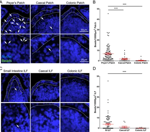
dramati-FIG 8Large intestinal GALT is relatively deficient in the uptake of particulate antigen. Mice were orally gavaged with 2⫻1011200-nm fluorescent
microbeads. At 24 h following gavage, the Peyer’s patches, small intestine, cecum, and colon were collected. Cryosections of each were prepared and counterstained with DAPI to detect cell nuclei (blue). (A) Images of Peyer’s, cecal, and colonic patches showing microbead (green) accumulation. Microbeads within patches are highlighted with arrows. The follicle-associated epithelium is defined by dotted lines. (B) The number of beads in sections of Peyer’s (n⫽96), cecal (n⫽28), and colonic (n⫽18) patches from 3 or 4 mice was determined, and the area of GALT was measured to determine the relative microbead density. Each dot represents the microbead density of an individual patch follicle. Bars display means⫾standard errors of the means (SEM). (C) Images of small intestinal, cecal, and colonic isolated lymphoid follicles (ILF) showing microbead (green) accumulation. Microbeads within ILF are highlighted with arrows. The follicle-associated epithelium is defined by dotted lines. (B) The number of beads in small intestinal (n⫽95), cecal (n⫽11), and colonic (n⫽64) ILF from 4 mice was determined, and the area of GALT was measured to determine the relative microbead density. Each dot represents the microbead density of an individual ILF. Bars display means⫾SEM. Significant differences were determined by nonparametric ANOVA (Kruskal-Wallis test) with a Dunn’s multiple comparisonpost hoctest. ***,P⬍0.001.
on November 7, 2019 by guest
http://jvi.asm.org/
[image:13.585.43.545.67.508.2]cally reduced susceptibility of aged mice to oral prion infection
(
58
) coincides with a significant reduction in the number of
ma-ture M cells in Peyer’s patches and disturbances to lymphoid tissue
microarchitecture (
59
,
60
). Conversely, chronic inflammation,
through the formation of ectopic FDC-containing B-cell follicles
(tertiary lymphoid tissues), may expand the tissue distribution of
prions within infected hosts (
61–63
). It is plausible that damage to
the LI mucosa and the associated immune pathology may also
affect oral prion disease pathogenesis (
64
). Although intestinal
helminth infections are common in animals and humans and
cause significant morbidity in cattle, sheep, and goats, nothing was
known about their effects on oral prion disease. Congruent
infec-tion with
T. muris
did not influence neuroinvasion or disease
sus-ceptibility irrespective of the time at which the mice were
coex-posed with prions, highlighting the important role of SI GALT in
oral prion pathogenesis. Our data appear to contradict those in an
independent study that reported that
Salmonella enterica
serovar
Typhimurium-induced colitis exacerbated oral prion disease (
65
).
However, while
T. muris
is restricted to the LI, subsequent data
have shown that
S
. Typhimurium infection can also have a
dra-matic effect on M cells and classical dendritic cells in the SI (
66
,
67
), which have key roles in oral prion pathogenesis (
26
,
27
). This
may have significantly influenced prion uptake in the SI,
enhanc-ing disease susceptibility independent of the effects on the LI.
In conclusion, our data demonstrate that the GALT in the SI,
not the LI, are the major early sites of prion accumulation and
neuroinvasion after oral exposure. This has important
implica-tions for our understanding of the factors that influence the risk of
infection and the preclinical diagnosis of disease. Although LI
GALT are not early sites of infection, the detection of PrP
Scwithin
the RAMALT and appendix has proved to be a useful method to
detect prion-infected individuals during the preclinical phase (
17
,
18
,
68
) and has been used in the United Kingdom to gain insight
into the possible prevalence of vCJD in the human population (
19
,
20
). However, our data suggest that the time at which these tissues
are sampled in relation to prion exposure may dramatically affect
the sensitivity of these assays. For instance, humans with
subclin-ical vCJD infection may have only minimal PrP deposition in
appendiceal tissue (
69
). Together, these data show that analyses of
such biopsy specimens may miss individuals in the early stages of
oral prion infection and underestimate the disease prevalence.
ACKNOWLEDGMENTS
We thank Bob Fleming, Barry Bradford, Dave Davies, Fraser Laing, Simon Cumming, Julia Oh, and the Pathology Services Group (University of Edinburgh, Edinburgh, United Kingdom) for helpful discussions and ex-cellent technical support, Jeffrey Browning (Boston University School of Medicine, Boston, MA) for provision of LTR-Ig, and Christine Farquhar (University of Edinburgh, Edinburgh, United Kingdom) for provision of pAb 1B3.
This work was supported by project funding (grant numbers BB/ G003947/1, BB/J014672/1) and Institute Strategic Programme Grant funding (grant number BB/J004332/1) from the Biotechnology and Bio-logical Sciences Research Council.
REFERENCES
1.Legname G, Baskakov IV, Nguyen H-OB, Riesner D, Cohen FE, De-Armond SJ, Prusiner SB.2004. Synthetic mammalian prions. Science
305:673– 676.http://dx.doi.org/10.1126/science.1100195.
2.Horiuchi M, Furuoka H, Kitamura N, Shinagawa M.2006. Alymphop-lasia mice are resistant to prion infection via oral route. Jpn J Vet Res
53:149 –157.
3.Glaysher BR, Mabbott NA.2007. Role of the GALT in scrapie agent neuroinvasion from the intestine. J Immunol178:3757–3766.http://dx .doi.org/10.4049/jimmunol.178.6.3757.
4.Prinz M, Huber G, Macpherson AJS, Heppner FL, Glatzel M, Eugster H-P, Wagner N, Aguzzi A.2003. Oral prion infection requires normal numbers of Peyer’s patches but not of enteric lymphocytes. Am J Pathol
162:1103–1111.http://dx.doi.org/10.1016/S0002-9440(10)63907-7. 5.Mabbott NA, Young J, McConnell I, Bruce ME.2003. Follicular
den-dritic cell dedifferentiation by treatment with an inhibitor of the lympho-toxin pathway dramatically reduces scrapie susceptibility. J Virol77:6845– 6854.http://dx.doi.org/10.1128/JVI.77.12.6845-6854.2003.
6.Andreoletti O, Berthon P, Marc D, Sarradin P, Grosclaude J, van Keulen L, Schelcher F, Elsen J-M, Lantier F.2000. Early accumulation of PrPScin gut-associated lymphoid and nervous tissues of susceptible sheep
from a Romanov flock with natural scrapie. J Gen Virol81:3115–3126. 7.Sigurdson CJ, Williams ES, Miller MW, Spraker TR, O’Rourke KI,
Hoover EA. 1999. Oral transmission and early lymphoid tropism of chronic wasting disease PrPresin mule deer fawns (Odocoileus hemionus).
J Gen Virol80:2757–2764.
8.Beekes M, McBride PA.2000. Early accumulation of pathological PrP in the enteric nervous system and gut-associated lymphoid tissue of ham-sters orally infected with scrapie. Neurosci Lett278:181–184.http://dx.doi .org/10.1016/S0304-3940(99)00934-9.
9.Kujala P, Raymond C, Romeijn M, Godsave SF, van Kasteren SI, HW, Prusiner SB, Mabbott NA, Peters PJ. 2011. Prion uptake in the gut: identification of the first uptake and replication sites. PLoS Pathog
7:e1002449.http://dx.doi.org/10.1371/journal.ppat.1002449.
10. McBride PA, Schulz-Shaeffer WJ, Donaldson M, Bruce M, Diringer H, Kretzschmar HA, Beekes M. 2001. Early spread of scrapie from the gastrointestinal tract to the central nervous system involves autonomic fibers of the splanchnic and vagus nerves. J Virol75:9320 –9327.http://dx .doi.org/10.1128/JVI.75.19.9320-9327.2001.
11. Glatzel M, Heppner FL, Albers KM, Aguzzi A. 2001. Sympathetic innervation of lymphoreticular organs is rate limiting for prion neuroinvasion. Neuron 31:25–34. http://dx.doi.org/10.1016/S0896 -6273(01)00331-2.
12. Glaysher BR, Mabbott NA.2007. Isolated lymphoid follicle maturation induces the development of follicular dendritic cells. Immunology120:
336 –344.http://dx.doi.org/10.1111/j.1365-2567.2006.02508.x.
13. Hamada H, Hiroi T, Nishiyama Y, Takahashi H, Masunaga Y,
Hachimura S, Kaminogawa S, Takahashi-Iwanaga H, Iwanaga T, Kiyono H, Yamamoto H, Ishikawa H.2002. Identification of multiple isolated lymphoid follicles on the antimesenteric wall of the mouse small intestine. J Immunol 168:57– 64. http://dx.doi.org/10.4049 /jimmunol.168.1.57.
14. Lorenz RG, Chaplin DD, McDonald KG, McDonough JS, Newberry
RD.2003. Isolated lymphoid follicle formation is inducible and depen-dent upon lymphotoxin-sufficient B lymphocytes, lymphotoxin re-ceptor, and TNF receptor 1 function. J Immunol170:5474 –5482.
15. Kweon MN, Yamamoto M, Rennert PD, Park EJ, Lee A-Y, Chang
S-Y, Hiroi T, Nanno M, Kiyono H.2005. Prenatal blockage of lym-photoxinreceptor and TNF receptor p55 signaling cascade resulted in the acceleration of tissue genesis for isolated lymphoid follicles in the large intestine. J Immunol 174:4365– 4372.http://dx.doi.org/10 .4049/jimmunol.174.7.4365.
16. Donaldson DS, Bradford BM, Artis D, Mabbott NA.2015. Reciprocal development of lymphoid tissue development in the large intestine by IL-25 and IL-23. Mucosal Immunol8:582–595.http://dx.doi.org/10.1038 /mi.2014.90.
17. González L, Dagleish MP, Bellworthy SJ, Sisó S, Stack MJ, Chaplin MJ, Davis LA, Hawkins SAC, Hughes J, Jeffrey M.2006. Postmortem diag-nosis of preclinical and clinical scrapie in sheep by the detection of disease-associated PrP in their rectal mucosa. Vet Rec158:325–331.http://dx.doi .org/10.1136/vr.158.10.325.
18. Wolfe LL, Spraker TR, González L, Dagleish MP, Sirochman TM,
Brown JC, Jeffrey M, Miller MW.2007. PrPCWDin rectal lymphoid tissue
of deer (Odocoileusspp.). J Gen Virol88:2078 –2082.http://dx.doi.org/10 .1099/vir.0.82342-0.
19. Hilton DA, Ghani AC, Conyers L, Edwards P, McCardle L, Ritchie D, Penney M, Hegazy D, Ironside JW.2004. Prevalence of lymphoreticular prion protein accumulation in UK tissue samples. J Pathol203:733–739. http://dx.doi.org/10.1002/path.1580.
20. Gill ON, Spencer Y, Richard-Loendt A, Kelly C, Dabaghian R, Boyes L,
Oral Prion Pathogenesis and the GALT
on November 7, 2019 by guest
http://jvi.asm.org/
Lineham J, Simmons M, Webb P, Bellerby P, Andrews N, Hilton DA, Ironside JW, Beck J, Poulter M, Mead S, Brandner S.2013. Prevalent abnormal prion protein in human appendixes after bovine spongiform encephalopathy epizootic: large scale survey. BMJ347:f5675.http://dx.doi .org/10.1136/bmj.f5675.
21. van Keulen LJ, Schreuder BE, Vromans ME, Langeveld JP, Smits MA.
2000. Pathogenesis of natural scrapie in sheep. Arch Virol Suppl16:57–71. 22. Gonzalez L, Martin S, Siso S, Konold T, Ortiz-Pelaez A, Phelan L, Goldmann W, Stewart P, Saunders G, Windl O, Jeffrey M, Hawkins SAC, Dawson M, Hope J.2009. High prevalence of scrapie in a dairy goat herd: tissue distribution of disease-associated PrP and effect ofPRNP ge-notype and age. Vet Res40:65.http://dx.doi.org/10.1051/vetres/2009048. 23. Thomsen BV, Schneider DA, O’Rourke KI, Gidlewski T, McLane J, Allen RW, McIsaac AA, Mitchell GB, Keane DP, Spraker TR, Balachan-dran A.2012. Diagnostic accuracy of rectal mucosa biopsy testing for chronic wasting disease within white-tailed deer (Odocoileus virginianus) herds in North America: effects of age, sex, polymorphism atPRNPcodon 96, and disease progression. J Vet Diagn Invest24:878 – 887.
24. Force WR, Walter BN, Hession C, Tizard R, Kozak CA, Browning JL, Ware CF. 1995. Mouse lymphotoxin-beta receptor. J Immunol155:
5280 –5288.
25. Rennert PD, Browning JL, Hochman PS.1997. Selective disruption of lymphotoxin ligands reveals a novel set of mucosal lymph nodes and unique effects on lymph node cellular organization. Int Immunol9:1627– 1639.http://dx.doi.org/10.1093/intimm/9.11.1627.
26. Raymond CR, Aucouturier P, Mabbott NA.2007.In vivodepletion of CD11c⫹cells impairs scrapie agent neuroinvasion from the intestine. J Immunol 179:7758 –7766. http://dx.doi.org/10.4049/jimmunol.179 .11.7758.
27. Donaldson DS, Kobayashi A, Ohno H, Yagita H, Williams IR, Mabbott NA.2012. M cell depletion blocks oral prion disease pathogenesis. Muco-sal Immunol5:216 –225.http://dx.doi.org/10.1038/mi.2011.68. 28. Fraser H, Dickinson AG.1968. The sequential development of the brain
lesions of scrapie in three strains of mice. J Comp Pathol78:301–311. http://dx.doi.org/10.1016/0021-9975(68)90006-6.
29. Wakelin D.1967. Acquired immunity toTrichuris murisin the albino laboratory mouse. Parasitology 57:515–524. http://dx.doi.org/10.1017 /S0031182000072395.
30. Else KJ, Wakelin D, Wassom DL, Hauda KM.1990. The influence of genes mapping with the major histocompatibility complex on resistance toTrichuris murisinfections in mice. Parasitology101:61– 67.http://dx .doi.org/10.1017/S0031182000079762.
31. Farquhar CF, Somerville RA, Ritchie LA.1989. Postmortem immuno-diagnosis of scrapie and bovine spongiform encephalopathy. J Virol Methods24:215–222.http://dx.doi.org/10.1016/0166-0934(89)90023