Effects of an In-Frame Deletion of the
6k
Gene Locus from the
Genome of Ross River Virus
Adam Taylor,aJulian V. Melton,b Lara J. Herrero,a,cBastian Thaa,fLiis Karo-Astover,ePeter W. Gage,bMichelle A. Nelson,c Kuo-Ching Sheng,aBrett A. Lidbury,dGary D. Ewart,bGerald M. McInerney,fAndres Merits,e Suresh Mahalingama,c
Emerging Viruses and Inflammation Research Group, Institute for Glycomics, Griffith University, Gold Coast, QLD, Australiaa
; Division of Molecular Biosciences, The John Curtin School of Medical Research, The Australian National University, Canberra, ACT, Australiab
; Virus and Inflammation Research Group, Faculty of Applied Science, University of Canberra, Canberra, ACT, Australiac
; Department of Genome Biology, The John Curtin School of Medical Research, The Australian National University, Canberra, ACT, Australiad
; Institute of Technology, University of Tartu, Tartu, Estoniae
; Karolinska Institutet, Department of Microbiology, Tumor, and Cell Biology (MTC), Stockholm, Swedenf
ABSTRACT
The alphaviral
6k
gene region encodes the two structural proteins 6K protein and, due to a ribosomal frameshift event, the
trans-frame protein (TF). Here, we characterized the role of the
6k
proteins in the arthritogenic alphavirus Ross River virus (RRV) in
infected cells and in mice, using a novel
6k
in-frame deletion mutant. Comprehensive microscopic analysis revealed that the
6k
proteins were predominantly localized at the endoplasmic reticulum of RRV-infected cells. RRV virions that lack the
6k
proteins
6K and TF [RRV-(
⌬
6K)] were more vulnerable to changes in pH, and the corresponding virus had increased sensitivity to a
higher temperature. While the
6k
deletion did not reduce RRV particle production in BHK-21 cells, it affected virion release
from the host cell. Subsequent
in vivo
studies demonstrated that RRV-(
⌬
6K) caused a milder disease than wild-type virus, with
viral titers being reduced in infected mice. Immunization of mice with RRV-(
⌬
6K) resulted in a reduced viral load and
acceler-ated viral elimination upon secondary infection with wild-type RRV or another alphavirus, chikungunya virus (CHIKV). Our
results show that the
6k
proteins may contribute to alphaviral disease manifestations and suggest that manipulation of the
6k
gene may be a potential strategy to facilitate viral vaccine development.
IMPORTANCE
Arthritogenic alphaviruses, such as chikungunya virus (CHIKV) and Ross River virus (RRV), cause epidemics of debilitating
rheumatic disease in areas where they are endemic and can emerge in new regions worldwide. RRV is of considerable medical
significance in Australia, where it is the leading cause of arboviral disease. The mechanisms by which alphaviruses persist and
cause disease in the host are ill defined. This paper describes the phenotypic properties of an RRV
6k
deletion mutant. The
ab-sence of the
6k
gene reduced virion release from infected cells and also reduced the severity of disease and viral titers in infected
mice. Immunization with the mutant virus protected mice against viremia not only upon exposure to RRV but also upon
chal-lenge with CHIKV. These findings could lead to the development of safer and more immunogenic alphavirus vectors for vaccine
delivery.
A
lphaviruses like Ross River virus (RRV), Barmah Forest virus
(BFV), Semliki Forest virus (SFV), Sindbis virus (SINV),
chi-kungunya virus (CHIKV), and O’nyong-nyong virus (ONNV) are
small, enveloped RNA viruses of the family
Togaviridae
. They
fre-quently cause human diseases manifesting with signs and
symp-toms such as encephalitis, acute or persistent arthritis, fever,
ar-thralgia, and myalgia (
1
). RRV is a prevalent disease-causing
alphavirus in the Australasian region and a major health hazard,
with 5,000 cases of Ross River virus disease (RRVD) reported
an-nually (
2
). Patients infected with RRV can develop a febrile illness,
rash, polyarthritis, and myalgia (
3
). A number of patients also
experience chronic arthralgia and debilitating joint pain that can
last for months. Despite this, the pathogenic determinants of RRV
have not been extensively studied.
The alphaviral genome comprises the 5
=
region, encoding the
nonstructural proteins, and the 3
=
region, encoding the structural
proteins. The structural proteins, capsid protein (C),
glycopro-teins E1, E2, and E3, 6K protein, and transframe protein (TF), are
derived from progressive cleavage of the major polyproteins,
C-E3-E2-6K-E1, and minor polyproteins, C-E3-E2-TF (
1
). TF
shares the same N-terminal sequence as the 6K protein but
con-tains an extended C terminus as a result of ribosomal
frameshift-ing that occurs durframeshift-ing the translation of the
6k
gene (
2
). C, E2, and
E1 are the obligatory components of mature virions that
morpho-logically appear as a nucleocapsid enveloped by a lipid bilayer that
is studded with E1/E2 trimers.
Although the 6K and TF
6k
proteins are translated alongside
the other structural proteins, they are generally present at very low
Received5 January 2016 Accepted2 February 2016
Accepted manuscript posted online10 February 2016
CitationTaylor A, Melton JV, Herrero LJ, Thaa B, Karo-Astover L, Gage PW, Nelson MA, Sheng K-C, Lidbury BA, Ewart GD, McInerney GM, Merits A, Mahalingam S. 2016. Effects of an in-frame deletion of the6kgene locus from the genome of Ross River virus. J Virol 90:4150 –4159.doi:10.1128/JVI.03192-15.
Editor:T. S. Dermody
Address correspondence to Suresh Mahalingam, [email protected].
A.T., J.V.M., and L.J.H. contributed equally to this article and should be considered joint first authors.
With great sadness we note that both Julian V. Melton and Peter W. Gage passed away before completion of this research study. The other authors dedicate this paper to these two outstanding researchers.
Copyright © 2016, American Society for Microbiology. All Rights Reserved.
on November 7, 2019 by guest
http://jvi.asm.org/
levels within virus particles (
4–6
). Both
6k
proteins are
mem-brane-associated proteins, containing hydrophobic (supposed
transmembrane) domains and numerous acylated amino acid
res-idues (
4
,
7
). The role of the
6k
proteins in alphaviral disease
re-mains unclear, although they have been shown to be involved in
processes of viral replication, assembly, and release. For example,
mutations of SINV
6k
resulting from either an in-frame insertion
of 15 amino acids in the
6k
gene (
8
) or a deletion of 21 amino acids
from the
6k
gene (
9
) led to defective glycoprotein processing,
traf-ficking, and viral budding. These findings are in line with a
previ-ous report demonstrating the involvement of an SFV
6k
protein
sequence in the insertion of E1 into the endoplasmic reticulum
(ER) membrane (
10
). Interestingly, complete deletion of the SFV
6k
gene had little effect on glycoprotein processing and transport
but significantly impaired the release of virus particles, suggesting
a role for the
6k
proteins in viral assembly or budding (
5
,
11
).
Similarly, disruption of SINV TF production reduced infectious
particle release during infection (
6
). The severity of disease
expe-rienced by mice infected with SINV TF mutants was also greatly
reduced relative to the severity of disease experienced by mice
infected with the wild-type virus in a model of SINV
neuropatho-genesis (
6
). The size, hydrophobicity, and structural
characteris-tics of the alphaviral
6k
proteins led to the hypothesis that they
could function as viral ion channels, or viroporins. In this model,
the
6k
proteins are thought to be responsible for increasing host
cell plasma membrane permeability, a common phenomenon of
cytolytic viral infections (
12–14
). A number of studies also
sup-ported a role for the alphaviral 6K protein in enhancing
mem-brane permeability through alternative mechanisms (
15–18
).
The
6k
region is not highly conserved among the alphaviruses.
In particular, the relevance of the
6k
proteins for the replication
and pathogenesis of RRV has not been assessed, despite the
im-portance of this virus. Hence, we have constructed a 6K/TF
dele-tion mutant of RRV [RRV-(
⌬
6K)] containing an in-frame
dele-tion of the entire
6k
gene region, thereby ensuring that the
translation of proteins encoded by sequences on either side of the
6k
gene region remains unaffected. Therefore, the potential
cave-ats associated with partial gene deletions or insertions, as were
done previously for SINV
6k
(
8
,
9
), are avoided. Using this
mu-tant, we demonstrate that the
6k
proteins are involved in virus
release in BHK-21 cells. Furthermore, we show that RRV-(
⌬
6K)
induces only mild disease in mice compared to the severity of
the disease induced by its wild-type (WT) counterpart and that
the viral load is reduced. RRV-(
⌬
6K)-immunized mice
pos-sessed the capacity to significantly reduce viremia upon
alpha-virus challenge. These results demonstrate the major role of the
6k
proteins in RRV pathogenesis and the potential of
6k
deletion as
a vaccination strategy against RRV.
MATERIALS AND METHODS
Cells.Baby hamster kidney (BHK-21; ATCC CCL-10) cells were main-tained at 37°C in F15 medium supplemented with nonessential amino acids, 5% fetal calf serum (FCS), 10% tryptose phosphate broth, and 2 mM glutamine. Vero cells (ATCC CCL-81) were cultured in Opti-MEM medium supplemented with 10% FCS. Human osteosarcoma (HOS) cells (ATCC CRL-1543) were maintained in Dulbecco’s modified Eagle’s me-dium (DMEM) with 10% FCS, and HOS cells stably transfected with enhanced green fluorescent protein (EGFP)-tagged LC3 (19) were kept in DMEM with 10% FCS and 1 g/liter G418.
Construction of an RRV 6kdeletion mutant.The fragment of pRR64, an RRV T48 infectious cDNA clone, spanning from the RsrII to the SmaI
restriction site was cloned into pKS⫹(Stratagene, Germany). The se-quence corresponding to the6kregion was removed by polymerase in-complete primer extension PCR (20) using oligonucleotides with the se-quences 5=-TGC TCG TAT GCG TTC GCC CTC GGT GCG CAG C-3=
and 5=-GAA CGC ATA CGA GCA CAC AGC CAC AAT TCC GAA TGT GG-3=as the primers and Phusion DNA polymerase (Thermo Scientific, USA) as the enzyme. The fragment containing the correct deletion was used to replace the corresponding fragment of pRR64. The sequence of the obtained clone, designated pRR64-(⌬6K), was verified by sequencing.
Viruses.Infectious RRV RNA transcripts were generated as described by Kuhn et al. (21). Briefly, pRR64 and pRR64-(⌬6K) were linearized with SacI and transcribed using SP6 RNA polymerase, to generate full-length transcripts that were 5=capped by the inclusion of m7G(5=)ppp(5=)A in
the transcription reaction. BHK-21 cells were transfected with these RNAs by electroporation, and both WT RRV and the6kdeletion mutant [RRV-(⌬6K)] were harvested at 24 to 48 h posttransfection. The deletion of the
6kregion was confirmed by RT-PCR, and the lack of 6K protein expres-sion was verified by immunofluorescence. CHIKV (Bali isolate) was kindly provided by Linda Hueston (Centre for Infectious Diseases & Mi-crobiology Laboratory Services, Westmead Hospital, Sydney, Australia) and propagated in Vero cells.
Infection and replication in cell culture.Cells were infected with vi-ruses in Hanks’ buffered salt solution (HBSS) at a multiplicity of infection (MOI) of 0.1 PFU/cell. Following adsorption of virus for 1 h at 37°C, cell monolayers were washed and fresh growth medium was added. The cul-ture medium was assayed for the presence of infectious particles at various time points. To determine the amount of cell-associated virus, cell mono-layers were washed and lysed by osmotic shock and four freeze-thaw cy-cles to release internal virus particy-cles. The cell lysate was then assayed for infectious particles by a plaque assay or for viral RNA by reverse transcrip-tion-quantitative PCR (RT-qPCR).
Temperature sensitivity.Plaque assays were performed on Vero cells incubated at 37°C, 39°C, or 41°C. Cells were incubated with WT RRV or RRV-(⌬6K) for 1 h, the virus inoculum was removed, and an overlay of semisolid medium (0.16% agarose in DMEM with 2% FCS) was added to the cells. The cells were incubated at the temperatures specified below for 48 h prior to staining with crystal violet. To assay the thermal stability of WT RRV or RRV-(⌬6K) particles, 100 PFU of each virus was incubated at 52°C or 56°C for 0, 6, or 12 min and cooled on ice. BHK-21 cells were incubated with virus for 1 h at 37°C and overlaid with semisolid medium (2% carboxymethyl cellulose [CMC] in Glasgow minimum essential me-dium [GMEM]). At 72 h postinfection (p.i.), the cells were fixed with 10% formaldehyde for 20 min and stained with crystal violet. The experiments were repeated three times.
pH sensitivity assessment.WT RRV or RRV-(⌬6K) (100 PFU) was treated at room temperature with 0.3 M morpholinoethanesulfonic acid (MES) at pH 5.5 for 0, 10, 20, 30, 40, 50, or 60 s and neutralized with 1 M NaOH. BHK-21 cells were incubated with virus for 1 h at 37°C and over-laid with semisolid medium (2% CMC in GMEM). At 72 h p.i., the cells were fixed with 10% formaldehyde for 20 min and stained with crystal violet. The experiments were repeated three times.
Virus quantification. (i) Infectious viral particle quantification by plaque assay.All plaque assays on Vero cells were performed as described previously (22).
(ii) Viral load quantification by RT-qPCR.RNA extraction was per-formed using the TRIzol reagent (Life Technologies) following the man-ufacturer’s instructions. Extracted RNA was reverse transcribed using random nonamers and Moloney murine leukemia virus reverse transcrip-tase (Sigma-Aldrich) according to the manufacturer’s instructions. A standard curve was generated using serial dilutions of pRR64 as described previously (23). Quantification of the viral load was performed using SsoAdvanced universal probes supermix (Bio-Rad) in a 12.5-l reaction volume to detect the nsP3 region (23). Primers nsp3 Forward (5=-CCG TGG CGG GTA TTA TCA AT-3=) and nsp3 Reverse (5=-AAC ACT CCC GTC GAC AAC AGA-3=) and fluorogenic probe RRV nsp3 (5=-ATT AAG
on November 7, 2019 by guest
http://jvi.asm.org/
AGT GTA GCC ATC C-3=) were used All reactions were performed using a Bio-Rad CFX96 Touch real-time PCR detection system in 96-well plates. Cycler conditions were as follows: (i) a PCR initial activation step of 95°C for 15 min for 1 cycle and (ii) 3-step cycling at 94°C for 15 s, followed by 55°C for 30 s and 72°C for 30 s for 40 cycles. A dissociation curve was acquired using CFX Manager software to determine the specificity of the amplified products. A standard curve was plotted, and the copy numbers of the amplified products were interpolated from the standard curve using GraphPad Prism software to determine the viral load.
Immunofluorescence.Cells were grown on glass coverslips and in-fected with virus as described above. Nonpermeabilized cells were fixed on ice for 30 min with 2% paraformaldehyde in phosphate-buffered saline (PBS), or, alternatively, cells were permeabilized and fixed with 2% para-formaldehyde containing 0.1% Triton X-100 in PBS, followed by blocking in 5% horse serum in PBS at 4°C for at least 16 h. RRV6kproteins and E2 were labeled using rabbit polyclonal antibodies raised against the RRV 6K N-terminal region or the RRV E2 protein; mouse monoclonal antibodies against double-stranded RNA (monoclonal antibody J2; English and Sci-entific Consulting), calnexin (monoclonal antibody AF18; catalog num-ber MA3027; Pierce/Thermo), GM130 (catalog numnum-ber 610822; Becton Dickinson), EEA1 (catalog number 610456; Becton Dickinson), and LAMP1 (catalog number ab25630; Abcam) were employed to stain repli-cation complexes, the ER,cis-Golgi network, early endosomes, and lyso-somes, respectively. Human cells had to be used for this analysis because not all these organelle marker antibodies reacted with the hamster pro-teins in BHK-21 cells (data not shown). Autophagosomes were detected by fluorescence of EGFP-tagged LC3 in HOS cells stably transfected with this marker (19). Antibodies were diluted in 5% horse serum-PBS; incu-bation was performed for 2 to 5 h at 21°C, followed by washing in PBS; and secondary antibodies were applied for 30 min at 21°C; suitable fluorescein isothiocyanate (FITC)-, Alexa Fluor 488-, or Alexa Fluor 555-labeled anti-rabbit and anti-mouse immunoglobulin secondary antibodies plus DRAQ5 (Thermo) were used to stain the nuclei. Samples were imaged with either an Olympus FV1000 confocal microscope or a Leica TCS SP5 X confocal microscope equipped with a supercontinuum pulsed white laser.
Mouse studies.The mouse model of RRV infection was established as described previously by Lidbury et al. (22). To assess the pathogenic po-tentials of WT RRV and RRV-(⌬6K), 17-day-old outbred Swiss mice were infected intraperitoneally (i.p.) with 103PFU of each virus and the clinical
manifestations were monitored for 20 days postinfection. Mock-inocu-lated animals were injected with the diluent alone. Clinical scores and body weights were recorded as previously described (24). To evaluate the prophylactic potential of RRV-(⌬6K), 21-day-old C57BL/6 mice were infected with 104PFU RRV-(⌬6K). At day 30 postinfection, the C57BL/6
mice were challenged with 106PFU RRV T48 or CHIKV. Viral titers in
sera were evaluated by plaque assay at days 1, 2, and 3 postchallenge. All experiments were approved by the animal ethics committees of the Uni-versity of Canberra and The Australian National UniUni-versity.
Statistical analysis.The statistical significance between experimental groups was determined by the Student’s unpairedttest (P⬍0.05). Data are presented as the mean⫾standard error of the mean (SEM). For disease scores, experimental results were analyzed by the Mann-Whitney test. Statistical analyses were performed using GraphPad Prism software (version 4.0b; GraphPad Software Inc.). Differences in means were con-sidered significant at aPvalue of⬍0.05.
RESULTS
Subcellular localization of the
6k
proteins in RRV-infected cells.
We first characterized the distribution and localization of the RRV
6k
proteins in RRV-infected BHK-21 cells at 12 h postinfection
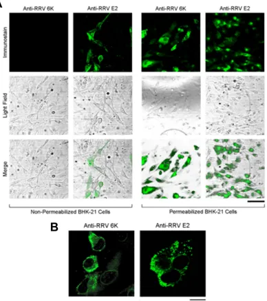
(p.i.) by immunofluorescence. When infected cells were processed
by fixation, permeabilization, and immunostaining for either the
RRV
6k
proteins or RRV E2, we readily detected these proteins
(
Fig. 1A
, right), while nonpermeabilized infected cells were
posi-tive for E2 but not the
6k
proteins (
Fig. 1A
, left). This suggests that
the RRV
6k
proteins were not exposed to the extracellular space.
When examined with a higher magnification, both the
6k
proteins
and E2 were predominantly present at the periphery of the nuclei
with punctate appearances (
Fig. 1B
), reminiscent of localization
to the vesicular organelles.
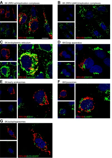
We then performed higher-resolution confocal microscopy to
determine the subcellular localization of the
6k
proteins (
Fig. 2
). A
punctate intracellular distribution of the
6k
proteins without
dis-cernible accumulation at the plasma membrane was observed.
These intracellular foci did not represent replication complexes, as
evidenced by costaining for the replication intermediate
double-stranded RNA (
Fig. 2A
). The lack of
6k
signals in cells infected
with RRV-(
⌬
6K) demonstrates the specificity of the staining (
Fig.
2B
). Further, colocalization analysis with organelle markers
showed that the
6k
protein foci in cells infected with WT RRV
extensively colocalized with calnexin, a marker for the ER (
Fig.
2C
), while the
6k
proteins did not colocalize to any considerable
extent with markers for the
cis
-Golgi network (
Fig. 2D
), early
en-dosomes (
Fig. 2E
), or lysosomes (
Fig. 2F
). Also, the
6k
proteins
were not targeted to autophagosomes (
Fig. 2G
), which
accumu-late in alphavirus-infected cells (
25
). Thus, this in-depth
micro-scopic analysis shows that the
6k
proteins were predominantly
localized to the ER in RRV-infected cells.
Effect of the
6k
deletion on RRV temperature and pH
sensi-tivity.
We then went on to assess whether the
6k
proteins affect the
sensitivity of RRV virions to variations in temperature or pH, as
such a role has been suggested in the context of other alphavirus
6k
mutations (
26
). To assess the thermostability of virions, WT RRV
and RRV-(
⌬
6K) particles were treated at elevated temperatures
(52 or 56°C) for 0, 6 and 12 min, before being cooled to 4°C and
assayed by plaque assay. WT RRV and RRV-(
⌬
6K) were both
completely inactivated following incubation at 56°C, and no
dif-ference in the amount of infectious virus after incubation at 52°C
was found (data not shown), indicating that there was no
differ-ence in the thermal stability of RRV-(
⌬
6K) and WT virus
par-ticles. To examine the temperature sensitivity of RRV-(
⌬
6K)
during replication, 10-fold dilutions of either WT RRV or
RRV-(
⌬
6K) were overlaid onto Vero cells and incubated at
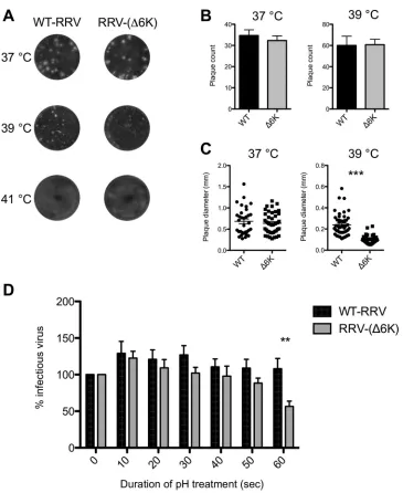
differ-ent temperatures. Variations in temperature did not differdiffer-entially
affect the infectivity of the WT and mutant viruses at 37°C and
39°C, where similar plaque numbers were counted, while no
plaques were detected at 41°C (
Fig. 3A
and
B
). The plaque size of
RRV-(
⌬
6K) was, however, significantly reduced compared to that
of WT RRV at 39°C but not at 37°C (
Fig. 3C
). Thus, unlike the
stability of virions and their ability to infect cells, a subsequent step
of the RRV replication cycle was clearly affected by the deletion of
the
6k
region.
To examine the effect of deletion of the
6k
proteins on the pH
sensitivity of virions, WT RRV and RRV-(
⌬
6K) particles were
incubated at pH 5.5 for various times prior to neutralization and
subsequent plaque assay. WT RRV was stable at pH 5.5 with no
reduction of infectivity (
Fig. 3D
). In contrast, RRV-(
⌬
6K) virions
were sensitive to low pH, as their infectivity was significantly
re-duced following incubation at pH 5.5 for 60 s compared to that of
WT RRV. In summary, we observed a tendency for RRV to have a
higher vulnerability to temperature and pH when it lacked the
6k
proteins.
6k
deletion does not affect virus particle production but
af-fects the release of virus particles from BHK-21 cells.
Next, we
on November 7, 2019 by guest
http://jvi.asm.org/
asked whether deletion of the
6k
proteins affected RRV
replica-tion. To this end, BHK-21 cells were infected with either WT RRV
or RRV-(
⌬
6K), and the supernatants were harvested at various
times postinfection and titrated by plaque assay. At early times
postinfection (0 to 16 h), no significant difference in the amount
of infectious cell-associated WT RRV or RRV-(
⌬
6K) was
ob-served, suggesting that deletion of the
6k
gene did not affect the
rate of replication of RRV (
Fig. 4A
). However, at late times (
⬎
24
h) postinfection, the amount of cell-associated RRV-(
⌬
6K)
signif-icantly increased compared to the amount of WT RRV, indicating
that virions of RRV-(
⌬
6K) were not efficiently released and
accu-mulated within and/or at the surface of infected cells.
Addition-ally, the significant increase in the amount of WT RRV released
from BHK-21 cells compared to the amount of RRV-(
⌬
6K)
re-leased, which was especially evident at 16 h p.i. and later time
points, also supports the conclusion that the release of
RRV-(
⌬
6K) was compromised (
Fig. 4B
). Complementary RT-qPCR
analysis of culture supernatants and lysates containing
cell-as-sociated virus also revealed evidence of the disturbed release of
RRV-(
⌬
6K); the cell-associated viral genome copy numbers of
RRV-(
⌬
6K) significantly increased at 24 h p.i. with respect
to the cell-associated viral genome copy numbers of WT RRV
FIG 1Cellular distribution of the RRV6kand E2 proteins in WT RRV-infected BHK-21 cells. BHK-21 cell monolayers were infected with WT RRV at an MOI of 0.1 for 1 h, placed in fresh growth medium, and incubated for 12 h. (A) Cells were either fixed with paraformaldehyde (nonpermeabilized) or permeabilized and fixed with paraformaldehyde containing 0.1% Triton X-100. The cellular location of the6kproteins and the E2 protein was visualized using rabbit polyclonal antibodies raised against the RRV 6K N-terminal region and the RRV E2 protein, respectively, followed by FITC-conjugated secondary antibodies. Cells were examined using fluorescence (top row) and light (middle row) microscopy, and merged images (bottom row) were generated. Bar⫽40m. (B) RRV6kproteins and the E2 protein exhibit a punctate distribution in BHK-21 cells. BHK-21 cell monolayers were infected with WT RRV, fixed, and permeabilized at 12 h p.i. and stained as described in the legend to panel A. Bar⫽20m.on November 7, 2019 by guest
http://jvi.asm.org/
[image:4.585.103.491.70.507.2](
Fig. 4C
), a finding which correlates with the larger amount of
infectious cell-associated RRV-(
⌬
6K) detected (
Fig. 4A
). The
ge-nome copy numbers of RRV-(
⌬
6K) were significantly reduced in
culture supernatants at 16 h p.i. though not at 24 h p.i. (
Fig. 4D
).
This may result from increased survival of RRV-(
⌬
6K)-infected
cells, allowing a prolonged or more efficient synthesis of viral
RNA. To rule out the possibility that this effect may be caused by
a reduced genome copy number/PFU ratio of RRV-(
⌬
6K), such
ratios were determined for both WT RRV and RRV-(
⌬
6K) (data
not shown). No difference was found, indicating that the high
FIG 2Subcellular localization of the RRV6kproteins. (A and B) BHK-21 cell monolayers were infected with WT RRV (A) or RRV-(⌬6K) (B) at an MOI of 0.1 for 1 h, placed in fresh growth medium, and incubated for 12 h. Cells were permeabilized and fixed with paraformaldehyde containing 0.1% Triton X-100. The subcellular localization of the6kproteins was visualized using rabbit polyclonal antibodies raised against the RRV 6K N-terminal region (pseudocolored red), combined with staining of the indicated organelles (green). dsRNA, double-stranded RNA. Bar⫽10m. (C to G) HOS cells were infected with WT RRV as indicated and fixed and permeabilized, followed by costaining for the6kproteins (red) and the indicated organelle marker (green). Mouse monoclonal antibodies against calnexin, GM130, EEA1, and LAMP1 were used to stain the ER (C),cis-Golgi network (D), early endosomes (E), and lysosomes (F), respectively. (G) Autophagosomes were detected by fluorescence of EGFP-tagged LC3 in HOS cells stably transfected with this marker.on November 7, 2019 by guest
http://jvi.asm.org/
[image:5.585.112.478.65.580.2]genome copy number of RRV-(
⌬
6K) in the cell culture
superna-tant at 24 h p.i. may be due to the release of virus RNA-containing
cell debris into the culture medium. Together these data suggest
that, although virus production was not reduced, deletion of
6k
inhibited the release of RRV from host cells.
RRV-(
⌬
6K)-infected mice show a reduced severity and
dura-tion of RRV disease.
Several
in vivo
studies examining the role of
the alphavirus structural proteins have outlined the importance of
the envelope glycoproteins in viral pathogenesis (
27
,
28
).
How-ever, little is known about the effect of the
6k
proteins on
alphavi-ral virulence. To examine this for RRV, Swiss mice were mock
infected or infected with 10
3PFU of WT RRV or RRV-(
⌬
6K) and
monitored for disease signs and body weight. WT RRV-infected
mice developed severe disease signs, including a loss of hind limb
grip strength and dragging of the hind limbs. These disease signs
peaked at day 10 p.i. and resolved by day 20 (
Fig. 5A
).
Further-more, WT RRV-infected mice failed to gain weight, showing signs
of wastage, which also peaked at day 10 p.i. (
Fig. 5B
). In contrast,
RRV-(
⌬
6K)-infected mice did not present the same severity of
disease as WT RRV-infected mice, showing only mild hind limb
weakness and a low level of lethargy. This mild disease peaked at
day 6 and resolved by day 12 p.i. (
Fig. 5A
). RRV-(
⌬
6K)-infected
mice also started to gain weight by day 8 (
Fig. 5B
). In addition, a
significant decrease in the viral load was found in both the
quad-riceps muscle (
Fig. 5C
) and the ankle joints (
Fig. 5D
) of
RRV-(
⌬
6K)-infected mice. While peak viral titers in WT RRV-infected
FIG 3RRV-(⌬6K) temperature and pH sensitivity. (A) Representative plaque phenotypes of WT RRV and RRV-(⌬6K) at the specified temperatures. (B) Average plaque count for WT RRV and RRV-(⌬6K) at 37°C and 39°C. The plaque count was zero at 41°C. The data represent the means⫾SEMs from three independent determinations. (C) Average plaque diameter for WT RRV and RRV-(⌬6K) at 37°C and 39°C. (D) Relative amount of infectious WT RRV and RRV-(⌬6K) after exposure to pH 5.5 for the indicated times normalized to the amount at time zero (100%). The data represent the means⫾SEMs from three independent determinations.Pvalues were determined by Student’s unpairedttest. **,P⬍0.01; ***,P⬍0.001.on November 7, 2019 by guest
http://jvi.asm.org/
[image:6.585.111.476.60.506.2]mice were observed at day 5 p.i., RRV-(
⌬
6K) titers in the
quadri-ceps muscle peaked at day 3 and were markedly lower than those
of WT RRV. A significant decrease in the RRV-(
⌬
6K) titer in
comparison to the WT RRV titer was observed in both quadriceps
muscle and ankle joint tissues from day 5 p.i. RRV-(
⌬
6K) was
cleared from the ankle joints of infected mice at day 8 p.i. In
addition, we detected viral RNA in the ankle joints of mice up to 1
month p.i. with WT RRV but not with RRV-(
⌬
6K) (
Fig. 5E
),
indicating the persistence of WT RRV but not the
6k
mutant.
RRV-(
⌬
6K) immunization results in a reduced viral burden
during alphaviral infection.
As RRV-(
⌬
6K) showed significantly
reduced virulence in the host, we tested its prophylactic efficacy.
Mice were immunized with 10
4PFU of RRV-(
⌬
6K) or mock
im-munized with PBS and at day 30 p.i. were challenged with a high
dose (10
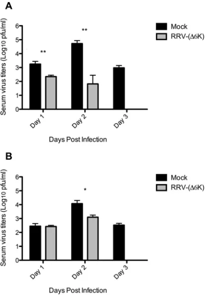
6PFU) of WT RRV or CHIKV. As shown in
Fig. 6
,
RRV-(
⌬
6K)-immunized mice showed an overall reduced viral load in
serum after infection with RRV (
Fig. 6A
) or CHIKV (
Fig. 6B
).
Immunization with RRV-(
⌬
6K) resulted in the effective
reduc-tion of the RRV load already by day 1 p.i. (
Fig. 6A
). When the
RRV-(
⌬
6K)-immunized mice were challenged with CHIKV, viral
titers at day 2 were reduced 10-fold compared to those in
mock-immunized mice (
Fig. 6B
), indicating cross protection. By day 3
postchallenge, infectious virus was cleared from the serum of
RRV-(
⌬
6K)-immunized mice but not control mice in both
RRV-and CHIKV-challenged mice (
Fig. 6
). These findings suggest that
RRV-(
⌬
6K) immunization offers protection against reinfection
with the same or even a closely related alphavirus by reducing the
viral burden in the host.
DISCUSSION
Little is known about the importance and role of the RRV
6k
proteins, 6K and TF, during virus infection and replication. A
number of previous studies have used partial gene deletions or
insertions to investigate the role of the alphaviral 6K protein (
8
,
9
).
However, in such mutants, it is not known whether protein
func-tion is completely abolished. Also, it is not known whether these
approaches result in the expression of a novel protein with
differ-ent properties and what effects that this might have on processes
such as viral replication and host cell metabolism (
29
,
30
).
Fur-thermore, the recent discovery of TF, the result of a translational
frameshift in the
6k
gene, brings added uncertainty to the validity
of these approaches (
2
,
31
). Additionally, the
6k
gene is not highly
conserved between alphaviruses, making it difficult to compare
findings in different alphaviruses. In this study, an RRV mutant
containing an in-frame deletion of the entire
6k
gene region was
constructed, thereby ensuring that the translation of proteins
en-coded by sequences on either side of the
6k
gene region remained
unaffected. This mutant is directly analogous to the alphavirus
6k
deletion mutant of SFV described previously (
11
).
Immunofluorescence detection of the
6k
proteins showed a
punctate perinuclear distribution, suggesting that these proteins
were largely retained within the cell. The
6k
proteins were not
detectable in RRV-infected nonpermeabilized cells by
immuno-fluorescence, indicating that the epitope for the
6k
antibody, the
N-terminal end of the
6k
proteins, was not detectably exposed at
the cell surface in infected cells. The predominant localization of
the RRV
6k
proteins was at the ER, in line with the findings of
FIG 4Deletion of RRV6khas no effect on RRV particle production in BHK-21 cells but inhibits particle release. BHK-21 cells were infected with either WT RRV or RRV-(⌬6K) at an MOI of 0.1 for 1 h and placed in growth medium. (A and B) Quantification of infectious cell-associated virus (A) and virus released into the cell culture supernatant (B) by plaque assay. The limit of detection for the plaque assay was 10 PFU/ml. (C and D) Determination of viral genome copy numbers by RT-qPCR at the indicated times p.i. for cell-associated virus (C) and virus in the culture supernatant (D).Pvalues were determined by Student’s unpairedttest. *,P⬍0.05; **,P⬍0.01; ***,P⬍0.001.
on November 7, 2019 by guest
http://jvi.asm.org/
[image:7.585.83.502.63.326.2]previous studies on SINV (
4
) and SFV (
7
)
6k
proteins. At the ER,
the alphaviral
6k
proteins may function as ion channels, thereby
disrupting calcium homeostasis by the sustained release of
cal-cium from the ER store (
13
,
31
). The sustained release of calcium
from the ER store leads to increased cytoplasmic calcium levels,
triggering cell shrinkage and cell permeabilization, which are
likely to play a role in the later stages of alphavirus replication,
such as viral budding and release (
15
). The subcellular localization
of the
6k
proteins was, however, not completely congruent with
the ER, implying some shuttling of 6K/TF to other vesicular
or-ganelles and/or the plasma membrane.
The reduced infectious RRV-(
⌬
6K) titers for released but not
cell-associated virus suggest that the RRV
6k
proteins are required
for efficient viral dissociation from the cell. The inefficient cell
dissociation of RRV-(
⌬
6K) virions may be linked to the formation
of ion channels by the
6k
proteins (
16
). Membrane
permeabiliza-tion by ion channel formapermeabiliza-tion leads to changes in the local
mem-brane potential, creating an environment thought to promote
al-phaviral budding (
9
,
32
). This defect in cell dissociation was most
likely enhanced at 39°C, as at this temperature RRV-(
⌬
6K) made
the same number of plaques as WT RRV, but the plaques were
smaller than those of WT RRV. With the 6K protein of SFV
thought to play a role in the organization and stability of the
het-erodimeric E1-E2 glycoprotein complex, it is possible that
dele-tion of the RRV
6k
proteins may influence the downstream
pro-cessing and/or trafficking of the RRV glycoproteins and ultimately
affect virus assembly and replication (
31
,
33
). Notably, in contrast
to RRV-(
⌬
6K), the SFV
6k
deletion mutant demonstrated a
dra-matic decrease in the rate of viral replication (
5
,
11
). In addition, a
6k
deletion mutant of salmonid alphavirus (SAV) was found to be
nonviable (
34
), outlining the functional divergence of the
6k
pro-teins in different alphaviruses.
FIG 5RRV-induced disease in mice infected with WT RRV and RRV-(⌬6K). Seventeen-day-old outbred Swiss mice were infected with 103PFU of WT RRV or
RRV-(⌬6K) by intraperitoneal injection. Mock-inoculated mice were injected with PBS alone. (A) Mice were scored for lethargy and hind limb dysfunction on the basis of the following scale: 0, no disease signs; 1, ruffled fur; 2, very mild hind limb weakness; 3, mild hind limb weakness; 4, moderate hind limb weakness and dragging of hind limbs; 5, severe dragging; 6, complete loss of hind limb function; 7, moribund state. Data for disease scores were analyzed by the Mann-Whitney test. *,P⬍0.05; **,P⬍0.01. (B) Mice were monitored daily for changes in weight. Values are the means⫾SEMs for 8 mice per group. *,P⬍
0.05. (C and D) On days 1, 3, 5, and 8 p.i., the quadriceps muscle (C) and ankle joints (D) were collected and the amount of infectious virus was determined by plaque assay. The limit of detection for the plaque assay was 10 PFU/ml. The data represent the means⫾SEMs for 4 mice per group and are representative of those from two separate experiments.Pvalues were determined by Student’s unpairedttest.*,P⬍0.05; **,P⬍0.01; ***,P⬍0.001. (E) RT-PCR analysis of the E2 region of viral RNA in the ankle joint tissue of WT RRV- or RRV-(⌬6K)-infected mice at different times after infection. The hypoxanthine-guanine phospho-ribosyltransferase (HPRT) housekeeping gene was used as a loading control. Lanes 1, day 5 p.i.; lanes 2, day 10 p.i.; lanes 3, day 15 p.i.; lanes 4, day 30 p.i.
on November 7, 2019 by guest
http://jvi.asm.org/
[image:8.585.81.506.61.425.2]In animal studies, RRV-(
⌬
6K)-infected mice did not show
dis-ease of the same severity as that in WT infected mice.
RRV-(
⌬
6K) induced mild disease, which culminated and resolved 4 and
8 days earlier, respectively, than WT RRV disease. RRV-(
⌬
6K)-infected mice continued to gain weight, in contrast to WT
RRV-infected mice. These observations are in line with those of
previ-ous studies into the role of SINV TF during infection
in vivo
,
which found that mice infected with an SINV TF deletion mutant
developed a disease less severe than that in mice infected with WT
SINV (
6
). The attenuated disease in mice infected with the SINV
TF deletion mutant was associated with a reduced viral burden in
the target organs (
6
), much like the reduced viral titers observed
here in the quadriceps muscle and ankle joint of RRV-(
⌬
6K)-infected mice. It is therefore likely that the inefficient release of
RRV-(
⌬
6K) from the host cell, observed
in vitro
, contributed to
the reduced viral burden in RRV-(
⌬
6K)-infected mice and,
sub-sequently, the reduced inflammation and disease. In this regard,
no RRV RNA was detectable within joint tissue of RRV-(
⌬
6K)-infected mice at 30 days postinfection.
The attenuated nature of infection with RRV-(
⌬
6K) provides
the possibility for using this virus as a vaccine vector, avoiding
severe RRV disease. Similarly, Hallengärd and colleagues reported
that CHIKV with a
6k
deletion was also attenuated
in vivo
, induced
high levels of neutralizing antibodies, and protected animals
against viremia and joint swelling upon challenge with WT
CHIKV (
35
). Thus, the deletion of
6k
has the potential to generally
attenuate alphaviruses and may be used as a potential vaccination
strategy against RRV. Importantly, our results show that
immu-nization with RRV-(
⌬
6K) also protected against disease upon
challenge with the related arthritogenic alphavirus CHIKV. This is
evidence for cross protection, a phenomenon reported for the
respective wild-type viruses and may be caused by the induction of
broadly neutralizing antialphavirus antibodies (
36
,
37
). Thus,
vaccination with a
6k
deletion virus has the potential to protect
against disease by several related alphaviruses.
In conclusion, we describe the phenotypic properties of an
RRV
6k
deletion mutant, whereby the
6k
gene locus likely plays a
key role in the enhanced pathogenicity exhibited by RRV.
Under-standing the viral genetic loci that govern pathogenesis could
ul-timately lead to the development of safer and more immunogenic
alphavirus vectors for vaccine delivery. In this regard,
manipulat-ing the
6k
gene locus could be a potential strategy for future viral
vector-based vaccine development.
ACKNOWLEDGMENTS
We thank Pontus Aspenström, Karin Loré, and Lisa Westerberg (Karo-linska Institutet, Stockholm, Sweden) for the generous provision of re-agents.
Part of this work was supported by a National Health and Medical Research Council grant to S.M. (grant 399700) and a Swedish Research Council grant (621-2014-4718) to G.M.M. A.T. is the recipient of a Na-tional Health and Medical Research Council early career research fellow-ship (1073108). L.J.H. is the recipient of an Australian Research Council Discovery early career researcher award (140101493). S.M. is the recipient of a National Health and Medical Research Council senior research fel-lowship (1059167).
FUNDING INFORMATION
Department of Health | National Health and Medical Research Council (NHMRC) provided funding to Suresh Mahalingam under grant num-bers 399700, 1047252, and 1059167. Svenska Forskningsrådet Formas (Swedish Research Council Formas) provided funding to Gerald Michael McInerney under grant number 621-2014-4718.
The funders had no role in study design, data collection and interpreta-tion, or the decision to submit the work for publication.
REFERENCES
1.Strauss JH, Strauss EG.1994. The alphaviruses: gene expression, repli-cation, and evolution. Microbiol Rev58:491–562.
2.Russell RC.2002. Ross River virus: ecology and distribution. Annu Rev Entomol47:1–31. http://dx.doi.org/10.1146/annurev.ento.47.091201 .145100.
3.Harley D, Sleigh A, Ritchie S. 2001. Ross River virus transmission, infection, and disease: a cross-disciplinary review. Clin Microbiol Rev
14:909 –932.http://dx.doi.org/10.1128/CMR.14.4.909-932.2001. 4.Gaedigk-Nitschko K, Schlesinger MJ.1990. The Sindbis virus 6K protein
can be detected in virions and is acylated with fatty acids. Virology175:
274 –281.http://dx.doi.org/10.1016/0042-6822(90)90209-A.
5.Loewy A, Smyth J, von Bonsdorff CH, Liljestrom P, Schlesinger MJ.
1995. The 6-kilodalton membrane protein of Semliki Forest virus is in-volved in the budding process. J Virol69:469 – 475.
6.Snyder JE, Kulcsar KA, Schultz KL, Riley CP, Neary JT, Marr S, Jose J, Griffin DE, Kuhn RJ. 2013. Functional characterization of the alphavirus TF protein. J Virol 87:8511– 8523.http://dx.doi.org/10 .1128/JVI.00449-13.
7.Welch WJ, Sefton BM.1980. Characterization of a small, nonstructural viral polypeptide present late during infection of BHK cells by Semliki Forest virus. J Virol33:230 –237.
FIG 6Primary RRV-(⌬6K) infection reduces the viral burden upon second-ary exposure to alphavirus in mice. C57BL/6 mice were immunized with 104
PFU RRV-(⌬6K) or mock immunized with PBS alone. At day 30 p.i., the mice were challenged with 106PFU WT RRV (A) and CHIKV (B). Viral titers in sera
were evaluated by the plaque assay at days 1, 2, and 3 postchallenge. The limit of detection for the plaque assay was 10 PFU/ml. Each data point represents the mean⫾SEM for 4 mice per group, and the data are representative of those from two separate experiments.Pvalues were determined by Student’s un-pairedttest. *,P⬍0.05; **,P⬍0.01.
on November 7, 2019 by guest
http://jvi.asm.org/
[image:9.585.62.270.66.360.2]8.Schlesinger MJ, London SD, Ryan C.1993. An in-frame insertion into the Sindbis virus 6K gene leads to defective proteolytic processing of the virus glycoproteins, a trans-dominant negative inhibition of normal virus formation, and interference in virus shut off of host-cell protein synthesis. Virology193:424 – 432.http://dx.doi.org/10.1006/viro.1993.1139. 9.Sanz MA, Carrasco L.2001. Sindbis virus variant with a deletion in the 6K
gene shows defects in glycoprotein processing and trafficking: lack of com-plementation by a wild-type 6K gene intrans. J Virol75:7778 –7784.http: //dx.doi.org/10.1128/JVI.75.16.7778-7784.2001.
10. Liljestrom P, Garoff H. 1991. Internally located cleavable signal se-quences direct the formation of Semliki Forest virus membrane proteins from a polyprotein precursor. J Virol65:147–154.
11. Liljestrom P, Lusa S, Huylebroeck D, Garoff H.1991. In vitro mutagen-esis of a full-length cDNA clone of Semliki Forest virus: the small 6,000-molecular-weight membrane protein modulates virus release. J Virol65:
4107– 4113.
12. Carrasco L.1995. Modification of membrane permeability by animal viruses. Adv Virus Res45:61–112.http://dx.doi.org/10.1016/S0065-3527 (08)60058-5.
13. Carrasco L.1977. The inhibition of cell functions after viral infection. A proposed general mechanism. FEBS Lett76:11–15.
14. Carrasco L, Perez L, Irurzun A, Lama J, Martinez-Abarca F, Rodrigez P, Guinea R, Castrillo JL, Sanz MA, Ayala MJ. 1993. Modification of membrane permeability by animal viruses, p 283–305.InCarrasco L, Sonenberg N, Wimmer E (ed), Regulation of gene expression in animal viruses. Plenum Publishing Corp, New York, NY.
15. Antoine A-F, Montpellier C, Cailliau K, Browaeys-Poly E, Vilain J-P, Dubuisson J.2007. The alphavirus 6K protein activates endogenous ionic conductances when expressed in Xenopus oocytes. J Membr Biol215:37– 48.http://dx.doi.org/10.1007/s00232-007-9003-6.
16. Melton JV, Ewart GD, Weir RC, Board PG, Lee E, Gage PW.2002. Alphavirus 6K proteins form ion channels. J Biol Chem277:46923– 46931.
http://dx.doi.org/10.1074/jbc.M207847200.
17. Sanz MA, Perez L, Carrasco L.1994. Semliki Forest virus 6K protein modifies membrane permeability after inducible expression inEscherichia colicells. J Biol Chem269:12106 –12110.
18. Madan V, Redondo N, Carrasco L.2010. Cell permeabilization by po-liovirus 2B viroporin triggers bystander permeabilization in neighbouring cells through a mechanism involving gap junctions. Cell Microbiol12:
1144 –1157.http://dx.doi.org/10.1111/j.1462-5822.2010.01460.x. 19. Eng KE, Panas MD, Hedestam GBK, McInerney GM.2010. A novel
quantitative flow cytometry-based assay for autophagy. Autophagy
6:634 – 641.http://dx.doi.org/10.4161/auto.6.5.12112.
20. Klock HE, Koesema EJ, Knuth MW, Lesley SA.2008. Combining the polymerase incomplete primer extension method for cloning and mu-tagenesis with microscreening to accelerate structural genomics efforts. Proteins71:982–994.http://dx.doi.org/10.1002/prot.21786.
21. Kuhn RJ, Niesters HGM, Hong Z, Strauss JH.1991. Infectious RNA transcripts from Ross River virus cDNA clones and the construction and characterization of defined chimeras with Sindbis virus. Virology182:
430 – 441.http://dx.doi.org/10.1016/0042-6822(91)90584-X.
22. Lidbury BA, Simeonovic C, Maxwell GE, Marshall ID, Hapel AJ.2000. Macrophage-induced muscle pathology results in morbidity and mortal-ity for Ross River virus-infected mice. J Infect Dis181:27–34.http://dx.doi .org/10.1086/315164.
23. Shabman RS, Rogers KM, Heise MT.2008. Ross River virus envelope
glycans contribute to type I interferon production in myeloid dendritic cells. J Virol82:12374 –12383.http://dx.doi.org/10.1128/JVI.00985-08. 24. Lidbury BA, Rulli NE, Suhrbier A, Smith PN, McColl SR, Cunningham
AL, Tarkowski A, van Rooijen N, Fraser RJ, Mahalingam S. 2008. Macrophage-derived proinflammatory factors contribute to the develop-ment of arthritis and myositis after infection with an arthrogenic alphavi-rus. J Infect Dis197:1585–1593.http://dx.doi.org/10.1086/587841. 25. Eng KE, Panas MD, Murphy D, Hedestam GBK, McInerney GM.2012.
Accumulation of autophagosomes in Semliki Forest virus-infected cells is dependent on expression of the viral glycoproteins. J Virol86:5674 –5685.
http://dx.doi.org/10.1128/JVI.06581-11.
26. McInerney GM, Smit JM, Liljestrom P, Wilschut J.2004. Semliki Forest virus produced in the absence of the 6K protein has an altered spike struc-ture as revealed by decreased membrane fusion capacity. Virology325:
200 –206.http://dx.doi.org/10.1016/j.virol.2004.04.043.
27. Santagati MG, Maatta JA, Itaranta PV, Salmi AA, Hinkkanen AE.1995. The Semliki Forest virus E2 gene as a virulence determinant. J Gen Virol
76(Pt 1):47–52.
28. Gorchakov R, Wang E, Leal G, Forrester NL, Plante K, Rossi SL, Partidos CD, Adams AP, Seymour RL, Weger J, Borland EM, Sherman MB, Powers AM, Osorio JE, Weaver SC.2012. Attenuation of chikun-gunya virus vaccine strain 181/clone 25 is determined by two amino acid substitutions in the E2 envelope glycoprotein. J Virol86:6084 – 6096.http: //dx.doi.org/10.1128/JVI.06449-11.
29. Firth AE, Chung BY, Fleeton MN, Atkins JF.2008. Discovery of frame-shifting in alphavirus 6K resolves a 20-year enigma. Virol J5:108.http://dx .doi.org/10.1186/1743-422X-5-108.
30. Chung BY, Firth AE, Atkins JF.2010. Frameshifting in alphaviruses: a diversity of 3=stimulatory structures. J Mol Biol397:448 – 456.http://dx .doi.org/10.1016/j.jmb.2010.01.044.
31. Yao JS, Strauss EG, Strauss JH.1996. Interactions between PE2, E1, and 6K required for assembly of alphaviruses studied with chimeric viruses. J Virol70:7910 –7920.
32. Waite MR, Pfefferkorn ER.1970. Inhibition of Sindbis virus production by media of low ionic strength: intracellular events and requirements for reversal. J Virol5:60 –71.
33. Lusa S, Garoff H, Liljestrom P.1991. Fate of the 6k membrane-protein of Semliki Forest virus during virus assembly. Virology185:843– 846.http: //dx.doi.org/10.1016/0042-6822(91)90556-Q.
34. Guo TC, Johansson DX, Haugland O, Liljestrom P, Evensen O.2014. A 6K-deletion variant of salmonid alphavirus is non-viable but can be res-cued through RNA recombination. PLoS One9:e100184.http://dx.doi .org/10.1371/journal.pone.0100184.
35. Hallengärd D, Kakoulidou M, Lulla A, Kummerer BM, Johansson DX, Mutso M, Lulla V, Fazakerley JK, Roques P, Le Grand R, Merits A, Liljestrom P.2014. Novel attenuated chikungunya vaccine candidates elicit protective immunity in C57BL/6 mice. J Virol88:2858 –2866.http: //dx.doi.org/10.1128/JVI.03453-13.
36. Gardner J, Anraku I, Le TT, Larcher T, Major L, Roques P, Schroder WA, Higgs S, Suhrbier A.2010. Chikungunya virus arthritis in adult wild-type mice. J Virol84:8021– 8032.http://dx.doi.org/10.1128/JVI .02603-09.
37. Fox JM, Long F, Edeling MA, Lin H, van Duijl-Richter MK, Fong RH, Kahle KM, Smit JM, Jin J, Simmons G, Doranz BJ, Crowe JE, Jr, Fremont DH, Rossmann MG, Diamond MS.2015. Broadly neutralizing alphavirus antibodies bind an epitope on E2 and inhibit entry and egress. Cell163:1095–1107.http://dx.doi.org/10.1016/j.cell.2015.10.050.
on November 7, 2019 by guest
http://jvi.asm.org/