Tunable Pentapeptide Self-Assembled
b
-Sheet Hydrogels
⇤⇤
David E. Clarke, Christopher D.J. Parmenter, Oren A. Scherman
⇤Abstract: Oligopeptide-based supramolecular hydrogels hold promise in a range of applications. The gelation of these systems is hard to control with minor alterations in the peptide sequence significantly influencing the self-assembly process. This makes sequence design difficult whereby typical self-assembly rules cannot be applied. We explored the design of pentapeptide sequences with different charge distributions and discovered that they formed robust, pH-responsive hydrogels. Through altering the concentra-tion and charge distribuconcentra-tion of the peptide sequence, we demonstrated that the stiffness of the hydrogels can be tuned across two orders of magnitude (2-200 kPa). Also, through the reassembly of the b-sheet interactions, the hydrogels can both selfheal and shear thin. Using spectroscopic and cryo-imaging techniques, we investigated the relationship between peptide sequence, molecular structure and how these influence the mechanical properties of the hydrogel. These pentapetide hydrogels attributed with tunable mor-phology and mechanical properties have promise in tissue engineering, injectable delivery vectors and 3D printing applications.
T
he self-assembly of oligopeptide sequences into nanos-tructures holds promise for a range of applications in biomedicine, food science, cosmetics and nanotechnol-ogy.[1–3]These materials can be easily synthesized,provid-ing hydrogel systems with robust mechanical properties.[3]
Experimental and computational approaches have yielded a selection of di- and tri-peptide sequences,[3–7] which have
been proven to assemble into nanostructures and hydro-gels in aqueous conditions, generating nanospheres,[8]
fi-brous and plate-like assemblies,[9,10]heterogeneous
nanos-tructures,[4,11,12]and micelles and nanotubes.[13–15] To
im-prove gelation characteristics, these small molecules often require either the inclusion of aromatic amino acid residues or a synthetic terminal group.[1,16–19] This introduces p
-⇤⇤This research was supported by the EPSRC (EP/L022494/1 and
‘NOtCH’ EP/L027151/1), Marie Curie FP7 SASSYPOL ITN (607602), Leverhulme Trust (‘Natural material innovation for sustainable living’), and the ERC starting investigator grant (ASPiRe 240629).
⇤ Dr. D. E. Clarke, Prof. O. A. Scherman
Melville Laboratory for Polymer Synthesis, Department of Chemistry, University of Cambridge, Lensfield Road, Cambridge, CB2 1EW, UK E-mail: oas23@cam.ac.uk
Dr. C. D. J. Parmenter
Nottingham Nanoscale and Microscale Research Centre, University of Nottingham, University Park, Nottingham, NG7 2RD, UK
Supporting information for this article can be found under https://doi.org/10.1002/anie.20xxxxxxx.
p stacking and hydrophobic interactions, which promote self-assembly and gelation.[3] However, synthetic terminal
groups are not inherently biodegradable and therefore, are less likely to be suitable for biological applications. Addi-tionally, minor alterations in the sequence can significantly influence the self-assembly process, which makes both de-sign and further functionalization difficult, whereby typical self-assembly rules cannot be applied.
The native tripeptide sequences discovered to self-assemble into stable hydrogels have contained aromatic amino acids such as the KYF and DFY motifs.[3,20]
Oligopeptides that consist of amino acids with aliphatic side chains have received less attention.[21,22] Furthermore,
out-side of tripeptide assemblies, there have only been a few studies which focused on oligopeptide sequences that are slightly extended in length (4-8 amino acids). In a few cases, these studies have been based on short peptide fragments of larger polypeptides, which are already known to self-assemble into nanostructures, such as NFGAIL[5,23]
(frag-ment of human islet polypeptide) and KLVFFAE[24] (part
of amyloidb16 22). Most recently, Pappas et al. utilized a
dynamic combinatorial peptide library with dipeptide inputs and discovered that sequences of 4 residues (W4, F2L2) and 6 residues (F6, L6) formed higher order assemblies. Addi-tionally, the 8 residue FDFSFDFS sequence was also able to form a self-supporting hydrogel.[22]
We hypothesized that exploring the self-assembly of pentapeptides would provide flexibility in chemical design and gelation propensity, whilst allowing for simplicity in synthesis for future applications. We report three pen-tapeptide sequences that are free of aromatic groups and can form highly robust hydrogels with stiffnesses that span two orders of magnitude from 2-200 kPa (Figure 1). The peptide sequences discovered were found to contain three aliphatic isoleucine (Ile) residues, an amino acid with a high propensity to form b-sheets.[25,26] These aliphatic amino
sequences and their different architectures, we aimed to ex-plore the relationship between amino acid sequence, molec-ular structure and how these influence the mechanical prop-erties of the hydrogel.
Figure 1Structures of the 3 pentapeptide sequences, which can form robust hydrogels (left) and image of the D2I3 peptide hydro-gel (2 wt%) being held with tweezers (right).
In an initial screen, we trialled different peptide designs and sequence lengths, which yielded differences in solubil-ity and gelation. These included an additional pentapetide sequence (DI4), a tetrapeptide (DI2D) and a valine variant (DV3D). The DI4 sequence was not soluble in aqueous me-dia and could not be purified. The DI2D and DV3D se-quences could be solubilized in aqueous media, but no obvi-ous self-assembly or gel formation was witnessed. From this initial screen, a ratio of 2 Asp to 3 Ile within a pentapeptide sequence proved most successful, enabling both purification of the peptides and subsequent assembly into robust hydro-gels.
Peptide stock solutions were dissolved at 1 and 2 wt% in a basic aqueous media at pH=10 through sonication. These stock solutions were then aliquoted onto a hydrophobic sur-face and a small volume of HCl pipetted onto each droplet to achieve a final pH of 7. Upon the HCl addition, the pep-tide solution gelled and could be manipulated with tweezers (Figure 1).
The mechanical properties of the hydrogels were studied using oscillatory shear rheology. Hydrogel formation was verified as the storage modulus (G’) exceeded the loss mod-ulus (G”) at both 1 and 2 wt% (Figures S2 and S3). The frequency sweeps show that the mechanical properties of all the hydrogels are independent of oscillation frequency and this is consistent across the three sequences studied (Figures S2A and S3A). The hydrogels were also evaluated under the application of shear strain, the moduli remained in the linear elastic region up to strains of around 1% with little change
[image:2.595.53.291.112.330.2]in G’, followed by a significant decrease in G’ for strains exceeding 2% (Figures S2B and S3B).
Figure 2A) Storage moduli taken from frequency sweeps at 0.1% strain, hydrogel stiffness can be controlled using both concentration and the charge distribution of the peptide sequence. Error bars rep-resent±S.D and *p<0.05. B) Sequential step strain sweeps, 0.1% strain (0-30 s), 200% strain (30-60 s) followed by a 45 min recov-ery period (0.1% strain), all steps were performed at an oscillation frequency of 6.283 rad s 1and demonstrate that all gels are able to recover their mechanical properties after failure.
The stiffness of the hydrogels is dependent on both hy-drogel concentration and the charge distribution of the pep-tide sequence (Figure 2A). At both 1 and 2 wt%, the D2I3 sequences generated the stiffest gels and under the same con-ditions the IDIDI hydrogels exhibited the lowest G’. Com-paring IDIDI (1 wt%) to D2I3 (2 wt%) hydrogels, G’ in-creases 2 orders of magnitude from 2 to 200 kPa, respec-tively. These stiffness values are in the region of many soft tissues and compare well to previously published pep-tide hydrogel systems, including aromatic peppep-tides[4,17]and
peptide-amphiphile hydrogels.[27,28] Having the ability to
One of the primary benefits of using non-covalent inter-actions is the ability to reform after deformation, allowing self-assembled hydrogels to recover their mechanical prop-erties after the application of high strains.[31,32] To
inves-tigate the self-healing performance of these systems, a se-ries of step strain measurements were carried out (Figure 2B). All the hydrogels displayed a steep incline in modulus, recovering around 50% of G’ within 5 min followed by a plateau and a complete recovery between 10-20 min. These self-healing properties can also be cycled, as demonstrated in Figure S4. The ability to repeatedly recover mechani-cal properties highlights the dynamic nature of these hydro-gels, where theb-sheets can adopt more energetically favor-able and mechanically robust conformations over time.[32]
The dynamic nature of these systems is further supported by their shear-thinning characteristics, which were evaluated using flow sweeps (Figures S5A and B). All the hydrogels displayed typical shear-thinning behavior with viscosity de-creasing linearly with inde-creasing shear stress. The combina-tion of both self-healing and shear-thinning capabilities ren-ders these hydrogels ideal for biomedical applications that require recovery after significant deformation, such as in-jectable therapies or 3D printing.
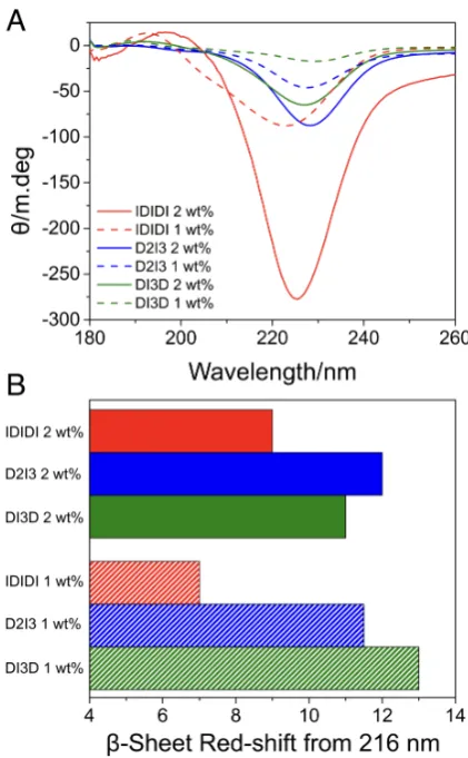
To investigate the relationship between supramolecular structure and mechanical properties, the secondary structure of the peptide assemblies in the hydrogels were studied us-ing spectroscopic techniques. CD experiments demonstrated that all the hydrogels had spectra that resembled ab-sheet, with a minima between 220-230 nm (Figures 3A, S5A and S6A). This was supported by the FTIR spectra of the amide I region (Figures S6B and S7B), where all the hydrogels dis-play a prominant peak at 1630 cm 1 indicating a b-sheet
conformation.[33,34]However, comparing the CD and FTIR
spectra for each of the sequences, distinctly different signa-tures are evident. The CD spectra differ in both intensity and are red-shifted relative to those of model b-sheets, which typically have a maximum at 195 nm and a minimum at 216 nm.[34]
The CD signatures ofb-sheets are known to have greater variability than other peptide secondary structures.[28] b
-sheets have both significant intermolecular and intrastrand hydrogen bonding.[35] Furthermore, peptides can form
an-tiparallel, parallel, or mixedb-sheets, which will influence both the strands in the assemblies as well as the networks they form.[28] When analyzing the relative red-shifts in the
CD minima of the different hydrogels, the IDIDI sequence provides the softest gels and has the smallest degree of red-shift at both 1 and 2 wt% (Figures 3A and B). In contrast, DI3D and D2I3 materials have similar red-shifts with no significant difference in G’ at 1 wt%. However, at 2 wt% the D2I3 sequence is significantly stiffer and has the greatest red-shift in the CD spectra at this concentration (Figure 3A and B). Previous studies have suggested that a red-shift in the CD spectra ofb-sheets is thought to be representative of more twisted and distorted arrangements.[28,36,37]The degree
of twisting ofb-sheets is centered around the middle of the sequence.[34]In twistedb-sheets, the hydrogen-bonding
dis-tance increases as the angle between two peptides increases, weakening the intermolecular forces and hydrogen bonds on the periphery of theb-sheet.[38,39]This will influence the
in-termolecular forces between individual peptide sequences in theb-sheet and the morphology of the structures present in the hydrogel.[35] A difference in b-sheet peak intensity at
[image:3.595.317.528.199.540.2]220-230 nm was also observed. The CD measurements are performed at the concentration found in the hydrogel and in some cases the hydrogels are partially opaque. This is likely to result in some fraction of the light being scattered, influ-encing peak intensity.
Figure 3A) Circular dichroism of the pentapeptide hydrogels at 1 and 2 wt%, the minima between 220 nm and 230 nm is typically indicative ofb-sheet formation. B) Theb-sheet red-shift from 216 nm taken from the circular dichroism spectra.
tem-Figure 4Cryo-FIB scanning electron micrographs of the hydro-gels. A) IDIDI 2 wt%, B) IDIDI 1 wt%, C) D2I3 2 wt%, D) D2I3 1 wt%, E) DI3D 2 wt%, and F) DI3D 1 wt%. The scale bar for all the images is 2µm.Note:C) has a reduced magnification.
perature of the stage to 100 C causes water to slowly sub-lime away from this face, revealing the underlying physi-cal structure (Figure S10). This technique allows for imag-ing of the hydrogels in their native state and in the presence of bound water, overcoming major artefacts associated with drying and water removal (more details of this technique can be found in the ESI).[40]
From the electron micrographs collected, it is evident that the charge distribution in the peptide sequence influ-ences the microstructures of the hydrogels (Figures 4, S8 and S9). The IDIDI hydrogels are comprised of high as-pect ratio nano-fibers, which at 2 wt% are several microns in length, extending to the height of the trench milled by the FIB (Figure 4A). At a lower concentration (1 wt%), the IDIDI hydrogels still maintain the same nano-fibrillar mor-phology but the fibers are shorter in length (Figure 4B). In comparison both the D2I3 and DI3D sequences have more entangled microstructures. The D2I3 materials are formed
from plate-like assemblies interconnected by some fibrous domains (Figures 4C and D), these observations were fur-ther supported by cryo-Transmission Electron Microscopy images of the D2I3 hydrogels at 2 wt% (Figure S11). Simi-larly, the DI3D hydrogels are comprised of some nano-fibers but mostly contain dense regions of fibrous bundles (Figures 4E and F). In summary, it can be observed that the more en-tangled structures have a greater degree of interconnectivity between the assemblies.
Recently, it has been reported that Asp positioning can influence the stacking orientation of tripeptide b-sheet as-semblies.[20]Shifting the Asp from the C- to the N-terminus
was shown to invert the conformation from a parallel to an anti-parallelb-sheet.[20]Similarly, both the D2I3 and DI3D
peptides contain charged Asp species situated at the termini of the sequence with regions of 3 repeat Ile residues. Pre-vious studies on polyisoleucines reported that sequential Ile rich structures are more stable in twisted parallelb-sheet ar-rangements.[35,41]The FTIR spectra for the D2I3 and DI3D
sequences have two minor peaks at 1655 cm 1 and 1675
cm 1(Figures S6B and S7B). It has been shown that twisted b-sheets (both in parallel and antiparallel conformations) can display an amide I splitting with a peak between 1680-1690 cm 1and also, a peak at 1650 cm 1.[28,34] While the
D2I3 and DI3D sequences here cannot be explicitly defined as being in an antiparallel or a parallel orientation, these ob-servations are in agreement with the red-shifted CD spectra found in this study, which suggests that both the D2I3 and DI3D hydrogels contain more twistedb-sheets.
The terminal charged groups coupled with weakened hy-drogen bonds on the periphery of the D2I3 and DI3D b -strands will result in a greater potential to form ionic inter-actions and further hydrogen bonds with other neighboring strands. This will give rise to the entangled and intercon-nected assemblies attributed to the D2I3 and DI3D hydro-gels (Figures 4C, D, E, F and S11). In the IDIDI sequence, the Asp residues are positioned more centrally with singular
b-sheet forming amino acids (Ile) in the middle and at the termini. Given that this arrangement does not contain a se-ries of repeat Ile residues, it is likely to provide less twisted
b-sheets. These types of structure will have less entropy and disorder, with hydrogen bonds between sequences being equal in length across the peptide chain. This is likely to fa-cilitate planar stacking arrangements and result in the high aspect ratio nano-fiber assemblies in Figures 4A and B.
[image:4.595.58.285.61.449.2]peptide designs demonstrate that by altering the position of theb-sheet forming amino acids and charge distribution of the sequence serves as a unique approach to control the mor-phology and tune the mechanical properties of the resultant hydrogel. Both substrate stiffness and substrate shape have been shown to influence cellular behavior.[29,30,42]
There-fore, with control over both of these parameters, the hydro-gels have potential to act as tissue engineering scaffolds and matrices.
We report three pentapeptide sequences free of aromatic groups, which can form robust hydrogels with gelation in-duced through changes in pH. We demonstrated that the stiffness of the hydrogels can be tuned across 2 orders of magnitude (2-200 kPa) by altering the concentration and charge distribution of the peptide sequence. Being formed through non-covalent interactions, the hydrogels can both selfheal and shear thin through the reassembly of the phys-ical crosslinks. To explore the relationship between molec-ular design and the mechanical properties of the hydrogel, we utilized spectroscopic techniques, which verified theb -sheet structure. Depending on the peptide sequence and its charge distribution, different degrees of red-shift were ev-ident in the CD spectra, which corresponded to the differ-ent morphologies of the self-assembled structures within the hydrogels. Cryo-FIB SEM identified that the IDIDI hydro-gels were formed from high-aspect ratio nanofibers. In con-trast, the D2I3 and DI3D hydrogels had more entangled and interconnected structures, generating the stiffest hydrogels. These pentapetide self-assembled hydrogels attributed with tunable morphology and mechanical properties, along with their ability to selfheal and shear thin, provides a promising platform for tissue engineering, injectable delivery vectors and 3D printing applications.
Acknowledgements
The authors thank the Nanoscale and Microscale Research Centre (nmRC) for providing access to instrumentation. We also thank Dr. Aniello Palma and Dr. Guanglu Wu for their suggestions and useful discussions.
References
[1] S Fleming, R. V. Ulijn,Chem. Soc. Rev.2014,43, 8150–8177. [2] G. Fichman, E. Gazit,Acta Biomater.2014,10, 1671–1682. [3] P. W.J. M. Frederix, G. G. Scott, Y. M. Abul-Haija, D. Kalafatovic,
C. G. Pappas, N. Javid, N. T. Hunt, R. V. Ulijn, T. Tuttle,Nat. Chem.
2014,7, 30–37.
[4] S Marchesan, C. D. Easton, K. E. Styan, L. J. Waddington, F Kushkaki, L Goodall, K. M. McLean, J. S. Forsythe, P. G. Hartley,
Nanoscale2014,6, 5172–5180.
[5] C. A. E. Hauser, R. Deng, A. Mishra, Y. Loo, U. Khoe, F. Zhuang, D. W. Cheong, A. Accardo, M. B. Sullivan, C. Riekel, J. Y. Ying, U. A. Hauser,Proc. Natl. Acad. Sci. USA2011,108, 1361–1366. [6] N. S. de Groot, T. Parella, F. X. Aviles, J. Vendrell, S. Ventura,
Bio-phys. J.2007,92, 1732–1741.
[7] P. Moitra, Y. Subramanian, S. Bhattacharya,J. Phys. Chem. B2017,
121, 815–824.
[8] C. Guo, Y. Luo, R. Zhou, G. Wei,Nanoscale2014,6, 2800–2811. [9] M. Reches, E. Gazit,Nano Lett.2004,4, 581–585.
[10] P. Tamamis, L. Adler-Abramovich, M. Reches, K. Marshall, P. Siko-rski, L. Serpell, E. Gazit, G. Archontis,Biophys. J.2009,96, 5020– 5029.
[11] S. Marchesan, C. D. Easton, F. Kushkaki, L. Waddington, P. G. Hart-ley,Chem. Commun.2012,48, 2195–2197.
[12] S. Marchesan, L. Waddington, C. D. Easton, D. A. Winkler, L. Goodall, J. Forsythe, P. G. Hartley,Nanoscale2012,4, 6752–6760. [13] J. James, A. B. Mandal,J. Colloid Interface Sci.2011,360, 600–
605.
[14] M. Reches, E. Gazit,Science2003,300, 625–627.
[15] P. Moitra, K. Kumar, P. Kondaiah, S. Bhattacharya,Angew. Chem. Int. Ed.2014,53, 1113–1117.
[16] J. Smadbeck, K. H. Chan, G. A. Khoury, B. Xue, R. C. Robin-son, C. A. E. Hauser, C. A. Floudas,PLoS Comput. Biol.2014,10, e1003718.
[17] A. Lakshmanan, D. W. Cheong, A. Accardo, E. Di Fabrizio, C. Riekel, C. A. E. Hauser,Proc. Natl. Acad. Sci. USA2013,110, 519– 524.
[18] A. K. Das, P. P. Bose, M. Drew, A Banerjee,Tetrahedron2007,63, 7432–7442.
[19] C. Subbalakshmi, S. V. Manorama, R. Nagaraj,J. Pept. Sci.2012,
18, 283–292.
[20] A. Lampel, S. A. McPhee, H.-A. Park, G. G. Scott, S. Humagain, D. R. Hekstra, B. Yoo, P. W.J. M. Frederix, T.-D. Li, R. R. Abzal-imov, S. G. Greenbaum, T. Tuttle, C. Hu, C. J. Bettinger, R. V. Ulijn,
Science2017,356, 1064–1068.
[21] H. Erdogan, E. Babur, M. Yilmaz, E. Candas, M. Gordesel, Y. Dede, E. E. Oren, G. B. Demirel, M. K. Ozturk, M. S. Yavuz, G. Demirel,
Langmuir2015,31, 7337–7345.
[22] C. G. Pappas, R. Shafi, I. R. Sasselli, H. Siccardi, T. Wang, V. Narang, R. Abzalimov, N. Wijerathne, R. V. Ulijn,Nat. Nanotech-nol.2016,11, 960–967.
[23] K. Tenidis, M. Waldner, J. Bernhagen, W. Fischle, M. Bergmann, M. Weber, M.-L. Merkle, W. Voelter, H. Brunner, A. Kapurniotu,J. Mol. Biol.2000,295, 1055–1071.
[24] D Thirumalai, D. K. Klimov, R. I. Dima,Curr. Opin. Struct. Biol.
2003,13, 146–159.
[25] P. Y. Chou, G. D. Fasman,Biochemistry1974,13, 222–245. [26] M. Levitt,Biochemistry1978,17, 4277–4285.
[27] M. A. Greenfield, J. R. Hoffman, M. O. de la Cruz, S. I. Stupp, Lang-muir2010,26, 3641–3647.
[28] E. T. Pashuck, H Cui, S. I. Stupp,J. Am. Chem. Soc.2010,132, 6041–6046.
[29] D. E. Discher, P Janmey, Y Wang,Science2005,310, 1139–1143. [30] A. J. Engler, S. Sen, H. L. Sweeney, D. E. Discher,Cell2006,126,
677–689.
[31] M. Guvendiren, H. D. Lu, J. A. Burdick,Soft Matter2012,8, 260– 272.
[32] D. E. Clarke, E. T. Pashuck, S. Bertazzo, J. V. M. Weaver, M. M. Stevens,J. Am. Chem. Soc.2017,139, 7250–7255.
[33] D. M. Byler, H. Susi,Biopolymers1986,25, 469–487.
[34] J. Kubelka, T. A. Keiderling,J. Am. Chem. Soc.2001,123, 12048– 12058.
[36] M. C. Manning, M. Illangasekare, R. W Woody,Biophys. Chem.
1988,31, 77–86.
[37] S. E. Paramonov, H. W. Jun, J. D. Hartgerink,J. Am. Chem. Soc.
2006,128, 7291–7298.
[38] F. R. Salemme, D. W. Weatherford,J. Mol. Biol.1981,146, 119– 141.
[39] F. R. Salemme,Prog. Biophys. Mol. Biol.1983,42, 95–133.
[40] C. Parmenter, A. Baki, K. Shakesheff in European Microscopy Congress 2016: Proceedings, Wiley-VCH,2016, pp. 682–683. [41] K. C. Chou, G Nemethy, H. A. Scheraga,J. Mol. Biol.1983,168,
389–407.
COMMUNICATION
Entry for the Table of Contents (Please choose one layout)
COMMUNICATION
Three pentapeptide sequences free from aromatic groups are able to form robust hydrogels with gelation induced via changes in pH. Through simple alterations in the chemical design of the peptide sequence, both the morphology and the mechanical properties of the resultant hydrogel can be tuned.
David E. Clarke, Christopher D.J. Parmenter, Oren A. Scherman*
Page No. – Page No.