0022-538X/08/$08.00⫹0 doi:10.1128/JVI.00358-08
Copyright © 2008, American Society for Microbiology. All Rights Reserved.
Recombinant Adeno-Associated Viral Vectors Are Deficient in
Provoking a DNA Damage Response
䌤
Michalis Fragkos,
1Madlaina Breuleux,
1† Nathalie Cle
´ment,
2and Peter Beard
1*
Ecole Polytechnique Fe´de´rale de Lausanne, Swiss Institute for Experimental Cancer Research, CH-1066 Epalinges, Switzerland,1
and Department of Gene and Cell Medicine, Mount Sinai School of Medicine, New York, New York 100292
Received 19 February 2008/Accepted 30 April 2008
Adeno-associated virus type 2 (AAV2) provokes a DNA damage response that mimics a stalled replication fork. We have previously shown that this response is dependent on ataxia telangiectasia-mutated and Rad3-related kinase and involves recruitment of DNA repair proteins into foci associated with AAV2 DNA. Here, we investigated whether recombinant AAV2 (rAAV2) vectors are able to produce a similar response. Surprisingly, the results show that both single-stranded and double-stranded green fluorescent protein-expressing rAAV2 vectors are defective in producing such a response. We show that the DNA damage signaling initiated by AAV2 was not due to the virus-encoded Rep or viral capsid proteins. UV-inactivated AAV2 induced a response similar to that of untreated AAV2. This type of DNA damage response was not provoked by other DNA molecules, such as single-stranded bacteriophage M13 or plasmid DNAs. Rather, the results indicate that the ability of AAV2 to produce a DNA damage response can be attributed to the presence ofcis-acting AAV2 DNA sequences, which are absent in rAAV2 vectors and could function as origins of replication creating stalled replication complexes. This hypothesis was tested by using a single-stranded rAAV2 vector containing the p5 AAV2 sequence that has previously been shown to enhance AAV2 replication. This vector was indeed able to trigger DNA damage signaling. These findings support the conclusion that efficient formation of AAV2 replication complexes is required for this AAV2-induced DNA damage response and provide an explanation for the poor response in rAAV2-infected cells.
Adeno-associated virus (AAV) is a replication-defective sin-gle-stranded DNA virus of theParvoviridaefamily. Productive infection of AAV requires the presence of a helper virus, such as adenovirus (Ad) or herpes virus (1, 2, 38, 41). AAV has a genome size of 4.7 kb and encodes two types of protein: the Rep polypeptides involved in its replication and the capsid proteins used for its encapsidation (16). The AAV genes are flanked by inverted terminal repeats (ITRs), which form a characteristic hairpin structure (13, 17). Infections by viruses are frequently followed by a host cell response that may either protect the host against the viral invasion or, alternatively, help the viral life cycle (21). In the case of AAV, a DNA damage response is provoked in the host cell, which potentially could be explained as a reaction to the single-stranded DNA part of the virus. It has been reported that transfection of cells with a random oligonucleotide can cause a p53-dependent apoptotic response (29). Alternatively, the hairpin structures of AAV DNA could be responsible for this type of response (6).
It has previously been shown by ourselves and others that treatment of AAV2 with UV light (UV-AAV2) inactivates the Rep and Cap genes and that subsequent infection in the ab-sence of helper virus leads to a significant DNA damage re-sponse and to perturbation of the cell cycle (19, 33, 42). This involves an arrest of the cell cycle at G2and an increase of the
levels of the p53 and p21 proteins (33). This DNA damage signaling pathway has been further examined and has been shown to resemble the response provoked by chemically in-duced stalled DNA replication forks. It is demonstrated by the formation of foci, which include AAV2 DNA localized with proteins involved in replication, such as replication protein A (RPA) and DNA polymerase␦(19). RPA has also been found to be associated with AAV2 replication fork progression (8, 27, 36). The AAV2-induced DNA damage response is also fol-lowed by activation of ataxia telangiectasia-mutated (ATM) and Rad3-related (ATR) proteins, leading to phosphorylation of Chk1 (19). Other indicators of this type of DNA damage response include phosphorylated H2AX (␥-H2AX) and hyper-phosphorylated RPA32 (11, 12, 34, 44). In the case of p53-deficient cells, infection with UV-AAV2 leads to cell death (33). This phenomenon has been attributed to mitotic catas-trophe as a result of proteosome-dependent degradation of Chk1 (18). The wild-type (WT) AAV2 virions can also activate a DNA damage response pathway that is dependent on Rep proteins and involves a cell cycle arrest at S phase (3).
AAV2 is widely used in gene therapy research due to its low immunogenicity, integration ability, and the absence of patho-genicity (4, 10, 15, 20). Despite these advantages, recombinant AAV2 (rAAV2) vectors are unable to integrate site specifically (24, 26, 32). Moreover, they give low levels of transgene ex-pression, at least partly due to the inability of the recombinant virus to replicate efficiently. Tullis and Shenk (37) showed that DNA sequences in the AAV2 early region can act in cisto enhance viral replication, and these have been narrowed down to an element within the p5 promoter of AAV2 (14, 37). Gene expression from rAAV2 vectors was enhanced when Ad or genotoxic stress was applied, and this was attributed to the fact
* Corresponding author. Mailing address: Swiss Institute for Exper-imental Cancer Research, EPFL SV ISREC, Chemin des Boveresses 155, Case postale, CH-1066 Epalinges, Switzerland. Phone: 41 21 6925921. Fax: 41 21 6526933. E-mail: peter.beard@epfl.ch.
† Present address: Basilea Pharmaceutica, Grenzacherstrasse 487, CH-4005 Basel, Switzerland.
䌤Published ahead of print on 7 May 2008.
7379
on November 8, 2019 by guest
http://jvi.asm.org/
observed with the WT AAV2 or UV-AAV2. Because rAAV2 vectors are used in gene therapy research as well as in clinical trials, it is important to know the reaction of the infected cells to the virus. Both types of vector DNA contain hairpin struc-tures, which could potentially lead to a DNA damage response. Furthermore, the single-stranded rAAV2 vector could lead to DNA damage signaling through its single-stranded DNA struc-ture. However, our results show that neither of the two rAAV2 vectors is capable of inducing a significant DNA damage re-sponse. We attribute this deficiency to the reduced ability of rAAV2 vectors to replicate and therefore to the absence of AAV2-stalled replication forks. Indeed, a single-stranded rAAV2 vector containing the p5 region of AAV2 that has been shown to function in the initiation of viral DNA replication was able to induce a DNA damage response.
MATERIALS AND METHODS
Cell lines, chemicals, and nucleic acids.All experiments were performed using U2OS osteosarcoma cells, which were grown in Dulbecco’s modified Eagle’s medium supplemented with 10% fetal bovine serum (FBS), penicillin/strepto-mycin, and ciprofloxacin (Ciproxin; Bayer). HeLa cells were used for the pro-duction of virus and were maintained in the same medium. Doxorubicin, DAPI (4⬘,6⬘,diamidino-2-phenylindole), and RNase A were obtained from Sigma. Doxorubicin was used at a final concentration of 50 nM. Proteinase K (Boeringer) was used at a final concentration of 100g/ml. M13 DNA was obtained from Fermentas (M13mp18). The plasmid used in the study was ob-tained from Promega (pRL-TK). The 36-Cy3 oligonucleotide (AAG-TGT-TAC-CGA-TAG-ACC-AGA-CCT-GAG-CTA-TGG-GAG) was designed randomly using the Vector-NTI program. The 55-Cy3 oligonucleotide (CTG-GGT-ATT- TAA-GCC-CGA-GTG-AGC-ACG-CAG-GGT-CTC-CAT-TTT-GAA-GCG-GGA-GGT-T) contains the AAV2 nucleotides 250 to 304.
Virus stocks, vectors, and infections.AAV2 was used for all experiments. Virus was produced following the protocol described by Q. Xie et al. (43). Briefly, HeLa cells were transfected with a linearized AAV2 plasmid, followed by infection with Ad type 5 (Ad5). When a full cytopathic effect was observed, the cell lysate was harvested and used to perform a secondary infection of HeLa cells. At 2 to 3 days after the secondary infection, the cells were lysed, and virus was pelleted by high-speed centrifugation. The AAV2 particles were finally obtained after two or three cycles of CsCl gradient centrifugation and stored at ⫺70°C. The single-stranded green fluorescent protein (ssGFP)-AAV2 and dou-ble-stranded GFP (dsGFP)-AAV2 vectors have been described before (23, 39). The p5GFP-AAV2 vector was made by substituting the chicken-actin promoter of ssGFP-AAV2 vector with the AAV2 p5 promoter (AAV2 nucleotides 162 to 324).
AAV2 samples were heat treated at 60°C for 45 min to inactivate any remain-ing Ad (the effectiveness of this treatment was verified) and then used to infect cells in a small quantity of plain Dulbecco’s modified Eagle’s medium for 3 to 5 h. Complete medium was then added to the cells, which were analyzed 1 to 4 days postinfection. AAV2 was used at a multiplicity of infection (MOI) of 10,000, whereas the dsGFP-AAV2 vector was used at an MOI of 20,000, and the
cells were then washed three times with PBS and incubated in blocking buffer (0.5% NP-40, 5% milk powder, 1% FBS) for 30 min. After being washed once with PBS, cells were incubated in buffer containing 5% milk powder, 1% FBS, and primary antibodies for 45 to 60 min. Coverslips were washed with PBS three times and then incubated with the appropriate secondary antibodies for 45 to 60 min. Cells were washed twice with PBS and then stained with 0.1g/ml DAPI for 45 s. Finally, they were washed with PBS and distilled water and mounted onto diazabicyclooctane-glycerol (50%).
The primary antibodies used were the following: anti-phospho-RPA32 S4/S8 (05-636; Bethyl), anti-RPA32 (NA18; Oncogene Research), anti-␥H2AX Ser139 (JBW301; Upstate Cell Signaling), and anti-Rep (a kind gift from P. Saudan). The secondary antibodies used were Alexafluor-488, Alexafluor-568 (Molecular Probes), and Cy3 (Jackson Immunoresearch) immunoglobulin G conjugates. Images were obtained using a Zeiss Axioplan fluorescence microscope.
Propidium iodide staining and fluorescence-activated cell sorting (FACS) analysis.Cells were trypsinized, washed with PBS, and then resuspended and incubated in 70% ethanol at⫺70°C for 60 min. The samples were washed with PBS and then resuspended in 0.1 mg/ml RNase A solution. Cells were incubated at 37°C for 15 to 20 min and then mixed with an equal volume of 20g/ml of propidium iodide. The samples were finally analyzed using a flow cytometer (Beckton-Dickinson).
Western blotting.Cells were collected, washed with PBS, and resuspended in 2.5 volumes of reporter lysis buffer (Promega) supplemented with a cocktail of protease inhibitors (Calbiochem). Samples were incubated on ice for 30 min and then centrifuged at 16,000⫻gfor 20 min at 4°C. Protein concentrations were measured using the Bradford method (Bio-Rad), and 40g of total protein from each sample was resolved on a 10% sodium dodecyl sulfate-polyacrylamide gel. The protein samples were then transferred (semidry transfer) onto a nitrocellu-lose membrane (Bio-Rad), which was then blocked overnight in blocking solu-tion (5% milk powder–0.1% Tween in PBS). The membrane was incubated with primary antibodies for 1 h, washed three times with PBS–0.1% Tween, incubated with secondary antibodies for 1 h, and finally washed three times with PBS–0.1% Tween. The blots were visualized using an ECL assay (Amersham) following the manufacturer’s instructions. The primary antibodies used were the following: MCM3 (minichromosome maintenance 3 complex) (ab4460; Abcam), anti-Rep, and anti-AAV2 viral protein (VP) (61058; Progen). Horseradish peroxi-dase-conjugated immunoglobulin G antibodies (Jackson Immunoresearch) were used as secondary antibodies.
RESULTS
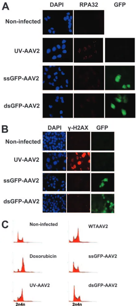
rAAV2 vectors do not provoke a DNA damage response typical of a stalled replication fork. In order to investigate whether the rAAV2 vectors can lead to DNA damage signal-ing, we compared a single-stranded AAV2 vector expressing the GFP gene under the control of a chicken-actin promoter (ssGFP-AAV2) and a double-stranded AAV vector expressing the GFP gene under the control of a cytomegalovirus promoter (dsGFP-AAV2) to WT AAV2 (Fig. 1A). Both rAAV2 vectors contain the 145-bp AAV2 ITR sequence at both ends. We initially infected U2OS osteosarcoma cells with either WT AAV2 or UV-AAV2 (UV-treated AAV2 to inactivate Rep
on November 8, 2019 by guest
and Cap genes). Infected cells were fixed and immunostained 1 day postinfection for phosphorylated RPA32 (Fig. 1B). Both WT AAV2 and UV-AAV2 infections resulted in phosphory-lation and accumuphosphory-lation of RPA32 into foci, indicating that a DNA damage response was activated that was due to the AAV2 DNA and not its protein products (Fig. 1B). DNA damage signaling was also examined by staining the cells for the phosphorylated version of histone variant H2AX (␥ -H2AX) 4 days after infection (Fig. 1C). Phosphorylation of H2AX is a widely used indicator of different types of response
to DNA damage (12). Based on␥-H2AX staining, WT AAV2
caused a significant DNA damage response, similar to that
[image:3.585.310.530.67.561.2]provoked by AAV2. Moreover, both WT AAV2 and UV-AAV2 caused cell cycle checkpoint activation and a subse-quent G2arrest, as can be concluded from the inhibited growth and the increased nuclear size of the infected cells 4 days postinfection (Fig. 1C) and FACS analysis (Fig. 2).
FIG. 1. AAV2 infection induces a DNA damage response. (A) De-scription of the viruses used in this study. (B) U2OS cells infected with WT AAV2 or UV-AAV2 and stained with phospho-RPA32 antibody at 1 day postinfection. Cell nuclei were stained with DAPI. (C)
In-fected cells were stained with␥-H2AX antibody 4 days postinfection. FIG. 2. rAAV2 vectors do not induce a significant DNA damage response. (A) U2OS cells infected with UV-AAV2, ssGFP-AAV2, or dsGFP-AAV2 and examined 1 day postinfection by staining with RPA32 antibody. Cell nuclei were stained with DAPI. GFP was used to identify the infected cells. (B) Infected cells were also examined 4 days postinfection by staining with␥-H2AX antibody. (C) U2OS cells infected with either WT AAV2 or rAAV2 vectors, stained with pro-pidium iodide, and analyzed by FACS 1 day postinfection. UV-AAV2-infected and doxorubicin-treated cells served as controls.
on November 8, 2019 by guest
http://jvi.asm.org/
[image:3.585.69.253.70.518.2]nuclei of the UV-AAV2-infected cells (Fig. 2B).
The host cell response to rAAV2 vectors was also assayed by propidium iodide staining and FACS analysis (Fig. 2C). U2OS cells were infected with the rAAV2 vectors, WT AAV2, and UV-AAV2 and analyzed 1 day postinfection. Doxorubicin treatment was also used as a control, as it is known to produce a significant G2cell cycle arrest. As shown in Fig. 2C, rAAV2 vectors were deficient in arresting the infected cells in G2, whereas the WT AAV2 and UV-AAV2 were capable of pro-ducing a significant G2cell cycle arrest.
The DNA damage response is independent of Rep.In order to investigate further the source of the DNA damage response provoked by UV-AAV2, we examined whether treatment of the virus with UV light inactivated the viral genes. We infected U2OS cells with WT AAV2 or UV-AAV2 with or without Ad5 coinfection and then stained the cells 1 day postinfection for Rep proteins and for␥H2AX to identify infected cells (Fig. 3A). Cells infected with WT AAV2, both in the presence or absence of helper virus, expressed the Rep gene. On the other hand, UV-AAV2-infected cells, as well as Ad5-infected and noninfected cells, did not express Rep in any case (Fig. 3A). This indicates that the DNA damage signaling observed in U2OS cells after infection with UV-AAV2 cannot be attrib-uted to Rep proteins.
Rep expression was also examined by Western blotting. U2OS cells were infected with WT AAV2 or UV-AAV2, with or without the presence of Ad5 helper virus, and protein was extracted 1 day after infection. Protein samples were separated on a sodium dodecyl sulfate-polyacrylamide gel and blotted against Rep antibody (Fig. 3B). This experiment again shows that no detectable Rep is expressed from the UV-AAV2. Un-infected U2OS cells or cells Un-infected with Ad5 did not express any Rep either (Fig. 3B). Taken together, these data indicate that AAV2 DNA itself can cause a significant DNA damage response, independently of Rep.
The DNA damage response is independent of the viral cap-sid.The next step in our study was to investigate whether the AAV2 capsid has a role in the induced DNA damage response. The role of the capsid in this kind of response was assayed by treating WT virus with proteinase K to destroy the capsid polypeptides. The proteinase K-treated AAV2 virions were further treated with UV light to inactivate any potential gene expression. The UV-AAV2 DNA was then used to transfect U2OS cells by lipofection, and transfected cells were finally
immunostained for␥-H2AX and phospho-RPA32 1 day
post-transfection. As shown in Fig. 4A, the viral capsid is dispens-able for the production of the DNA damage response, and the AAV2 genome alone is sufficient to induce this response. U2OS cells treated with Lipofectamine did not show any signs of DNA damage signaling (Fig. 4A).
[image:4.585.310.529.68.461.2]In order to assay the effectiveness of the proteinase K treat-ment on the AAV2 capsid proteins, U2OS cells were infected with AAV2 or AAV2 treated with proteinase K and then coinfected with Ad5. Total protein was extracted 3 days postin-fection and used for Western blotting against AAV2 capsid proteins (VP polypeptides). Cells infected with proteinase K-treated AAV2, as well as Ad5-infected and noninfected cells, did not show any signs of AAV2 capsid proteins (Fig. 4B). On
FIG. 3. Rep is not responsible for the UV-AAV2-induced DNA damage response. (A) Immunofluorescence experiment showing that UV-AAV2-infected U2OS cells initiate a DNA damage response (␥ -H2AX staining) without expressing Rep. Cells were infected with AAV2 either treated or not with UV light in the presence or absence of Ad5 and stained 1 day postinfection. Cells infected with Ad5 only were used as controls. (B) Western blot showing that UV-AAV2-infected U2OS cells do not express Rep 1 day after infection. MCM3 served as a loading control. NI, noninfected; WT, wild-type AAV2; UV, UV-AAV2.
on November 8, 2019 by guest
the other hand, the cells infected with AAV2 and Ad5 ex-pressed AAV2 capsid protein VP3. These data indicate that the proteinase K treatment was functional and therefore con-firms the conclusion that the viral capsid is not responsible for the DNA damage response observed in UV-AAV2-infected cells.
The DNA damage response is independent of the UV treat-ment per se.We then examined whether the UV treatment per se could be responsible for the DNA damage response
ob-served in UV-AAV2-infected cells. UV treatment of the virus can create protein-DNA links that could potentially provoke such a response. rAAV2 vectors as well as WT AAV2 were treated with UV light and then used to infect U2OS cells. Cells were stained with propidium iodide 1 day postinfection and then analyzed by FACS (Fig. 5A). The UV-treated rAAV2 vectors were unable to initiate a DNA damage signaling path-way, as assayed by cell cycle analysis.
The DNA damage response in the cells infected with the
FIG. 4. AAV2 capsid is not responsible for the UV-AAV2-induced DNA damage response. (A) U2OS cells transfected with proteinase K-treated UV-AAV2 particles and immunostained for both␥-H2AX and phospho-RPA32 1 day posttransfection. Cell nuclei were stained with DAPI. Lipofectamine-treated cells served as controls. (B) Control Western blotting to show that proteinase K treatment of AAV2 was functional. Presence of AAV2 particles was assayed in AAV2- or proteinase K-treated AAV2-infected (Ad5 coinfected) U2OS cells by staining for AAV2 capsid protein VP3 (61 kDa). The Ad5-infected U2OS sample served as a control. NI, noninfected.
FIG. 5. UV treatment is not responsible for the UV-AAV2-induced DNA damage response. (A) U2OS cells infected with UV-treated rAAV2 vectors, stained with propidium iodide, and analyzed by FACS do not arrest at G2. UV-AAV2-infected cells served as a control. (B) Infected cells
were stained with␥-H2AX antibody 4 days postinfection to show the absence of a DNA damage response from the UV-treated rAAV2 vectors. Cell nuclei were stained with DAPI. (C) U2OS cells infected with UV-treated rAAV2 vectors and stained for phospho-RPA32. Cells infected with UV-AAV2 were used as a control for phospho-RPA32 staining. Cells infected with the dsGFP-AAV2 vector were used as a control for GFP staining.
on November 8, 2019 by guest
http://jvi.asm.org/
is observed in U2OS cells infected with UV-AAV2 and that this type of response is probably AAV2 DNA specific.
The DNA damage response is specific to AAV2 DNA. We next addressed the question of whether the observed DNA damage response is specific to the AAV2 DNA sequence. For this reason U2OS cells were transfected with either bacterio-phage M13 or plasmid DNA, both of which were treated with UV light prior to transfection to inactivate any potential gene expression. M13 DNA is a 7.3-kb single strand, whereas the plasmid used was a double-stranded 4-kb DNA plasmid (see Materials and Methods). Cells were immunostained for ␥H2AX and phospho-RPA32 1 day posttransfection (Fig. 6A). Neither the M13 nor the plasmid DNA could initiate a DNA damage response comparable to the one produced by UV-AAV2. This indicates that the response observed in U2OS cells after infection with UV-AAV2 is specific to an AAV2 DNA sequence and cannot be produced by any DNA frag-ment.
To validate this finding, we transfected U2OS cells with a randomly designed oligonucleotide (36-mer–Cy3). The oligo-nucleotide used was Cy3-tagged so as to identify the trans-fected cells. Transtrans-fected cells were examined 1 day posttrans-fection by immunofluorescence analysis for the DNA damage indicators␥H2AX and phospho-RPA32. As shown in Fig. 6B this random piece of single-stranded DNA was not capable of initiating a DNA damage response, confirming the conclusion that the DNA damage signaling cascade cannot be initiated by any kind of DNA fragment.
The next step in our study was to investigate whether a single-stranded AAV2 DNA sequence itself is able to lead to a DNA damage response. We have shown that the DNA damage response induced by UV-AAV2 is similar to the response provoked by chemically induced stalled replication forks (19). It has also been demonstrated that the p5 AAV2 sequence acts incisto enhance viral replication (37). This sequence has been suggested to act as an origin of replication for AAV2 and is considered to be important for AAV2 site-specific integration (28, 30, 31). Thiscis-acting replication element (CARE) has been narrowed down to 55 nucleotides (AAV2 DNA nucleo-tides 250 to 304) containing the p5 TATA box, the terminal resolution site, and the Rep binding site (14). We thus de-signed an oligonucleotide comprised of this 55-nucleotide AAV2 DNA element tagged with Cy-3 (55-mer–Cy3) and used it to transfect U2OS cells and test whether this sequence itself can cause a DNA damage response. We selected this AAV2
[image:6.585.323.515.68.586.2]sequence to design the oligonucleotide because the p5 se-quence is the AAV2 part that is present in WT AAV2 and not in the rAAV2 vectors and could therefore account for the observed DNA damage response. The samples were assayed 1
FIG. 6. The DNA damage response is specific to AAV2 DNA. (A) Im-munofluorescence experiment showing that the DNA damage response can-not be caused by any DNA fragment. U2OS cells were transfected with UV-treated M13 or plasmid DNA and compared to UV-AAV2-infected cells. Cells were stained 1 day posttreatment for␥-H2AX and phospho-RPA32. Lipofectamine-treated cells were used as a control. (B) U2OS cells were transfected with two different oligonucleotides conjugated to Cy3 and then stained with␥-H2AX and phospho-RPA32 antibodies. UV-AAV2-infected cells were used as a control. Merged pictures include DAPI staining of nuclei.
on November 8, 2019 by guest
day posttransfection by staining for immunofluorescence with phospho-H2AX and phospho-RPA32 antibodies (Fig. 6B). The results show that the oligonucleotide containing the 55-nucleotide AAV2 sequence cannot lead to a significant DNA damage response. This indicates that the AAV2 55-nucleotide element is not sufficient to cause a DNA damage response and cannot explain, by itself, the massive response observed in UV-AAV2-infected U2OS cells.
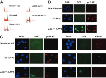
An rAAV2 vector containing p5 provokes a DNA damage response.We then constructed a single-stranded rAAV2 vec-tor expressing GFP under the control of the AAV2 p5 pro-moter (p5GFP-AAV2), which contains the 55-nucleotide CARE sequence, and asked the question whether this virus can provoke a DNA damage response. To answer this ques-tion, U2OS cells were infected with the p5GFP-AAV2 vector (UV treated or not) and stained with propidium iodide 1 day postinfection. Subsequent analysis by FACS shows that the cells infected with the p5GFP-AAV2 vector arrested at the G2 phase of the cell cycle (Fig. 7A). The effect of the p5GFP-AAV2 infection on the host cell was also examined by immu-nostaining infected U2OS cells for the DNA damage response marker␥-H2AX 1 day after infection (Fig. 7B). The p5GFP-AAV2-infected cells showed enhanced staining of ␥-H2AX, similar to the staining seen in the control UV-AAV2-infected
cells. The p5GFP-AAV2 vector is therefore able to induce a DNA damage response.
U2OS cells infected with p5GFP-AAV2 were also examined for AAV2-induced DNA repair focus formation by staining the cells for proteins known to be found in AAV2-induced stalled replication forks. Cells were infected and stained for RPA32 and phospho-RPA32 1 day postinfection. As shown in Fig. 7C, infection with the p5GFP-AAV2 vector led to phospho-RPA32 staining, although this was not within clear foci, as seen with WT AAV2. These data altogether show that the rAAV2 vector containing the p5 sequence is capable of inducing the DNA damage signaling pathway, though without leading to formation of DNA repair foci. The observed DNA damage response in UV-AAV2-infected cells can be therefore attrib-uted in large part to the p5 DNA sequence of AAV2.
DISCUSSION
[image:7.585.81.502.68.379.2]The data presented here indicate that AAV2 can induce a significant DNA damage response that is provoked by the AAV2 DNA sequence itself. The DNA damage signaling path-way initiated after AAV2 infection cannot be attributed to the capsid or to single-stranded DNA, as ssGFP-AAV2 virions include single-stranded DNA enclosed inside a protein capsid
FIG. 7. The rAAV2 vector containing the p5 sequence of AAV2 provokes a DNA damage response. (A) FACS analysis of U2OS cells infected with p5GFP-AAV2 (UV treated or not) indicating an AAV2-provoked cell cycle arrest. (B) Immunofluorescence staining of p5GFP-AAV2-infected cells with phospho-H2AX antibody. DAPI staining was used to stain the nuclei, and GFP was used to indicate the p5GFP-AAV2-infected cells. UV-AAV2-infected cells were used as a control for␥-H2AX staining. (C) Cells stained with RPA32 and phospho-RPA32 antibodies to identify AAV2-induced DNA repair foci. UV-AAV2-infected cells were used as a positive control for such foci.
on November 8, 2019 by guest
http://jvi.asm.org/
rAAV2 vectors without p5 did not have a significant effect on the infected cells. Altogether these data indicate that AAV2 contains a sequence, other than its ITRs, that is responsible for the induction of a DNA damage signaling pathway.
Tullis and Shenk (37) noted that rAAV2 vectors often rep-licate poorly and identified an AAV2 element in the left part of the genome that is essential for efficient AAV2 replication. Nony et al. (28) and Franc¸ois et al. (14) have recently located this element to a 55-nucleotide sequence within the p5 AAV2 promoter (AAV2 nucleotides 250 to 304), named CARE. Apart from enhancing replication, this element has also been shown to have a role in AAV2 integration (30). Given that the AAV2-induced DNA damage response is biochemically simi-lar to the response caused by stalled replication forks, we hypothesized that this element might function as an origin of replication, creating stalled replication forks and thereby in-ducing the DNA damage response. To test this hypothesis, we initially designed a 55-nucleotide-long oligonucleotide com-prised of the CARE sequence tagged with Cy3. This sequence by itself could not induce a DNA damage response. This could be because the CARE fragment alone may not be sufficient to create a stalled replication fork and thereby initiate a DNA damage response. This notion would agree with a study show-ing that M13 sshow-ingle-stranded DNA was able to lead to check-point activation only when it was primed (22). Activation of the replication checkpoint by aphidicolin was also shown to re-quire RNA primer synthesis (25). Therefore, the CARE se-quence might be able to provoke a DNA damage response only in the context of AAV2, which could provide the primer re-quired for the production of stalled replication forks.
To test whether an AAV2-derived virus containing the CARE fragment would lead to a DNA damage response, a single-stranded rAAV2 vector was constructed containing the AAV2 p5 sequence driving the expression of a GFP transgene. The p5-containing rAAV2 vector was indeed able to lead to DNA damage signaling. Specifically, p5GFP-AAV2 was able to arrest cells at the G2phase of the cell cycle, as well as to lead to H2AX phosphorylation. This vector triggered staining of the infected cells by phospho-RPA32 antibody but not the forma-tion of clear AAV2 DNA repair foci. This could mean that the AAV2-induced foci represent incomplete AAV2 replication centers that cannot be formed by p5GFP-AAV2, possibly due to the requirement for another AAV2 sequence that is not present in the p5GFP-AAV2 vector. Alternatively, the DNA damage signaling pathway induced by this rAAV2 vector might
be incomplete and not involve formation of DNA repair foci, which could be a downstream step of the DNA damage sig-naling pathway. The ability of the p5GFP-AAV2 vector to induce a DNA damage response without producing foci is an interesting observation that deserves further study.
The effect of the p5 region of AAV2 on the DNA damage response exhibited by AAV-infected cells can be explained by the presence of a potential origin of replication within this sequence. The p5 AAV2 promoter contains the CARE se-quence that has been thought to function as a replication origin (14, 28). RPA, which has been shown to colocalize with AAV2 replication centers, might recognize and bind to this potential origin of replication (8, 36, 40). Binding of RPA may then lead on to formation of a stalled replication fork, because of either defective AAV2 replication in the absence of helper virus or UV-induced lesions if the virus is UV inactivated. As a conse-quence, ATR protein becomes activated, and proteins such as RPA32 and H2AX become phosphorylated, leading to check-point activation and cell cycle arrest (Fig. 8). Therefore, initi-ation of repliciniti-ation at an adequate level is required for a DNA damage response to be produced. The rAAV2 vectors that do not contain the p5 sequence are unable to achieve this and are therefore unable to induce such a DNA damage signaling pathway.
AAV2 seems able to interact with cellular DNA damage signaling in several ways. An association of the MRN (Mre11/ Rad50/Nbs1) complex with AAV2 DNA has recently been reported (5, 35), and such binding acts to limit viral replication and transduction. Choi et al. (6) have also examined the role of cellular DNA recombination and repair pathways in AAV2 genome processing and found that, in this case, cellular DNA repair proteins including Mre11 and Nbs1 are required for efficient circularization of self-complementary recombinant AAV2 genomes. The MRN complex is a sensor of double-strand breaks and is part of the ATM signaling pathway (9). We have shown previously that the DNA damage response we observed in AAV2-infected cells, involving cell cycle arrest and H2AX phosphorylation, is independent of the ATM pathway (19). We therefore propose that the AAV2 hairpins of either the WT AAV2 or the rAAV2 vectors may attract the MRN complex, but the induced DNA damage response in that case is not as strong as the one seen when stalled DNA replication
provokes a DNA damage response. Pol␦, DNA polymerase␦.
on November 8, 2019 by guest
forks are formed. The DNA damage signaling pathway in-duced by p5 promoter-containing AAV2, which includes acti-vation of ATR and Chk1 proteins, is possibly so strong and dominant that under these conditions it does not allow any other signaling pathway to be easily detected.
Summarizing the above results, we have shown that the p5 sequence of AAV2 is required for the production of a DNA damage response in the AAV2-infected cells. rAAV vectors that do not include this sequence are unable to provoke DNA damage signaling, whereas the vector containing this sequence can initiate a DNA damage response. The data therefore sug-gest that rAAV2 gene therapy vectors not containing the p5 AAV2 promoter sequence are less likely to affect adversely the host cells by induction of cellular DNA damage signaling.
ACKNOWLEDGMENTS
We thank N. Paduwat for technical help, as well as past and present members of the ISREC Virology group for many invaluable discus-sions, and M. Linden for helpful advice.
This work was supported by Recherche Suisse contre le Cancer and the Fonds National Suisse de la Recherche Scientifique.
REFERENCES
1.Berns, K. I.1996.Parvoviridae: the viruses and their replication, p. 2173– 2197.InB. N. Fields, D. M. Knipe, P. M Howley, D. E. Griffin, R. A. Lamb, M. A. Martin, B. Roizman, and S. E. Straus (ed.), Fields virology, 3rd ed. Lippincott, Williams & Wilkins, Philadelphia, PA.
2.Berns, K. I., and R. M. Linden.1995. The cryptic life style of adeno-associated virus. Bioessays17:237–245.
3.Berthet, C., K. Raj, P. Saudan, and P. Beard.2005. How adeno-associated virus Rep78 protein arrests cells completely in S phase. J. Virol.102:13634– 13639.
4.Burger, C., O. S. Gorbatyuk, M. J. Velardo, C. S. Peden, P. Williams, S. Zolotukhin, P. J. Reier, R. J. Mandel, and N. Muzyczka.2004. Recombinant AAV viral vectors pseudotyped with viral capsids from serotypes 1, 2 and 5 display differential efficiency and cell tropism after delivery to different regions of the central nervous system. Mol. Ther.10:302–317.
5.Cervelli, T., J. A. Palacios, L. Zentilin, M. Mano, R. A. Schwartz, M. D. Weitzman, and M. Giacca.2008. Processing of recombinant AAV genomes occurs in specific nuclear structures that overlap with foci of DNA-damage-response proteins. J. Cell Sci.121:349–357.
6.Choi, V. W., D. M. McCarty, and R. J. Samulski.2006. Host cell DNA repair pathways in adeno-associated viral genome processing. J. Virol.80:10346– 10356.
7.Choi, V. W., R. J. Samulski, and D. M. McCarty.2005. Effects of adeno-associated virus DNA hairpin structure on recombination. J. Virol.79:6801– 6807.
8.Christensen, J., and P. Tattersall.2002. Parvovirus initiator protein NS1 and RPA coordinate replication fork progression in a reconstituted DNA repli-cation system. J. Virol.76:6518–6531.
9.D’Amours, D., and S. P. Jackson.2002. The Mre11 complex: at the cross-roads of DNA repair and checkpoint signaling. Nat. Rev. Mol. Cell. Biol.
3:317–327.
10.Emery, D. W., T. Nishino, K. Murata, M. Fragkos, and G. Stamatoyanno-poulos.2002. Hematopoietic stem cell gene therapy. Int. J. Hematol.75:
228–236.
11.Fanning, E., V. Klimovich, and A. R. Nager.2006. A dynamic model for replication protein A (RPA) function in DNA processing pathways. Nucleic Acids Res.34:4126–4137.
12.Fernandez-Capetillo, O., A. Lee, M. Nussenzweig, and A. Nussenzweig.2004. H2AX: the histone guardian of the genome. DNA Repair (Amst.)3:959–967. 13.Flotte, T. R., and K. I. Berns.2005. Adeno-associated virus: a ubiquitous
commensal of mammals. Hum. Gene Ther.16:401–407.
14.Francois, A., M. Guilbaud, R. Awedikian, G. Chadeuf, P. Moullier, and A. Salvetti.2005. The cellular TATA binding protein is required for rep-de-pendent replication of a minimal adeno-associated virus type 2 p5 element. J. Virol.79:11082–11094.
15.Hendrie, P. C., and D. W. Russell.2005. Gene targeting with viral vectors. Mol. Ther.12:9–17.
16.Hunter, L. A., and R. J. Samulski.1992. Colocalization of adeno-associated virus rep and capsid proteins in the nuclei of infected cells. J. Virol.66:317–324. 17.Im, D. S., and N. Muzyczka.1989. Factors that bind to adeno-associated
virus terminal repeats. J. Virol.63:3095–3104.
18.Jurvansuu, J., M. Fragkos, C. Ingemarsdotter, and P. Beard.2007. Chk1
instability is coupled to mitotic cell death of p53-deficient cells in response to virus-induced DNA damage signaling. J. Mol. Biol.372:397–406. 19.Jurvansuu, J., K. Raj, A. Stasiak, and P. Beard.2005. Viral transport of
DNA damage that mimics a stalled replication fork. J. Virol.79:569–580. 20.Kotin, R. M., M. Siniscalco, R. J. Samulski, X. D. Zhu, L. Hunter, C. A.
Laughlin, S. McLaughlin, N. Muzyczka, M. Rocchi, and K. I. Berns.1990. Site-specific integration by adeno-associated virus. J. Virol.87:2211–2215. 21.Lilley, C. E., R. A. Schwartz, and M. D. Weitzman.2007. Using or abusing:
viruses and the cellular DNA damage response. Trends Microbiol.15:119–126. 22.MacDougall, C. A., T. S. Byun, C. Van, M. C. Yee, and K. A. Cimprich.2007. The structural determinants of checkpoint activation. Genes Dev.21:898–903. 23.McCarty, D. M., P. E. Monahan, and R. J. Samulski.2001. Self-comple-mentary recombinant adeno-associated virus (scAAV) vectors promote ef-ficient transduction independently of DNA synthesis. Gene Ther.8:1248– 1254.
24.McCarty, D. M., S. M. Young, Jr., and R. J. Samulski.2004. Integration of adeno-associated virus (AAV) and recombinant AAV vectors. Annu. Rev. Genet.38:819–845.
25.Michael, W. M., R. Ott, E. Fanning, and J. Newport.2000. Activation of the DNA replication checkpoint through RNA synthesis by primase. Science
289:2133–2137.
26.Miller, D. G., G. D. Trobridge, L. M. Petek, M. A. Jacobs, R. Kaul, and D. W. Russell.2005. Large-scale analysis of adeno-associated virus vector integra-tion sites in normal human cells. J. Virol.79:11434–11442.
27.Ni, T. H., W. F. McDonald, I. Zolotukhin, T. Melendy, S. Waga, B. Stillman, and N. Muzyczka.1998. Cellular proteins required for adeno-associated virus DNA replication in the absence of adenovirus coinfection. J. Virol.
72:2777–2787.
28.Nony, P., J. Tessier, G. Chadeuf, P. Ward, A. Giraud, M. Dugast, R. M. Linden, P. Moullier, and A. Salvetti.2001. Novelcis-acting replication ele-ment in the adeno-associated virus type 2 genome is involved in amplification of integrated rep-cap sequences. J. Virol.75:9991–9994.
29.Nur-E-Kamal, A., T. K. Li, A. Zhang, H. Qi, E. S. Hars, and L. F. Liu.2003. Single-stranded DNA induces ataxia telangiectasia mutant (ATM)/p53-depen-dent DNA damage and apoptotic signals. J. Biol. Chem.278:12475–12481. 30.Philpott, N. J., C. Giraud-Wali, C. Dupuis, J. Gomos, H. Hamilton, K. I.
Berns, and E. Falck-Pedersen.2002. Efficient integration of recombinant adeno-associated virus DNA vectors requires a p5-rep sequence incis. J. Vi-rol.76:5411–5421.
31.Philpott, N. J., J. Gomos, K. I. Berns, and E. Falck-Pedersen.2002. A p5 integration efficiency element mediates Rep-dependent integration into AAVS1 at chromosome 19. Proc. Natl. Acad. Sci. USA99:12381–12385. 32.Ponnazhagan, S., D. Erikson, W. G. Kearns, S. Z. Zhou, P. Nahreini, X. S.
Wang, and A. Srivastava.1997. Lack of site-specific integration of the re-combinant adeno-associated virus 2 genomes in human cells. Hum. Gene Ther.8:275–284.
33.Raj, K., P. Ogston, and P. Beard.2001. Virus-mediated killing of cells that lack p53 activity. Nature412:914–917.
34.Redon, C., D. Pilch, E. Rogakou, O. Sedelnikova, K. Newrock, and W. Bonner.2002. Histone H2A variants H2AX and H2AZ. Curr. Opin. Genet. Dev.12:162–169.
35.Schwartz, R. A., J. A. Palacios, G. D. Cassell, S. Adam, M. Giacca, and M. D. Weitzman.2007. The Mre11/Rad50/Nbs1 complex limits adeno-associated virus transduction and replication. J. Virol.81:12936–12945.
36.Stracker, T. H., G. D. Cassell, P. Ward, Y. M. Loo, B. van Breukelen, S. D. Carrington-Lawrence, R. K. Hamatake, P. C. van der Vliet, S. K. Weller, T. Melendy, and M. D. Weitzman.2004. The Rep protein of adeno-associated virus type 2 interacts with single-stranded DNA-binding proteins that en-hance viral replication. J. Virol.78:441–453.
37.Tullis, G. E., and T. Shenk.2000. Efficient replication of adeno-associated virus type 2 vectors: acis-acting element outside of the terminal repeats and a minimal size. J. Virol.74:11511–11521.
38.Vihinen-Ranta, M., S. Suikkanen, and C. R. Parrish.2004. Pathways of cell infection by parvoviruses and adeno-associated viruses. J. Virol.78:6709– 6714.
39.Wang, Z., H. I. Ma, J. Li, L. Sun, J. Zhang, and X. Xiao.2003. Rapid and highly efficient transduction by double-stranded adeno-associated virus vec-tors in vitro and in vivo. Gene. Ther.10:2105–2111.
40.Ward, P., F. B. Dean, M. E. O’Donnell, and K. I. Berns.1998. Role of the adenovirus DNA-binding protein in vitro adeno-associated virus DNA rep-lication. J. Virol.72:420–427.
41.Weitzman, M. D., K. J. Fisher, and J. M. Wilson.1996. Recruitment of wild-type and recombinant adeno-associated virus into adenovirus replica-tion centers. J. Virol.70:1845–1854.
42.Winocour, E., M. F. Callaham, and E. Huberman.1988. Perturbation of the cell cycle by adeno-associated virus. Virology167:393–399.
43.Xie, Q., J. Hare, J. Turnigan, and M. S. Chapman.2004. Large-scale pro-duction, purification and crystallization of wild-type adeno-associated vi-rus-2. J. Virol. Methods122:17–27.
44.Zou, Y., Y. Liu, X. Wu, and S. M. Shell.2006. Functions of human replication protein A (RPA): from DNA replication to DNA damage and stress re-sponses. J. Cell Physiol.208:267–273.