I p
i
i
I 1:
I "
•1 B
I ,_
NONSYMBIOTIC PLANT HAEMOGLOBIN GENES
Carol Rae Andersson
February 1994
A thesis submitted for the degree of Doctor of Philosophy of
This thesis contains no material which has been accepted for the award of any
other degree or diploma in any University. To the best of my knowledge, this thesis
contains no material previously published, or the result of any work by another person,
except where due reference is made in the text.
The research described in this thesis is my own original work, with the following
exceptions.
Celia Miller embedded, sectioned and mounted tissue for dark field microscopy
(Chapter 2). S.B. Narasimhulu made some of the Parasponia haemoglobin promoter deletions (P-235 and below, Chapter 2). Janice Norman constructed and screened the
soybean genomic library and isolated the soybean nonsymbiotic haemoglobin gene
(Chapter 4). Erik Ostergaard Jensen conducted the Northern analysis of the soybean
nonsymbiotic haemoglobin gene (Chapter 4). Danny Llewellyn cloned the Arabidopsis
ACKNOWLEDGEMENTS
There are many people in the lab and throughout the Division who have helped
me at some stage during my PhD, either with practical help, advice or encouragement.
To them my grateful thanks. There are several people I would particularly like to thank.
Special thanks to my supervisors, Liz Dennis and Danny Llewellyn for their
support and advice throughout my PhD, from practical help to comforting words in
times of crisis. Also to Janice Norman for her encouragement and for teaching me
many techniques, and especially for practical help during the last few months. Thanks
to Jim Peacock for spurring me on and for critical reading of much of this thesis.
Thanks to Celia Millar for all her help with preparing tissue sections for dark field
microscopy, Stuart Craig for instruction and advice, Fraser Bergersen and lnara Licis
for generously providing soybean plants, Paul Keese for enlightening discussions,
Leigh Farrell and Tony Ashton for computing help, Robert Hill (University of Manitoba)
for making information regarding the barley haemoglobin available prior to publication
and to Cyril Appleby for his continued enthusiasm. Special thanks to Ron Wier for
keeping track of PhD progress and formal requirements.
Part of the work described in this thesis was conducted during a six month visit
to Professor Kjeld Marckers laboratory, University of Aarhus, Denmark. My thanks to
him, Jens Stougaard, Erik Ostergaard Jensen, Peter Lauridsen and Niels Bech
Laursen for vectors and bacterial strains and helpful advice about high efficiency
transformation of Lotus. Special thanks to Jens Stougaard for initiating the soybean
lbps1 study. Thanks to Erik Ostergaard Jensen for useful discussions and his
involvement in the soybean nonsymbiotic haemoglobin work.
Special thanks to Mary Clarke and Erika Merkel, whose efficiency kept the lab
running smoothly and made life easier for the rest of us.
Finally, thanks to my family for their encouragement and patience.
This research was conducted in the Division of Plant Industry, CSIRO.
acknowledge financial support from an Australian Postgraduate Research Award and
ABSTRACT
Haemoglobin is present in the nitrogen-fixing root nodules of both legumes and the non-legume Parasponia, where it facilitates the diffusion of oxygen to the respiring bacteria at a free oxygen concentration too low to inhibit the oxygen sensitive nitrogenase complex (reviewed by Appleby, 1984; Appleby et al, 1988). A closely related non-nodulating relative of Parasponia, Trema tomentosa, has a haemoglobin gene that is expressed in roots (Bogusz et al, 1988). Both the Parasponia and Trema
haemoglobin promoters direct expression of a reporter gene in the nodules of a transgenic legume (Bogusz et al, 1988). However, this thesis demonstrates that, unlike expression of the leghaemoglobins and endogenous Parasponia haemoglobin, these promoters confer high expression in uninfected tissues of Lotus nodules, and low expression in the bacteroid containing cells. This nodule expression pattern is similar to that of a nonsymbiotic haemoglobin gene from Casuarina, analysed concurrently with this study (Jacobsen-Lyon et al, in preparation), which is expressed in parenchyma of both roots and nodules. Thus, regulation of the Parasponia and Trema haemoglobin genes in a transgenic legume reflects aspects of both symbiotic and nonsymbiotic expression and suggests distinct signals control organ versus cell specificity.
The discovery of haemoglobin and haemoglobin genes in phylogenetically diverse non-legume genera, including Parasponia, Trema, Casuarina and Hordeum
(barley) (Appleby et al, 1983; Bogusz et al, 1988; Fleming et al, 1987; Christensen et al, 1991; Taylor et al, 1993) led to the proposal that haemoglobin may have a fundamental, but as yet unknown, function in all plants independent of its role in
symbiotic nodules (Appleby et al, 1988; 1990). This thesis explores the suggestion that legumes must have a haemoglobin gene expressed in nonsymbiotic tissue in addition to the nodule specific leghaemoglobin genes (Appleby et al, 1988; 1990). The apparent pseudogene /bps 1 has been suggested as the soybean nonsymbiotic haemoglobin gene (Nap and Bisseling, 1990b,c). However, no evidence of expression of lbps1, either in soybean or transgenic Lotus, was found in this study and it is likely
This thesis describes the isolation and characterisation of a novel soybean
haemoglobin gene that shows significant homology to other nonsymbiotic haemoglobin
genes and which is expressed in a variety of tissues. This haemoglobin gene
represents the elusive nonsymbiotic legume haemoglobin and supports the proposal
that the nodule specific leghaemoglobins arose from a preexisting nonsymbiotic
haemoglobin by gene duplication (Appleby et al, 1988; 1990). In addition, this thesis
describes the isolation of a haemoglobin gene fragment from Arabidopsis, which will
provide a powerful tool for the analysis of the general function of plant haemoglobin.
The taxonomic distribution of haemoglobins and the regulation of both symbiotic
and nonsymbiotic haemoglobin genes has implications both for the origin of plant
CONTENTS page
TITLE PAGE
CANDIDATES STATEMENT II
ACKNOWLEDGEMENTS 111
ABSTRACT IV
CONTENTS VI
LIST OF FIGURES XII
LIST OF TABLES XIII
CHAPTER 1
GENERAL INTRODUCTION 1
LEGHAEMOGLOBIN AND THE LEGUME-(BRADY)RHIZOBJUM SYMBIOSIS 1
The legume-(Brady)Rhizobium symbiosis 1
Leghaemoglobin facilitates oxygen diffusion within nitrogen fixing nodules 4 Leghaemoglobin as the archetype symbiotic protein
NODULINS ARE PLANT PROTEINS SPECIFICALLY INVOLVED
IN NODULATION
Early nodulins are involved in nodule formation
EN002 AND EN0012 ARE CELL WALL PROTEINS
Late nodulins are involved in nodule function
ENZYMES INVOLVED IN NITROGEN AND CARBON METABOLISM
PERIBACTEROID MEMBRANE NODULINS
Origin of nodulins
PARASPONJA HAEMOGLOBIN AND THE
PARASPONIA-(BRADY)RHIZOBIUM SYMBIOSIS
The Parasponia/(Brady)Rhizobium symbiosis
Haemoglobin is expressed in nodules of Parasponia and
6
9
9
10
11
1 1
12
13
13
HAEMOGLOBIN IN ACTINORHIZAL NODULES AND THE ACTINORHIZAL
SYMBIOSES 15
The actinorhizal symbioses 15
Haemoglobin in actinorhizal nodules 16
NONSYMBIOTIC PLANT HAEMOGLOBINS AND POSSIBLE FUNCTION 17 THE COMMON FUNCTION OF HAEMOGLOBIN IS REVERSIBLE
BINDING OF OXYGEN 18
Some haemoglobins are involved in oxygen transport 18
Some haemoglobins are linked to enzymatic domains 19
E.COLI AND YEAST HAEMOGLOBINS HAVE A REDUCTASE ACTIVITY 19
ASCARIS HAEMOGLOBIN CHANNELS OXYGEN INTO BIOSYNTHETIC PATHWAYS
Other haem proteins are also known to act as oxygen sensors
Rhizobium Fixl is part of a two-component regulator of
20 21
nitrogen fixation genes 21
ALL HAEMOGLOBINS EVOLVED FROM A COMMON ANCESTOR 22
Plant and vertebrate haemoglobins have a similar gene structure 23
Evolution of plant haemoglobin 25
HAEMOGLOBIN GENE REGULATION CAN BE DISSECTED USING TRANSGENIC PLANTS
Expression of leghaemoglobin genes involves quantitative
and qualitative promoter elements
SOYBEAN lbc3 SOYBEAN Iba
SESBANIA ROSTRATA g/b3
Gene expression requires trans-acting protein factors
A RHIZOBIAL PROTEIN INTERACTS SPECIFICALLY WITH A
27
28 28 30 32
33
LEGHAEMOGLOBIN PROMOTER 33
NODULE SPECIFIC HMG-LIKE FACTORS MAY BE SCAFFOLD PROTEINS 34
CASUARINA SYMBIOTIC HAEMOGLOBIN GENE REGULATION
PARASPONIA AND TREMA HAEMOGLOBIN GENES GENERAL AIMS OF THIS THESIS
CHAPTER 2
EXPRESSION OF THE PARASPONIA AND TREMA HAEMOGLOBIN PROMOTERS IN TRANSGENIC LOTUS REFLECTS ASPECTS OF BOTH
34 35
35
SYMBIOTIC AND NONSVMBIOTIC HAEMOGLOBIN EXPRESSION 37
INTRODUCTION 37
RESULTS 39
The Parasponia and Trema haemoglobin promoters confer expression
in nodules of Lotus 39
A distal positive element is required for high level, nodule
specific expression
The consensus motif GAAGAG is required for high levels of nodule
41
expression 46
5' deletion of the Parasponia and Trema haemoglobin promoters alters
the spatial pattern of GUS expression 46
The Parasponia and Trema haemoglobin promoters are also expressed
in the indeterminate nodules of white clover
DISCUSSION
Expression of the Parasponia and Trema haemoglobin promoters in
transgenic Lotus reflects aspects of both symbiotic and
nonsymbiotic haemoglobin expression
The promoter motif GAAGAG is involved in high level expression
The Parasponia and Trema haemoglobin promoters contain multiple,
47 48
48 50
interacting regulatory domains 51
Is the repressor control mechanism functionally significant? 52
The Parasponia and Trema haemoglobin genes are qualitatively similar 53
METHODS
Plasmids and Constructions
Site directed mutagenesis
Plant transformations
GUS histochemical assay
GUS fluorometric assay
CHAPTER 3
THE SOYBEAN LEGHAEMOGLOBIN GENE LBPS1 IS NOT THE PUTATIVE NONSYMBIOTIC HAEMOGLOBIN GENE
INTRODUCTION RESULTS
No /bps 1 transcript is detectable outside nodules
The Jbps1 promoter is unable to direct GUS expression in transgenic
Lotus roots or nodules DISCUSSION
METHODS
Plasmids
Northern analysis
Lbps1 promoter-GUS analysis in transgenic Lotus
CAT assays
Reporter gene analysis
CHAPTER 4
Amplification of a fragment of the soybean nonsymbiotic haemoglobin
gene by PCR 71
Isolation of soybean nonsymbiotic haemoglobin genomic clones 73
Soybean nonsymbiotic haemoglobin genome organisation 73
Expression pattern of the soybean nonsymbiotic haemoglobin gene 7 4
Isolation of a nonsymbiotic haemoglobin gene from Arabidopsis thaliana 75
Isolation of an Arabidopsis haemoglobin PCR fragment 75
Arabidopsis haemoglobin genome organisation 75
DISCUSSION 76
Novel haemoglobin genes from soybean and Arabidopsis 76
Evolution of plant haemoglobins 77
Possible functions of nonsymbiotic plant haemoglobin 79
MATERIALS AND METHODS 81
Isolation of genomic DNA 81
PCR reactions 82
Southern analysis 83
Isolation of soybean genomic clones 83
Northern analysis 83
CHAPTER 5
EVOLUTION OF PLANT HAEMOGLOBINS, NODULINS AND NODULATION 85
PLANT HAEMOGLOBIN GENE REGULATION 85
WHICH IS THE ANCESTRAL HAEMOGLOBIN GENE EXPRESSION
PATTERN IN SYMBIOTIC ROOT NODULES?
EVOLUTION OF PLANT HAEMOGLOBIN GENES
EVOLUTION OF NODULINS
REGULATION OF OTHER NODULINS AND THEIR HOMOLOGUES
POSSIBLE FUNCTION OF NONSYMBIOTIC HAEMOGLOBIN
87
88
91
93
EVOLUTION OF NODULATION 97 Symbiotic root nodules may have evolved as modifications of lateral roots 99
Nod factors may imitate plant signals 101
Nodulation as a controlled pathogenesis 101
Nitrogen-fixing symbioses involve both (Brady)Rhizobium and Frankia 103
CONCLUDING REMARKS 105
REFERENCES
107LIST OF FIGURES following page
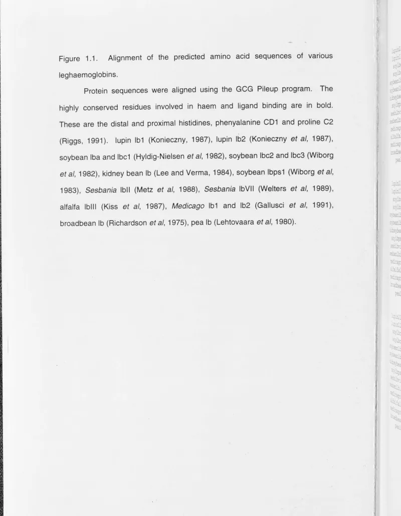
1.1. Alignment of the predicted amino acid sequences of various
leghaemoglobins. 4
1.2. Stages, sequence of events and involvement of nodulins in the
formation and function of a nitrogen-fixing root nodule. 9
1.3a. The basic 3-dimensional structure of the globins. 22
1.3b. The active centre of oxymyoglobin. 22
1.4. Alignment of key globin sequences of known structure. 22
1.5. Position of globin introns within the conserved a helices. 24
2.1. Alignment of the Parasponia andersonii and Trema tomentosa
haemoglobin promoter sequences. 39
2.2. Histochemical localisation of GUS activity. 40
2.3. Alignment of the 'nodulin box' motifs of haemoglobin gene promoters. 43
2.4. Site directed mutagenesis of promoter motifs. 43
2.5. GUS activity of mutant Parasponia and Trema haemoglobin promoters. 44
2.6. Summary of promoter elements involved in tissue specific regulation
of the Parasponia and Trema haemoglobin genes. 52
2.7. Schematic of Parasponia and Trema haemoglobin promoter-GUS
constructs. 56
3.1. Alignment of the predicted amino acid sequences of the
soybean leghaemoglobin genes. 59
3.2. Northern analysis of soybean /bps 1 expression. 61
3.3. GUS activity of the lbps1 promoter. 62
3.4. Alignment of the promoter regions of soybean lb genes. 64
3.5. Map of /bps 1 gene. 65
4.1. Alignment of the predicted amino acid sequences of various
plant haemoglobins. 72
4.3. Southern blot analysis of the soybean nonsymbiotic haemoglobin gene
and leghaemoglobin genes.
4.4. Two possible models of soybean nonsymbiotic haemoglobin
genome organisation.
4.5. Northern analysis of soybean nonsymbiotic haemoglobin gene
expression.
4.6. Sequence of the Arabidopsis haemoglobin 2f+4r2 PCR clone.
4.7. Southern blot analysis of the Arabidopsis haemoglobin gene.
4.8. Plant haemoglobin protein similarity tree.
4.9. Sequence alignment of the promoter regions of haemoglobin genes
from Casuarina, soybean, Parasponia and Trema.
5.1. A phylogenetic analysis of the polyphyletic class Hamamelidae.
LIST OF TABLES following page
2.1. Summary of GUS staining results for Parasponia and Trema
haemoglobin promoter constructs in transgenic Lotus.
4.1. Overall similarity between various plant haemoglobins.
5.1. Summary of nodule characteristics for legume, Parasponia and
actinorhizal plants.
73
74
74
75
76
77
78
90
41
71
page 4, line 14, change:
1984; to: 1984,
page 9, line 21, change:
15 40 to: 15, 40
page 15, line
12,
change:
taxanomically to: taxonomically
page 16, line 20-21, change: 'on
the tips of the hyphae of
Frankia' to: 'as terminal
swellings differentiating from short
branch
Frankia hyphae'.
page 16, line 25-26, change: 'which can
only fix nitrogen at very low ... ' to: 'which
cannot fix nitrogen even at very
low
...
'
page 20, line 3, change: et al to: et
al
page 30, line 26, change: requires
to: require
page 57, line 14, change: pIV21
to: pIV20
(Ramlov
et al, 1993)
page 58, line 18, change: 2-3um to:
2-3µm
following page 59, Figure 3.1
legend, line 5, change: proximal to: distal. line 6,
change: distal to: proximal.
page 65, line 14, change: absorbence
to: absorbance
page 102, line 9, change:
tumafaciens
to:
tumefaciens
t,:.:,,
'
..
:-;~
,
..
'w".~.
'i!
"" !,
i:
I
.
.,~a
"terminal
'
to:
'whlcht~. line o,
CHAPTER 1
GENERAL INTRODUCTION
LEGHAEMOGLOBIN AND THE LEGUME-(BRADY)RHIZOBIUM SYMBIOSIS
The legume-(Brady)Rhizobium symbiosis
The nitrogen-fixing root nodules of legumes develop as the result of a complex
interaction between the host plant and the invading bacteria. Within the controlled
environment of the nodule, the bacteria reduce atmospheric nitrogen to ammonia, thus
freeing the host plant from limitations in the soil nitrogen. In return, the bacteria are
supplied with carbohydrate and an environment compatible with nitrogen fixation.
The family Leguminosae (Fabaceae) is divided into three subfamilies:
Caesalpinioideae, Mimosoideae and Papilionoideae. Nodulation is general in the
Papilionoideae and Mimosoideae, but is less common in the Caesalpinioideae, with
only 23°/o of species examined found to nodulate (de Faria et al, 1989; Corby, 19_88).
The nodulating plants within the Papilionoideae have been the most extensively
studied, particularly members of the Loteae, Phaseloeae, Trifolieae and Viceae.
Nodule formation is induced by three genera of the Rhizobiaceae; Rhizobium,
Bradyrhizobium and Azorhizobium. These genera are quite distinct and are more
similar to nonsymbiotic relatives than to each other. For example, Rhizobium is closely
related to the plant pathogen Agrobacterium (Denarie et al, 1992). These diverse
bacteria are grouped in a single family by virtue of 'their common ability to establish a
nitrogen-fixing symbiosis with legumes' (Denarie et al, 1992).
Rhizobial genetics has been intensively studied (for review, Long, 1989).
Several groups of symbiotic genes have been defined, mostly by mutation. Nod
(nodulation) genes are required for nodule formation (Nod+ phenotype). Both nit and
fix genes are required for nitrogen fixation. Mutations in either produce ineffective
nodules (Nod+, Fix- phenotype). The nit genes probably encode components of the
nitrogen fixation machinery, while the fix genes are thought to be required for symbiotic
The interaction between legume species and Rhizobium strains is specific.
Bacterial strains nodulate a particular range of plants, having either a broad or narrow
host range. For example, R. meliloti nodulates alfalfa, B. japonicum nodulates
soybean, while Rhizobium sp. NG R234 is capable of nodulating a variety of tropical
legumes and Parasponia (Denarie et al, 1992).
Nodule formation is initiated by molecular signalling between a compatible
bacteria and plant. The legume produces flavonoids that induce expression of the
bacterial nodulation (nod) genes. Different legumes produce flavonoids with distinct
modifications that act as host determinants. Plant secreted flavonoids induce
expression qf the bacterial nodD gene, which in turn activates expression of other nod
genes, such as nodABC. nodABC are common nod genes, whereas other
host-specific nod genes are involved in determining host range (reviewed in Denarie et al,
1992; Spaink, 1992; Denarie and Cullimore, 1993). Together, the products of these
genes produce bacterial Nod factors, capable of initiating nodule organogenesis in the
host plant. Several bacterial Nod factors have been purified and shown to be
lipooligosaccharides (Lerouge et al, 1990; Spaink et al, 1991 ). Different bacterial species produce oligomers of ~-1,4-linked N-acetylglucosamine with different
substitutions, which have been shown to be important in determining host range.
The bacterial host-specific signals initiate homologous processes in different
legumes (Fisher and Long, 1992). The bacterial signal compound induces curling of root hairs in a susceptible zone behind the growing root tip. Only a small region close
to the zone of elongation is susceptible to infection by Rhizobia. Bacteria attach and invade the root hair by localised degradation of the cell wall. Formation of a specialised
structure called the infection thread allows the bacteria to penetrate the root.
The bacterial Nod factors also induce morphological changes in the plant root,
prior to bacterial infection. Cortical cell divisions are induced, giving rise to the nodule primordia prior to release of bacteria within the plant. Pericycle divisions are also
induced, and the two meristems fuse to produce a cluster of dividing cells (Brewin,
Infection threads enter individual nodule primordium cells. Endocytosis of the infection thread leads to formation of discrete structures, the symbiosomes, within the host cell cytoplasm. Within the symbiosomes, bacteria differentiate into bacteroids, their endosymbiotic form, and remain separated from the host cytoplasm by the host derived peribacteroid membrane (reviewed by Verma, 1992).
The mature nodule is derived from two cell lineages, the cortex and pericycle/ endodermis (Brewin, 1991 ). Differentiation of the nodule produces vascular connections and the various cell types needed for nodule function. The central nodule tissue contains both uninfected and infected, bacteroid containing cells. This tissue is surrounded by the nodule parenchyma, the nodule endodermis and the nodule cortex. The vascular bundles lie within the parenchyma.
The morphology and physiology of the mature root nodule creates an environment compatible with nitrogen fixation by the bacteroids. The tightly packed cells of the nodule inner cortex are thought to be important in the mechanical control of oxygen diffusion into the symbiotic zone (reviewed by Hunt and Layzell, 1993). Expression of the bacterial nitrogenase enzyme occurs late in the process. The mature nodule maintains a symbiotic zone where infected and uninfected cells assume different roles in nitrogen fixation, assimilation and transport. Fixed nitrogen in the form of ammonia is transported from the bacteroids to the plant cell cytoplasm and then to other parts of the plant (Nap and Bisseling, 1990b).
There are two main categories of nodule type (Nap and Bisseling, 1990b) although more complex classifications exist (Corby, 1988). Tropical legumes such as soybean, Lotus and Phaseo/us develop a spherical, determinate nodule, where the different stages in nodule development are separated temporally. Cell division is initiated in the outer cortex of the root and stops during nodule development and growth is a result of cell expansion rather than cell division. The vascular bundles meet,
persistent apical meristem that generates new cells that continue to be infected. In this
case, all developmental stages of nodule formation are present in a single nodule and
are separated spatially. A longitudinal section through an indeterminate nodule reveals
the entire series of developmental stages. The apical meristematic zone I is followed
consecutively by the zone of infection or prefixing zone II, the amyloplast-rich interzone
11/111, the nitrogen-fixing zone Ill, and the senescent zone IV (Vasse et al, 1990). Lupin
nodules are distinct in having an extensive attachment to the root, resulting in a
collar-like nodule that girdles the root (Corby, 1988).
Leghaemoglobin facilitates oxygen diffusion within nitrogen-fixing nodules
Plant haemoglobin was first identified in the nitrogen-fixing root nodules of
legumes (Kubo, 1939). The paradox of symbiotic nitrogen fixation is the requirement of
the bacteria for oxygen for respiration to meet the energy demands of nitrogen fixation
combined with a requirement for exclusion of free oxygen from the oxygen sensitive
nitrogenase enzyme complex (reviewed by Appleby, 1984; in press). Leghaemoglobin
facilitates the controlled diffusion of oxygen within symbiotic root nodules to respiring
bacteria while maintaining the local free oxygen as low as 10-20 nM. Within this low
oxygen environment, the concentration of leghaemoglobin-bound oxygen may be as
high as 600µM (Appleby, in press). The bound oxygen is available to the bacteroids
for respiration as it dissociates from the leghaemoglobin, but the free oxygen
concentration remains non-toxic.
All legumes so far examined appear to express high levels of nodule specific
leghaemoglobin (Konieczny, 1987; Marcker et al, 1984; Lehtovaara and Ellfolk, 1975;
Bogusz et al, 1987; Kiss et al, 1987; Gallusci et al, 1991; Richardson et al, 1975;
Lehtovaara et al, 1980). The amino acid sequences of many leghaemoglobins have
been determined, and all show extensive homology (see Figure 1.1 ). Leghaemoglobin
is the most abundant nodule protein, comprising up to 15-20°/o of total protein (Fuller et
leghaemoglobins.
Protein sequences were aligned using the GCG Pileup program. The
highly conserved residues involved in haem and ligand binding are in bold.
These are the distal and proximal histidines, phenyalanine CD1 and proline C2
(Riggs, 1991 ). lupin lb1 (Konieczny, 1987), lupin lb2 (Konieczny et al, 1987),
soybean Iba and lbc1 (Hyldig-Nielsen et al, 1982), soybean lbc2 and lbc3 (Wiborg et al, 1982), kidney bean lb (Lee and Verma, 1984), soybean lbps1 (Wiborg et al,
1983), Sesbania lbll (Metz et al, 1988), Sesbania lbVII (Welters et al, 1989), alfalfa lblll (Kiss et al, 1987), Medicago lb1 and lb2 (Gallusci et al, 1991 ),
broadbean lb (Richardson et al, 1975), pea lb (Lehtovaara et al, 1980).
,: .. r·r;:r,
,. •. -1·· ::i'~:;: .. :e:c .. t, ....
.
:;:"'a:'. r ... -:~· ,.;.,. ....
•• - .. 1,,, .. ;::-·
t:a.:a.: ""fl',.~,. ... ... •;··
~, ... , ""':.~· ;,.." ...
-·-;:: .. : [image:19.815.6.806.29.1058.2].... " .. ...
..
~=1·.x:
:;!·:tt .. ~c :•·>?1;:• ,.,. ... ! ...
"""'' 1°1::· \.,..,c ~ "' ..
~:: .. ~s .. ::::
...
. ,,::~:.:.: .. : ...
·:::::;: ..
..
...
: .. :t:: ... ·::::a;:. ::":~~t "::: ..!" ...
...
"•1 .. ,.~ .. : "1 ... ••
"!" ... :
~,:1,:::;
"~·· "]· ...
"··
~~:e;:-~::c
...
.. t,::
::\r~;:-~> ...
soylbc3 GAFTDKQEAL VSSSFEAFKT NIPQYSWFY TSILEKAPVA KDLFSFLAN . . GVDPTNPKL soylbc2 GAFTEKQEAL VSSSFEAFKA NIPQYSWFY TSILEKAPAA KDLFSFLSN . . GVDPSNPKL m. The soybeanlba VAFTEKQDAL VSSSFEAFKA NIPQYSWFY TSILEKAPAA KDLFSFLAN . . GVDPTNPKL
soybeanlbc GAFTEKQEAL VSSSFEAFKA NIPQYSWFY NSILEKAPAA KDLFSFLAN. . GVDPTNPKL . In cold. kidney bean GAFTEKQEAL VNSSWEAFKG NIPQYSWFY TSILEKAPAA KNLFSFLAN. .GVDPTNPKL
soylbpsl GAFTEKQEAL VNSSFEAFKA NLPHHSWFF NSILEKAPAA KNMFSFLGD . . AVDPKNPKL
11 e C2
sesblbvii .GFTEKQEAL VNASYEAFKQ NLPGNSVLFY SFILEKAPAA KGMFSFLKDF DEVPQNNPSL sesbanlbii .GFTDKQEAL VNASYEAFKK NLPGHSVLFY SFILEKEPAA KGLFSFLKDS DGVPQNNPSL
I, 1987), medicago2
.GFTEKQEAL VNSSWELFKQ N.PGNSVLFY TIILEKAPAA KGMFSFLKDT AGV.QDSPKL
alfalfalb .GFTDKQEAL VNSSWESFKQ N.PGNSVLFY TIILEKAPAA KGMFSFLKDS AGV.QDSPKL medicagol .SFTDKQEAL VNSSYEAFKQ NLSGYSVFFY TVILEKAPAA KGLFSFLKDS AGV.QDSPQL
rll!oor~
broadbean .GFTEQQEAL VNSSSQLFKQ NPSNYSVLFY TIILQKAPTA KAMFSFLKDS AGV.VDSPKL pealb .GFTDKQEAL VNSSSE.FKQ NLQGYATLFY TIILEKAPAA KGLFSFLKDT AGV.EDSPKL~r~ el al,
61 120
1 , 1989),
lupin2lb QAHAGKVFKL VYEAAIQLQV TGVWTDATL KNLGSVHVSK GVADAHFPW KEAILKTIKE lupinllb QAHAGKVFKL TYEAAIQLQV NGAVASDATL KSLGSVHVSK GVVDAHFPW KEAILKTIKE soylbc3 TGHAEKLFGL VRDSAGQLKA SGTW.IDA. .ALGSIHAQK AITDPQFVW KEALLKTIKE
I 1991), soylbc2 TGHAEKLFGL VRDSAGQLKA NGTW.ADA. .ALGSIHAQK AITDPQFVW KEALLKTIKE
soybeanlba TGHAEKLFAL VRDSAGQLKA SGTW.ADA. .ALGSVHAQK AVTDPQFVW KEALLKTIKA soybeanlbc TGHAEKLFAL VRDSAGQLKT NGTW.ADA. .ALVSIHAQK AVTDPQFVW KEALLKTIKE kidney bean TAHAESLFGL VRDSAAQLRA NGAW.ADA. .ALGSIHSQK GVNDSQFLW KEALLKTLKE soylbpsl AGHAEKLFGL VRDSAVQLQT KGLW.ADA. .TLGPIHTQK GVTDLQFAW KEALLKTIKE sesblbvii QAHAEKVFGL VRDSAAQLRA TGVWLADA. .SLGSVHVQK GVLDPHFVW KEALLKTLKE sesbanlbii QAHAEKVFGL VHDAAGQLRA TGVWLADA. .SLGSVHVQK GVTDPHFVW KEALLKTLKE medicago2 QSHAEKVFGM VRDSAVQLRA TGGWLGDA. .TLGAIHIQK GVVDPHFVW KEALLKTIKE alfalfalb QSHAEKVFGM VRDSAAQLRA TGGWLGDA. .TLGAIHIQK GVVDPHFAW KEALLKTIKE medicagol QAHAEKVFGL VRDSASQLRA TGGWLGDA. .ALGAIHIQK GVVDPHFVW KEALLKTIKE broadbean GAHAEKVFGM VRDSAVQLRA TGEWL.DG. .KDGSIHIQK GVLDPHFVW KEALLKTIKE pealb QAHAEQVFGL VRDSAAQLRT KGEWLGNA. .TLGAIHAGK GVTNPHFVW KEALLQTIKK
121 155
lupin2lb WGAKWSEEL NSAWTIAYDE LAIVIKKEMN DAA .. lupinllb WGDKWSEEL NTAWTIAYDE LAIIIKKEMK DAA .. soylbc3 AVGDKWSDEL SSAWEVAYDE LAAAIKKAF. .
....
soylbc2 AVGDKWSDEL SSAWEVAYDE LAAAIKKAF. .
....
soybeanlba AVGDKWSDEL SRAWEVAYDE LAAAIKKA .. .
....
soybeanlbc AVGGNWSDEL SSAWEVAYDE LAAAIKKA .. .
....
kidneybean AVGDKWTDEL STALELAYDE LAAAIKKAYA ... soylbpsl AVEDKWSEEL SNAWEVAYDE IAAAIKKAMA IGSLV sesblbvii AGGATWSDEV SNAWEVAYDE LSAAIKKAMS
...
sesbanlbii AAGATWSDEV SIAWEVAYDG LAAAIKKAMS...
medicago2 VSGDKWSEEL STAWEVAYDA LAAAIKKAMG ... alfalfalb VSGDKWSEEL NTAWEVAYDA LATAIKKAMV
...
rnedicagol AAGDKWSEEL STAWEVAYDA LATEIKKAMS ... broadbean ASGDKWSEEL SAAWEVAYDG LATAIKAA .. .....
cells (Verma and Long, 1983; Appleby, 1984), prior to the onset of nitrogen fixation
(Marcker et al, 1984; Verma et al, 1979; de Billy et al, 1991 ).
The soybean leghaemoglobins have been well characterised. Soybean nodules
contain four major leghaemoglobins, Lba, Lbc1, Lbc2 and Lbc3, as well as four minor
components, Lbb, Lbd1, Lbd2 and Lbd3, which result from N-terminal acetylation of the
major components (Fuschmann and Appleby, 1979). These leghaemoglobins are
encoded by a small family of four functional genes, one apparent pseudogene and two
or three truncated genes (Brisson and Verma, 1982; Hyldig-Nielsen et al, 1982; Wiborg
et al, 1982; 1983). The genes are arranged in two independent clusters, one
comprising 5_' Iba - /bc1 - lbps1 - lbc3 - 3' and the other 5' - lbps2- /bc2- 3' where lbps2
is a truncated gene (Bojsen et al, 1983; Brisson and Verma, 1982). The genes show
extensive homology extending into the 5' upstream region. The genes in both clusters
have the same transcriptional polarity (Bojsen et al, 1983). Induction of the four
soybean leghaemoglobin genes occurs sequentially in the opposite order to their
arrangement on the chromosome (Marcker et al, 1984). Transcription of lbc3, lbc2 and
lbc1 starts slightly before the appearance of visible nodules while Iba initiates after
another day or two. At around day 12, there is a dramatic increase in the level of
transcription of Iba, lbc3 and /be 1, concomitant with the transcription initiation of several
other nodule specific genes and prior to detectable nitrogenase activity. All four
leghaemoglobins continue to be expressed for as long as the nodule is functional. The
functional significance of this differential regulation of transcriptional activation is not
known. Expression of the apparent pseudogene /bps 1 has not been detected
(Marcker et al, 1984; Wiborg et al, 1983), but as the gene encodes an apparently
functional leghaemoglobin (Wiborg et al, 1983) it has been proposed that /bps 1 may
represent a functional haemoglobin gene (Nap and Bisseling, 1990b,c). This possibility
is examined in Chapter 3.
The leghaemoglobins of the tropical legume Sesbania rostrata have also been
extensively studied. The symbiosis between Sesbania and Azorhizobium caulinodans
stem nodules are unusual in that chloroplast containing cells are immediately adjacent
to bacteroid containing cells. Oxygen evolution associated with photosynthesis may
put an additional stress on the oxygen-sensitive nitrogenase enzyme complex, and it is
possible that stem nodules have adapted to the higher levels of oxygen by producing
modified or higher levels of leghaemoglobin (Dehio and de Bruijn, 1992). Sesbania
contains seven major leghaemoglobins in its stem and root nodules, of which at least
six are separate gene products (Bogusz et al, 1987). One form, LbVI appears to be
differentially regulated as it is five times more abundant in stem nodules than root
nodules (Bogusz et al, 1987) suggesting that it may play a specific role in stem
nodules. Two Sesbania leghaemoglobin genes have been isolated and studied, Srglb2
(encoding LbVI) and Srglb3 (LbVII) (Metz et al, 1988; Welters et al, 1989). The two
genes show a high level of homology, with only 6 nucleotide differences, leading to 4
predicted amino acid changes (Metz et al, 1988). The homology between the two
Sesbania genes, as well as the soybean leghaemoglobin genes, extends into the 5'
untranslated region.
Leghaemoglobin as the archetype symbiotic protein
The functional leghaemoglobin holoprotein consists of the monomeric globin
apoprotein and a haem prosthetic group. It has been clearly established that the
apoprotein is plant encoded, but the origin of the haem moiety is still unclear.
Biochemical studies localising 8-aminolevulinic acid synthase activity, the first
committed step in the haem biosynthetic pathway, to the bacteroid fraction of root
nodules (Nadler and Avissar, 1977; Cutting and Schulman, 1969) led to the idea that
leghaemoglobin was comprised of plant encoded globin and Rhizobium provided
haem. Leghaemoglobin has come to be regarded as the classic symbiotic protein, with
components provided by each symbiotic partner to produce a functional holoprotein.
Several studies have used haem deficient (Bradl)Rhizobial mutants in an effort
A 8. japonicum hemA mutant, deficient in 8-aminolevulinic acid synthetase,
induced fully effective soybean nodules that contained haem (Guerinot and Chelm,
1986). In contrast, a hemH mutant, deficient in protoporphyrinogen oxidase activity,
the penultimate step in haem biosynthesis, induced ineffective soybean nodules
(O'Brian et al, 1987). These conflicting results can be reconciled by evidence that the
hemA mutant bacteroids can synthesise haem from imported plant 8-aminolevulinic
acid (Sangwan and O'Brian, 1991 ). Nodules induced by the hemH mutant contained
the globin apoprotein but not the holoprotein, implying that bacterial haem synthesis is
required for leghaemoglobin but not apoleghaemoglobin synthesis (O'Brian et al, 1987).
This is the strongest evidence that the leghaemoglobin haem is provided by the
bacteria.
An R. meliloti hemA mutant formed ineffective nodules lacking leghaemoglobin
on inoculation of alfalfa (Leong et al, 1982). de Bruijn et al (1989) also found that
alfa.lfa nodules induced by a hemA mutant did not express leghaemoglobin. However,
Mohapatra and Puhler (1986) found that a hemA mutant of R. meliloti, although
ineffective, induced nodules apparently containing near wild-type levels of
haemoglobin. A hemA mutant of Azorhizobium caulinodans ORS571 also induced
ineffective root nodules lacking leghaemoglobin on inoculation of Sesbania (Pawlowski
et al, 1993).
These results were seen as further evidence that the haem group is bacterial in
origin. However, a recent reexamination of the R. meliloti hemA mutants used by
Leong et al (1982), Mohapatra and Puhler (1986) and de Bruijn et al (1989) found that
these nodules failed to express any of six nodule specific genes tested, including
leghaemoglobin (Dickstein et al, 1991 ). So it is unlikely that the ineffective symbiosis is
a "direct consequence of the failure of R. meliloti to supply the heme moiety of
hololeghemoglobin.11
Dickstein et al (1991) confirmed that symbiotic effectiveness of
the hemA mutants could be restored by an exogenous supply of 8-aminolevulinic acid
(Leong et al, 1982). They propose that the hemA mutant may be unable to properly
mutants induce nodules with atypical morphology and infection thread formation ( either
empty nodules with no infection threads or infection threads that failed to release
bacteria) (Dickstein et al, 1991; Pawlowski et al, 1993) and other evidence shows that
leghaemoglobin expression is dependent on bacterial release from the infection
threads (Dunn et al, 1988; Norris et al, 1988).
It appears -that bacterial haem synthesis is essential for an effective symbiosis
in the alfalfa-A. meliloti and Sesbania-A. caulinodans symbioses. However, in both
these cases the hemA mutation has multiple effects and its influence on
leghaemoglobin production appears general rather than specific. There are several
alternative explanations for the importance of bacterial haem in establishing an
effective symbiosis. Rhizobial bacteroids synthesise a terminal oxidase with high
oxygen affinity in order to function in the low free oxygen environment of the symbiotic
nodule (reviewed by Appleby, 1984). The B. japonicum symbiotic oxidase appears to
comprise several haem containing subunits including a haem binding oxidase subunit,
a dihaem cytochrome c and a monohaem cytochrome c (Preisig et al, 1993).
Mutations in the oxidase gene cluster, fixNOQP, result in defective nodules that are
white or greenish and contain few bacteroids, a phenotype similar to that of hemA
mutants. Also, the oxygen sensing regulatory system which triggers expression of
most of the genes required for nitrogen fixation includes an oxygen binding
haemoprotein, Fixl (discussed further on page 21 ). A haem-dependent
symbiosis-specific oxidase complex and haem-dependent activation of nitrogen fixation genes is
likely to account for the ineffective phenotype of haem deficient (BradY)Rhizobium
mutants.
Recent evidence demonstrating that plant synthesis of coproporphyrinogen
oxidase and 8-aminolevulinic acid increases concomitant with leghaemoglobin
expression, suggests the plant increases haem production to meet increased demand
in the nodules (Madsen et al, 1993; Sangwan and O'Brian, 1992) and thus that the
NODULINS ARE PLANT PROTEINS SPECIFICALLY INVOLVED IN NODULATION Nodule formation is the result of a coordinated reponse of both the bacteria and
the plant which requires the sequential induction of both bacterial and plant genes.
Many plant genes are required for the development and function of the nodule.
Host-encoded nodule-specific proteins, such as the well characterised leghaemoglobins, are
referred to as nodulins. By definition, nodulins are exclusively expressed during
development of the symbiosis, although there are many proteins that show enhanced,
rather than specific, nodule expression. Nodulins are distinguished for convenience on
the basis of time of expression. Early nodulin genes are expressed during
development 9f the nodule structure. Their gene products are thought to be involved in
bacterial invasion and in nodule formation, although the precise function of most early
nodulins is not known. Late nodulin gene expression, such as that of leghaemoglobin,
generally follows release of bacteria but precedes the onset of nitrogen fixation. Late
nodulins probably function in establishing and maintaining a nodule environment
compatible with nitrogen fixation (reviewed by Nap and Bisseling, 1990b,c; Verma and
Miao, 1992). The relationship of nodulin expression and nodule development and
function is summarised in Figure 1.2. Nodulins have been most extensively studied in
the legume-(Brady)Rhizobium symbiosis.
Early nodulins are involved in nodule formation
Several early nodulins (ENODs) have been isolated and characterised,
including ENOD2, 3, 5, 13, 15 40 and 55 (Franssen et al, 1987, 1990; Scheres et al,
1990a, b; Vijn et al, 1993; de Blank et al, 1993). Activation of early nodulin expression seems to be independent of the attachment of bacteria as purified Nod signal compounds can induce some of these genes, as can auxin-transport inhibitors or
'
PRElNFECTION INFECTION ANO NODULE FORMATION
STAGE I STAGE lI
-·· .;_
-
-~-~
·---~
V
,;sJ.;,
UCTERCIO OEVELO,.MENT.A TT .A CHM ENT
/ / IACTERI.AC. )olUl Tll'LJCA. TION
RELEASE OF BACTERIA MOOUL( IHITI.A TIOH
IHF'ECTION THR(.AO GROWTH
ll'IF(C TION THRE.AO FOR>,4A TIOH
CORTICJ.C. cnL OIVlSIOHS
NODULE FUNCTION ANO MAINTENANCE
STAGE m
NODULE SENESCENC~
STAGE I7
MEMBIUN[ OISIH rEGIU TIOH
CC:Ll OISIN fEC.RA TIOH
HOOULE BRE.AXOOWH
Figure 1.2. Stages, sequence of events and involvement of nodulins in the formation
and function of a nitrogen-fixing legume root nodule. From Gloudemans and Bisseling
(1989). Many early and late nodulins have been characterised (see text for details).
ENOD12 appears to be involved in the preinfection stage (Scheres et al, 1990b; Pichon
et al, 1992). A protease inhibitor expressed specifically in the infected cells of
senescent nodules has been identified (Manen et al, 1991).
[image:26.839.16.822.21.909.2]developmental stages in pea nodules. ENOD12 transcript is 'present in every cell of
the invasion zone, whereas ENOD5, ENOD3 and ENOD14 transcripts were restricted
to the infected cells in successive but partially overlapping zones of the central tissue'
(Scheres et al, 1990a).
ENOD2 AND ENOD12 ARE CELL WALL PROTEINS
ENOD2 is expressed exclusively in the parenchyma (inner cortex) of soybean,
pea and alfalfa nodules (van de Wiel et al, 1990a). mRNA expression is detectable as
soon as meristem cells differentiate into inner cortical cells. In soybean, expression is
transient, wh~reas in pea, ENOD2 transcript accumulates during development. This
may reflect the different developmental pattern of determinate nodules, where mitotic
activity ceases shortly after the onset of nitrogen fixation, in contrast to the persistent
meristem of indeterminate nodules (Nap and Bisseling, 1990b). Expression of alfalfa
ENOD2 is also induced in empty (bacteria-free) alfalfa nodules formed by R. meliloti
mutants defective in exopolysaccharide synthesis, in pseudo-nodules induced by auxin
transport inhibitors (Van de Wiel et al, 1990b), and in the spontaneous nodules formed
on some alfalfa roots in the absence of Rhizobial infection (Truchet et al, 1989).
Expression of the ENOD2 gene of Sesbania is induced by a variety of cytokinins (Dehio
and de Bruijn, 1992).
The presence of a signal transit peptide and the characteristic praline-rich
pentapeptide repeat of ENOD2 (Franssen et al, 1987; 1990) resembles that of
extensin, a major structural component of the cell wall, and a soybean praline rich
protein, 1A10, expressed in the cell wall of germinating seedlings. It is therefore likely
that ENOD2 is a structural cell wall protein. The expression of ENOD2 in the nodule parenchyma, as well as its expression in soybean nodules devoid of bacteria and infection threads (Franssen et al, 1987) suggests a role in the formation of the nodule
de Bruijn, 1992; Verma and Miao, 1992) possibly by limiting cell wall expansion,
producing the small tightly packed cells of the inner cortex (Brewin, 1991 ).
ENOD12 also contains a putative signal peptide and a series of two
pentapeptide repeats (Govers et al, 1991 ). ENOD12 is expressed in the root cortical
cells containing the infection thread, in cells preparing for infection thread passage and
in dividing inner cortical cells that form the nodule primordium (Scheres et al, 1990b;
Pichon et al, 1992). ENOD12 is expressed transiently in root hairs of the infection
zone, while expression is enhanced and maintained in root hairs that are subsequently
infected (Pichon et al, 1992). In nodules, transcript is present in the invasion zone and
at decreasing_ levels in the early symbiotic zone. Because of its homology with ENOD2
and 1A10, and as all root and nodule cells containing ENOD12 transcript are sites of
new cell wall synthesis, ENOD12 is assumed to also be a cell wall protein (Scheres et
al, 1990b). As ENOD12 is expressed at a different stage and in different cell types to
ENOD2 it probably fulfils a different role in nodule formation.
Late nodulins are involved in nodule function
Leghaemoglobin is the most abundant and best characterised late nodulin.
Many other late nodulins function as the key enzymes of nitrogen assimilation and
carbon metabolism. Factors involved in the activation of late nodulins are clearly
different from the early nodulins. Expression of many late nodulins occurs prior to nitrogen fixation. While expression does not occur without the formation of infection
threads and release of bacteria, it does occur in nodules induced by some
Fix-mutants. Induction is triggered either by bacterial infection or in some way by the
unique nodule environment created during the symbiotic state. The expression and
regulation of late nodulins has been reviewed recently by Sanchez et al, 1991; Verma and Miao, 1992; Nap and Bisseling 1990b, c.
There are nodule-specific or nodule-abundant forms of several enzymes of the
nitrogen and carbon assimilation pathways. These include glutamine synthetase,
glutamate synthase, sucrose synthase, phosphoenolpyruvate carboxylase, aspartate
aminotransferase, uricase, malate dehydrogenase and xanthine dehydrogenase.
Glutamine synthetase (GS) catalyses the first reaction in the assimilation of
ammonia produced by the bacteroids. Cytosolic GS is encoded by a small family of
genes. Several legumes, such as soybean, alfalfa and lupin, have nodule specific
isoforms (Roche et al, 1993; Dunn et al, 1988). However, in pea, Phaseolus and
Medicago truncatula, GS isoforms show enhanced nodule expression but are also
expressed i~ other parts of the plant (Forde et al, 1989; Stanford et al, 1993). It
appears that the nodule GS form is located exclusively in the cytosol of the infected
cells, while another, highly homologous GS is expressed in uninfected nodule tissue
and in other parts of the plant (Forde et al, 1989). Differentially expressed GS genes
produce functionally similar enzymes (Nap and Bisseling, 1990b).
A nodule specific form of sucrose synthase has been isolated from soybean
nodules (Thummler and Verma, 1987). Sucrose synthase is the key enzyme involved
in breakdown of sucrose to the monosaccharides glucose and fructose and plays three
key roles in nodules: to sustain respiration in the plant and bacteroids; to provide
substrate for cell wall synthesis and; to provide carbon skeletons for the assimilation of
fixed nitrogen (Thummler and Verma, 1987).
PERIBACTEROID MEMBRANE NODULINS
The host-derived peribacteroid membrane (PBM) surrounding the bacteroids is the main control point for nutrient and signal exchanges between the Rhizobium and the host. The PBM nodulins are likely to be involved in transport across the membrane, as is the dicarboxylic acid transporter identified in the soybean PBM
N-26 is a transmembrane protein with six membrane-spanning domains. The
protein is cotranslationally glycosylated and inserted into membranes and is
phosphorylated near the carboxy terminus (Miao et al, 1992). The phosphorylation may be directly related to the function of N-26. Based on the homology of N-26 to
several eukaryotic and prokaryotic intrinsic channel-type proteins, it has been
suggested that N-2b may form a channel translocating specific molecules between the
bacteroids and the host cytoplasm (Miao et al, 1992; Miao and Verma, 1993).
Origin of nodulins
It ha~ been hypothesised that nodulin genes evolved by gene duplication of
preexfsting plant genes to fit the developmental and physiological requirements of
symbiotic nitrogen fixation (Nap and Bisseling, 1990b,c; Verma and Miao, 1992).
Preexisting genes may have been modified and brought under nodule developmental
control. These nodulin genes remain recognisably homologous to the progenitor gene
(Nap and Bisseling, 1990b). It is therefore expected that all nodulin genes will have a
nonsymbiotic counterpart expressed in non-nodule tissue. This is the case for many of
the well-characterised nodulins that have been assigned a biochemical role, such as
glutamine synthetase, glutamate synthase and sucrose synthase, as well as ENOD2
and N-26 (Dehio and de Bruijn, 1992; Miao and Verma, 1993). The nonsymbiotic form
of the archetype nodulin, leghaemoglobin, has been difficult to identify. This thesis describes the isolation of a soybean haemoglobin gene homologous to the
nonsymbiotic haemoglobin genes previously identified in nonlegumes (Chapter 4).
PARASPON/A HAEMOGLOBIN AND THE PARASPON/A-(BRADY)RHIZOB/UM
SYMBIOSIS
The Parasponia/(Brady)Rhizobium symbiosis
legumes, infection of Parasponia roots by (Brady')Rhizobium occurs in the zone of elongating cells directly behind the root tip which is transiently, highly susceptible to
infection (Scott and Bender, 1990). However, there is no root hair curling or formation
of infection threads (Lancelle and Torrey, 1984; Scott and Bender, 1990).
Caetano-Anolles and Gresshoff (1991) are of the opinion that bacteria invade the host by taking
advantage of the emergence of a lateral root. However, others suggest that cells
exposed by emerging lateral roots are not susceptible to invasion (Lancelle and Tor~ey,
1984; Scott and Bender, 1990). Scott and Bender (1990) have observed degradation
of the mucilage layers in the primary cell walls of epidermal cells in association with
bacterial contact. Erosion of the root surface results in localised root swelling
producing a break in the epidermis (Lancelle and Torrey, 1984; Scott and Bender,
1990). The bacteria induce cortical cell divisions, forming the prenodule, which
emerges through the epidermal break, forming an exposed site for intercellular
colonisation by bacteria. These initial cortical divisions cease and are followed by
pericycle divisions, giving rise to the nodule lobe primordia, which is invaded by
Rhizobium (Lancelle and Torrey, 1985). The mature nodule tissue is derived from these pericycle, rather than cortical, cell divisions. Thus, the Parasponia nodule has the same ontogeny, and so similar morphology, to that of a lateral root (Caetano-Anolles and Gresshoff, 1991 ). The mature nodule has a central vascular bundle and
persistent apical meristem (Trinick, 1979; Lancelle and Torrey, 1985; Scott and Bender,
1990). The central zone of the nodule contains both infected and uninfected cells. Within the infected cells, the Rhizobia remain within host-derived thin-walled 1
infection1
threads, more appropriately called fixation threads, rather than differentiating into
symbiosomes (Trinick, 1979; Lancelle and Torrey, 1985; Scott and Bender, 1990).
Haemoglobin is expressed in nodules of Parasponia and also in roots of Parasponia and Trema
conserved amino acid residues (Appleby et al, 1983; Kortt et al, 1988). The
Parasponia haemoglobin has similar binding kinetics to the leghaemoglobins (Appleby
et al, 1988) and functions, like leghaemoglobin, to provide oxygen to the bacterial
symbiont at a low free oxygen concentration. lmmunogold labelling has localised
Parasponia haemoglobin expression to the infected cells within the nodule (Trinick et
al, 1989). P. andersonii has a single haemoglobin gene which is differentially
expressed to give high expression in nodules (Landsmann et al, 1986), and also
approximately 1000 fold lower expression in roots (Bogusz et al, 1988).
The genus Parasponia (7 species) is the only member of the subfamily
Celtidoideae (12 genera) to form nodules. The related genus Trema (55 species) is so
morphologically close to Parasponia that the two genera were initially confused
taxanomically (Akkermans et al, 1978a,b). Trema tomentosa is non-nodulating in the
wild and also lacked nodule formation after inoculation with 98 representative test
strains of (Brady)Rhizobium under controlled conditions (Bogusz et al, 1988). Trema
tomentosa also has a single haemoglobin gene with 93°/o homology to that of Parasponia, which is also expressed in the roots (Bogusz et al, 1988). This represents
the first isolation of a functional haemoglobin gene from a non-nodulating plant. The
close taxonomic relationship of Parasponia and Trema suggests the possibility that
Trema has recently lost the ability to nodulate, but still retains a functional haemoglobin
gene homologous to that of Parasponia. However, a Parasponia haemoglobin DNA
probe also detected haemoglobin-like sequences in the genome of Ce/tis australis, another non-nodulating member of the Ulmaceae (Bogusz et al, 1988).
HAEMOGLOBIN IN ACTINORHIZAL NODULES AND THE ACTINORHIZAL SYMBIOSES
The actinorhizal symbioses
Well known members of the group include Casuarina glauca (river she-oak), Myrica
gale (sweet gale) and A/nus rubra (alder). The actinorhizal plants rival legumes in
overall amounts of biological nitrogen fixation, but the symbiosis is less well understood (reviewed by Newcomb and Wood, 1987).
The nitrogen-fixing endophyte, Frankia, is a gram positive, filamentous
prokaryote which often exhibits hyphal growth. It is not yet clear whether
host-specificity exists to the same extent as in the (Brady)Rhizobia, although there are at
least two major inoculation groups (Newcomb and Wood, 1987).
As in the legumes, the first step in nodule initiation is usually invasion of a
deformed root hair, but can also occur by intercellular penetration of the root epidermis
(Newcomb and Wood, 1987). As in Parasponia nodules, transitory cell division is stimulated in the root cortex, forming the prenodule, followed by pericycle divisions,
giving rise to the nodule primordium. Actinorhizal nodules also resemble modified
lateral roots. The nodules are generally multi-lobed with each lobe being a modi.fied
root with a central vascular cylinder {Tjepkema et al, 1986). The nodules are
indeterminate, having a persistent apical meristem. As the nodule differentiates, the
intercellular bacteria invade the newly divided cells of the nodule cortex (Mullin, 1993).
Frankia hyphae are separated from the host cytoplasm by a host-derived structure resembling an infection thread (Newcomb and Wood, 1987).
The nitrogenase enzyme complex is expressed in symbiotic vesicles formed on
the tips of the hyphae of Frankia. These vesicles are also formed in vitro under
aerobic, but not hypoxic, conditions. Cultures of Frankiae are capable of fixing nitrogen
at atmospheric concentrations of oxygen, despite the oxygen sensitivity of the nitrogenase. It appears that the cell wall layers of the vesicles provide a physical
barrier against oxygen diffusion. This is in contrast to free-living Rhizobia, which can only fix nitrogen at very low concentrations of oxygen.
The observation that Frankia can fix nitrogen at atmospheric concentrations of oxygen may suggest that actinorhizal symbioses do not require haemoglobin to act as
an oxygen buffer. Haemoglobin is required in legume nodules, not only because
Rhizobia have no means of buffering against oxygen, but because the structural barrier of the legume nodule results in a low free oxygen concentration, requiring facilitated
transport of oxygen to the endophyte within the nodule. Many actinorhizal nodules do
not have such a diffusion barrier and have atmospheric concentrations of oxygen within
the cortex. There is no need for facilitated oxygen diffusion and haemoglobin would
presumably have little effect. However, haemoglobin is found in varying concentrations
in actinorhizal nodules, and is known to occur at relatively high concentrations in the
nodules of Casuarina, A/nus and Myrica (Tjepkema, 1983; Tjepkema et al, 1986). This correlates with the low free oxygen concentration known to exist in the nodules of
Myrica and Casuarina (Tjepkema, 1983b; Newcomb and Wood, 1987).
Casuarina glauca, an Australian tree, has a small family of symbiotic haemoglobin genes expressed exclusively in the root nodules (Fleming et al, 1987; Jacobsen-Lyon et al, in preparation). C. glauca also has a distinct haemoglobin gene expressed in roots and at a lower level in leaves/stems and nodules (Christensen et al,
1991; Jacobsen-Lyon et al, in preparation). The predicted amino acid similarity between the symbiotic and nonsymbiotic haemoglobins is 64°/o.
NONSYMBIOTIC PLANT HAEMOGLOBINS AND POSSIBLE FUNCTION
Recently, the serendipitous discovery of a haemoglobin cDNA from barley
(Hordeum vulgare) was reported (Taylor et al, 1993). The deduced amino acid sequence shows significant homology to other nonlegume haemoglobins as well as to haemoglobins generally (Taylor et al, in press). The barley haemoglobin gene is expressed in the roots as well as coleoptiles and the aleurone layer. Presence of a functional haemoglobin gene in a monocot, as well as expression of the Parasponia
function other than in nitrogen-fixing symbioses, and that haemoglobin is likely to be
present throughout the angiosperms (Appleby et al, 1988; 1990).
The function of nonsymbiotic plant haemoglobin is not known. Two possible
roles have been proposed: as a facilitator of oxygen diffusion in rapidly dividing cells, or
as an oxygen sensor involved in switching plant metabolism to anaerobic pathways
during hypoxic stress (Appleby et al, 1988; Bogusz et al, 1990). These possibilities will
be discussed further in Chapters 4 and 5. The possible functions of nonsymbiotic plant
haemoglobins may best be considered with regard to the known functions of
haemoglobins in other systems.
THE COMMON FUNCTION OF HAEMOGLOBIN IS REVERSIBLE BINDING OF
OXYGEN
Haemoglobin, present in vertebrates, many invertebrate phyla, bacteria, fungi
and some higher plants, can be broadly defined as a haem protein capable of
reversibly binding oxygen. The precise function of haemoglobin varies in different
systems, ranging from oxygen storage and transport, sensing of oxygen tension and
acting as a terminal oxidase (reviewed by Riggs, 1991; Wittenberg and Wittenberg, 1990).
Some haemoglobins are involved in oxygen transport
Almost all vertebrates have a tetrameric haemoglobin, comprised of two a and
two ~ subunits, which is expressed in erythrocytes and functions in intercellular oxygen
transport through the cardiovascular system. Vertebrates also have a monomeric myoglobin that facilitates diffusion of oxygen to the mitochondria of muscle and heart
tissue, making oxygen available to the respiratory machinery at low free oxygen tensions (reviewed by Wittenberg and Wittenberg, 1990). There is evidence that
The filamentous bacterium Vitreoscilla produces a cytoplasmic homodimeric
haemoglobin that shows 24°/o amino acid sequence homology to that of lupin
leghaemoglobin (Wakabayashi et al, 1986). Haemoglobin levels increase 50-fold when
the bacteria are grown under hypoxic conditions. Expression of Vitreoscil/a
haemoglobin is regulated by oxygen at the level of transcription (Dikshit et al, 1989).
The cooperative binding of Vitreoscilla dimeric haemoglobin has led to the suggestion
that the deoxy/oxy state of the protein may be the sensor molecule of oxygen
concentration leading to stimulation of its own production (Dikshit et al, 1989). An
associated protein, NADH-methaemoglobin reductase, serves to keep haemoglobin in
the active, ferrous form (Dikshit et al, 1989). The Vitreoscilla globin gene expressed in
E. coli allows the cells to grow faster and to greater cell densities (Khosla and Bailey,
1988). Vitreoscilla haemoglobin may function as an oxygen trap, facilitating diffusion to
the respiratory membranes (Wakabayashi et al, 1986).
Some haemoglobins are linked to enzymatic domains
Several haemoglobins are known to be two-domain proteins with an N-terminal
haem binding group linked to domains with various enzymatic activities:
dihydropteridine reductase in Escherichia coli (Vasudevan et al, 1991 ), and a
flavoprotein reductase in Alcaligenes (Weihs et al, 1989; Zhu and Riggs, 1992),
Saccharomyces (Zhu and Riggs, 1992) and Candida (lwaasa et al, 1992). Ascaris
haemoglobin also has a reductase activity that has not yet been localised (Sherman et
al, 1992b). The fusion, or association, of a globin domain with an unrelated protein may serve as a molecular switch controlling the enzymatic activity of the second domain through conformational changes of the globin domain associated with its oxygenation status (Zhu and Riggs, 1992).