1
Investigation of Neonatal
Mouse Kidney and Endometrial
Cell Populations in Explant
Culture Combined With an
in-silico
Approach to the
Endometrium
Thesis submitted in accordance with the requirements
of the University of Liverpool for the degree of Master
in Philosophy
By
David Mathew
2
Abstract
Background
The endometrium is a highly dynamic tissue undergoing up to 400 cycles of proliferation, differentiation and breakdown in an average woman’s reproductive life. The mechanisms of this proliferative potential are the subject of active study and one candidate is Adult Stem Cells (ASCs). Endometriosis is a benign proliferative endometrial disease resulting from ectopic growth of endometrial tissue comprising glands and stroma with unknown pathogenesis. ASCs have also been implicated in the pathogenesis of endometriosis and attempts have been made in recent years to characterise and isolate this cell population within the endometrium. A great deal of investigation into cellular and molecular processes governing this disease has been undertaken with conflicting results. A useful tool to guide future investigation is the emerging discipline of bioinformatics.
Objectives:
One of the difficulties with investigating endometriosis and endometrial ASCs is the lack of suitable in-vitro models to study the benign yet abnormal growth of endometrial epithelial cells in culture. Ex-vivo culture models using a multicellular culture system are highly desirable for future work and the present study aimed to investigate two of the features associated with ASCs: proliferation and differentiation potential. A crudely fractionated epithelial population of endometrial cells in a neonatal kidney explant model was used to assess this.
3 interface for 2 hours (0 Days), 3 Days and 7 Days respectively before being fixed, processed and analysed with immunofluorescence and RT-qPCR to detect the presence of endometrial-derived glandular structures, and endometrial differentiation markers. Markers of interest for analysis of the chimeras included: Laminin, Progesterone Receptor and Mucin1. A human-specific cytoplasmic antibody was used to identify human cells in the chimeras.
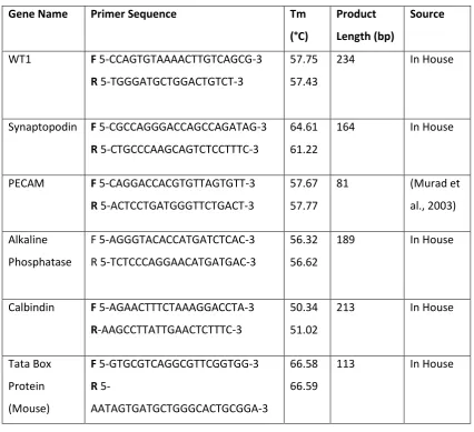
This work was further extended to assess nephrogenesis and endothelial cell survival in the neonatal kidney explants. In the study of the developmental processes governing the mammalian kidney progress has been achieved through the use of embryo explant models. These models are useful for studying early developmental processes such as reciprocal induction but are less suitable for studying later maturation events (e.g. foot process formation in podocytes), due to the fact that the endothelial cell population dies. Podocytes depend upon interactions with the glomerular basement membrane (GBM) to achieve maturity and the GBM is known to be formed, at least in part, from factors secreted by endothelial cells. If an explant model could be established with an intact endothelial population there is potential to study these maturation processes. Markers of interest for neonatal kidney explants included: WT1, Pecam, Synaptopodin, Laminin and Calbindin.
Bioinformatics tools were employed to examine existing microarray data sets of normal endometrium during various stages of the menstrual cycle with the aim of identifying molecular pathways of interest and generating suitable hypotheses. The same process was carried out for endometrium from patients with endometriosis with the future goal of comparing these datasets.
Results:
Analysis of chimeric explants indicated that a large proportion of the endometrial cells died in culture with only a small population present by Day 7 that showed no evidence of forming glandular structures. A degree of nephrogenesis appeared to have occurred in the neonatal kidney explant model with evidence of endothelial cells surviving and forming cord like structures in culture.
4 Cytoscape. A large amount of output was obtained and specific pathways suitable for further investigation were identified including: Super Oxide Radicals Degradation, Tryptophan Degradation to 2-amino-3 Semi Aldehyde and Wnt/β Catenin Pathway.
Conclusions:
The three main conclusions were as follows. 1 Human endometrial cells were not able to survive in the chimeric model of ex-vivo culture, indicating that this is unlikely to be a suitable model system for studying the formation of endometrial cell-derived glandular structures. 2 Nephrogenesis may be occurring in the reaggreagted neonatal explants, and the endothelial population appeared to have survived and formed capillary like structures
5
Contents
Chapter 1: Introduction
1.1 Gross Structure of the Uterus………..………..…………..19
1.2 Microscopic Structure of the Endometrium………..………….…19
1.3 Embryology of the Uterus………...……...20
1.4 Menstrual Cycle……….….20
1.5 Ovarian Cycle………21
1.5.1 Follicular phase………21
1.5.2 Luteal Phase………..21
1.6 Endometrial Cycle……….………..…..…21
1.6.1 Proliferative Phase………..………..…..22
1.6.2 Secretory Phase………..…22
1.6.3 Menstruation……….…………..…23
1.7 Endometriosis……….…….…23
1.7.1 Epidemiology……….………..23
1.7.2 Clinical Features………..………..24
1.7.3 Investigations and Diagnosis………..………..24
1.7.4 Staging of Endometriosis………..…25
1.7.5 Management of Endometriosis……….………..…25
1.7.6 Endometriosis and Fertility……….26
1.8 Pathogenesis of Endometriosis………...26
1.8.1 Retrograde Menstruation………..………....27
1.8.2 Coelomic Metaplasia and Induction Theory……….……….…27
1.8.3 Mullerian Rest Theory……….………..27
1.8.4 Genetics of Endometriosis………..………27
1.9 Proposed Mechanism of Lesion Establishment……….……….………….……….…28
1.9.1 Continued Growth and Survival of Endometrial Cells………..28
1.9.2 Attachment to the Peritoneal Epithelium and Tissue Remodel…...29
1.9.3 Matrix Metalloproteinases………..29
1.9.4 Angiogenesis in Endometriosis……….………...30
1.9.5 Cytokines, Steroid Hormones and Cell Adhesion Molecules……...30
6
1.10 Future Directions of Endometriosis Research………..……..…….31
1.11 A Bioinformatics Approach to the Endometrium and Endometriosis…..…...31
1.11.1 Bioinformatics……….………….32
1.12 Stem Cells………33
1.12.1 Embryonic Stem Cells………..…..34
1.12.2 Adult Stem Cells………..…34
1.12.3 Adult Stem Cell Plasticity………..…………36
1.12.4 Transit Amplifying Cells……….…37
1.13 Evidence for ASCs in the Endometrium……….38
1.14 Characterisation of Endometrial Progenitor Cells………..…………39
1.14.1 Label Retaining Technique………..40
1.14.2 Side Population Method………...………..41
1.14.3 Specific Marker Identification………..……….…..42
1.14.4 In vivo reconstitution Assay………43
1.15 The Future of Stem Cells in the Endometrium……….45
1.16 Disease Models of Endometriosis……….………45
1.16.1 in vitro Models………..………..45
1.16.2 Animal Models……….……46
1.17 The Kidney………..………..47
1.17.1 Embryology of the Mammalian Kidney………..48
1.17.2 Reciprocal Induction……….………..…48
1.17.3 Angiogenesis in the Developing Kidney……….49
1.17.4 Maturation Processes of Podocytes are not Fully Understood…….50
1.17.5 Neonatal Mouse Kidneys………..…50
1.17.6 Generating an Explant Model of Nephrogenesis Using Neonatal Mouse Kidneys ………51
1.17.7 Disaggregation and Reaggregation of Mouse Kidneys………...51
1.18 Specific Markers of Nephron Development and Endometrial Cells…………..52
1.18.1 Kidney Explant Culture………..…...52
1.18.2 Wilms Tumour Protein (Wt1)………...52
1.18.3 Synaptopodin ………..…53
1.18.4 Laminin……….………..……….53
1.18.5 Platelet Endothelial Cell Adhesion Molecule 1 (Pecam)……….53
7
1.19 Chimera Culture………....54
1.19.1 Mucin1………..…………54
1.19.2 Progesterone Receptor……….………54
1.19.3 Human Specific Antibody……….54
1.19.4 Laminin………..………..55
1.20 Aims and Objectives of the Study………..…56
1.20.1 Aims……….………..……….56
1.20.2 Objectives………56
Chapter 2: Materials and Methods 2.1 Sample Collection and Processing………….……….………59
2.1.1 Sample Collection………..…59
2.1.2 Sample Processing………..……..59
2.2 Primary Culture of Endometrial Cells………...60
2.2.1 Crude Fractionation of Epithelial and Stroma Cell Populations…...61
2.2.2 Vybrant Labelling of Human Endometrial Cells…………..……….…62
2.2.3 Trypsinisation of Human Endometrial Cells………....63
2.3 Disaggregation of Neonatal Mouse Kidneys………..….63
2.4 Re-aggregation of Human Endometrial and Mouse Kidney Cells……….………64
2.5 Gelatine Fixing and Sectioning of Chimeras………....65
2.5.1 Preparation of Gelatin Solution……….………..65
2.5.2 Preparing Fixed Rudiments……….65
2.5.3 Gelatine Embedding and Generation of Frozen Section……..……….66
2.6 Immunofluorescence……….66
2.6.1 Slide Preparation………66
2.6.2 Primary Antibodies………..….66
2.6.3 Secondary Antibodies……….………68
2.6.4 DAPI Staining and Mounting………..…68
2.7 Polymerase Chain Reaction………..…..69
2.7.1 RNA Extraction and Quantification………...69
2.7.2 DNase Treatment of RNA………..…..70
2.7.3 Complementary DNA Synthesis: Preparation Stage………..…70
2.7.4 Complimentary DNA Synthesis: Enzymatic Stage………...70
8
2.7.6 Polymerase Chain Reaction Conditions………..…………72
2.7.7 Gel Electrophoresis………..…………...73
2.7.8 Calculation of Relative Gene Expression………74
2.7.9 Relative Quantity………..….74
2.7.10 Relative Gene Expression ………...……….75
2.7.11 Standard Deviation of relative Gene Expression and Standard Error………..……….76
2.8 Bioinformatics Methods………76
2.8.1 Normal Endometrium’ Literature search ………..…..76
2.8.2 Further ‘Normal Endometrium’ Dataset Refinement……….…….77
2.8.3 Endometriosis Literature Search……….………….……..78
2.8.4 Further Endometriosis Dataset refinement………..……….79
2.8.5 Identification and Isolation of Differentially Regulated Genes.…….80
2.8.6 Analysis of Differentially Regulated Genes and Cellular Networks in ‘Normal’ and Endometriosis’ Data Series………..80
3 Chapter 3: Optimisation and Validation of Methods 3.1 Optimization of ‘Vybrant’ Labeling………...83
3.2 Optimisation of Human Specific Antibody Staining……….………...84
3.3 Optimisation of Kidney Explant Model: Complete or Partial Disaggregation…..85
3.3.1 Further Modifications to Disaggregation-Reaggregation Protocol……….87
3.4 RT-qPCR………..……….….87
3.4.1 Validation of RNA extraction and cDNA synthesis…..………87
3.4.2 Identification of Human and Mouse Specific Primer………88
3.4.3 Human and Mouse Specific Housekeeping Genes…….………88
3.4.4 Genes of Interest: Progesterone Receptor and Mucin1………….…...89
3.4.5 Kidney Explant PCR, House Keeping Genes and Genes of Interest………..91
4 Chapter 4: Results 4.1 Chimera Results………..94
9 4.1.2 Minimal Co-Localisation is Detected Between Vybrant and
Anti-Cytoplasmic Staining at Day 7 of Culture………..………96
4.1.3 ‘Bright’ Vybrant Staining Does Not Represent Viable Cells….……....98
4.1.4 Endometrial Chimera Experiment Including the Stromal Fraction………..………..99
4.1.5 Human Cells Do Not Form Glandular Structures Within the Chimera……….………...101
4.1.6 Laminin Structures in Chimeras Appear Disorganised Compared With Kidney Explants……….…..102
4.1.7 Relative Gene Expression within the Chimera………...……103
4.2 Immunofluoresence Analysis of Neonatal Kidney Explants………...…..106
4.2.1 WT1/Laminin……….……….……..106
4.2.2 Pecam/Synaptopodin………..…108
4.2.3 Calbindin/Laminin………..111
4.2.4 Relative Gene Expression of Kidney Explants………..…112
4.3 Bioinformatics Results……….113
4.3.1 Cycle Phase Specific Differentially regulated Genes: Normal Endometrium………..………..113
4.3.2 Cycle Phase Specific Differentially Regulated Genes: Endometriosis………114
4.3.3 Ingenuity Pathways Analysis Output: Normal Endometrium…..…115
4.3.4 Super Oxide Radicals Degradation………..………116
4.3.5 Tryptophan Degradation to 2-amino-3-carboxymuconate Semi Aldehyde……….………...118
4.3.6 Endometriosis Results……….………119
5 Chapter 5: Discussion 5.1 Impaired Survival of Endometrial Cells……….……..121
5.2 Invasion and Formation of Glandular Structures……….…124
5.3 Experimental Refinements and RT-qPCR Analysis……….……….125
5.4 Neonatal Kidney Explant………...127
5.5 Bioinformatics: Normal Endometrium……….………130
5.6 Bioinformatics: Endometriosis………..…..….…132
5.7 Limitations of the Study………..……..133
10
5.9 Conclusions………...……..……137
6 Chapter 6: Appendix 6.1 References………..……….………...139
6.2 Patient Information Form……….……….151
6.3 Patient Consent Form……….……….154
6.4 Patient Clinical Data Collection Form……….………155
6.5 Patient Demographic Information……….……….156
6.6 Additional Immunofluorescence Images………...157
6.7 Reagent Formulations………..162
6.8 Reagent Specifications………...………….163
6.9 Equipment Specifications………..………167
6.10 Concentration of RNA Samples Used in Study………..…………..………….168
6.11 Appendices for Bioinformatics Study 6.11.1 Data Series from ‘Normal Human Endometrium’ Literature Search Excluded from the Study………170
6.11.2 Data Series from ‘Human Endometriosis’ Literature Search Excluded from the Study………...………175
6.11.3 Further analysis of datasets initially identified for inclusion in Endometriosis Study……….177
11
Acknowledgements
First of all I would like to thank my supervisors Dr Dharani Hapangama and Dr Patricia Murray for their guidance, encouragement and enthusiasm throughout this year. They have always been on hand to provide advice on the more challenging aspects of the project and for that I am extremely grateful.
I would like to acknowledge the Jean Shanks Foundation for selecting me to receive an Intercalated Bursary. Their financial support has been exceptionally helpful in enabling me to complete this research year.
I would also like to thank from the Liverpool Womens Hospital: Anthony Valentijn, Jo Drury, Kelly Harper, Lisa Heathcoate, Nicola Tempest, Jane Harrold and from the University of Liverpool Stem Cell Laboratory: Sandra Rak-Raszewska, Sofia Pereira, Virginie Mournetas and Arthur Taylor for all the advice and seemingly inexhaustible amounts of help they have given me throughout the course of the project. I would also like to thank Dr Quentin Nunes and Dr Brian Lane for their expert help and advice in the bioinformatics section of this project.
12
List of Figures
Figure 1 Anatomy of the Female Reproductive System………..……….19
Figure 2 Microscopic Structure of the Endometrium……….………19
Figure 3 Ovarian and Menstrual Cycles……….………20
Figure 4 Ovarian Cycle………..21
Figure 5 Common Sites and Morphology of Endometriotic Lesions…..……..24
Figure 6 Explanations for apparent ASC Transdifferentiation Observed Experimentally……….37
Figure 7 Dual Secretion of GBM Components.………49
Figure 8 Disaggregation and Reaggregation of Neonatal Kidneys Allows Formation of Chimeras and Neonatal Explants………52
Figure 9 Threshold Value for Quantification of Relative Gene Expression...74
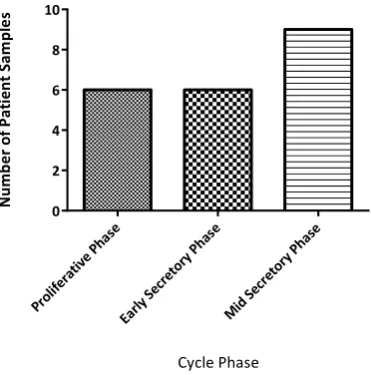
Figure 10 Number of Patient Samples Within Each Phase of the Menstrual Cycle: Normal Endometrium……….77
Figure 11 Number of Patient Samples Within Each Phase of the Menstrual Cycle: Endometriosis………..………79
Figure 12 No difference can be demonstrated between Vybrant and Paraformaldehyde Fixation………84
Figure 13 Trialing Human Specific Antibodies……….…….86
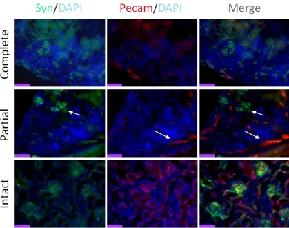
Figure 14 Pecam/Synaptopodin Immunofluorescence: Complete or Partial Disaggregation?...87
13 Figure 16 RT-qPCR Analysis of RNA, DNase Treated RNA and cDNA from
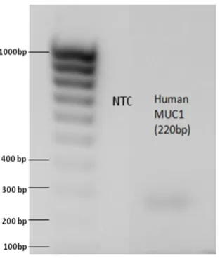
Neonatal Kidney Explant………..89 Figure 17 Gel Electrophoresis of Housekeeping Genes………..91 Figure 18 Identification of Mouse Specific MUC1, Progesterone Receptor
and Human Specific Progesterone Receptor primers……….………91 Figure 19 Identification of Human Specific MUC1 Primers……….92 Figure 20 Identification of Specific Primers for Kidney Explant Analysis…..93 Figure 21 Distribution of Vybrant Staining Changes With Increased Time in
Culture………..96 Figure 22 Anti Cytoplasmic Immunofluorescence Staining of Vybrant
Labeled Chimeras……….98 Figure 23 Day 3 and Day 7 Vybrant Labeled Chimera Frozen Sections
Stained With Human Anti-Cytoplasmic Antibody………..99 Figure 24 Anti-Cytoplasmic Immunostaining of Chimeras cultured with Both Stromal and Epithelial……….…101 Figure 25 Day 0, Day 3 and Day 7 Vybrant Labelled Chimera Frozen Sections Stained With Ant-Laminin Antibody……….103 Figure 26 Comparison of Chimera and Kidney Explant Frozen Sections
Stained With Anti-Laminin Antibody at Day 0 and Day 7 of
Culture……….….104 Figure 27 Relative Gene Expression of Human and Mouse Genes of interest within the Endometrial – Neonatal Mouse Kidney Chimera………106 Figure 28A Wt1/ Laminin Immunofluorescence of Kidney explant Frozen
Sections……….108
o Figure 28B Enlarged Images of Glomeruli from Wt1/Laminin
14 Figure 29A PECAM/Synaptopodin Immunofluorescence of Kidney Explant
Frozen Sections………110
o Figure 29B Enlarged Glomeruli Images from Day 0 and Day 3 Pecam/Synaptopodin Immunofluorescence………..111
Figure 30 Comparison of Day 0 and Day 3 Culture of Calbindin/Laminin Immunostaining………..112
Figure 31 Analysis of Relative Gene Expression in Neonatal Mouse Kidney Explants……….114
Figure 32 Cycle Phase Specific Differentially Regulated Genes: Normal Endometrium………115
Figure 33 Cycle Phase Specific Differentially Regulated Genes: Endometriosis………116
Figure 34 Top Enriched Canonical Pathways to the Mid Secretory Intracellular Dataset and Super Oxide Radicals Degradation Pathway……118
Figure 35 Top Enriched Canonical Pathways to Late Secretory Intracellular Dataset and Tryptophan Degradation to 2-amino-3-carboxymuconate Semi Aldehyde Pathway……….119
Figure 36 Cytoscape Output Cluster from Mid-Secretory Dataset………….120
Figure 37 CD1 Chimera: Anti Cytoplasmic Staining Repeat……….158
Figure 38 CD1 Chimera: Laminin Staining Repeat……….158
Figure 39 CD1 WT1 Laminin Repeat………...159
Figure 40 CD1 WT1 Laminin Repeat 2………...159
Figure 41 Black 6 WT1 Laminin Repeat 1……….159
Figure 42 CD1 PECAM Synaptopodin Repeat 1………160
Figure 43 CD1 PECAM Synaptopodin Repeat 2………...160
15
Figure 45 CD1 Calbindin Laminin Repeat 1………....161
Figure 46 CD1 Calbindin Laminin Repeat 2……….161
Figure 47 Black 6 Calbindin Laminin Repeat 1………..161
16
Glossary
AMM. Amniotic Membrane Model
ASC. Adult Stem Cell
BM. Basement Membrane
BrdU.
Bromodeoxyuridine
CAM. Chorioallantotic Membrane
cDNA. Complementary
DNA
CFDA SE. Carboxy
Fluorescein Diacetate, succinimidyl ester
CFU. Colony Forming Unit
Ct. Cycle to Threshold
DAPI.
4',6-diamidino-2-phenylindole
DAVID. Database for
Annotation Visualisation and Integrated Discovery
DMEM. Dulbecco’s
Modified Eagle Medium
DMSO.
Dimethylsulfoxide
dNTP.
Deoxyribonucleotide Triphosphate
DTT. Dithiotheritol
ECM. Extra Cellular Matrix
EF. Epithelial Fraction
ELISA. Enzyme Linked
Immuno-absorbant Assay
ESCs. Embryonic Stem
Cells
FACS. Fluorescence
Activated Cell Sorting
FBS. Fetal Bovine Serum
FGF. Fibroblast Growth Factor
FISH. Fluorescence In
Situ Hybridisation
FSH. Follicle Stimulating Hornone
g. Relative Centrifugal Force
GBM. Glomerular Basement Membrane
GE. Glandular Epithelium
GnRH. Gonadotrophin
Releasing Hormone
HCG. Human Chorionic Gonadotrophin
IDO. Indoleamine 2,3-dioxygenase
IHC.
Immunohistochemistry
IPA. Ingenuity Pathways Analysis
IVF. In Vitro Fertilization
KCM. Kidney Culture Medium
LE. Luminal Epithelium
LH. Leutinising Hormone
LRC. Label Retaining Cells
LRT. Label Retaining Technique
MEME. Minimum
Essential Medium Eagle
MET. Mesenchymal to Epithelial Transition
MIP. Metastasis Inducing Protein
MM. Metanephric Mesenchyme
MMPs. Matrix
Metalloproteinases
MUC1. Mucin1
NBF. Neutral Buffered Formalin
NCBI. National Centre
for Biotechnology Information
17
NSAIDs. Non Steroidal
Anti Inflammatory Drugs
NSG. Nod SCID (Severe Combined Immune Deficiency) Gamma
NTC. No Template Control
PBS. Phosphate Buffered Saline
PC. Progenitor Cell
PCR. Polymerase Chain Reaction
PDGF-β. Platelet derived
Growth Factor Beta
Pecam. Platelet
Endothelial Cell Adhesion Molecule 1
PGR. Progesterone Receptor
PTEN. Phosphate and
Tensin Homologue
RANTES. Regulated on
Activation, Normal T Cell Expressed and Secreted
RCF. Regulators of Cell Fate
ROS. Reactive Oxygen Species
RT-qPCR. Real Time
quantitative Polymerase Chain Reaction
RQ. Relative Quantity
SCID. Severe Combined
Immunodeficiency
SD. Standard Deviation
SF. Stromal Fraction
SOD2. Superoxide
Dismutase 2
SP. Side Population
SPC. Stem/Progenitor Cell
SSEA-1. Stage Specific
Embryonic Antigen 1
TA. Transit Amplifying
TAE. Tris-Acetate EDTA
Tbp. Tata Box Protein
TGF-β. Transforming
Growth Factor Beta
TIMPs. Tissue Inhibitors
of Metalloproteinases
UB. Ureteric Bud
VEGF. Vascular
Endothelial Growth Factor
19
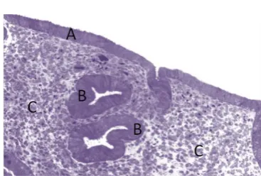
Figure 2: Microscopic Structure of the Endometrium
Fig2. Microstructure of the human endometrium. A: Luminal Epithelium, B: Glandular Epithelium, C: Stroma. The functionalis and basalis layers are not histologically distinct. (Gargett, 2008)
1.1 Gross Structure of the Uterus
The human uterus is a thick walled muscular organ located within the pelvic cavity. It is roughly 7.5 cm in length and is continuous superiorly with the fallopian tubes, which are encased in the broad ligament. Inferiorly the uterus is continuous with the cervix, vagina and vulva (Figure 1) (Drake RL, 2010). The structure of the uterus is made up of three layers with the outermost layer being parietal peritoneum continuous with the broad ligament. The middle layer is myometrium composed of smooth muscle fibers and the innermost layer of the uterus is the endometrium (Drake RL, 2010).
1.2 Microscopic Structure of the Endometrium
The endometrium is the reproductive tissue lining the uterine cavity. It possesses immense proliferative potential evidenced by the fact that it is able to undergo up to 400 cycles of growth differentiation and shedding within an average woman’s reproductive years (Gargett and Masuda, 2010). The endometrium is functionally divided into a superficial ‘functionalis’ layer which is shed during menstruation and a deeper ‘basalis’ layer which is retained and from which the new functionalis is regenerated each month.
Histologically the endometrium can be separated into two distinct cell populations, the Epithelium and the Stroma (Figure 2). The epithelium is further subdivided into the Luminal Epithelium (LE, Figure 2A), which lines the uterine cavity serving as the point of contact for the implanting blastocyst and the Glandular Epithelium (GE, Figure 2B), which lines the glands penetrating deep into the basalis (Jabbour et al., 2006). The second population is the stroma (Figure 2C), which constitutes the supporting parenchyma of the endometrium. The stroma and the basalis glands form a rough boarder with the myometrial cells, which make up the body of the uterus (Uduwela et al., 2000).
Fig1. Anatomy of the human female reproductive system (Drake RL, 2010)
[image:19.595.388.572.491.615.2]20
1.3 Embryology of the Uterus
The uterus, cervix, fallopian tubes and cranial portion of the vagina are formed from the paired Paramesonephric or ‘Mullerian’ ducts which were first described in 1825 and derive from the intermediate mesoderm (Gargett). Both the reproductive and urinary systems are derived from this structure which lies between the paraxial and the lateral plate mesoderm along the posterior wall of the abdominal cavity (Sadler, 2004). The luminal epithelium emerges first with the glands being a late fetal development shallow in nature until further development occurs postnatally (Ma, 1973). The stromal cells and myometrium are derived from the urogenital ridge mesenchyme and there is evidence that their differentiation is regulated by the surrounding epithelium (Brody and Cunha, 1989). This highlights the idea of crosstalk between these structures and indicates the importance of studying both cell populations when investigating endometrial physiology (Brody and Cunha, 1989).
1.4 Menstrual Cycle
The human menstrual cycle refers to a set of dynamic changes involving proliferation and differentiation that occur within the premenopausal endometrium according to the corresponding cyclical ovarian hormonal changes, for which the endometrium is the target organ (Figure 3). The cycle can be split
into events occurring in the ovary (Figure 3A), and events occurring in the endometrium (Figure 3B). The standardised menstrual cycle is 28 days in duration with day 1 defined as the start of the menstrual phase. Ovulation occurs at approximately day 14 which signifies the transition from proliferative to secretory phase
[image:20.595.318.547.459.619.2](Gargett, 2008). Fig3. The histological and hormonal changes associated with the human ovarian and menstrual cycles.
Figure 3: Ovarian and Menstrual Cycles
21
1.5 The Ovarian Cycle
The ovaries are the female gonads and are responsible for producing the ova which will form the maternal portion of the zygote, they undergo a series of cyclical changes in tandem with the endometrium releasing Oestrogen and Progesterone under the influence ofFollicle Stimulation Hormone (FSH) and Luteinising Hormone (LH) (Impey, 2012, Gargett, 2008). The ovarian cycle is divided into follicular and luteal phases (Figure 4).
1.5.1 Follicular phase
The follicular phase is characterised by the pulsatile release of FSH and LH from the anterior pituitary under the influence of Gonadotrophin releasing Hormone (GnRH) (Figure 4A). FSH acts to stimulate the development of several primordial follicles which release oestrogen and inhibin to inhibit FSH in a negative feedback response. As oestrogen levels increase however they begin to exert a positive feedback response stimulating a ‘surge’ of LH at day 14 (Figure 4A) with follicle rupture and ovulation occurring usually within 36 hours of this event (Gargett, 2008).
1.5.2 Luteal Phase
Ovulation signifies the beginning of the luteal phase as the ruptured follicle degenerates to form the corpus luteum. This structure secretes both oestrogen and progesterone, though progesterone is dominant, along with inhibin A, the levels of which peak around day 21 (Figure 4B). Progesterone acts to prevent degeneration of the oestrogen primed endometrium and stimulates decidualization (Impey, 2012). Unless the corpus luteum is maintained by Human Chorionic Gonadotrophin (HCG) released as the result of implantation it will degenerate with the subsequent fall in progesterone triggering menstruation (Figure 4B).
1.6 Endometrial Cycle
[image:21.595.404.550.222.404.2]The endometrial cycle can be split into three broad phases: Proliferative, Secretory and Menstrual each of which are characterised by structural and functional changes highlighting the varied and dynamic nature of this tissue.
Figure 4: Ovarian Cycle
Fig4. Illustration of cyclical hormonal changes during the ovarian cycle. A: Hormones acting on the ovaries, FSH, LH. B: Hormones released by the ovaries, Oestrogen (E2) and
22
1.6.1 Proliferative Phase
The endometrial changes seen in the proliferative phase are thought to be regulated by the dominant hormone in the tissue at this time, oestrogen. The early proliferative phase is characterised by initial repair of the denuded endometrial surface, a process commenced by epithelial cells suggesting that this population is required for stromal responsiveness later in the cycle (Bigsby, 2002). As the proliferative phase continues all cell types multiply under the influence of oestrogen which is thought to assert its effects via Insulin-like Growth Factors along with Epidermal Growth Factors and Transforming Growth Factor Beta (TGF-β) (Gargett et al., 2008). A vast array of additional molecules have also been implicated in the proliferation of the endometrium at this time including: growth factors, cytokines, fibronectin, integrins and matrix metalloproteinases (MMPs) (Thie and Denker, 2002). An interesting aspect of these repair and proliferation processes is that they enable the endometrium to heal without scarring, a phenomenon only previously documented in fetal skin wounds (Samuels and Tan, 1999).
1.6.2 Secretory Phase
The Secretory Phase commences at around day 14 of the cycle and is characterised by functional differentiation of the endometrial tissue to promote an environment suitable for embryo implantation (Gargett, 2008). The key hormone driving these processes is progesteronewhich acts on oestrogen primed cells. The secretory phase can be histologically subdivided into: Early, Mid and Late phases based on the criteria of Noyes et al (Noyes and Haman, 1953).
Early Secretory Phase: sub-nuclear vacuoles are observed in epithelium
Mid Secretory Phase: increased gland tortuosity with increased secretory activity. This phase also represents the implantation window (Psychoyos, 1973).
Late Secretory Phase: the differentiation of stromal cells known as decidualization occurs. This spontaneous non reversible change involves fibroblast type cells close to spiral arterioles becoming enlarged and rounded. Progesterone receptor A is dominant at this time (Alexander et al., 1996).
23
1.6.3 Menstruation
Menstruation is the term used for the sequence of tissue breakdown and endometrial bleeding occurring at the end of each menstrual cycle during which the functionalis layer of the endometrium is shed leaving the basalis intact (Gargett et al., 2008). These changes are initiated by a fall in oestrogen and progesterone preceded by the breakdown of the corpus luteum. Various mechanisms governing menstruation have been proposed, one of which is hypoxia (Markee, 1978) however is it also likely that menstruation is an inflammatory process characterised by tissue oedema, inflammatory cell recruitment and pro inflammatory cytokine release (Finn, 1986). As the endometrial vasculature may play a key role in the tissue breakdown associated with menstruation, studies have considered various pro inflammatory and vasoactive agents associated with this structure e.g. Prostaglandin F2 alpha, Cyclo-oxygenase 2 and Endothelin 1 (Salamonsen et al., 1999a, Salamonsen et al., 1999b).
1.7 Endometriosis
A wide variety of factors contributing to the regenerative capacity of the endometrium have been proposed and research is ongoing. In addition, widening the understanding of normal physiology may provide insights into pathological processes and there are several endometrial diseases which share this characteristic of a large proliferative potential. One such disease is endometriosis which is defined as the presence and growth of tissue similar to endometrium comprising glands and stroma outside of the uterus and represents a major detriment on the quality of life of affected patients (Impey, 2012).
1.7.1 Epidemiology
24
1.7.2 Clinical Features
The clinical features of endometriosis are broad and highly varied but common signs and symptoms include: pelvic pain, dysmenorrhoea, deep dyspareunia, dyschezia, subfertility and infertility. Other rare symptoms that may suggest extensive disease include: cyclical haematuria, rectal bleeding and bleeding from the umbilicus (Impey, 2012).
1.7.3 Investigations and Diagnosis
Serum CA 125 levels may be raised in patients with endometriosis however this is of limited diagnostic value and reliable biomarkers of the disease are currently lacking. Direct visualisation of endometriotic lesions at laparoscopy is required for definitive diagnosis with or without tissue biopsy and histological analysis (Guideline, 2008). If there is clinical evidence of deeply infiltrating endometriosis, bladder and bowel involvement are assessed with MRI, plus or minus intravenous pyelogram and Barium studies (Guideline, 2008). On inspection of the abdominal cavity lesions may be diffusely spread and thorough investigation is necessary for accurate staging. Common sites include: utero-sacral ligaments, utero-vesical fold, utero-rectal pouch, sigmoid colon and the ovary (endometrioma) (Impey, 2012) (Figure 5A).
There are four different characteristic appearances of endometriotic lesions commonly visualised at laparoscopy (Figure 5B) which may represent different stages of disease activity and different cellular processes. More active lesions appear as red vesicles with white scars or brown spots (powder burn) followed by fibrotic lesions representing more longstanding disease (Impey, 2012). Another laparoscopic finding more commonly found in endometriosis patients is the Allen Masters Peritoneal Window which may be the result of a prolonged inflammatory state (Burney and Giudice, 2012).
Figure 5: Common Sites and Morphology of Endometriotic Lesions
Fig5. Common sites and phenotypes associated with endometriosis lesions. A. Common sites of endometriotic lesions within the pelvic cavity. (Impey, 2012) B. Phenotypic subtypes of peritoneal endometriosis. (A) Red vesicular lesion. (B) Powder-burn lesion. (C) Fibrotic lesion. (D) Allen-Masters peritoneal defect (Burney., 2012).
25
1.7.4 Staging of Endometriosis
The revised American fertility society criteria for endometriosis scores the disease extent based on the presence and location of lesions. There are four stages (Impey, 2012):
Grade 1 (Minimal) Grade 2 (Mild) Grade 3 (Moderate) Grade 4 (Severe)
It should be noted that the severity of disease assessed by the grading system does not correlate well with patient symptoms.
1.7.5 Management of Endometriosis
The treatment of endometriosis can be divided into conservative, medical and surgical treatment. However this is governed primarily by patient symptoms as no treatment is required for asymptomatic lesions found incidentally at laparoscopy. The exception to this are endometriomas which are occasionally removed in this situation as there is a small risk of misdiagnosing ovarian cancer (Guideline, 2008).
Medical Management
26
Surgical Management
Surgical management aims to remove the endometriotic lesions from the abdominal cavity and is only considered if medical therapy has been ineffective. At laparoscopy laser or bipolar diathermy can be used to ablate lesions. Other options include radical dissection of lesions or hysterectomy with bilateral salpingo-opherectomy however these options should be considered as a last resort as they entail major surgery and are associated with adhesion formation. Laparoscopy itself has also been associated with endometriosis lesion induction (Harirchian et al., 2012).
1.7.6 Endometriosis and Fertility
Endometriosis is a disease associated with sub-fertility, for example lesions are found in 25% of laparoscopies carried out for subfertility (Impey, 2012); the mechanisms of this sub fertility are not yet clear, though implantation failure is thought to be one mechanism, and this affects management. A Cochrane review has found that medical treatment will not increase fertility if the fallopian tubes are unaffected but that laparoscopic ablation may, particularly when adhesions are present (Jacobson et al., 2002). Many patients with advanced endometriosis will be of reproductive age and the best chance for achieving pregnancy will be In Vitro Fertilisation (IVF). With this in mind it is essential to attempt to understand the processes behind this disease as it impacts upon so many aspects of a patient’s life and several different approaches need to be considered.
1.8 Pathogenesis of Endometriosis
Whilst the clinical features of endometriosis are well described the cause of this proliferative disease is still unknown. Several theories have been proposed and these include:
27 These proposed mechanisms of pathogenesis can be broadly divided into those where ectopic tissue implants onto the abdominal wall e.g. retrograde menstruation, and those where the endometrial tissue arises de novo e.g. Coelomic Metaplasia (Gargett, 2008).
1.8.1 Retrograde Menstruation
Retrograde menstruation is the most widely accepted theory for endometriotic lesion induction and was first proposed by Sampson in 1927 (Sampson, 1927). It is suggested that at the point of menstruation some of the shed endometrium, instead of passing through the cervix and vagina, enters the abdominal cavity via the fallopian tubes and implants there, the result being a lesion (Sampson, 1927). There is some clinical evidence to support this theory in the increased prevalence of endometriosis noted in women with uterine outflow obstruction e.g. cervical stenosis (Jenkins et al., 1986).
1.8.2 Coelomic Metaplasia and Induction Theory
Coelomic metaplasia and induction theory are two interrelated ideas implicating metaplasia of extra-uterine cells in the pathogenesis of endometriosis. Coelomic metaplasia states that normal peritoneal tissue is transformed by an as yet undefined stimulus to ectopic endometrial tissue. Similarly induction theory proposes that extra-uterine metaplasia may be induced by either a hormonal or immunological factor, while study is ongoing these ‘factors’ have not yet been defined (Levander and Normann, 1955). Some indirect evidence for these theories can be found in the form of case reports of ovarian endometriosis in patients with Mayer-Rokitansky-Kuster-Hauser syndrome, a condition characterised by uterine agenesis including absence of the endometrium (Yan et al., 2011).
1.8.3 Mullerian Rest Theory
This theory relates to the development of the female reproductive tract through the Mullerian duct. It is suggested that cells derived from the Mullerian duct maintain a differentiation potential, giving them the capacity to transform into endometrial cells at the onset of puberty under hormonal stimulation (Longo, 1979).
1.8.4 Genetics of Endometriosis
28 is limited due to the requirement for laparoscopy to make a diagnosis (Zondervan et al., 2001, Kennedy et al., 2001). Some correlation has been found but the trait is likely to be complex (Simpson and Bischoff, 2002).
1.9 Proposed Mechanism of Lesion Establishment
Sampson’s theory of retrograde menstruation is the most widely excepted model of endometriosis; however it poses several questions that need to be examined in more detail to fully understand the pathogenesis of this disease.
One such issue is the observation that ‘retrograde menstruation’ occurs in up to 90% of pre-menopausal females (Halme et al., 1984) however only 5-10% experience the disease (Cramer and Missmer, 2002, Missmer and Cramer, 2003), which would suggest other factors are important in determining lesion establishment. In a recent review, Burney and Guidice summarised these as: escape from immune clearance, attachment to peritoneal epithelium, invasion of the epithelium, establishment of local neurovascularity, and continued growth and survival (Burney and Giudice, 2012). Looking at these processes and how they may vary between women with and without the disease has yielded a group of potential biomarkers. However, investigation in this area is still early with many publications offering conflicting evidence. The variation of these putative biomarkers throughout the menstrual cycle is also not always considered in their analysis, a factor which must be taken into account to ensure findings are relevant (May et al., 2011). Bioinformatics is a useful tool which could help gain a ‘whole view’ of the endometrium as it can consider vast amounts of data, however there is a difference between identifying biomarkers and confirming their role in pathogenesis. The work presented below indicates that further investigation is required in order to more fully understand the pathogenesis of this disease.
1.9.1 Continued Growth and Survival of Endometrial Cells
29 anti-apoptotic environment in patients with endometriosis characterised by increased MCL-1 and reduction in Bak expression in secretory phase glandular epithelium (Burlev et al., 2006). Another group has demonstrated up-regulation of the anti-apoptotic gene BCL-2 in the endometrium of affected patients (Wingfield et al., 1995). Telomerase activity is another factor increasing the survival potential of endometrial cells and has been detected increased in secretory phase endometrium of patients with the disease, it was also noted that mean telomere length may also be increased (Hapangama et al., 2008, Hapangama et al., 2009). A further consideration is the presence of endometrial stem cells in endometriosis lesions.
1.9.2 Attachment to the Peritoneal Epithelium and Tissue Remodelling
It is thought that damaged peritoneum could be a risk factor for implantation of ectopic tissue, one in vitro study has shown that fragments of endometrial tissue were only adherent in a model of ectopic implantation when extracellular matrix (ECM) was exposed due to induced damage (Gerhard et al., 1991). This is also consistent with work in baboons demonstrating that laparoscopy may be a risk factor for lesion establishment (Harirchian et al., 2012).
1.9.3 Matrix Metalloproteinases
30 remodelling. This group of enzymes has also been investigated in endometriosis with similarly conflicting results (Estellés et al., 2003, Ramón et al., 2005, Gilabert-Estellés et al., 2007).
1.9.4 Angiogenesis in Endometriosis
It has been observed in cancer that any tissue growth above a critical mass must establish a blood supply for itself. For this reason angiogenesis has been investigated in endometriosis and interest in this area is enhanced by the finding of a chimeric circulation in the work performed by the Masuda group (Masuda et al., 2007). Vascular Endothelial Growth Factor (VEGF) is a molecule that has been extensively investigated with increased expression detected in the late secretory epithelium of patients with the disease (Donnez et al., 1998).
A subtype of the molecule VEGF-A is has also been found upregulated in women with endometriosis as well as down regulation of VEGF receptor 1 and 2 possibly indicating decreased sensitivity (Bourlev et al., 2006). These studies indicated that VEGF might be elevated at a certain point in the cycle in endometriosis, which would make sense as it is a hormone responsive disease. More recent investigation however has suggested increased VEGF expression might persist throughout the cycle (Gilabert-Estellés et al., 2007).
1.9.5 Cytokines, Steroid Hormones and Cell Adhesion Molecules
Many other intra and extracellular molecules have been investigated with regard to their involvement in the pathogenesis of endometriosis. Interleukins are a group of cytokines thought to be differentially expressed in endometriosis. For example IL-1R Type II has demonstrated downregulation in epithelium throughout several studies (Akoum et al., 2001, Kharfi and Akoum, 2001, Kharfi et al., 2002, Lawson et al., 2008) whereas IL-8 has been found up-regulated in epithelial cells (Ulukus et al., 2005). The chemokine ‘Regulated
on Activation, Normal T Cell Expressed and Secreted’ (RANTES) has been found
differentially expressed in endometriosis, but further work is needed to fully elucidate its
association with the disease as its expression appears to also be cycle phase dependent
31
1.9.6 Stem Cells in Endometriosis
The cellular and molecular processes investigated in relation of endometriosis often relate to increased cell survival within the peritoneal cavity and proliferation of a cell population in response to a stimulus (in this case, oestrogen). These are properties shared by Adult Stem Cells (ASCs) or Progenitor Cells (PCs) and it is for this reason that ASCs have been hypothesised to play a role in the pathogenesis of endometriosis. Some experimental findings have backed up this hypothesis including investigation of epithelial glands in endometriosis which were monoclonal in some lesions suggesting they descend from a single PC (Gargett and Masuda, 2010). Experimental models of lesion induction in baboons have also demonstrated that introducing endometrial cells into the abdominal cavity was associated with lesion development (Harirchian et al., 2012). As progenitor cells are thought to reside in the basalis it has been suggested that in women with endometriosis these may become inappropriately dislodged and refluxed (Leyendecker et al., 2002).
1.10 Future Directions of Endometriosis Research
It is clear that potential molecules involved in the pathogenesis of endometriosis have been extensively studied using laboratory techniques with potential applications including peripheral blood tests to aid diagnosis and the development of disease modifying pharmacological interventions. While this work is undoubtedly useful there is a great variability in results and to a degree it may be beneficial to introduce a standardised method within the field e.g. in accounting for cycle phase. Another tool that would be useful in guiding this research, and could provide useful insights into cellular and molecular processes occurring in both normal endometrium and endometriosis, is bioinformatics.
1.11 A Bioinformatics Approach to the Endometrium and Endometriosis
32 Studies using mouse models have provided useful insights into potential disease processes in endometriosis (Masuda et al., 2007). Humans however are more biologically complex organisms and although this is not demonstrated by an increased number of genes within the genome, it can be seen in Protein Superfamily expansion, particularly in the extracellular compartment (Vogel and Chothia, 2006). This is logical as the extracellular compartment is a far less stable environment than the intracellular compartment and as a result adaptations must be more rapid. Given that many of the markers postulated to play a role in the pathogenesis of endometriosis are associated with the extracellular compartment, it is important to observe their expression specifically in humans as the molecular processes may differ between species (Vogel and Chothia, 2006). A potential tool to help overcome these challenges is bioinformatics.
1.11.1 Bioinformatics
Bioinformatics can be most simply described as the ‘combination of biology and information technology’ and can incorporate any tools or methods used to analyse and manipulate large sets of data (Westhead DR, 2002). This rapidly growing area of investigational biology uses a multidisciplinary approach to increase understanding of complex organisms in a quantitative and discrete way not previously possible. Bioinformatic study consists of three main components:
The creation of databases allowing storage and manipulation of large biological datasets
The development of algorithms and statistics to determine relationships between members of these large datasets.
Use of these tools for analysis and interpretation of various types of biological data, including DNA, RNA and protein sequences, protein structures gene expression profiles and biochemical pathways (Westhead DR, 2002).
33 With this in mind the three components described are utilised in order to achieve three main objectives: (Arthur, 2008).
To understand integrated aspects of the biology of organisms, viewed as coherent complex organisations at microscopic and macroscopic levels
To interrelate sequence, three dimensional structure, expression pattern, interaction and function of individual proteins, nucleic acids and protein-nucleic acid complexes
To support applications in medicine, agriculture and technology (Arthur, 2008).
The potential of bioinformatics study to aid scientific discovery and influence clinical practice is becoming increasingly realised with several scientific papers in the field of Obstetrics and Gynaecology utilising this tool in recent years (Nguyen et al., 2012). Within this context the main focus is on drug target and biomarker discovery using tools such as: Microarrays, Mass Spectrometry and Proteomic Data Analysis (Arthur, 2008). As it is the case with bioinformatics study that a large amount of information is generated and recorded analysis is not always exhaustive. It was with this in mind that a component of this study was proposed in order to investigate differential gene expression in ‘normal’ endometrium and endometrium of patients with endometriosis. With this view it is thought that potential drug targets and biomarkers could be discovered and avenues for further ‘wet’ lab investigation identified.
1.12 Stem Cells
While hypotheses can be generated from in silico work it is also important to investigate current models of physiology and disease within the laboratory setting. One such model relating to both normal endometrial function and endometriosis is the idea of stem cells in the endometrium. Stem Cells are defined as a population of undifferentiated cells, the key characteristics of which are self renewal and differentiation into one or more descendant of highly differentiated cell lineage (Till and McCulloch, 1961). They can be subdivided according to their ability to reconstitute tissues associated with an organism: (Wagers and Weissman, 2004)
Totipotent: (All embryonic and extra embryonic cell types) Pluripotent (All cell types of the embryo proper)
34 Unipotent (Only one mature cell type)
Stem Cells can also be categorised as either Embryonic Stem Cells (ESCs) or Adult Stem Cells (ASCs) (Evans and Kaufman, 1981).
1.12.1 Embryonic Stem Cells
ESCs are defined as pluripotent cells isolated from the blastocyst inner cell mass before implantation and are capable of differentiating into structures constituting all 3 germ layers in vitro (Evans and Kaufman, 1981). ESCs are not widely used for investigation within the endometrium and given the associated ethical concerns this population is not utilised in this study.
1.12.2 Adult Stem Cells
Adult Stem Cells (ASCs) reside within mature tissues and demonstrate the stem cell properties of self renewal and limited differentiation potential, being either multipotent or unipotent (Wagers and Weissman, 2004). This means that whilst maintaining a steady population within their tissue of origin e.g. the endometrium, they are able to form cell colonies specific to their lineage, a concept arising from studies involving Haematopoietic Stem Cells (Harrison, 1980).
ASCs may be responsible for processes such as growth and repair after injury in mature tissue and are believed to be surrounded by a physiological microenvironment referred to as the ‘Stem Cell Niche’ (Schofield, 1978). This environment is thought to be comprised of a group of supporting ‘niche cells’ which communicate with their resident ASC stimulating either proliferation or differentiation (Gargett, 2007). Interactions between the ASC and its niche are vital for normal function of adult tissues, a process characterised in gut epithelial tissue (Gargett, 2007).
35 understanding and aid clinical management of a variety of endometrial pathologies including menstrual disorders and endometriosis.
One tissue in which ASCs have been extensively studied relevant to the endometrium is the intestine. In this tissue the epithelial cells are arranged in a glandular structure similar to that of the endometrium; the gland base, or crypt of Lieberkuhn, has been identified as the area where proliferation occurs. This is evidenced by studies demonstrating activation of the canonical Wnt/Beta Catenin signalling pathway in basal cells, a key regulator of an undifferentiated state associated with ASC activity (Fevr et al., 2007, Valentijn et al., 2013). Being located at the base of the crypt allows these ASC/progenitor cells to be surrounded by a niche, as described by Schofield, composed of extracellular matrix and other cells (Spradling et al., 2001). This would not be possible higher up the crypt as the more differentiated cells migrate upwards through the ‘transit amplifying zone’ before being shed (Spradling et al., 2001). Specific markers have also been identified in the intestine, notably Musashi1, Hes1 and CD133 (Iovino F, 2009). Any tissue containing ASCs may be associated with a rapid turnover of cells. One danger recognized with this rapid proliferation is the increased risk of mutations which may lead to tumour formation (Wagers and Weissman, 2004). It has been noted in Colon Cancer that tumours expressing the marker CD133 have been demonstrated residing at the base of normal glands leading to the hypothesis that the aberrant proliferation of ASCs may play an early role in the formation of these tumors (Salven et al., 2003, Yin et al., 1997). The progenitor cells isolated in the intestine also show a higher telomerase activity with longer telomere lengths, another ASC property currently being investigated in the endometrium (Schepers et al., 2011, Valentijn et al., 2013)
Given the similarities between endometrial glands and the small intestine epithelial glands this tissue has been used as a model for endometrial research. An example of this is the investigation of Sox9, a molecule associated with proliferation known to be present in the small intestine which a group at the University of Liverpool has recently investigated in the endometrium using IHC (Valentijn et al., 2013).
36 fibroblasts (Takahashi and Yamanaka, 2006). This was achieved via the retroviral induction of 4 transcription factors: Oct4, Klf4, c-myc and Sox2, the actions of which are thought to reprogram the cell to a pluripotent phenotype (Jaenisch and Young, 2008).
This is an emergent field and several modifications to the procedure including the induction of different transcription factors e.g. Nanog and different methods of induction have been used to reduce potential teratogenicity. Since 2006 iPS cells have also been generated from human cells (Yu et al., 2007).
There are a great number of potential therapeutic applications using this technology e.g. the generation of organs for transplant which, as they would be derived from a patients own cells, would potentially be free from the ethical concerns of using embryonic stem cells and the risks of immune rejection currently associated with transplant surgery. Indeed one recent development was the generation of ‘Liver buds’ using iPS cells to form hepatocytes (Takebe et al., 2013). However a number of concerns have been raised over the use of iPS cells, primarily the potential for tumour formation given the relatively undifferentiated state of the cells combined with a high proliferation capacity, and recent reports demonstrating immunogenicity of iPS cells (Zhao et al., 2011). There is a great amount of progress still to be made in the field of iPS cells however early results are promising and the potential therapeutic applications are many.
1.12.3 Adult Stem Cell Plasticity
One of the interesting potential benefits of isolating an ASC population within the endometrium centres on the concept of ‘Stem Cell Plasticity’. This property allows an ASC population to ‘transdifferentiate’, a process by which ASCs could contribute to cell types of different lineages (Wagers and Weissman, 2004). If this is possible then endometrial ASCs, easily obtained in the out-patient clinic and ethically safe, could represent a great source of autologous tissue for transplant. Endometrial ASCs could be extracted from a tissue sample, grown in vitro before differentiation into the population of choice e.g. dopaminergic neurons for to treat Parkinson’s disease and transplanted.
37 underlying this process. A number of alternate mechanisms need to be ruled out in order to accurately demonstrate this phenomenon (Figure 6) (Wagers and Weissman, 2004).
A. ASC Transdifferentiation: Lineage conversion of the cells occurs. A dormant lineage differentiation program is activated to alter the lineage specificity of the cell (Wagers and Weissman, 2004).
B. De-differentiation of an ASC occurs to a more primitive phenotype before re-differentiation This is a phenomenon observed in animal models (Brockes and Kumar, 2002).
C. A heterogeneous population of ASCs is present in the test population. D. A rare pluripotent stem cell is present in the tissue
E. Cell fusion is taking place. This process is associated with normal cell physiology e.g. in the formation of skeletal myofibroblasts (Anderson, 2000).
Figure 6: Explanations for apparent ASC Transdifferentiation Observed Experimentally
To date, many of the studies reporting ASC transdifferentiation within the endometrium have not satisfactorily ruled out these events. It is possible either that transdifferentiation within the endometrium is extremely rare, a finding consistent with findings from our laboratory in the University of Liverpool (Da Silva, 2012). Alternatively, progenitor cells within the endometrium could be unipotent; a property associated with the more terminally differentiated Transit Amplifying (TA) cells.
Fig6. Explanations for apparent ACS transdifferentiation observed in culture. When documenting ASC
38
1.12.4 Transit Amplifying Cells
Transit Amplifying cells represent a temporary cell population more differentiated than ASCs but which still have a limited self renewal capacity both in vitro and in vivo (Seaberg and van der Kooy, 2003). These cells are unipotent and hormone responsive; it is unclear to what extent they are identified in assays of ASC function in the endometrium (Gargett, 2008). In fact one theory is that TA cells and ASCs are in fact the same population of cells exhibiting a differing phenotype depending on the conditions of their niche (Quesenberry et al., 2005).
1.13 Evidence for ASCs in the Endometrium
The high regenerative capacity of the endometrium has long been a source of interest to clinicians and it has been thought for many years that a population of ASCs residing in this environment could account for the vast proliferation potential observed. The first publication recognising this possibility was Prianishnikov in 1978 who outlined a theory of ASCs within the endometrial epithelial population (Prianishnikov, 1978). This paper suggests there are three populations of ACSs in the endometrium divided by their response to oestrogen and progesterone:
ASCs responsive to oestrogen
ASCs responsive to oestrogen and progesterone ASCs responsive to progesterone only
Prianishnikov thought that the growth of these ‘stem cells’ was regulated by oestrogen and progesterone acting on the cells directly. It is possible that ASCs themselves are hormone independent but that their nearest descendants (TA cells) become hormone dependent and it was suggested that the ASC and TA cells receptor status would be mediated in an oestrogen dependent manner (Prianishnikov, 1978).
39 backed up with the experimental finding that oestrogen will induce the synthesis of progesterone receptors in uterine tissue cells thereby making them susceptible to progesterone (Wiest WG, 1971). Prianishnikov described the characteristics of this putative progenitor population and his theory states that these cells would be found in the base of endometrial glands which lie within the basalis layer not shed during menstruation. This is consistent with studies of ASC distribution in the human gut (Barker et al., 2007) and would seem logical as the ASCs would need to regenerate the endometrium after each menstrual cycle (Prianishnikov, 1978). Functional kinetic studies of human endometrial growth also suggest a progenitor population may be found in basalis tissue (Ferenczy et al., 1979). Further genomic investigation of the endometrium suggested that this basal population may be sparsely distributed, as endometrial glands have been characterised as monoclonal for the deletion of the Phosphate and Tensin Homologue (PTEN) gene. This finding suggests that all cells within this gland and surrounding glands had descended from a common progenitor (Lacey et al., 2008) and is backed up by analysis of endometriosis lesions demonstrating a similar monoclonality, a finding which also implies a stem cell origin of this disease (Van Gorp et al., 2004).
These studies seem to describe a sparsely distributed population of progenitor cells residing in the endometrial basalis responsible for cyclical proliferation; however this would be counter to the theory of induction of endometriosis by retrograde flow as only the functionalis is shed at menstruation. One potential explanation would be lesion induction by hormone responsive TA cells which may be located in the functionalis. Alternatively sections of the endometrial basalis may become dislodged inappropriately and implant in the abdominal cavity (Leyendecker et al., 2002).
In an attempt to both understand endometrial physiology and pathogenesis of endometriosis the characterisation of this progenitor population has been the subject of intense investigation in the 35 years since Prianishnikovs’ initial publication. It is currently thought that the endometrium contains two ASC populations, a stromal progenitor and an epithelial progenitor (Gargett, 2008).
1.14 Characterisation of Endometrial Progenitor Cells
40 renewal, Multi/Unipotency, Quiescence and high proliferative capacity (Snippert and Clevers, 2011).
Techniques used include:
Label Retaining Technique Side Population method Specific Marker Identification In vivo xenotransplantation studies.
The final method attempts to demonstrate the ‘Gold Standard’ for identifying a putative ASC population which is demonstration of in vivo re-constitution of the tissue from which the ASC population was isolated (Joseph and Morrison, 2005). The majority of this work has been performed using endometrial stromal cells. This is partly because it is thought that a relatively large population of ASCs will be found here (Holinka and Gurpide, 1987) but also due to the challenges posed by epithelial cell culture, particularly the need for 3D culture to more accurately reflect the in vivo environment. As a result work characterising the epithelial progenitor population is sparse.
1.14.1 Label Retaining Technique
The Label Retaining Technique (LRT) aims to identify Label Retaining Cells (LRCs) using the DNA binding dye Bromodeoxyuridine (BrdU) (Chan and Gargett, 2006). The cell nuclei are pulse labelled followed by a ‘chase’ period in which the tissue is analysed at varying time intervals. Rapidly dividing cells will lose their ’Label’ as the BrdU is diluted to undetectable levels. The LRT is based on the feature of ‘quiescence’ exhibited by ASCs meaning they will retain their ‘Label’ for longer (Chan and Gargett, 2006). Stained tissues can then be analyzed using Immunohistochemistry (IHC) to identify putative ASC populations and their surrounding niche (Chan and Gargett, 2006).
41 (Ferenczy et al, 1979). This is backed up by work demonstrating the monoclonality of endometrial epithelial glands with the HUMARA assay showing clonality of adjacent glands up to 1mm in diameter (Tanaka et al., 2003). The HUMARA assay is a laboratory technique examining monoclonality of cells e.g. in a tumour by looking at X chromosome inactivation. If a group of cells are monoclonal they will all have the same X-chromosome inactivated and are likely to have a common parent cell; this assay specifically examines the Human Androgen Receptor (HUMARA) on the X chromosome (Tanaka et al., 2003).
The LRT is a good method for identifying non-specific quiescent cells, a characteristic associated with ASC populations. Transit Amplifying calls are also of interest in the endometrium however and the LRT would not be suitable for detecting this population due to the rapid proliferation they exhibit (Gargett, Gargett, 2008). For this reason it would be sensible to also use alternative methods to investigate a progenitor population within the endometrium
1.14.2 Side Population Method
Side Population (SP) cells are a group of primitive cells thought to represent an ASC population characterised by the presence of ABCG2 transporter proteins which are not present in mature cells (Zhou et al., 2001). SP cells are identified by staining with Hoechst 33342 dye which is rapidly extruded by the ABCG2 proteins these cells posess but not by the surrounding more differentiated cells. This method has been validated in mouse models to isolate a putative ASC population (Goodell et al., 1996) and the SP phenotype is considered synonymous with an ASC population (Challen and Little, 2006).The labelled cells can then be isolated e.g. using Fluorescence Activated Cell Sorting (FACS).