Int. J. Electrochem. Sci., 8 (2013) 458 - 466
International Journal of
ELECTROCHEMICAL
SCIENCE
www.electrochemsci.org
Self-Assembling of Gold Nanoparticles Array for
Electro-Sensing Applications
Islam M. Al-Akraa1, Ahmad M. Mohammad1, 2, *, Mohamed S. El-Deab2,3, Bahgat E. ElAnadouli2,*
1
Department of Chemical Engineering, Faculty of Engineering, The British University in Egypt, Cairo 11837, Egypt
2
Chemistry Department, Faculty of Science, Cairo University, Cairo 12613, Egypt
3
Department of Electronic Chemistry, Interdisciplinary Graduate School of Science and Engineering, Tokyo Institute of Technology, Midori-ku, 4259 Nagatsuta, Yokohama 226-8502, Japan
*
E-mail: [email protected]; [email protected]
Received: 25 September 2012 / Accepted: 29 November 2012 / Published: 1 January 2013
A colloidal solution of citrate-stabilized gold nanoparticles (AuNPs) with an average size of ca. 2.6 nm has been prepared, characterized and further implemented in electro-sensing applications. This colloidal solution of AuNPs has been prepared via the reduction of NaAuCl4 with sodium
tetrahydroborate (NaBH4) using trisodium citrate as a stabilizer. The optical properties of this solution
have been studied with UV–Vis spectroscopy. Next, these AuNPs have been immobilized onto a polycrystalline Au (poly-Au) electrode with the assistance of benzenedimethanethiol (BDMT), which served as a binder. Attention has been taken to ensure the formation of a compact impermeable layer of BDMT on poly-Au electrode, in order isolate the ploy-Au surface from participating in the upcoming applications. Interestingly, the AuNPs-modified Au electrode has shown a better sensing capability for ascorbic acid than that of the bare poly-Au, which opens opportunities for future designing of nanoparticles-based biological sensors.
Keywords: Gold nanoparticles; Optical properties; Self-assembly; Benzenedimethanethiol; Ascorbic acid.
1. INTRODUCTION
compared to other metal nanoparticles mainly due to their ease of preparation, high stability and their shape and size-dependent catalytic activity [1, 7, 12-19].
Gold nanoparticles are defined as stable colloid solutions of clusters of gold atoms with sizes in the nanometer scale. At this nanoscale, AuNPs possess different physicochemical characteristics when compared to the bulk gold [2, 20] , most obvious example being the color change from yellow to ruby red when bulk gold is converted into nanoparticulate gold.
Generally, metallic nanoparticles have long been utilized in the modification of electrode surfaces to target specific applications [21-23]. One of the most interesting approaches implemented in this modification was the “Self-Assembly”, which first discovered by Bigelow et al. [24] . Basically, “assembly” means gathering things together and arranging them (fitting them together) to produce an organized structure. The goal of an ideal self-assembly process is for the same structure to be formed each time the constituent particles are mixed together as was imagined by von Foerster [25]. A typical example of the “self-assembly” process is the chemisorption of alkane thiols on metal surfaces such as those of gold, silver, and copper [26, 27].
Herein, the layer-by-layer technique is sought to assemble AuNPs onto a poly-Au substrate, and the BDMT is served as a linker to bind the AuNPs to the poly-Au substrate. A similar process has previously been reported [28, 29], however, the soaking time of the substrate in the BDMT solution was not sufficient to assemble a compact monolayer. Hence, BDMT did not entirely cover the substrate, which resulted in the direct participation of the substrate in the electrochemical applications. This definitely leads to a complication to understand the mechanism of electronic conduction of the nanoparticles-modified electrodes, as we will then have several conducting routes. We wish here to investigate and optimize the least soaking time required to align a full monolayer of BDMT molecules on the surface of poly-Au substrate, in a way to avoid the direct substrate influence in the electrochemical applications. The cyclic voltammetry is employed to track the formation of the covalently bonded monolayers of AuNPs and BDMT over the poly-Au electrode. Next, the electrochemical activity of AuNPs will be evaluated using potassium ferricyanide, K3[Fe(CN)6], redox
system, and their applications for the oxidation of ascorbic acid (AA) will be examined.
2. EXPERIMENTAL
Prior to use in the electrochemical measurements, the surface of poly-Au electrode was polished with a fine emery paper and subsequently with aqueous slurries of successively fine alumina powder with the help of a polishing microcloth. Then, the electrode was washed with doubly distilled water to remove the adsorbed alumina particles. After that, the electrode was cleaned electrochemically in 0.5 M H2SO4 by cycling the potential sweep between 0.2 and 1.5 V vs.
E / mV vs. Ag/AgCl/KCl (sat.)
-400 -200 0 200 400 600 800 1000 1200 1400 1600
/ m
A
cm
-2
[image:3.596.129.465.79.257.2]-4 -3 -2 -1 0 1 2
Figure 1. The characteristic CV response of the bare poly-Au electrode (d=1.6 mm) in 0.5 M H2SO4.
Potential scan rate: 100 mVs-1.
The preparation of the colloidal solution of AuNPs has previously been reported. Typically, 1 mL of 1 % NaAuCl4 was added to 90 mL deionized H2O at room temperature. After 1 min of stirring,
2 mL of 38.8 mM sodium citrate was added. Subsequently, 1 mL of 0.075% NaBH4 in 38.8 mM
sodium citrate was added. The prepared colloidal solutions was stirred for 510 min and stored in a dark bottle at 4°C. The average size of AuNPs in this colloidal solution is expected 2.6 nm in diameter [30].
The BDMT layer was self-assembled on the poly-Au substrate by soaking the clean poly-Au electrode in an ethanolic solution of 1 mM BDMT for different times. The as-prepared BDMT-modified poly-Au (BDMT/Au) electrode was subsequently washed well with copious amount of water and ethanol. To immobilize AuNPs onto the BDMT/Au electrode, the BDMT/Au electrode was kept in the Au colloidal solution for 12 h at room temperature (see scheme 1). The electrode was next rinsed again with water before measurements. The poly-Au electrode after modification with AuNPs will next be referred as AuNPs/BDMT/Au electrode.
The electrochemical measurements were performed at room temperature (25 + 1 ) in a two-compartment three-electrode cell with a Pt wire auxillary electrode and KCl-saturated Ag/AgCl reference electrode. The measurements were recorded using EG&G potentiostat (model 273A).
The UVVis. Spectrum of AuNPs colloidal solution was recorded by Shimadzu-lambda 4B spectrophotometer using 1 cm matched quartz cell.
3. RESULTS AND DISCUSSION
3.1.Optical properties and surface plasmon oscillation of gold nanoparticles
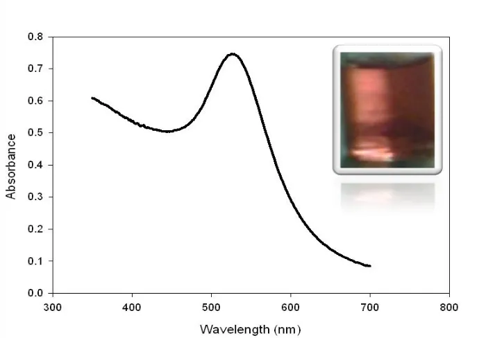
[image:4.596.123.473.458.702.2]Figure 2 displays the UV-Vis spectrum of the prepared AuNPs colloidal solution. The interesting change in color from yellow to ruby red when bulk Au is converted to AuNPs (see the inset of Fig. 2) is explained by the theory of surface plasmonics. According to this theory, the light absorption by metal nanoparticles physically originates from the coherent oscillation of the conduction band electrons induced by the interacting electromagnetic field. The absorption band results when the incident photon frequency is resonant with the collective oscillation of the conduction band electrons, and this is known as the surface plasmon resonance (SPR) [31]. In other words, the SPR refers to the coherent excitation of all the free electrons within the conduction band, leading to an in-phase oscillation [32]. The surface Plasmon band of AuNPs has an absorption peak in the visible region in the range of 500-600 nm and mainly this band is used as an indicator for the formation of AuNPs from their precursor salts. The sensitivity of plasmon band absorptivity is the basic detection mechanism involved in the AuNPs-based bio-sensors.
Figure 2. UV-Vis spectra for the gold nanoparticles colloidal solution prepared by the reduction of 1% sodium tetrachloroaureate with 1 ml of 0.075% NaBH4.38.8 mM Sodium citrate was used as a
3.2. Self assembling of BDMT and AuNps
E/mV vs. Ag/AgCl/KCl(sat.)
-1600 -1400 -1200 -1000 -800 -600 -400 -200
I/uA
-20 -15 -10 -5 0 5
1 h BDMT (a) 6 h BDMT (b) 12 h BDMT (c) a
[image:5.596.116.476.111.320.2]b c
Figure 3. The CVs for the reductive desorption of BDMT self-assembled for 1 (a), 6 (b), and 12 (c) h on poly- Au electrode measured in 0.5 M KOH solution. Potential scan rate: 50 mVs-1.
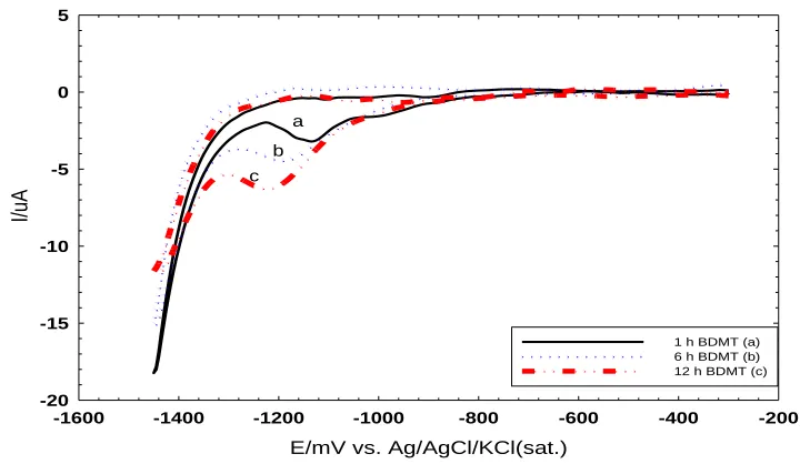
The soaking time of the poly-Au electrode in the BDMT solution is very critical as it determines the BDMT surface coverage and consequently the amount of AuNPs next. Therefore one should be able to evaluate the surface coverage of the BDMT on the poly-Au electrode in a way to assemble a complete monolayer.
The reductive desorption is one of the powerful techniques for tracking a monolayer formation. In this technique, the poly-Au is soaked in BDMT solutions for different periods of times (1, 6, and 12 h) and the CV for the reductive desorption of BDMT is measured at a scan rate of 50 mVs-1 in 0.5 M KOH solution. In all cases, the reductive desorption peak of BDMT appeared at around ca. 1.1 V [33] with a slightly negative shifted potentials at higher loading periods, which is needed to desorb more amount of the BDMT. However, the peak area depended greatly on the time of soaking in BDMT solution. As the soaking time increases, the peak area, which represents the amount of BDMT self-assembled on the Au electrode, increases (Fig. 3).
Our results indicate that 1 h soaking in the BDMT is not sufficient for the formation of full monolayer. This is matched as reported in literature [28, 29]. Moreover, the electrode surface of the poly-Au electrode is not fully covered with BDMT molecules if the soaking time is less than 12 h.
Soaking the poly-Au electrode in BDMT solutions for 12 h and more did not affect the area of the reductive desorption peak, which reveals the formation of a complete monolayer of BDMT on the surface of poly-Au electrode (see Fig. 3 c).
Alternatively, CV measurements of one of the redox markers (namely [Fe(CN)6]3-/4- couple)
E/ mV vs. Ag/AgCl/KCl(sat.)
-400 -200 0 200 400 600
I/uA
-6 -4 -2 0 2 4 6
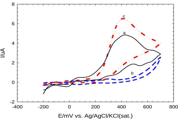
bare Au (a) BDMT 1h (b) BDMT 6h (c) BDMT 12h (d) AuNPs (e)
Figure 4. The CVs obtained at a scan rate of 150 mVs-1 for 1mM K3[Fe (CN)6] in 0.1M KCl at (a) bare
Au electrode, (b,c,d) BDMT/Au electrodes (diameter=1.6 mm) prepared by soaking the poly-Au electrode in BDMT solution for 1,6, and 12 h respectively.
Figure 4 shows the CVs in 1mM [Fe(CN)6]3-/4- at the bare poly-Au, and BDMT/Au electrodes
after soaking in BDMT ethanolic solution for different periods of times. A well-defined reversible voltammogram characteristic of a diffusion-controlled redox process was observed at the bare polycrystalline Au electrode with a ΔEp value of ca. 70 mV (Fig. 4 a). Whereas, after soaking in
BDMT solutions for a period less than 12 h, the BDMT-modified poly-Au electrodes showed another voltammetric response in [Fe(CN)6]3-/4- with a ΔEp>400 mV, indicating a sluggish electron-transfer
reaction at the these BDMT/Au electrodes (Fig. 4 b and c) [29]. When the bare poly-Au electrode was soaked in the BDMT solution for a period of 12 h or more, the reversible redox response was totally disappeared (Fig. 4 d), revealing the formation of a complete coverage of BDMT on the surface of poly-Au electrode. This means that the BDMT layer stopped the electron and mass transfer of ferricyanide toward the electrode surface.
We believe that the hydrophobic nature and the compactness of BDMT monolayer are hindering the permeation of the redox molecules to the electrode surface [28]. Once again, the results depicted in Figure 4 totally coincide with the reductive desorption investigation and both confirm that 12 h soaking of the bare poly-Au electrode in the BDMT solution is minimum to cover the poly-Au electrode surface with a monolayer of BDMT molecules. Other soaking times less than 12 h of poly-Au electrode in BDMT did not result in the formation of a complete BDMT monolayer on the poly- Au-substrate (data are not shown).
[image:6.596.142.452.75.268.2]
electrode, where no conduction was observed. Again, the conduction is restored if AuNPs were immobilized as a fourth layer onto BDMT/AuNPs/BDMT/Au electrode. This phenomenon is very interesting and can further be helpful in designing nanoelectronic sensors and nanoelectrical switches.
3.3. Electroanalysis of Ascorbic acid using AuNPs-modified electrodes
E/mV vs. Ag/AgCl/KCl(sat.)
-400 -200 0 200 400 600 800
I/uA
-2 0 2 4 6 8
c
a
b
Figure 5. The CVs obtained for 1 mM AA at (a) bare poly-Au, (b) BDMT/Au (12 h soaking in BDMT) and (c) AuNPs/BDMT/Au electrodes in 0.1 M PBS pH 7.2 at a scan rate of 50 mVs-1.
Ascorbic acid is always interfering in the detection of dopamine and other neurotransmitters, and in many cases, the bare electrodes very often suffer from fouling by its oxidation product. This, unfortunately, influences the detection of dopamine and the other neurotransmitters by a poor selectivity and reproducibility.
Several approaches, based on polymer-modified electrodes, pretreated electrodes and self-assembled monolayers, have been sought to solve this problem [35, 36]. Modifying the bare Au electrode with AuNPs could positively participate in solving this problem. The electrocatalytic activity of AuNPs toward the oxidation of ascorbic acid (AA) was recorded by measuring the CVs in 1 mM AA in 0.1 M PBS (pH 7.2) (Fig. 5). At the bare poly-Au electrode (Fig. 5 a), The AA oxidation occurred at ca. 0.5 V vs. Ag/AgCl/KCl (sat.) with a maximum peak current of 4.5 µA and the electron transfer kinetics was rather sluggish owing to the fouling of the electrode surface which is caused by the oxidation products of AA. The irreversible fouling of the electrode surface caused by the oxidation products resulted in a progressive deterioration in sensing capability. It is generally believed that the direct oxidation of AA at the bare electrodes is irreversible due to the rapid hydration of the oxidation products [37].
[image:7.596.138.443.189.394.2](Fig. 5 c), the electrode restored its electrical communication with the underlying electrode surface and the oxidation peak restored again but with a maximum peak current of 6.5 µA which is 2 µA higher than that at the bare one. This not the only enhancement observed but, in addition, the fouling of the bare electrode disappeared. This indicates that the AuNPs/BDMT/Au electrode can be used for the sensing reaction of AA.
4. CONCLUSION
AuNPs colloidal solution with an average diameter of 2.6 nm has been prepared by the chemical reduction of NaAuCl4 by NaBH4 using sodium citrate as stabilizer. This solution acquired a
ruby red color with SPR at about 520 nm. The soaking time of the poly-Au electrode in BDMT ethanoilc solution had a significant influence on the compactness of the BDMT self-assembled monolayer. A complete monolayer of BDMT has been achieved after 12 h soaking of the poly-Au electrode in BDMT solution. The BDMT layer in the BDMT/Au electrode acted as an inert electron and mass transfer blocking layer that hindered the diffusion of ferricyanide toward the electrode surface. The self-assembled AuNPs layer on the BDMT/Au electrode achieved a good electrical communication with the underlying electrode surface and further, worked as a renewed activated-electrode surface. In addition, the AuNPs/BDMT/Au activated-electrode could successfully be used for the ascorbic acid sensing.
References
1. P. Kalimuthu and S.A. John, J. Electroanal. Chem., 617 (2008) 164. 2. A.P. Alivisatos, Science, 271 (1996) 933.
3. M.C. Daniel and D. Astruct, Chem. Rev., 104 (2004) 293.
4. C. Burda, X. Chen, R. Narayanan and M.A. El-Sayed, Chem. Rev., 105 (2005) 1025. 5. K. Aslan, J.R. Lakowicz and C.D. Geddes, Anal. Chem., 77 (2005) 2007.
6. R. Elghanian, J.J. Storhoff, R.C. Mucic, R.L. Letsinger and C.A. Mirkin, Science, 277 (1997) 1078. 7. D.A. Stuart, A.J. Haes, C.R. Yonzon, E.M. Hicks and R.P.V. Duyne, Nanobiotechnology, 152
(2005) 13.
8. J.L. West and N.J. Halas, Annu. Rev. Biomed. Eng., 5 (2003) 285. 9. S. Bharathi, M. Nagami and O. Lev, Langmuir, 17 (2001) 2602. 10.B.K. Jena and C.R. Raj, Anal. Chem., 78 (2006) 6332.
11.G. Maduraiveeran and R. Ramaraj, J. Electroanal. Chem., 608 (2007) 52. 12.I.H. El-Sayed, X. Huange and M.A. El-Sayed, Nano Lett., 5 (2005) 829. 13.S.J. Hurst, A.K.R. Lytton-Jean and C.A. Mirkin, Anal. Chem., 78 (2006) 8313. 14.T.K. Sau and T.P. Anjali, J. Phys. Chem. B, 105 (2001) 9266.
15.X. Jiang, J. Jiang, Y. Jin, E. Wang and S. Dong, Biomacromolecules, 6 (2005) 46. 16.J.-P. Deng, W.-C. Shih and C.-Y. Mou, Chem. Phys. Chem., 6 (2005) 2021.
17.H. Tsunoyama, H. Sakurai, Y. Negishi and T. Tsukuda, J. Am. Chem. Soc., 127 (2005) 9374. 18.I.M. Al-Akraa, A.M. Mohammad, M.S. El-Deab and B.E. El-Anadouli, Chem. Lett., 40 (2011)
1374.
20.D. Feldheim , A. Colby and D. Marcel, J. Phys. Chem, 300 (2002) 11202.
21.D. Hernandez - Santos , M.B. Gonzalez - Garcea and A. Costa - Garcea, Electroanalysis, 14 (2002) 1225.
22.E. Katz , I. Willner and J. Wang, Electroanalysis, 16 (2004) 19.
23.C.M. Welch and R.G. Compton, Anal. Bioanal. Chem., 384 (2006) 601. 24.W.C. Bigelow , D.L. Pickett and W.A. Zisman, J. Colloid Sci., 1 (1946) 513 25.H. Von Foerster, Pergamon Press, 1960, p. 31.
26.R.G. Nuzzo , C.O. Fusco and D.L. Allara, J. Am. Chem. Soc., 109 (1987) 2358
27.H. Sellers , A. Ulman , Y. Shnidman and J.E. Eilers, J. Am. Chem. Soc., 115 (1993) 9389
28.A.I. Abdelrahman, A.M. Mohammad, T. Okajima and T. Ohsaka, J. Phys. Chem. B, 110 (2006) 2798.
29.C.R. Raj, A.I. Abderahman and T. Ohsaka, Electrochem. commun., 7 (2005) 888. 30.K.R. Brown, D.G. Walter and M.J. Natan, J. Chem. Matter, 12 (2000) 306.
31.S. Basu, S. Ghosh, S. Kundu, S. Panigrahi, S. Praharaj, S. Pande, S. Jana and T. Pal, J. Colloid Interface Sci., 313 (2007) 724.
32.S. Link and M.A. El-Sayed, Int . Rev. Phys. Chem., 19 (2000) 409± 453. 33.S.H. Othman, African J. of Pure and Applied Chemistry, 5 (2011) 278.
34.K.V.G.K. Murty, M. Venkataramanan and T. Pradeep, Langmuir, 14 (1998) 5446. 35.C.R. Raj and T. Ohsaka, J. Electroanal. Chem., 540 (2003) 69.
36.C.R. Raj, T. Okajima and T. Ohsaka, J. Electroanal. Chem., 543 (2003) 127. 37.C.R. Raj, K. Tokuda and T. Ohsaka, Bioelectrochemistry, 53 (2001) 183.
![Figure 4. The CVs obtained at a scan rate of 150 mVs-1 for 1mM K3[Fe (CN)6] in 0.1M KCl at (a) bare Au electrode, (b,c,d) BDMT/Au electrodes (diameter=1.6 mm) prepared by soaking the poly-Au electrode in BDMT solution for 1,6, and 12 h respectively](https://thumb-us.123doks.com/thumbv2/123dok_us/1904086.148650/6.596.142.452.75.268/obtained-electrode-electrodes-diameter-prepared-electrode-solution-respectively.webp)