R E S E A R C H
Open Access
Sub-microscopic malaria cases and mixed malaria
infection in a remote area of high malaria
endemicity in Rattanakiri province, Cambodia:
implication for malaria elimination
Nicolas Steenkeste
1*, William O Rogers
2, Lucy Okell
3, Isabelle Jeanne
4, Sandra Incardona
1, Linda Duval
5,
Sophy Chy
1, Sean Hewitt
6, Monidarin Chou
7, Duong Socheat
8, François-Xavier Babin
9, Frédéric Ariey
1,
Christophe Rogier
10Abstract
Background:Malaria microscopy and rapid diagnostic tests are insensitive for very low-density parasitaemia. This insensitivity may lead to missed asymptomatic sub-microscopic parasitaemia, a potential reservoir for infection. Similarly, mixed infections and interactions betweenPlasmodiumspecies may be missed. The objectives were first to develop a rapid and sensitive PCR-based diagnostic method to detect low parasitaemia and mixed infections, and then to investigate the epidemiological importance of sub-microscopic and mixed infections in Rattanakiri Province, Cambodia.
Methods:A new malaria diagnostic method, using restriction fragment length polymorphism analysis of the cytochrome bgenes of the four humanPlasmodiumspecies and denaturing high performance liquid chromatography, has been developed. The results of this RFLP-dHPLC method have been compared to 1) traditional nested PCR amplification of the18S rRNAgene, 2) sequencing of the amplified fragments of the cytochrome bgene and 3) microscopy.
Blood spots on filter paper and Giemsa-stained blood thick smears collected in 2001 from 1,356 inhabitants of eight villages of Rattanakiri Province have been analysed by the RFLP-dHPLC method and microscopy to assess the prevalence of sub-microscopic and mixed infections.
Results:The sensitivity and specificity of the new RFLP-dHPLC was similar to that of the other molecular methods. The RFLP-dHPLC method was more sensitive and specific than microscopy, particularly for detecting low-level parasitaemia and mixed infections. In Rattanakiri Province, the prevalences ofPlasmodium falciparumand
Plasmodium vivaxwere approximately two-fold and three-fold higher, respectively, by RFLP-dHPLC (59% and 15%, respectively) than by microscopy (28% and 5%, respectively). In addition,Plasmodium ovaleandPlasmodium malariaewere never detected by microscopy, while they were detected by RFLP-dHPLC, in 11.2% and 1.3% of the blood samples, respectively. Moreover, the proportion of mixed infections detected by RFLP-dHPLC was higher (23%) than with microscopy (8%).
Conclusions:The rapid and sensitive molecular diagnosis method developed here could be considered for mass screening and ACT treatment of inhabitants of low-endemicity areas of Southeast Asia.
* Correspondence: nicolas.steenkeste@gmail.com
1Unité d’Epidémiologie Moléculaire, Institut Pasteur du Cambodge, Phnom Penh, Cambodia
Background
The development of sensitive PCR-based methods for malaria diagnosis has highlighted the low sensitivity of malaria microscopy for very low-level parasitaemia (<50 parasites/μl). The low sensitivity of microscopy may have at least two important consequences for malaria control and eradication efforts. First, asymptomatic sub-micro-scopic parasitaemia may serve as a reservoir for infection even when very efficient rapid diagnosis and treatment programmes have been implemented. Second, mixed infections may be overlooked when one species is present at low parasitaemia, and clinically or epidemiologically important interactions between species may be missed.
Current malaria control methods in Southeast Asia rely largely on early detection and treatment; control based on insecticide-treated bed nets (ITN) may be less effective in areas where malaria is primarily acquired not at home but by working age men engaged in mining, hunting, or logging in the forest. Since indivi-duals with asymptomatic parasitaemia will not be identi-fied by early detection and treatment programmes, they may continue to serve as a source of infection for vector mosquitoes, complicating control measures. It is clear that PCR-based methods can detect sub-microscopic parasitaemia [1-4]; it remains to be determined how common such cases of parasitaemia are in the field under different ecological conditions and what effect they may have on transmission.
Accurate detection of mixed infections is important for both clinical and epidemiological reasons. The four Plasmodium species, which commonly infect humans, have somewhat different clinical characteristics. Plasmo-dium falciparum is the most lethal species, causing severe malarial anaemia and cerebral malaria and, con-sequently, the vast majority of malaria deaths. Plasmo-dium vivax and Plasmodium ovale may persist within the liver as hypnozoites causing relapses even after treatment with blood schizonticides. Plasmodium malariaemay cause chronic asymptomatic parasitaemia and may lead to renal failure via unclear mechanisms [5]. Finally, Plasmodium knowlesi is a newly emergent malaria parasite in Southeast Asia [6,7]. How these clini-cal characteristics are modified in mixed species infec-tions is not clear. Voza et alin 2005 found a species-specific inhibition of cerebral malaria in mice coinfected withPlasmodium spp[8]. In human, according to sev-eral studies in Ivory Coast [9], Sri Lanka [10] and Thai-land [11], the severity of infection may be modulated by mixed infections. In a study carried out in Vanuatu, P. falciparumappeared to be associated with a reduced P. vivaxparasitaemia [12,13], but the reverse effect has been observed in another study [14]. There seems to be a strong interaction between these two malaria species,
and P. vivax co-infection has even been reported to decrease treatment failures of P. falciparum [15]. In other studies,P. falciparum - P. vivaxmixed infections affected the clinical outcome in patients [13,16]. Exam-ples of P. malariae and P. ovale co-infections with P. falciparumprotecting against malaria symptoms have also been reported [17]. Studies with aPlasmodium ber-gheiandPlasmodium yoeliico-infection model in mice appear to confirm that there can be a strong cross-pro-tective effect of mixed species infections [18]. Mixed infections may have important epidemiological effects. For example, if P. vivax parasitaemia is suppressed by co-infection withP. falciparum, then effective control of P. falciparummalaria in an area might be followed by an increase inP. vivaxtransmission.
Studies of mixed species infections based only on microscopy may underestimate their importance. The frequencies of minority species, such asP. malariaeand P. ovale, are largely underestimated by microscopy [19-21]. PCR-based methods are more sensitive [1-4] and more readily detect mixed infections. Using PCR diagnosis, between one third and half of malaria infec-tions in a study in Thailand were found to be mixed species infections [22].
In order to investigate the epidemiological importance of sub-microscopic and mixed species infections, a highly sensitive PCR-based diagnostic method was developed using amplification of a fragment of the cyto-chrome bgene, followed by Restriction Fragment Length Polymorphism (RFLP) analysis using Denaturing High Performance Liquid Chromatography (dHPLC) to detect amplification and restriction products. Results of the RFLP-dHPLC method were first compared to traditional PCR and microscopy. This highly sensitive method was then used to analyze samples collected in 2001 in malaria prevalence surveys in eight villages in Rattana-kiri Province, a site of high malaria transmission in Cambodia, and assess the prevalence and importance of sub-microscopic and mixed infections in this population.
Methods
Study area and population
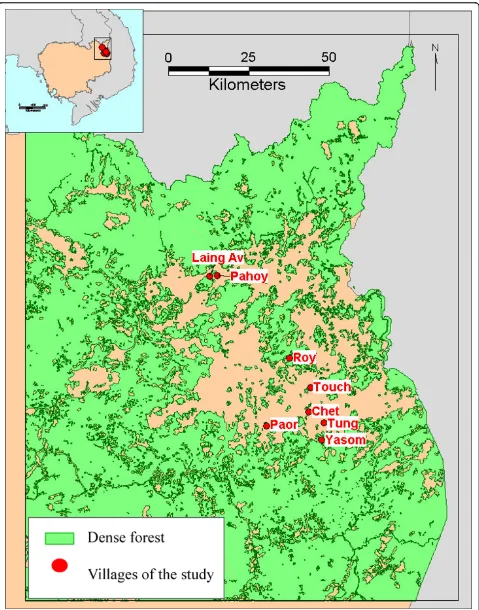
Rattanakiri province is located in North-Eastern Cambodia, close to the borders with Laos and Vietnam (Figure 1). Malaria is transmitted byAnopheles dirus,Anopheles macu-latusandAnopheles minimus[23,24]. In 2001, the number of slide-confirmed malaria cases confirmed in Rattanakiri province by the National Malaria Control Programme (CNM) was 1,165 forP. falciparum(71.9%), 424 forP. vivax(26.1%) and 31 forP. malariae(2%) [25].
Approximately 20 μL of finger prick blood were col-lected on Whatman 3 M filter paper. Thin and thick blood smears were prepared, and data on age, sex and temperature were collected. Blood spots were stored at -20°C.
For the present study, eight villages from the 2001 survey were randomly selected. Those villages belonged to three ethnic minorities; respectively, Tampuon (Yasom, 204 samples; Roy, 206 samples; Chet, 204 sam-ples and Paor, 209 samsam-ples), Jarai (Tung, 143 samsam-ples and Touch, 143 samples) and Brao (Laing Av, 132 sam-ples and Pahoy, 134 samsam-ples).
Microscopic diagnosis
Thin smears were fixed in methanol. Both thin and thick smears were stained with 3% Giemsa for 30 min-utes at room temperature. Examination was performed by experienced microscopists at the National Centre for Parasitology, Entomology and Malaria Control in Phnom Penh. At least 100 thick film fields with 1,000× magnification were examined before a slide was consid-ered negative. Parasite species and stages were con-firmed on the thin film. Parasite densities were classified according WHO recommendations: class 1 for 1-10 parasites per 100 thick film fields; class 2 for 11-100 parasites per 100 fields; class 3 for 1-10 parasites per single field; class 4 for 11-100 parasites per single field; class 5 for > 100 parasites per single field.
DNA extraction
Parasite DNA was extracted from a piece of 4 mm dia-meter of blood spots of Whatman 3 M filter paper, using the Instagene resin (Biorad, Germany) as pre-viously described [27].
18S rRNAspecies-specific nested PCR, standard PCR
The nested PCR method based on the18S rRNA gene marker [28] adapted for epidemiological studies was performed as described [29].
Restriction Fragment Length Polymorphism (RFLP) and dHPLC
Nested PCR amplification and Alu I restriction digestion of the Plasmodiumcytochrome bgene was performed as previously described [27,30]. Fiveμl of each restriction digested PCR product were injected with a 96 well auto-sampler for DHPLC analysis in the WAVE DNA Frag-ment Analysis System (Transgenomic, Santa Clara, CA) with a variable wave-length detector set at 260 nm. To separate DNA fragments, the analysis was performed on a DNASepTM column (Transgenomic, Inc.) at 50°C with a flow rate of 0.9 ml/min. DNA fragments were eluted with a linear gradient mixture of Buffer A: 0.1 M triethylammo-nium acetate (TEAA) pH 7.0 (Transgenomic) and Buffer
B: 0.1 M TEAA/25% acetonitrile (ACN) pH 7.0. Elution started from 46% to 59.1% of buffer B over a period 13.5 min. The column was regenerated with the buffer D: 75% ACN pH 7.0 and re-used.
Cytochrome bSNP identification
Sequencing reactions were performed on both strands of the cytochrome bPCR product using internal primers and ABI Prism BigDye Terminator chemistry. Sequen-cing reactions were run on ABI3730XL (Applied Biosys-tems) at Macrogen® (Korea). The analysis of the sequence was performed with Seqscape software v.2.0 (Applied Biosystems). An algorithm based on 11 selected SNPs has previously been developed based on an alignment of published cytochrome b reference sequences ofP. falciparum, P. vivax, P. malariae and P. ovale, allowing identification of each species [27].
Reference genomic DNA
Genomic DNA fromP. falciparumwas extracted from a continuous culture of the 3D7 strain. DNA from the P. vivax Belem strain was kindly provided by Peter David (Institut Pasteur, Paris, France). Plasmodium malariaeand P. ovale DNA as well as human control DNA from a non-infected person were kindly provided by Georges Snounou (Muséum d’Histoire Naturelle, Paris, France). DNA from patients with pureP. malariae infections were kindly provided by Eric Legrand (Institut Pasteur de Cayenne, French Guiana).
Statistical analysis
All statistical analyses were performed using the STATA 10 software (Stata Corporation, College Station, TX). Prevalence rates and their 95% confidence intervals were estimated using the svy commands to take into account the sampling design (clustered by village). McNemar’s exact test was used for paired analysis of the results of the diagnostic methods applied to the same blood sam-ples. The associations of independent variables with malaria prevalence were tested using logistic regression models with random effects taking into account the clustered sampling design.
Results
RFLP-dHPLCPlasmodiumspecies diagnosis validation
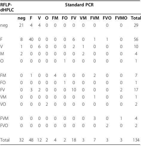
In order to ensure the accuracy of this approach, the RFLP-dHPLC method was first validated against the standard PCR methods (Table 1). All samples from one of the study sites (n = 134, Pahoy) were subjected to both methods. Comparison ability of the two methods to detect Plasmodium parasites, regardless of species, gave an agreement on 115 (21 negative and 94 positive). Eight samples were positive according to RFLP-dHPLC, but not according to the standard PCR and 11 were positive by the standard PCR, but not by RFLP-dHPLC. There was no evidence for the superiority of either method in detectingPlasmodium parasites (McNemar’s exact test p = 0.65). The only difference in species iden-tification between the two methods occurred in cases in which one test detected a mixed species infection and the other test identified only the predominant species. For example, the standard PCR approach identified eight mixed infections among 56 infections identified as P. falciparummono-infections by dHPLC, while dHPLC identified four mixed infections among 48 infections identified asP. falciparum mono-infections by standard PCR. Twenty-three mixed infections were detected by both techniques. There was no evidence for superiority
Figure 2dHPLC profile forPlasmodiumidentification.
Table 1 Comparison of results by RFLP-dHPLC and by standard PCR method in Pahoy
RFLP-dHPLC
Standard PCR
neg F V O FM FO FV VM FVM FVO FVMO Total
neg 21 4 4 0 0 0 0 0 0 0 0 29
F 8 40 0 0 0 0 6 0 1 1 0 56
V 1 0 6 0 0 0 2 1 0 0 0 10
M 2 0 0 0 0 0 0 2 0 0 0 4
O 0 0 0 0 0 1 0 0 0 0 0 1
FM 0 1 0 0 4 0 0 0 2 0 0 7
FO 0 0 0 0 0 1 0 0 0 0 0 1
FV 0 3 2 0 0 0 10 0 0 0 2 17
VM 0 0 0 0 0 0 0 0 1 0 0 1
VO 0 0 0 2 0 0 0 0 0 0 0 2
FVM 0 0 0 0 0 0 0 0 3 0 1 4
FVO 0 0 0 0 0 0 0 0 0 2 0 2
[image:5.595.304.540.480.723.2]of either technique in detecting mixed species infections including P. falciparum (McNemar’s exact test p = 0.39).
Because of the same pattern found for P. vivax and P. knowlesiin dHPLC technique, all isolates found to be infected by P. vivax were sequenced. No P. knowlesi infection was found.
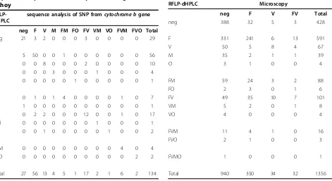
Since the RFLP-dHPLC method did not appear infer-ior to standard PCR diagnosis, validation was done on the sequence specificity of the new method by compar-ing the species identification made by RFLP-dHPLC detection with the results ofcytochrome b SNP analysis as determined by direct DNA sequencing ofcytochrome b PCR products [27]. For mono-infections there were no discrepancies between the methods. In the case of mixed infections, each technique occasionally missed a minor species identified by the other (Table 2). Overall, the two techniques gave the same species identification, including mixed species, in 85/99 (86%) of cases. In seven cases RFLP-dHPLC identified mixed infections missed by cytochrome b SNP analysis; in seven cases, cytochrome b SNP analysis identified mixed infections missed by RFLP-dHPLC. Since the RFLP-dHPLC method appeared no less sensitive than standard PCR and identified mixed infections as effectively as cyto-chrome bSNP analysis, the RFLP-dHPLC method was used to assess the prevalence of sub-microscopic and mixed infections in samples from eight villages in Rattanakiri.
Comparison of RFLP-dHPLC and microscopy results
A comparison between the numbers of Plasmodium infections detected by microscopy and by the RFLP-dHPLC method was done on 1,356 samples collected from eight villages in Rattanakiri province (Table 3). The prevalence ofPlasmodium infections measured by the RFLPdHPLC method (68.4%, 95%CI: 62.7% -74.1%) was more than twice that measured by micro-scopy (30.7%, 95%CI: 24.9% - 36.4%).
[image:6.595.66.537.476.733.2]Regardless of species, the two techniques agreed on 764 samples (376 positive and 388 negative according to both techniques). Nevertheless, 552 specimens negative by microscopy were positive by RFLP-dHPLC, and 40 negative by RFLP-dHPLC were positive by microscopy: 32 P. falciparum, five P. vivaxand three mixed infec-tionsP. falciparumwithP. vivax; theP. falciparum den-sity is distributed as follow: 16 in class 1 of parasite densities, 14 in class 2, four in class 3 and one in class 4. The RFLP-dHPLC method identified many more infections than microscopy (McNemar’s exact test p < 0.0001).
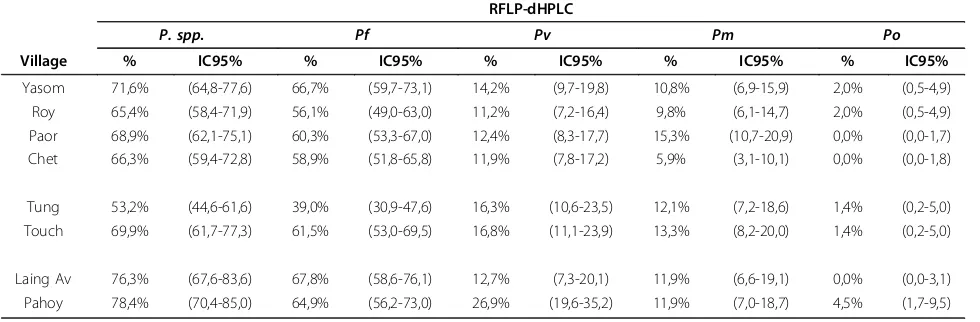
[image:6.595.299.539.476.727.2]Table 4 and 5 show the prevalence of malaria infec-tion in the individual villages as determined by both microscopy and RFLP-dHPLC. The prevalence of P. falciparumandP. vivax was approximately two-fold and three-fold higher, respectively, when measured by the RFLP-dHPLC (P. falciparum: 59.4%, 95%CI: 52.2% - 66.6%;P. vivax: 14.7%, 95%CI: 10.8% - 18.7%) than by microscopy (P. falciparum: 28.2%, 95%CI: 21.6%
Table 2 Comparison of results by RFLP-dHPLC and by sequence analysis of SNP fromcytochrome bgene in Pahoy
RFLP-dHPLC
sequence analysis of SNP fromcytochrome bgene
neg F V M FM FO FV VM VO FVM FVO Total
neg 21 3 2 0 0 0 3 0 0 0 0 29
F 5 50 0 0 1 0 0 0 0 0 0 56
V 0 0 8 0 0 0 2 0 0 0 0 10
M 0 0 0 3 0 0 0 1 0 0 0 4
O 0 0 0 0 0 1 0 0 0 0 0 1
FM 0 1 0 1 4 0 0 0 0 1 0 7
FO 1 0 0 0 0 0 0 0 0 0 0 1
FV 0 2 2 0 0 0 12 0 0 1 0 17
VM 0 0 0 0 0 0 0 1 0 0 0 1
VO 0 0 1 0 0 0 0 0 1 0 0 2
FVM 0 0 0 0 0 0 0 0 0 4 0 4
FVO 0 0 0 0 0 0 0 0 0 0 2 2
Total 27 56 13 4 5 1 17 2 1 6 2 134
Table 3 Comparison between microscopy and RFLP-dHPLC diagnostic methods
RFLP-dHPLC Microscopy
neg F V FV Total
neg 388 32 5 3 428
F 331 241 6 13 591
V 50 5 8 4 67
M 35 2 1 1 39
O 3 1 0 0 4
FM 59 24 3 2 88
FO 2 3 0 1 6
FV 49 35 10 7 101
VM 5 2 0 1 8
VO 4 0 0 0 4
FVM 11 4 1 0 16
FVO 2 1 0 0 3
FVMO 1 0 0 0 1
[image:6.595.59.309.491.731.2]- 34.7%;P. vivax: 4.8%, 95%CI: 3.1% - 6.6%). In addi-tion,P. ovaleandP. malariaewere never detected by microscopy while they were detected by RFLP-dHPLC (P. malariae: 11.2%, 95%CI: 8.2% - 14.2%; P. ovale: 1.3%, 95%CI: 0.04% - 2.6%).
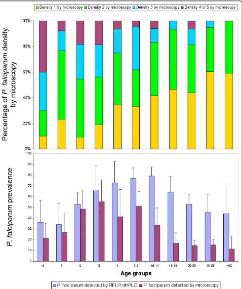
Prevalence of sub-microscopicP. falciparuminfection
In subjects less than five years old, microscopy and RFLP-dHPLC gave similarP. falciparumprevalences. In subjects aged five years or older, theP. falciparum pre-valence was higher when estimated by RFLP-dHPLC than microscopy (Figure 3). TheP. falciparum parasite densities estimated by microscopy decrease significantly with increasing age. No such association of age with prevalence or parasite density was found for the three otherPlasmodiumspecies.
Characterization of mixed infection
Due to the higher sensitivity of RFLP-dHPLC, the pro-portion of mixed infections was significantly higher with this method (24.5%, 227/928) than with microscopy (7.7%, 32/416) (Table 3). The frequency of mixed infec-tions detected by RFLP-dHPLC did not vary significantly depending on the positivity of the microscopy (p = 0.75) or the parasite density (p = 0.91) estimated by microscopy.
Sex and ethnic groups
Plasmodium falciparum was more prevalent in males than in females (p < 0.001). No such association was found for the other malaria species (Table 6). Between ethnic groups, the prevalence of P. falciparum and P. vivax, but not of P. malariae or P. ovale, differed significantly (Table 6). The prevalence of P. falciparum was significantly higher in the Brao than in the Tam-poun and in the TamTam-poun than in the Jaray ethnic groups, and the prevalence ofP. vivaxwas significantly higher in the Brao than in the Tampoun, without sig-nificant differences between the Jaray and the other ethnic groups.
Evidence of non-random species associations
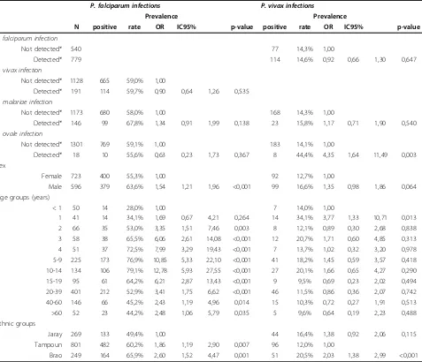
[image:7.595.58.291.134.295.2]Controlling for age and ethnic group in multivariate ran-dom effects logistic regression models,P. vivaxinfection was significantly associated withP. ovaleinfection (OR = 4.35, IC95 = 1.64 - 11.49, p = 0.003) (Table 6). There was also an association betweenP. falciparumandP. malar-iaeinfections (unadjusted OR = 1.61, IC95 = 1.12 - 2.32, p = 0.010) that was no more significant when controlling for age (adjusted OR = 1.34, IC95 = 0.91 - 1.99, p = 0.138). There was no association betweenP. falciparum andP. vivaxinfection (p = 0.535).
Table 4 Prevalence ofPlasmodium spp. by village diagnosed by microscopy (P. spp.: AllPlasmodium
species;Pf:Plasmodium falciparum;Pv:Plasmodium vivax;
Pm:Plasmodium malariae;Po:Plasmodium ovale) Microscopy
P. spp Pf Pv
Village N % IC95% % IC95% % IC95%
Yasom 204 38,7% (32,0-45,8) 36,8% (30,1-43,8) 8,3% (4,9-13,0) Roy 205 37,1% (30,4-44,1) 33,2% (26,8-40,1) 7,8% (4,5-12,4) Paor 209 25,4% (19,6-31,8) 23,0% (17,4-29,3) 3,8% (1,7-7,4) Chet 202 27,7% (21,7-34,4) 26,2% (20,3-32,9) 3,0% (1,1-6,4)
Tung 141 20,6% (14,2-28,2) 14,2% (8,9-21,1) 9,2% (5,0-15,3) Touch 143 35,0% (27,2-43,4) 34,3% (26,5-42,7) 1,4% (0,2-5,0)
Laing Av 118 28,8% (20,8-37,9) 26,3% (18,6-35,2) 2,5% (0,5-7,3) Pahoy 134 29,1% (21,6-37,6) 28,4% (20,9-36,8) 0,7% (0,0-4,1)
Table 5 Prevalence ofPlasmodium spp. by village diagnosed by RFLP-dHPLC (P. spp.: AllPlasmodiumspecies;Pf:
Plasmodium falciparum;Pv:Plasmodium vivax;Pm:Plasmodium malariae;Po:Plasmodium ovale) RFLP-dHPLC
P. spp. Pf Pv Pm Po
Village % IC95% % IC95% % IC95% % IC95% % IC95%
Yasom 71,6% (64,8-77,6) 66,7% (59,7-73,1) 14,2% (9,7-19,8) 10,8% (6,9-15,9) 2,0% (0,5-4,9)
Roy 65,4% (58,4-71,9) 56,1% (49,0-63,0) 11,2% (7,2-16,4) 9,8% (6,1-14,7) 2,0% (0,5-4,9)
Paor 68,9% (62,1-75,1) 60,3% (53,3-67,0) 12,4% (8,3-17,7) 15,3% (10,7-20,9) 0,0% (0,0-1,7)
Chet 66,3% (59,4-72,8) 58,9% (51,8-65,8) 11,9% (7,8-17,2) 5,9% (3,1-10,1) 0,0% (0,0-1,8)
Tung 53,2% (44,6-61,6) 39,0% (30,9-47,6) 16,3% (10,6-23,5) 12,1% (7,2-18,6) 1,4% (0,2-5,0)
Touch 69,9% (61,7-77,3) 61,5% (53,0-69,5) 16,8% (11,1-23,9) 13,3% (8,2-20,0) 1,4% (0,2-5,0)
Laing Av 76,3% (67,6-83,6) 67,8% (58,6-76,1) 12,7% (7,3-20,1) 11,9% (6,6-19,1) 0,0% (0,0-3,1)
[image:7.595.56.546.556.717.2]Discussion
The detection ofPlasmodiumspecies at very low parasi-taemia is difficult and requires a molecular approach. The performance of a relatively simple and semi-auto-mated technique has been evaluated here. It is a highly sensitive PCR-based diagnostic method using amplifica-tion of a fragment of thecytochrome b gene, followed by RFLP analysis using dHPLC to detect amplification and restriction products. Compared to the well established PCR typing of the18S rRNAgene for species detection, this method appeared reliable, sensitive (multi copy number of the cytochrome b gene), specific and rapid (two PCR instead of five) and give the ability to ampli-fied large number of species [27]. The dHPLC semi-automated analysis allowed the screening of a 96-well
plate in 48 h and to diagnose 1,356 samples in less than one month.
[image:9.595.61.538.111.521.2]Use of the RFLP-dHPLC method revealed a high valence of sub-microscopic infections. The overall pre-valence detected by the RFLP-dHPLC method was twice that detected by microscopy (68.4% versus 30.7%). Inter-estingly, the proportion of sub-microscopic P. falci-parum infections, was higher in older age groups. It is possible that acquired immunity favours maintenance of sub-microscopic, asymptomatic infections. This substan-tial population of adults with sub-microscopic, asympto-matic P. falciparum infections may represent a significant challenge to malaria control programmes. Such individuals will not seek treatment and even if they were included in a mass screening and treatment Table 6 Associations ofP. falciparumandP. vivaxinfection with other Plasmodium infections and demographics; results of logistic regression analysis allowing for random village effects. N = 1319 individuals from 8 villages
P. falciparum infections P. vivax infections
Prevalence Prevalence
N positive rate OR IC95% p-value positive rate OR IC95% p-value
P. falciparum infection
Not detected* 540 77 14,3% 1,00
Detected* 779 114 14,6% 0,92 0,66 1,30 0,647
P. vivax infection
Not detected* 1128 665 59,0% 1,00
Detected* 191 114 59,7% 0,90 0,64 1,26 0,535
P. malariae infection
Not detected* 1173 680 58,0% 1,00 168 14,3% 1,00
Detected* 146 99 67,8% 1,34 0,91 1,99 0,138 23 15,8% 1,17 0,71 1,90 0,540
P. ovale infection
Not detected* 1301 769 59,1% 1,00 183 14,1% 1,00
Detected* 18 10 55,6% 0,63 0,23 1,73 0,367 8 44,4% 4,35 1,64 11,49 0,003
Sex
Female 723 400 55,3% 1,00 92 12,7% 1,00
Male 596 379 63,6% 1,54 1,21 1,96 <0,001 99 16,6% 1,35 0,98 1,86 0,064
Age groups (years)
< 1 50 14 28,0% 1,00 7 14,0% 1,00
1 41 14 34,1% 1,69 0,67 4,21 0,264 14 34,1% 3,77 1,33 10,71 0,013
2 66 35 53,0% 3,35 1,51 7,46 0,003 8 12,1% 0,89 0,30 2,68 0,838
3 58 38 65,5% 6,06 2,61 14,08 <0,001 12 20,7% 1,71 0,60 4,85 0,313
4 51 37 72,5% 7,99 3,29 19,43 <0,001 7 13,7% 1,02 0,32 3,20 0,978
5-9 225 173 76,9% 10,85 5,33 22,10 <0,001 41 18,2% 1,45 0,59 3,57 0,418
10-14 134 106 79,1% 12,78 5,93 27,55 <0,001 27 20,1% 1,66 0,65 4,27 0,290
15-19 95 61 64,2% 6,21 2,87 13,43 <0,001 9 9,5% 0,69 0,23 2,02 0,494
20-39 401 212 52,9% 3,41 1,75 6,62 <0,001 46 11,5% 0,86 0,36 2,07 0,742
40-60 146 66 45,2% 2,43 1,19 4,96 0,014 15 10,3% 0,72 0,27 1,91 0,513
>60 52 23 44,2% 2,48 1,06 5,79 0,035 5 9,6% 0,64 0,19 2,23 0,488
Ethnic groups
Jaray 269 133 49,4% 1,00 44 16,4% 1,38 0,92 2,06 0,115
Tampoun 801 482 60,2% 1,86 1,19 2,90 0,007 96 12,0% 1,00
Brao 249 164 65,9% 2,60 1,52 4,47 0,001 51 20,5% 2,03 1,38 2,99 <0,001
campaign, their parasitaemia would remain invisible to light microscopy or rapid diagnostic tests. Moreover, sub-microscopicP. falciparum gametocyte densities may contribute importantly to mosquito infections and thus to maintaining transmission [27]. In order to have a major impact on malaria transmission, mass screening and treatment campaigns may need to use molecular screening techniques capable of identifying sub-micro-scopic parasitaemia.
The RFLP-dHPLC method detected many more mixed species infections than did microscopy (Table 3). Indeed, 24.5% of all malaria infections in the Rattanakiri survey included more than one species. No association was found between the level of parasitaemia and the chance that the infection would be mixed. Co-infection withP. vivax and P. ovale was more common than would be expected based on the individual prevalences of these species (OR = 4.35, IC95 = 1.64 - 11.49, p = 0.003). This association could be attributed to cross-immunity that determines susceptibility to both infections [31] or to the exposure to infective bites of common vectors that transmit both species. An alternative explanation might be that both these species can produce relapses from hypnozoites. Similarly,P. falciparumandP. malar-iae infection were associated (OR = 1.61, IC95 = 1.12 -2.32, p = 0.010). There was, however no association betweenP. vivaxand P. falciparuminfection.
Plasmodium falciparumand P. vivaxinfections were found at different prevalences in the different ethnic groups. Since the different groups lived in different indi-vidual villages, it is not possible to decide whether the observed differences reflect different genetic susceptibil-ities, as observed for some African ethnic groups [32] or simply to different local transmission intensities or other local factors.
Conclusions
Rapid diagnosis and treatment of malaria cases, vector control and protection against mosquito bites are the cornerstone of the malaria control strategy in Cambodia. Sub-microscopic infections may provide a reservoir of infection, which maintains transmission. This phenom-enon has already been highlighted in Guinea Bissau [1] in Brazil [33] and Gabon [34]. The high prevalence of sub-microscopic parasitaemia within Rattanakiri Pro-vince, a site of higher malaria transmission in Cambodia, is here shown. Such infections may hamper elimination efforts, since they are asymptomatic and would not be efficiently detected by conventional diagnosis methods, i.e. microscopy or rapid diagnostic tests. Thus, the rapid and sensitive molecular diagnosis method developed here could be considered for mass screening and ACT treatment of inhabitants of low-endemicity areas of Southeast Asia to face the urgent need to eradicate
malaria in Cambodia due to emerging artemisinin resis-tance in the country.
Ethical approval
Ethical approval for this study was granted by the National Ethics Committee of the Kingdom of Cambodia.
Acknowledgements
The authors thank the staff of the National Center for Parasitology, Entomology and Malaria Control as well as the staff of the European Commission National Malaria Control Programme for sample collection and for the examination of the blood slides.
Thanks go to Jonathan Cox who provided GPS position of the study villages. The work was funded by a grant from Institut Pasteur à Paris (Modipop project). The opinions expressed are the personal ones of the authors and do not purport to represent those of the U.S. Navy.
Author details 1
Unité d’Epidémiologie Moléculaire, Institut Pasteur du Cambodge, Phnom Penh, Cambodia.2Naval Medical Research Center Unit 2, Jakarta 10560 Indonesia.3London School of Hygiene & Tropical Medicine (LSHTM), London, UK.4Centre de recherche médicale et sanitaire (CERMES), Niamey, Niger. 5Laboratoire de Génétique de la réponse aux infections chez l’homme, Institut Pasteur, Paris, France.6European Commission National Malaria Control Program, Phnom Penh, Cambodia.7Rodolphe Mérieux Laboratory of Cambodia, University of Health Science, Phnom Penh, Cambodia.8National Center for Parasitology, Entomology and Malaria Control, Phnom Penh, Cambodia.9Fondation Mérieux, Phnom Penh, Cambodia.10Equipe « Moustiques et Maladies Emergentes » - UMR 6236 - URMITE, Unité de Recherche en Biologie et Epidémiologie Parasitaires - Institut de Recherche Biomédicale des Armées, Marseille, France.
Authors’contributions
NS, LD and FA conceived and designed the genus-specific nested PCR and SNP identification based on thecytochrome bgene, SH and SD lead the field work, NS, SI, MC, FX, SC managed the experimental procedure and performed the laboratory work, NS, CR, WR, LO, FA participated in the statistical analyses, NS, IJ participated in SIG work. NS, WR, LO, IJ, SI, LD, SC, SH, DS, FB, FA, CR drafted and critically revised the manuscript. All authors read and approved manuscript.
Competing interests
The authors declare that they have no competing interests.
Received: 19 September 2009 Accepted: 22 April 2010 Published: 22 April 2010
References
1. Snounou G, Pinheiro L, Goncalves A, Fonseca L, Dias F, Brown KN, do Rosario VE:The importance of sensitive detection of malaria parasites in the human and insect hosts in epidemiological studies, as shown by the analysis of field samples from Guinea Bissau.Trans R Soc Trop Med Hyg
1993,87:649-653.
2. Singh B, Cox-Singh J, Miller AO, Abdullah MS, Snounou G, Rahman HA: Detection of malaria in Malaysia by nested polymerase chain reaction amplification of dried blood spots on filter papers.Trans R Soc Trop Med Hyg1996,90:519-521.
3. Toma H, Kobayashi J, Vannachone B, Arakawa T, Sato Y, Nambanya S, Manivong K, Inthakone S:A field study on malaria prevalence in southeastern Laos by polymerase chain reaction assay.Am J Trop Med Hyg2001,64:257-261.
4. Alves FP, Durlacher RR, Menezes MJ, Krieger H, Silva LH, Camargo EP:High prevalence of asymptomaticPlasmodium vivaxandPlasmodium falciparuminfections in native Amazonian populations.Am J Trop Med Hyg2002,66:641-648.
5. Neri S, Pulvirenti D, Patamia I, Zoccolo A, Castellino P:Acute renal failure in
6. White NJ:Plasmodium knowlesi: the fifth human malaria parasite.Clin Infect Dis2008,46:172-173.
7. Cox-Singh J, Singh B:Knowlesi malaria: newly emergent and of public health importance?Trends Parasitol2008,24:406-410.
8. Voza T, Vigario AM, Belnoue E, Gruner AC, Deschemin JC, Kayibanda M, Delmas F, Janse CJ, Franke-Fayard B, Waters AP, Landau I, Snounou G, Renia L:Species-specific inhibition of cerebral malaria in mice coinfected withPlasmodium spp.Infect Immun2005,73:4777-4786.
9. Black J, Hommel M, Snounou G, Pinder M:Mixed infections with
Plasmodium falciparumandP. malariaeand fever in malaria.Lancet1994, 343:1095.
10. Gunewardena DM, Carter R, Mendis KN:Patterns of acquired anti-malarial immunity in Sri Lanka.Mem Inst Oswaldo Cruz1994,89(Suppl 2):63-65. 11. Luxemburger C, Ricci F, Nosten F, Raimond D, Bathet S, White NJ:The
epidemiology of severe malaria in an area of low transmission in Thailand.Trans R Soc Trop Med Hyg1997,91:256-262.
12. Molineaux L, Storey J, Cohen JE, Thomas A:A longitudinal study of human malaria in the West African Savanna in the absence of control measures: relationships between different Plasmodium species, in particular
P. falciparumandP. malariae.Am J Trop Med Hyg1980,29:725-737. 13. Maitland K, Williams TN, Newbold CI:Plasmodium vivaxandP. falciparum:
Biological interactions and the possibility of cross-species immunity.
Parasitol Today1997,13:227-231.
14. Mayxay M, Pukrittayakamee S, Newton PN, White NJ:Mixed-species malaria infections in humans.Trends Parasitol2004,20:233-240. 15. Price RN, Nosten F, Luxemburger C, van Vugt M, Phaipun L,
Chongsuphajaisiddhi T, White NJ:Artesunate/mefloquine treatment of multi-drug resistant falciparum malaria.Trans R Soc Trop Med Hyg1997, 91:574-577.
16. Smith T, Genton B, Baea K, Gibson N, Narara A, Alpers MP:Prospective risk of morbidity in relation to malaria infection in an area of high endemicity of multiple species of Plasmodium.Am J Trop Med Hyg2001, 64:262-267.
17. Alifrangis M, Lemnge MM, Moon R, Theisen M, Bygbjerg I, Ridley RG, Jakobsen PH:IgG reactivities against recombinant Rhoptry-Associated Protein-1 (rRAP-1) are associated with mixed Plasmodium infections and protection against disease in Tanzanian children.Parasitology1999, 119:337-342.
18. Sedegah M, Weiss WW, Hoffman SL:Cross-protection between attenuated
Plasmodium bergheiandP. yoeliisporozoites.Parasite Immunol2007, 29:559-565.
19. Snounou G, Viriyakosol S, Jarra W, Thaithong S, Brown KN:Identification of the four human malaria parasite species in field samples by the polymerase chain reaction and detection of a high prevalence of mixed infections.Mol Biochem Parasitol1993,58:283-292.
20. Zhou M, Liu Q, Wongsrichanalai C, Suwonkerd W, Panart K, Prajakwong S, Pensiri A, Kimura M, Matsuoka H, Ferreira MU, Isomura S, Kawamoto F:High prevalence ofPlasmodium malariaeandPlasmodium ovalein malaria patients along the Thai-Myanmar border, as revealed by acridine orange staining and PCR-based diagnoses.Trop Med Int Health1998,3:304-312. 21. Kawamoto F, Liu Q, Ferreira MU, Tantular IS:How prevalent are
Plasmodium ovaleandP. malariaein East Asia?Parasitol Today1999, 15:422-426.
22. Snounou G, White NJ:The co-existence of Plasmodium: sidelights from falciparum and vivax malaria in Thailand.Trends Parasitol2004, 20:333-339.
23. Denis MB, Meek SR:Malaria in Cambodia.Southeast Asian J Trop Med Public Health1992,23:23-28.
24. Socheat D, Denis MB, Fandeur T, Zhang Z, Yang H, Xu J, Zhou X, Phompida S, Phetsouvanh R, Lwin S, Lin K, Win T, Than SW, Htut Y, Prajakwong S, Rojanawatsirivet C, Tipmontree R, Vijaykadga S, Konchom S, Cong le D, Thien NT, Thuan le K, Ringwald P, Schapira A, Christophel E, Palmer K, Arbani PR, Prasittisuk C, Rastogi R, Monti F, Urbani C, Tsuyuoka R, Hoyer S, Otega L, Thimasarn K, Songcharoen S, Meert JP, Gay F, Crissman L, Cho Min Naing, Chansuda W, Darasri D, Indaratna K, Singhasivanon P, Chuprapawan S, Looareesuwan S, Supavej S, Kidson C, Baimai V, Yimsamran S, Buchachart K:Mekong malaria. II. Update of malaria, multi-drug resistance and economic development in the Mekong region of Southeast Asia.Southeast Asian J Trop Med Public Health2003,34:1-102.
25. Annual Progress Reports 2000 - 2007. National Center for Parasitology, Entomology and Malaria Control, Ministry of Health of Cambodia, Phnom Penh, Cambodia.
26. Sochantha T, Hewitt S, Nguon C, Okell L, Alexander N, Yeung S, Vannara H, Rowland M, Socheat D:Insecticide-treated bednets for the prevention of
Plasmodium falciparummalaria in Cambodia: a cluster-randomized trial.
Trop Med Int Health2006,11:1166-1177.
27. Steenkeste N, Incardona S, Chy S, Duval L, Ekala MT, Lim P, Hewitt S, Sochantha T, Socheat D, Rogier C, Mercereau-Puijalon O, Fandeur T, Ariey F: Towards high-throughput molecular detection of Plasmodium: new approaches and molecular markers.Malar J2009,8:86.
28. Snounou G, Viriyakosol S, Zhu XP, Jarra W, Pinheiro L, do Rosario VE, Thaithong S, Brown KN:High sensitivity of detection of human malaria parasites by the use of nested polymerase chain reaction.Mol Biochem Parasitol1993,61:315-320.
29. Singh B, Bobogare A, Cox-Singh J, Snounou G, Abdullah MS, Rahman HA:A genus- and species-specific nested polymerase chain reaction malaria detection assay for epidemiologic studies.Am J Trop Med Hyg1999, 60:687-692.
30. Duval L, Robert V, Csorba G, Hassanin A, Randrianarivelojosia M, Walston J, Nhim T, Goodman SM, Ariey F:Multiple host-switching of Haemosporidia parasites in bats.Malar J2007,6:157.
31. Collins WE, Jeffery GM:A retrospective examination of sporozoite-induced and trophozoite-sporozoite-induced infections withPlasmodium ovale: development of parasitologic and clinical immunity during primary infection.Am J Trop Med Hyg2002,66:492-502.
32. Modiano D, Petrarca V, Sirima BS, Nebie I, Diallo D, Esposito F, Coluzzi M: Different response toPlasmodium falciparummalaria in west African sympatric ethnic groups.Proc Natl Acad Sci USA1996,93:13206-13211. 33. Alves FP, Gil LH, Marrelli MT, Ribolla PE, Camargo EP, Da Silva LH:
Asymptomatic carriers ofPlasmodium spp. as infection source for malaria vector mosquitoes in the Brazilian Amazon.J Med Entomol2005, 42:777-779.
34. Dal-Bianco MP, Koster KB, Kombila UD, Kun JF, Grobusch MP, Ngoma GM, Matsiegui PB, Supan C, Salazar CL, Missinou MA, Issifou S, Lell B, Kremsner P: High prevalence of asymptomaticPlasmodium falciparuminfection in Gabonese adults.Am J Trop Med Hyg2007,77:939-942.
doi:10.1186/1475-2875-9-108
Cite this article as:Steenkesteet al.:Sub-microscopic malaria cases and mixed malaria infection in a remote area of high malaria endemicity in Rattanakiri province, Cambodia: implication for malaria elimination.
Malaria Journal20109:108.
Submit your next manuscript to BioMed Central and take full advantage of:
• Convenient online submission
• Thorough peer review
• No space constraints or color figure charges
• Immediate publication on acceptance
• Inclusion in PubMed, CAS, Scopus and Google Scholar
• Research which is freely available for redistribution