Open Access
Methodology
Human primary osteoclasts: in vitro generation and applications as
pharmacological and clinical assay
Mira Susa*, Ngoc-Hong Luong-Nguyen, David Cappellen, Natasa Zamurovic
and Rainer Gamse
Address: Arthritis and Bone Metabolism Disease Area, Novartis Institutes for BioMedical Research Basel, Novartis Pharma AG, CH-4002 Basel, Switzerland
Email: Mira Susa* - mira.susa_spring@pharma.novartis.com; Ngoc-Hong Luong-Nguyen - ngoc-hong.luong-nguyen@pharma.novartis.com; David Cappellen - david.cappellen@fmi.ch; Natasa Zamurovic - natasa.zamurovic@pharma.novartis.com;
Rainer Gamse - rainer.gamse@pharma.novartis.com * Corresponding author
osteoclastmethodosteoporosisscreeningbisphosphonatecathepsin Kestrogen
Abstract
Osteoclasts are cells of hematopoietic origin with a unique property of dissolving bone; their inhibition is a principle for treatment of diseases of bone loss. Protocols for generation of human osteoclasts in vitro have been described, but they often result in cells of low activity, raising questions on cell phenotype and suitability of such assays for screening of bone resorption inhibitors. Here we describe an optimized protocol for the production of stable amounts of highly active human osteoclasts. Mononuclear cells were isolated from human peripheral blood by density centrifugation, seeded at 600,000 cells per 96-well and cultured for 17 days in α-MEM medium, supplemented with 10% of selected fetal calf serum, 1 µM dexamethasone and a mix of macrophage-colony stimulating factor (M-CSF, 25 ng/ml), receptor activator of NFκB ligand (RANKL, 50 ng/ml), and transforming growth factor-β1 (TGF-β1, 5 ng/ml). Thus, in addition to widely recognized osteoclast-generating factors M-CSF and RANKL, other medium supplements and lengthy culture times were necessary. This assay reliably detected inhibition of osteoclast formation (multinucleated cells positive for tartrate-resistant acid phosphatase) and activity (resorbed area and collagen fragments released from bone slices) in dose response curves with several classes of bone resorption inhibitors. Therefore, this assay can be applied for monitoring bone-resorbing activity of novel drugs and as an clinical test for determining the capacity of blood cells to generate bone-resorbing osteoclasts. Isolation of large quantities of active human osteoclast mRNA and protein is also made possible by this assay.
Background
Osteoclasts are highly differentiated cells of hematopoi-etic origin that resorb bone in the organism, and are the only cell type able to resorb bone from the bone slices in vitro. Other main characteristics of osteoclasts are:
tar-trate-resistant acid phosphatase (TRAP) staining (shared with macrophages), multinuclearity, formation of actin ring structure and a polar cell body during resorption, and contraction in response to calcitonin. Osteoclasts express a number of molecular markers, such as calcitonin
Published: 16 March 2004
Journal of Translational Medicine 2004, 2:6
Received: 02 February 2004 Accepted: 16 March 2004
This article is available from: http://www.translational-medicine.com/content/2/1/6
receptor, RANK (receptor of RANKL, receptor activator of NFκB ligand), c-fms (receptor of M-CSF, macrophage-col-ony stimulating factor), cathepsin K, c-src, fosL1 and the vitronectin receptor (integrin αvβ3). Osteoporosis is a dis-ease of low bone mass and incrdis-eased fracture rate, commonly occurring in aged population and increasing in women after menopause. Drugs for osteoporosis treat-ment comprise several classes of compounds with anti-resorptive properties and a search for better drugs is ongo-ing. Therefore, an in vitro cell culture system for assaying the number and activity of osteoclasts would have an important role in discovering new osteoporosis drugs. Currently, no such system with isolated primary human osteoclasts has been described.
Classically, osteoclasts have been generated in co-cultures of osteoblasts or stromal cells and hematopoietic cells from spleen or bone marrow. Since the breakthrough dis-covery of RANKL (also named OPGL, ODF) in 1998, which was identified as osteoblast-produced ligand pro-moting osteoclast differentiation; it has become possible to generate bone-resorbing osteoclasts without the need for co-culture [1]. RANKL (together with M-CSF) could be added directly to the cultures of osteoclast precursors to generate mature, active osteoclasts. This provided impor-tant advantages over previous co-culture systems, as it enabled the work with the cells from one lineage and, at the end of the culture, with pure osteoclasts. Early reports on RANKL-dependent culture systems described the con-ditions for generation of human osteoclasts from the peripheral blood mononuclear cells (PBMNC) [2,3]. These protocols were similar in the use of RANKL, M-CSF and dexamethasone or hydrocortisone, but differed in the use of PBMNC fraction (adherent plus non-adherent or only adherent cells), the length of the assay (7–12 days vs. 14–21 days), concentration of M-CSF, seeding cell density and the source of RANKL cytokine. TGF-β1 was not included in the culture medium in both of these methods. While these reports were an important scientific advance by showing that it is possible to generate human osteo-clasts from PBMNC, the described conditions were not robust enough to ensure production of highly active cells in a relatively stable fashion. Similar results were obtained when a method for osteoclast formation from human bone marrow cells [4] was adapted to PBMNC and when M-CSF and RANKL were used instead of the co-culture with osteoblasts. The application of these protocols in our laboratory produced TRAP-positive cells, but their forma-tion was not RANKL-dependent and the bone resorpforma-tion activity of the cells was poor (only few percent of bone slice area resorbed). With the aim to set up a reproducible system that is amenable to screening and pharmacological characterization of drugs with bone resorbing properties, we have optimized the conditions for human osteoclast generation. Three different, simple read-outs were used to
measure osteoclast numbers and activity. This system has been used over extended periods of time, enabling phar-macological characterization of several classes of known bone resoprtion inhibitors, for the first time with human primary osteoclasts. There are several possible applica-tions of this assay: a) as a low to medium throughput screening system for anti-resorptive agents, b) as a source of human osteoclast RNA and protein, and c) as a clinical assay, whose a diagnostic value could be estimated by test-ing various patient populations.
Methods
Isolation of human peripheral blood mononuclear cells (PBMNC)
Either whole blood (about 50 ml) or the "buffy coat" cell preparation from about 450 ml of whole blood from male volunteers (up to 40 years of age) was provided by the Transfusion Center, Basel. These cells were once more purified over the Ficoll-Paque: 15 ml of whole blood or "buffy coat" was mixed with 15 ml of warm (37°C) phos-phate-buffered saline (PBS, without Ca2+ and Mg2+),
lay-ered over 15 ml of Ficoll-Paque (Amersham Pharmacia Biotech, Uppsala, Sweden) and centrifuged (1500 g, 30 min, room temperature, without the brake). The cell layer on top of the Ficoll-Paque was collected, resuspended in 10 ml of warm PBS (without Ca2+ and Mg2+), diluted with
40 ml PBS and centrifuged (1500 g, 15 min, room temper-ature, with the use of brake). Subsequently, cells were counted in a hematocytometer, and used immediately, or aliquoted, frozen and stored in liquid nitrogen.
Culture conditions for human osteoclastogenesis assays
Whole population of PBMNC (fresh or thawed) was plated in 96-well plates at 6 × 10⁵ cells per well in 0.2 ml
of medium (α-MEM, Gibco, pH 7.4, containing 10% FCS from Amimed, batch #S03485). Medium was also supple-mented with the following cytokines, growth factors and hormones: 25 ng/ml human M-CSF (R&D Systems, Abingdon, UK), 50 ng/ml human RANKL (Insight Bio-technology, Wembley, UK), 5 ng/ml human TGF-β1 (R&D Systems, Abingdon, UK), and 1 µM dexamethasone (Sigma, Buchs, Switzerland). The cells were re-fed twice weekly by demi-depletion (half of the medium with-drawn and replaced with the fresh medium). The culture duration was 17 days for both TRAP staining and pit assays.
For pit assays the cells were plated from the beginning of the culture on top of sterile bovine cortical bone or ivory dentin slices placed in 96-well plates. The culture condi-tions were as described above.
Remarks
necessary in our method, making the protocol simpler and cheaper. B) Serum was batch-selected. C) RANKL from Insight Biotechnology was more active than RANKL from PeproTech EC Ltd. (London, UK). D) Osteoclast for-mation phase lasted until day 8–10 (cell morphology, expression of molecular markers and the start of bone resorption). The rest of the culture period is necessary for osteoclast maturation and full activation.
TRAP and pit assays
TRAP staining of adherent cultures was done with a kit from Sigma (Buchs, Switzerland) exactly according to manufacturer's instruction (Procedure No. 386). The stained cells developed red color of different intensity. For measurement of resorbed area in the pit assay, the bovine cortical bone or dentin slices were washed with PBS, incu-bated in 5% sodium hypochlorite for 10 min, washed twice with water, and stained with 0.1% toluidine blue. The pits developed blue to purple color.
Quantification of TRAP-positive multinucleated cell number and area of resorbed bone
The number of TRAP-positive multinucleated cells and the area of resorbed bone in the pit assay were determined using the 1 × 1-mm grid placed in the ocular of the micro-scope. For TRAP assay, Zeiss Axiovert 100 microscope (Zeiss, Oberkochen, Germany) was used; and for pit assay Leitz Laborlux S microscope (Leica AG, Glattbrugg, Swit-zerland) was used.
In the TRAP assay, the number of TRAP-positive multinu-cleated cells (>2 nuclei per cell) was measured at predeter-mined sites of the area of 1 × 1 mm. Five sites were measured in a well of a 96-well plate, and a mean value was calculated. Four wells were measured in total per one condition and these results were expressed as mean ± SEM. Typical values for the number of TRAP-positive multinucleated cells in controls were about 400–600 cells per well (96-well plate).
In the bone resorption pit assay, four bovine cortical slices were assessed per one condition, each in one well on 96-well plate. Quantification was done on the whole bone slice area (3 × 3 mm on 96-well plates). The results were expressed as mean ± SEM. Typical values for resorbed areas in controls were between 25 and 50%. In some experiments, ivory dentin slices were used. All slices were prepared by cutting blocks of bone or dentin at 0.3 mm thickness with a low speed saw using water cooling (Iso-met, Buehler, Lake Bluff, Illinois, USA).
Remarks
A) Cell number was optimized in titration experiments using 1, 3, 6, and 10 × 105 PBMNC per well of a 96-well
plate. B) TRAP-stained area per well (represents both
mononuclear and multinuclear cells) was also measured, but it did not correlate with the bone resorption activity of osteoclasts. Thus, we do not recommend total TRAP meas-urements, either as areas or as enzyme activity in cell lysates, as read-outs for osteoclast formation. C) Pits pro-duced on dentin slices were easier to measure than pits on cortical bone slices (a more regular shape, absence of stained structures on dentin, and low background stain-ing). Osteoclasts released higher amounts of collagen fragments in dentin. However, bovine cortical slices were routinely used for screening procedures because of easier accessibility and lower costs. D) For drug screening pur-poses, the osteoclast resorption activity should be >10% of resorbed area. E) Higher resorption was obtained with culture duration of 17 days, than of 10 and 14 days.
Quantification of collagen fragments by CrossLaps ELISA
For CrossLaps assay with human osteoclasts, the condi-tioned culture medium was collected three days after medium change at the end of the culture (day 17). Colla-gen fragments (CTx) were quantified using the serum CrossLaps™ one step ELISA kit (Osteometer, Herlev, Den-mark). Aliquots of 50 µl were assayed directly or diluted 1:5 with the assay buffer, according to the instructions of the manufacturer. Samples of culture medium from wells containing bone slices without cells were also measured. If such values exceeded 100 pM, they were subtracted from values obtained for samples with cells. The occa-sional minus values in the assay were adjusted to zero. Typical values for control collagen fragment concentra-tion after 17 days were 10–35 nM, depending on the oste-oclast activity and the choice of slice material (bone or dentin).
Microphotography
Microphotography was done with Leica Fluovert FS microscope, Leica DC300 digital camera (Wetzlar, Ger-many) and Leica IM 50 software (Leica Imaging Systems, Cambridge, England).
Remarks
We recommend taking microphotographs of both cells and pits, at least at the method set-up stage. They provide valuable information on the quality of the assay.
Compounds
IC50 value determination
The IC50 values were calculated from dose response curves in each experiment using the curve fitting with the soft-ware Origin 7 (OriginLab Corporation, Northampton, USA).
RNA isolation and radioactive quantitative RT-PCR
Cells have been harvested in a guanidinium isothiocy-anate containing buffer and total RNA was extracted, treated with DNAse I and purified according to the manu-facturer's instructions (RNeasy mini kit, QIAGEN, Swit-zerland). The yields of RNA were 2–3 µg/well of a 6-well plate or about 200 µg per one buffy coat (about 450 ml blood). The yields of protein were 1 mg/10 cm plate or about 10 mg per one buffy coat.
Reverse transcription (RT)
First strand complementary DNA was synthesized accord-ing to standard protocols. Polymerase chain reaction (PCR): One µl of diluted cDNA or RT(-) controls was used. PCR was performed in a final volume of 25 µl, con-taining 100 µM of each dNTP, 1 µCi of α[32P]-dATP, 1 µM
of each primer and 1.25 units of "hot start" thermostable DNA polymerase and the corresponding reaction buffer (FastStart Taq, ROCHE Molecular Diagnostics, Switzer-land). The amplification protocol consisted of an initial step of 5 min at 95°C, then desired number of cycles of denaturation at 94°C for 1 min, annealing at 57°C (with some exceptions) for 1 min, and extension at 72°C for 2 min. The final cycle included incubation at 72°C for 10 min.
Aliquots of PCR products, supplemented with a loading buffer (final concentrations: 5% glycerol, 10 mM EDTA, 0.01% SDS, 0.025% xylene cyanol and bromophenol blue dyes), were fractionated on 8% polyacrylamide gels. The gels were vacuum-dried and exposed to phosphor-storage screens (Molecular Dynamics, USA). Gel pictures and quantification of signals have been obtained after scanning with PhosphorImager SF and ImageQuant soft-ware analysis (Molecular Dynamics, USA). Each primer set was first pre-tested in cycle curve experiments to deter-mine the linear range and the optimal cycle number.
Human primer sequences for osteoclast markers
18S rRNA: Forward: 5'-ACGGGGAATCAGGGTTCGA-3',
Reverse: 5'-CTCGAAAGAGTCCTGTATT-3'; product size 141 bp; annealing at 57°C, linear at 8–14 cycles;
TRAP: Forward: 5'-GATCCTGGGTGCAGACTTCA-3',
Reverse: 5'-GCGCTTGGAGATCTTAGAGT-3'; product size 210 bp; annealing at 57°C, 20 cycles;
Cathepsin K: Forward: 5'-ACCGGGGTATTGACTCTGAA-3',
Reverse: 5'-GAGGTCAGGCTTGCATCAAT-3'; product size 190 bp; annealing at 57°C, 22 cycles;
RANK/TNFRSF11A: Forward: 5'-TGTGGCACTGGAT-CAATGAG-3',
Reverse: 5'-GTCTTGCTGACCAATGAGAG-3'; product size: 262, annealing at 57°C, 33 cycles;
Calcitonin Receptor: Forward: 5'-GACAACTGCTGGCT-GAGTG-3',
Reverse: 5'-GAAGCAGTAGATGGTCGCAA-3', product size 321 bp, annealing at 57°C, 33 cycles;
c-src: Forward: 5'-CCAGGCTGAGGAGTGGTATT-3',
Reverse: 5'-CAGCTTGCGGATCTTGTAGT-3', product size 193 bp, annealing at 57°C, 27 cycles;
Fosl1: Forward: 5'-CAGGCGGAGACTGACAAACT-3',
Reverse: 5'-TGCTGGTGCCACTGGTACT-3', product size: 172 bp, annealing at 57°C, 33 cycles
c-fms: Forward: 5'-GGCTCCTGGGCCTTCATACC-3',
Reverse: 5'-CAAAGGCTCCAGCTCCGAGG-3', product size 301 bp, annealing at 66°C, 25 cycles;
Integrin beta3: Forward: 5'-TCGAGTTCCCAGTGAGT-GAG-3',
Reverse: 5'-GACAGGTCCATCAAGTAGTAG-3'; product size 202 bp, annealing at 60°C, 30 cycles.
Results
Generation of highly active human osteoclasts from peripheral blood mononuclear cells: a requirement for TGF-β1
Figure 1
Generation of highly active human osteoclasts from peripheral blood mononuclear cells: a requirement for TGF-β1. Human osteoclasts were generated from peripheral blood mononuclear cells (PBMNC) as described under Materials and Methods. The medium contained either a mix of 10% FCS, dexamethasone (1 µM), M-CSF (25 ng/ml) and RANKL (50 ng/ml) (- TGF-β1) or this mix with 5 ng/ml TGF-β1 (+ TGF-β1). Number of TRAP-positive multinucleated cells and area of resorbed bovine bone or dentin was quantified microscopically. Upper panel: Quantification of TRAP-positive multinucleated cell number and pit area. The results are shown as means ± SEM; n is a number of independent experiments. Middle panel: Microphotographs of TRAP-stained cells on plastic and of toluidine blue-stained pits on dentin. Bottom panel: Molecular characterization of cells during osteoclast differentiation from PBMNC. Total RNA was extracted at the indicated times and expression of 8 osteoclast markers and 18S rRNA was measured by radioactive quantitative RT-PCR. RT-: reverse transcription omitted. OSM: osteo-clast stimulating medium, containing the above mix of serum, dexamethasone, M-CSF, RANKL, and TGF-β1. No OSM cultures contained medium and FCS only. B: bone slices; D: dentin slices. Nd: not determined.
TGF-β1 Without With Fold
TRAP (MNC No.) 164±41 (7) 558±39 (31) 3.4x PIT (D, % area) 5.3±2.2 (7) 43.0±6.0 (16) 8.1x
PIT (B, % area) nd 41.0±4.0 (31) nd
- TGF-β1 + TGF-β1
TRAP
PIT
CalcR
RANK
c-Fms
TRAP
CathK
c-Src
FosL1
Intβ3
18S
OSM: - - + - + - + - + RT(-)
are most likely macrophage polykaryons. Subsequently, we tested the addition of TGF-β1 to the cultures, although controversial reports existed on its effects on human oste-oclast formation [7,8]. In the presence of 10 % FCS, M-CSF, RANKL and dexamethasone, TGF-β1 had a dramatic stimulating effect on the bone-resorbing activity of gener-ated human osteoclasts, enhancing their activity by 8-fold (Fig. 1 upper and middle panels). The osteoclasts gener-ated in the presence of TGF-β1 were also more multinucle-ated. They expressed a full set of eight osteoclast markers from day 8–17, confirming the cell phenotype at the molecular level (Fig. 1, lower panel, OSM+ samples). However, the cells without the medium supplements also expressed all markers from day 13–17, were partly TRAP-positive, but were not active in the pit assay (Fig. 1, lower panel, OSM- samples, and data not shown). Thus, the pit assay is necessary for the confirmation of mature osteo-clast phenotype. In all subsequent experiments, we used the above culture conditions to generate osteoclasts.
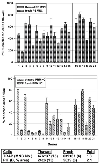
To run drug screening assays, it would be advantageous to have a large, stable pool of PBMNC to work with. There-fore, we examined the effect of cell freezing and pooling on the osteoclast generation and activity (Fig. 2). Fifteen frozen-thawed PBMNC batches from different donors exhibited variable potential in forming TRAP-positive multinuclear cells (Fig. 2, upper panel) and even larger variation in their ability to resorb bone (Fig. 2, middle panel). Importantly, these two parameters did not change in parallel. Six batches of freshly isolated PBMNC were analyzed and among them the variation was smaller. Freezing-thawing did not have a big effect on osteoclast formation or activity (Fig. 2, bottom panel). Thus, for large analyses we collected PBMNC from several donors, froze them, and tested them individually for osteoclast formation and activity. Then, we pooled only those PBMNC batches that showed good activity (>30% area of bone slice resorbed). This enabled us to test different com-pounds on the homogenously active cell population.
For some compounds, such as cathepsin K inhibitors, resorbed area may not be a sensitive read-out, as the pit depth and volume are affected more than the pit area [9]. Therefore, in addition to measuring TRAP-positive multi-nucleated cell number and area of resorbed bone, we established a third read-out, reflecting the pit volume and a degree of collagen degradation in the bone slice. We used human-specific CrossLaps ELISA assay for measuring CTx collagen fragments. This assay was effective with both ivory dentin and bovine bone (Fig. 3). The highest signal was obtained in the conditioned medium collected at the end of culture period (day 14–17). The bisphosphonate zoledronic acid inhibited collagen fragment release in all conditions examined (Fig. 3).
The effects of several known bone resorption inhibitors were measured in the human osteoclast assay. We used representative compounds for bisphosphonates (zoledronic acid), selective estrogen receptor modulators (SERM, raloxifene) and cathepsin K inhibitors (E-64 and AAR494). These compounds have different mechanisms of action and it was interesting to evaluate whether the assay would detect these differences. The control osteo-clast cultures at day 17 contained only TRAP-positive cells and were almost confluent (Fig. 4, TRAP). Osteoclasts were of various sizes: very large cells, smaller multinucle-ated cells, and small mononuclemultinucle-ated cells (Fig. 4, TRAP, C). In the control cultures on bone slices, pits covered most of the bone slice surface and formed characteristic resorption tracks, resulting from osteoclast migration (Fig. 4, PIT, C). The bisphosphonate zoledronic acid (50 nM) and selective estrogen receptor modulator (SERM) raloxifene (100 nM) reduced osteoclast number, leaving the culture largely depleted from mononuclear and smaller multinuclear cells (Fig. 4, TRAP, Z.A., Ral.). At these concentrations both compounds partially inhibited pit formation (Fig. 4, PIT, Z.A., Ral.). Two cathepsin K inhibitors were tested, a non-selective cysteine protease inhibitor E-64 and a selective inhibitor AAR494. Both compounds did not affect osteoclast formation, but partially inhibited pit formation (Fig. 4, E-64, AAR). AAR494 in particular induced changes in cell morphol-ogy, evident as densely TRAP-stained regions within cells. In addition, the remaining pits after treatment with this compound did not have a track appearance, but were clus-ter-like, containing small round pits.
The same bone resorption inhibitors were analyzed for their efficacy and potency in dose-response curves for three assay read-outs: osteoclast formation (multinucle-ated cells), osteoclast activity as resorbed area and as collagen fragments (Fig. 5). All three parameters changed in parallel upon treatment with zoledronic acid and raloxifene; however, raloxifene exerted only a partial effect, while zoledronic acid was fully effective. By con-trast, cathepsin K inhibitors produced dose response curves that diverged. Multinucleated cell number curve stayed unaffected at all concentrations. The resorbed area dose response curve showed a decline, but maximal effects were still partial. As expected, dose response curve for collagen fragments was most sensitive towards cathep-sin K inhibitors. The non-selective inhibitor E-64 was more potent in suppressing collagen fragment release than the selective cathepsin K inhibitor AAR494.
Individual variation in osteoclast generation and activity from different donors, and the effects of cell freezing-thawing
Figure 2
Individual variation in osteoclast generation and activity from different donors, and the effects of cell freezing-thawing. Human osteoclasts were generated from peripheral blood mononuclear cells (PBMNC) as described under Materials and Methods. The results from 21 donors are presented as quantification of TRAP-positive multinucleated cell number (upper panel), and as resorbed area in the pit assay (middle panel). The results are summarized in the bottom panel. The results are shown as means ± SEM; n is a number of independent experiments. PBMNC were used either after freezing-thawing or as freshly isolated cells. B: bone slices.
%
resor
b
ed
ar
ea
/
s
lice
0 20 40 60 80
100 thawed PBMNC fresh PBMNC
Donor
1 2 3 4 5 6 7 8 9 10 11 12 13 14 15 16 17 18 19 20 21
m
u
lti-nucleated
cells
/
9
6-well
0 200 400 600 800 1000
thawed PBMNC fresh PBMNC
1 2 3 4 5 6 7 8 9 10 11 12 13 14 15 16 17 18 19 20 21
Cells
Thawed
Fresh
Fold
Discussion
We have optimized culture conditions for generation of human osteoclasts from human PBMNC to produce up to 2,200 cells/cm2 that were active by resorbing up to 80%
area of the bone slice. Main distinguishing features of our protocol are: a) use of non-purified PBMNC, b) presence of a combination of TGF-β1 and dexamethasone from the start of the culture, in addition to well known osteoclas-togenic factors M-CSF and RANKL, c) use of only commer-cial and not self-made reagents, enabling reproducibility, d) the length of cultures (>14 days), and e) pre-testing of cells from various donors to avoid individual variation.
A number of cytokines has been described to stimulate human osteoclast generation and /or activity, such as prostaglandin E2, interleukin-1α, tumor necrosis factor-α [10]. TGF-β, however, was reported to either inhibit or stimulate human osteoclast formation [7,8]. Our data show that TGF-β1, but not TGF-β3 (data not shown), in presence of dexamethasone, M-CSF and RANKL, has a major positive impact on osteoclast activity and a smaller effect on the osteoclast formation. During the course of our work others have also reported stimulatory effects of TGF-β1, but in the absence of dexamethasone [11]. The removal of non-adherent PBMNC was shown to increase human osteoclast formation to the level induced by
TGF-β [11]. The removal of non-adherent cells would have
[image:8.612.76.539.119.433.2]Establishment of a collagen fragment release read-out with dentin and bone slices
Figure 3
The effects of several bone resorption inhibitors on human osteoclast and resorption pit morphology
Figure 4
been a cheaper and simpler protocol for production of human osteoclasts than the addition of TGF-β1. However, we did not detect any positive impact of the removal of non-adherent cells (data not shown), suggesting that the effect of TGF-β1 is not due to the presence of lymphocytes in blood cell cultures, as proposed. Thus, we continued to use TGF-β1. In agreement with a small positive effect on osteoclast formation, TGF-β1 did not enhance osteoclast markers (data not shown). The mechanism used by
TGF-β1 to stimulate osteoclast activity remains to be determined.
The described assay is the first human primary osteoclast assay that has been adapted for drug screening. Previous osteoclast assays for measurement of compound activity were either done with non-human cells (mouse, rat, rab-bit) [12-14], or, if they used human cells, were done with a human monocytic cell line in co-cultures with osteob-lastic cells [15], or used human bone marrow-derived
Dose response curves for zoledronic acid, raloxifene, E-64 and AAR494 in the human osteoclast assay: measurements of oste-oclast formation, pit area and collagen fragment release
Figure 5
osteoclasts [4], or human tumor osteoclastoma-derived cells [9]. Although useful, these methods do not use active primary human osteoclasts. Furthermore, obtaining tissues required for osteoclast precursor isolation in these methods is more difficult than obtaining human blood.
To test reproducibility and predictability of human osteo-clast assay, the IC50 values were determined for three known classes of bone resorption inhibitors, some of which have already been tested in clinical studies: bisphosphonates, SERM, and cathepsin K inhibitors. A Src kinase inhibitor was also tested and was active, but it was not characterized in detail (data not shown). Zoledronic acid is the most potent bisphosphonate known [16]. In primary human osteoclast assay, all three read-outs decreased in parallel, indicating that main effect of zoledronic acid was on osteoclast number, in agreement with the notion that N-containing bisphosphonates induce osteoclast apoptosis [17].
Estradiol and the SERM raloxifene are known inhibitors of bone resorption in vivo, which were recently demon-strated to exert direct effects on mouse osteoclasts [18]. Such effects have not been shown so far with human oste-oclasts. Both compounds were active in human osteo-clasts (estradiol data not shown), but, in contrast to bisphosphonates, the maximal inhibition by raloxifene and estrogen remained partial. Interestingly, this in vitro result correlates well with a better efficacy of bisphospho-nates as compared to estrogen and SERM in the retrospec-tive analyses of clinical studies [19]. Although other factors, such as pharmacokinetics and adverse effects, influenced the outcome of clinical studies, our in vitro data show that even at the level of osteoclasts, there is a limited potential of SERM to inhibit bone resoprtion.
A non-specific and a specific cathepsin K inhibitor were tested in the human osteoclast assay. Both compounds did not affect osteoclast number, in agreement with the notion that their target is a protease involved in bone resorption, but not in osteoclast formation. Human oste-oclast activity was dose-dependently inhibited, with collagen release showing stronger inhibition, again in agreement with the mechanism of action of these inhibi-tors. E-64 was more potent, but not more effective than AAR494, suggesting that other cathepsins play a minor role in bone resorption. Our results with E-64 differ from those generated in rat osteoclasts, where resorption area was almost unaffected by the compound [14]. This appar-ent discrepancy may represappar-ent a difference in species or in experimental conditions, as our human assay is much longer than the rat assay. We detected a peculiar, cluster-like morphology of pits remaining after treatment with AAR494, suggesting that the compound caused osteo-clasts to interrupt the process of resorption. This
phenom-enon appears to be due to inhibition of osteoclast motility and deserves further examination.
An additional application of the human osteoclast assay could be in the clinic. Already limited analyses of about 20 samples from a rather homogenous population of male blood donor volunteers younger than 40 years showed a considerable variation in the ability of PBMNC to form active osteoclasts. As small amounts of blood (<10 ml) suffice for generation of osteoclasts, patients could be screened for the ability of their circulating osteo-clast precursors to generate osteoosteo-clasts. Osteoporotic, normal and other patient categories could be examined and a correlation between activity of osteoclast precursors and disease occurrence and progression could be evaluated.
Conclusions
We conclude that modified conditions for in vitro gener-ation of human osteoclasts from peripheral blood result in a stable generation of active osteoclasts. As documented by testing several classes of bone-resorption inhibitors, this assay can be used for screening novel drugs. As this assay can be easily performed with a small volume of human blood, it can also be used to monitor the capacity of blood cells to generate bone-resorbing osteoclasts.
Competing interests
The authors are employed by the Novartis Institutes for BioMedical Research.
Authors' contributions
N-H. L-N. performed all experimental studies, except measurements of collagen fragments, and prepared the Figures. RG supervised collagen fragment assays. DC designed the primers for osteoclast markers and tested most of them by radioactive quantitative RT-PCR. NZ supervised N-H. L-N. in radioactive quantitative PCR. MS conceived the study, supervised the experimental work and wrote the manuscript. All authors read and approved the final manuscript.
Acknowledgements
We thank Roger Vuille for his technical assistance with the CrossLaps ELISA, Martin Missbach and Rene Lattmann for the synthesis of AAR494 and information on its selectivity, and Yves Seltenmeyer for the help in preparation of bone and dentin slices. Kind advice on bone marrow osteo-clast generation and bone resorption assays by Usha Sarma, John Scopes, Helen Massey and Cheryl Lader (Flanagan laboratory, St. Mary's Hospital, London, UK) during the visit of MS in 1999 is gratefully acknowledged.
References
Publish with BioMed Central and every scientist can read your work free of charge "BioMed Central will be the most significant development for disseminating the results of biomedical researc h in our lifetime."
Sir Paul Nurse, Cancer Research UK
Your research papers will be:
available free of charge to the entire biomedical community
peer reviewed and published immediately upon acceptance
cited in PubMed and archived on PubMed Central
yours — you keep the copyright
Submit your manuscript here:
http://www.biomedcentral.com/info/publishing_adv.asp
BioMedcentral
2. Matsuzaki K, Udagawa N, Takahashi N, Yamaguchi K, Yasuda H, Shima N, Morinaga T, Toyama Y, Yabe Y, Higashio K, Suda T: Oste-oclast differentiation factor (ODF) induces osteOste-oclast-like cell formation in human peripheral blood mononuclear cell cultures.Biochem Biophys Res Commun 1998, 246:199-204. 3. Shalhoub V, Faust J, Boyle WJ, Dunstan CR, Kelley M, Kaufman S,
Scully S, Van G, Lacey DL: Osteoprotegerin and osteoprote-gerin ligand effects on osteoclast formation from human peripheral blood mononuclear cell precursors.J Cell Biochem
1999, 72:251-261.
4. Sarma U, Edwards M, Motoyoshi K, Flanagan AM: Inhibition of bone resorption by 17beta-estradiol in human bone marrow cultures.J Cell Physiol 1998, 175:99-108.
5. Mbalaviele G, Jaiswal N, Meng A, Cheng L, Van Den Bos C, Thiede M:
Human mesenchymal stem cells promote human osteoclast differentiation from CD34+ bone marrow hematopoietic progenitors.Endocrinology 1999, 140:3736-3743.
6. Nicholson GC, Malakellis M, Collier FM, Cameron PU, Holloway WR, Gough TJ, Gregorio-King C, Kirkland MA, Myers DE: Induction of osteoclasts from CD14-positive human peripheral blood mononuclear cells by receptor activator of nuclear factor kappaB ligand (RANKL).Clin Sci (Lond) 2000, 99:133-140. 7. Weitzmann MN, Cenci S, Haug J, Brown C, DiPersio J, Pacifici R: B
lymphocytes inhibit human osteoclastogenesis by secretion of TGFbeta.J Cell Biochem 2000, 78:318-324.
8. Scopes J, Massey HM, Flanagan AM: TGFb enhances human oste-oclast formation and bone resorption at the expense of macrophages.J Bone Miner Res 2000, 15:s265.
9. James IE, Lark MW, Zembryki D, Lee-Rykaczewski EV, Hwang SM, Tomaszek TA, Belfiore P, Gowen M: Development and charac-terization of a human in vitro resorption assay: demonstra-tion of utility using novel antiresorptive agents.J Bone Miner Res 1999, 14:1562-1569.
10. Lader CS, Flanagan AM: Prostaglandin E2, interleukin 1alpha, and tumor necrosis factor-alpha increase human osteoclast formation and bone resorption in vitro. Endocrinology 1998,
139:3157-3164.
11. Massey HM, Scopes J, Horton MA, Flanagan AM: Transforming growth factor-beta1 (TGF-beta) stimulates the osteoclast-forming potential of peripheral blood hematopoietic precur-sors in a lymphocyte-rich microenvironment. Bone 2001,
28:577-582.
12. David JP, Neff L, Chen Y, Rincon M, Horne WC, Baron R: A new method to isolate large numbers of rabbit osteoclasts and osteoclast-like cells: application to the characterization of serum response element binding proteins during osteoclast differentiation.J Bone Miner Res 1998, 13:1730-1738.
13. Taranta A, Brama M, Teti A, De Luca V, Scandurra R, Spera G, Agnus-dei D, Termine JD, Migliaccio S: The selective estrogen receptor modulator raloxifene regulates osteoclast and osteoblast activity in vitro.Bone 2002, 30:368-376.
14. Parikka V, Lehenkari P, Sassi ML, Halleen J, Risteli J, Harkonen P, Vaananen HK: Estrogen reduces the depth of resorption pits by disturbing the organic bone matrix degradation activity of mature osteoclasts.Endocrinology 2001, 142:5371-5378. 15. Kung Sutherland MS, Lipps SG, Patnaik N, Gayo-Fung LM,
Kham-mungkune S, Xie W, Brady HA, Barbosa MS, Anderson DW, Stein B:
SP50 a novel SERM, blocks osteoclastogenesis in a human bone cell model: role of IL-6 and GM-CSF. Cytokine 0263,
23:1-14.
16. Green JR: Preclinical pharmacology of zoledronic acid.Semin Oncol 2002, 29:3-11.
17. Reszka AA, Rodan GA: Bisphosphonate mechanism of action.
Curr Rheumatol Rep 2003, 5:65-74.
18. Shevde NK, Bendixen AC, Dienger KM, Pike JW: Estrogens sup-press RANK ligand-induced osteoclast differentiation via a stromal cell independent mechanism involving c-Jun repression.Proc Natl Acad Sci U S A 2000, 97:7829-7834.