City, University of London Institutional Repository
Citation:
Ntonia, I. (2016). Modulation of lateralised responses to primary affect.(Unpublished Doctoral thesis, City, University of London)
This is the accepted version of the paper.
This version of the publication may differ from the final published
version.
Permanent repository link:
http://openaccess.city.ac.uk/16213/Link to published version:
Copyright and reuse: City Research Online aims to make research
outputs of City, University of London available to a wider audience.
Copyright and Moral Rights remain with the author(s) and/or copyright
holders. URLs from City Research Online may be freely distributed and
linked to.
MODULATION OF
LATERALISED RESPONSES TO
PRIMARY AFFECT
IRO NTONIA
A THESIS SUBMITTED TO THE DEPARTMENT OF
PSYCHOLOGY
CITY UNIVERSITY LONDON
FOR THE DEGREE OF DOCTOR OF PHILOSOPHY
City, University of London Northampton Square London EC1V 0HB United Kingdom
T +44 (0)20 7040 5060
THE FOLLOWING PARTS OF THIS THESIS HAVE BEEN REDACTED
FOR COPYRIGHT REASONS:
p. 48:
Figs 2.1 & 2.2:
Images from ‘Pictures of Facial Affect.’
p. 50:
Fig. 2.3:
Images from ‘Pictures of Facial Affect.’
p. 51:
Fig. 2.4:
Images from ‘Pictures of Facial Affect.’
p. 52:
Fig. 2.5:
Images from ‘Pictures of Facial Affect.’
p. 116:
Fig. 4.2:
Images from ‘Pictures of Facial Affect’
p. 135:
Fig. 4.8:
Images from ‘Pictures of Facial Affect’
p. 136:
Fig. 4.9:
Images from ‘Pictures of Facial Affect’
First and foremost, I would like to express my deepest gratitude to my supervisor, Dr Elliot Freeman, for his continuous support, extremely helpful feedback and invaluable insight throughout my doctoral studies. Without his support, advice and patience, this work would not have been possible.
I am forever grateful to my wonderful parents, Stamatis and Zizi, who believed in me from the beginning and supported me throughout my life (and my studies). I wish my mum, whom I sadly lost in May 2014, was still around to see this work come to
completion.
My deepest thanks go to my wonderful and supportive husband, Jamie, who has stuck by me and made life during the PhD that little bit more manageable.
I am grateful to my colleagues at LEaD, City University London, for giving me the opportunity to further my academic career whilst completing my thesis.
I would also like to thank Dr Marinella Cappelletti, Dr Markus Bauer, and Dr Christian Kluge for giving me the opportunity to further my research training alongside my PhD studies.
DECLARATION
ABSTRACT
Do we use one cerebral hemisphere or both to process positive and negative
emotions? Is it more physiologically economical for the brain to initiate responses to both types of primary affect from a unilateral locus, or does our readiness to react to emotional stimuli depend on the differential contribution of each hemisphere based on the approach or avoidance behaviours positive and negative affect elicit? This thesis is concerned with these questions that have so far remained unanswered even though they form a key part of emotional perception research. The behavioural literature has provided evidence for both unilaterally (right hemisphere) and
bilaterally derived responses to different types of emotional stimuli, with the directionality of response patterns changing depending on stimulus type and task demands. The neuroimaging literature has addressed whether there is a functional need for the lateralised processing of basic emotional stimuli by mapping subcortical and cortical emotional attention networks, specific to different variants of only
negative affect (i.e. fear, sadness). How this subcortically originating lateralisation manifests into observable behaviour however still remains to be established. This research therefore posits that hemispheric lateralisation may be a modulated process, and aims to explore how this modulation guides the directionality of our behavioural responses to primary affect. The thesis introduces a novel methodology that provides the first evidence of the modulation of emotional lateralisation by establishing a behavioural paradigm that can effectively investigate hemispheric lateralisation through measures of response efficiency. The thesis further
investigates whether subcortically originating lateralisation may be inferred through its resulting behavioural response, by examining visual field asymmetries in
TABLE OF CONTENTS
1 CHAPTER 1: GENERAL INTRODUCTION ... 14
1.1. THEORETICAL BACKGROUND & OVERVIEW ... 14
1.2 DEFINITIONS OF AFFECT AND LATERALITY ... 15
1.3 REVIEW OF EVIDENCE FOR THE LATERALISATION OF PRIMARY AFFECT ... 18
1.3.1 BEHAVIOURAL LATERALISATION LITERATURE ... 18
1.3.2 NEUROIMAGING LATERALISATION LITERATURE ... 21
1.3.3 PRELIMINARY CONCLUSIONS ON LATERALISATION... 22
1.3.4 MIGHT LATERALITY PATTERNS BE DEPENDENT ON METHODOLOGY?... 23
1.4 IS THERE A FUNCTIONAL NEED FOR LATERALISATION? ... 25
1.4.1 MIGHT EMOTIONAL FACIAL EXPRESSIONS ENHANCE THE FUNCTIONAL NEED FOR LATERALITY? ... 27
1.4.2 MIGHT DISTRIBUTION ASYMMETRIES OF CORTICAL ATTENTION RESULT IN LATERALISED RESPONSES? ... 29
1.4.2 MIGHT THE ASYMMETRIC DISTRIBUTION OF CORTICAL EMOTIONAL ATTENTION RELATE TO PHYSIOLOGICAL CONTRALATERALITY? ... 32
1.5 SUMMARY AND THESIS OUTLINE ... 34
2 CHAPTER 2: MODULATION OF LATERALISED RESPONSES TO PRIMARY AFFECT ... 36
2.1 INTRODUCTION ... 36
2.1.2. IMPACT OF METHODOLOGY ON THE INTERPRETATION OF LATERALISED PROCESSING OF AFFECT ... 37
2.1.2.1 THE PROBLEM WITH NEGATIVE AFFECT... 37
2.1.2.2 MIGHT RESPONSES TO AFFECT BE TASK-DEPENDENT? ... 40
2.1.3 HOW MIGHT EMOTIONAL LATERALISATION TRANSLATE INTO APPROACH/AVOIDANCE BEHAVIOURS? ... 42
2.1.4. SUMMARY ... 44
2 PILOT PROTOCOL FOR STIMULUS SELECTION ... 45
2.2.1 STIMULUS SELECTION METHODOLOGY ... 45
2.2.1.1 OVERVIEW ... 45
2.2.1.2 STIMULUS SELECTION PROTOCOL PARTICIPANTS ... 46
2.2.1.3 STIMULUS SELECTION PROTOCOL APPARATUS ... 46
2.2.1.4 STIMULUS IMAGE PROCESSING & PRESENTATION ... 47
49 2.2.1.4 STIMULUS SELECTION PROTOCOL ... 51
2.2.1.5 STIMULUS SELECTION PROTOCOL PROCEDURE ... 51
2 EXPERIMENT 2.2 ... 57
2.2.2 METHODS ... 57
2.2.2.1 SUBJECTS ... 57
2.2.2.2 APPARATUS ... 57
2.2.2.3 STIMULI ... 57
2.2.2.4 DESIGN... 58
2.2.2.5 PROCEDURE ... 58
2.2.2.6 DATA PREPARATION AND ANALYSIS ... 59
2.2.2 RESULTS ... 61
2.2.2.1 REACTION TIME ... 61
2.2.2.2 ACCURACY ... 63
2.2.2.3 THRESHOLD ANALYSIS ... 65
2.2.3 SUMMARY ... 67
2.3 DISCUSSION ... 68
2.3.1 EMOTIONAL MODULATION OF BEHAVIOURAL LATERALISATION ... 70
2.3.2 CONCLUSION ... 74
3 CHAPTER 3: NASAL-TEMPORAL ASYMMETRIES IN SUPRATHRESHOLD FACIAL EXPRESSIONS OF PRIMARY AFFECT ... 76
3.1 INTRODUCTION ... 76
3.1.1. ASYMMETRIC DISTRIBUTION OF SUBCORTICAL LOCI OF ACTIVITY IN EMOTIONAL ATTENTION ... 78
3.1.2 NASAL-TEMPORAL ASYMMETRIES AS AN INDEX OF SUBCORTICAL DISTRIBUTION OF ATTENTION ... 81
3.1.2.1. MIGHT NASAL-TEMPORAL ASYMMETRIES ACT AS AN INDEX OF SUBCORTICAL ASYMMETRICAL DISTRIBUTION OF EMOTIONAL ATTENTION? ... 83
3.1.2.2. HOW MIGHT THE LINK BETWEEN NASAL-TEMPORAL ASYMMETRIES AND THE RETINOTECTAL PATHWAY INFLUENCE ASYMMETRY? ... 84
3.1.2 SUMMARY ... 86
3 EXPERIMENT 3.1 ... 88
3.2.1. METHODS ... 88
3.2.1.1. SUBJECTS ... 88
3.2.1.2 APPARATUS ... 88
3.2.1.3. STIMULI ... 88
3.2.1.4. DESIGN ... 88
3.2.1.5. PROCEDURE ... 89
3.2.1.6. DATA PREPARATION AND ANALYSIS ... 90
3.2.2 RESULTS ... 91
3.2.2.1 REACTION TIME ... 91
3.2.2.2. ACCURACY ... 92
3.2.2.3. THRESHOLD ANALYSIS ... 94
3.2.3 SUMMARY ... 95
3.3 DISCUSSION ... 96
3.3.1 MIGHT THE PRESENCE OF NASAL-TEMPORAL ASYMMETRIES BE METHODOLOGY-DEPENDENT? ... 97
3.3.1.1 RESPONSE TYPE ... 97
3.3.1.2 PARADIGM TYPE ... 99
3.3.2 MIGHT NASAL-TEMPORAL ASYMMETRIES BE SENSITIVE TO FACES OVERALL, BUT NOT TO DIFFERENT TYPES OF FACIAL AFFECT? ... 100
3.3.3 CONCLUSION ... 101
4 CHAPTER 4: AUDITORY THREAT AND GAZE DIRECTION MODULATE LATERALISED RESPONSES TO AFFECT ... 103
4.1 GENERAL INTRODUCTION ... 103
4.1.1 MODULATING EFFECTS OF SOUND ON EMOTIONAL ATTENTION AND BEHAVIOUR ... 105
4.1.1.1 SUBCORTICAL LOCI OF ACTIVITY IN THE PROCESSING OF AUDITORY EMOTION ... 108
4.1.1.2 LOOMING SOUNDS AS ECOLOGICALLY-VALID EMOTIONAL STIMULI ... 110
4 EXPERIMENT 4.1 ... 112
OVERVIEW ... 112
4.2.1. METHODS ... 113
4.2.1.1. SUBJECTS ... 113
4.2.1.2 APPARATUS ... 113
4.2.1.3. STIMULI ... 113
4.2.1.4. DESIGN ... 114
4.3.1.2 ACCURACY ... 121
4.3.1.3 THRESHOLD ANALYSIS ... 124
4.3.1 EXPERIMENT 4.1 SUMMARY ... 125
4.1.2 MODULATING EFFECTS OF GAZE DIRECTION ON EMOTIONAL ATTENTION ... 126
4.1.2.1 MIGHT THE MODULATING EFFECT OF GAZE DIRECTION DIFFER DEPENDING ON TYPE OF EMOTION? ... 127
4.1.3 REFLEX VERSUS REFLECT: RESPONSE PATTERN DIFFERENCES DEPEND ON DURATION OF GAZE-MANIPULATED FACIAL AFFECT ... 132
4 EXPERIMENT 4.2 ... 133
OVERVIEW ... 133
4.2.2 METHODS ... 134
4.2.2.1 SUBJECTS ... 134
4.2.2.2. APPARATUS ... 134
4.2.2.3. STIMULI ... 134
4.2.2.4. DESIGN ... 138
4.2.2.5. PROCEDURE ... 138
4.2.2.6. DATA PREPARATION AND ANALYSIS ... 138
4.2.2.2 RESULTS ... 139
4.2.2.2.1 REACTION TIME ... 139
4.2.2.2.2 ACCURACY ... 142
4.2.3 EXPERIMENT 4.2 SUMMARY ... 145
4.3 GENERAL DISCUSSION ... 146
4.3.1 METHODOLOGICAL CONSIDERATIONS ... 147
4.3.2 ALERTING SOUNDS AND DIRECT GAZE MODULATE LATERALISATION OF RESPONSES TO POTENTIAL THREAT ... 149
4.3.3 CONCLUSION ... 151
5 CHAPTER 5: INDIVIDUAL VARIABILITY IN ANXIETY MODULATES LATERALISED RESPONSES TO AFFECT ... 152
5.1 INTRODUCTION ... 152
5.1.1 INDIVIDUAL VARIABILITY AND EMOTIONAL PERCEPTION... 154
5.1.1.1 NEURAL UNDERPINNINGS OF INDIVIDUAL DIFFERENCES IN EMOTIONAL ATTENTION ... 156
5.1.2 ANXIETY AND EMOTIONAL PERCEPTION ... 158
5.1.3 MIGHT INDIVIDUAL DIFFERENCES IN ANXIETY LEVELS PREDICT PATTERNS OF RESPONSE EFFICIENCY? ... 160
5.1.4 SUMMARY ... 162
5 EXPERIMENT 5.1 ... 164
5.2.1 METHODS ... 164
5.2.1.1 SUBJECTS ... 164
5.2.1.2 APPARATUS & MATERIALS ... 164
5.2.1.3 STIMULI & PROCEDURE ... 165
5.2.1.4 DESIGN... 165
5.2.1.5 DATA PREPARATION & ANALYSIS ... 165
5.3.1 RESULTS ... 166
5.3.1.1 REACTION TIME ... 166
5.3.1.2 ACCURACY ... 175
5.3.1.3 THRESHOLDS ... 180
5.3.1.4 EXPERIMENT 5.1 SUMMARY ... 183
5 EXPERIMENT 5.2 ... 184
OVERVIEW ... 184
5.2.2 METHODS ... 184
5.2.2.1 SUBJECTS ... 184
5.2.2.3 STIMULI & PROCEDURE ... 185
5.2.2.4 DESIGN... 185
5.2.2.5 DATA PREPARATION & ANALYSIS ... 185
5.3.2 RESULTS ... 186
5.3.2.1 REACTION TIME ... 186
5.3.2.2 ACCURACY ... 189
5.3.2.3 EXPERIMENT 5.2 SUMMARY ... 195
5 EXPERIMENT 5.3 ... 197
OVERVIEW ... 197
5.2.3 METHODS ... 197
5.2.3.1 SUBJECTS ... 197
5.2.3.2 APPARATUS & MATERIALS ... 197
5.2.3.3 STIMULI & PROCEDURE ... 198
5.2.3.4 DESIGN... 198
5.2.3.5 DATA PREPARATION & ANALYSIS ... 198
5.3.3 RESULTS ... 199
5.3.3.1 REACTION TIME ... 199
5.3.3.2 ACCURACY ... 199
5.3.3.3 EXPERIMENT 5.3 SUMMARY ... 203
5.4 DISCUSSION ... 204
5.4.1 AUTOMATICITY OF RESPONSES TO BASIC AFFECT DEPENDS ON INDIVIDUAL ANXIETY ... 206
5.4.2 CONCLUSION ... 209
6 CHAPTER 6: GENERAL DISCUSSION ... 210
6.1 CHAPTER OVERVIEW ... 210
6.2 SUMMARY OF EXPERIMENTAL CHAPTER FINDINGS ... 211
6.3 LATERALISATION ... 215
6.4 POSITIVITY BIAS ... 217
6.5 ANGER SUPERIORITY OVER FEAR ... 218
6.7 CONCLUSION OF THE THESIS ... 224
REFERENCES ... 225
LIST OF FIGURES & TABLES
Table 1. Summary table outlining task details and findings from a selection of behavioural lateralisation studies that have informed the rationale of the present thesis. ... 19 Figure 1. Diagram displaying the contralateral distribution of the human visual system (Figure adapted from Felten & Shetty, 2011). ... 33 Figure 2.1. Angry stimuli from one Ekman & Friesen poser, displayed on grey background as used in the task. From left to right, expression salience starts from completely neutral (intensity 0) and morphs
gradually from highly ambiguous (intensity 1) up to a highly exaggerated expression (intensity 5). ... 48 Figure 2.2. Example of stimulus before and after windowing; the far-left picture shows the original
photograph whereby poser-specific elements (such as the poser’s hairline) could render the photograph easily identifiable. The middle graphic displays the oval-shaped window used to superimpose on the original photograph so as to eliminate poser-specific identifiable features. Aside from the window, the graphic also displays two horizontal and one vertical guides used to align features from each face so as to equate positioning of eyes and mouth across stimuli. The far-right graphic shows the final version of the
photograph, post-windowing. ... 48 Figure 2.3. Two example stimulus pairs where the target photograph is shown on the right visual field on the top image, and on the left visual field on the bottom image. This example shows an emotional
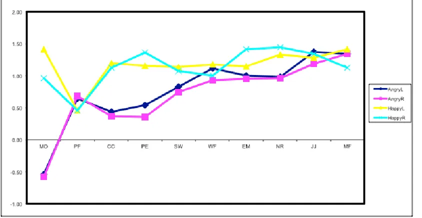
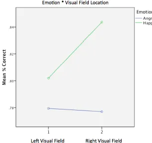
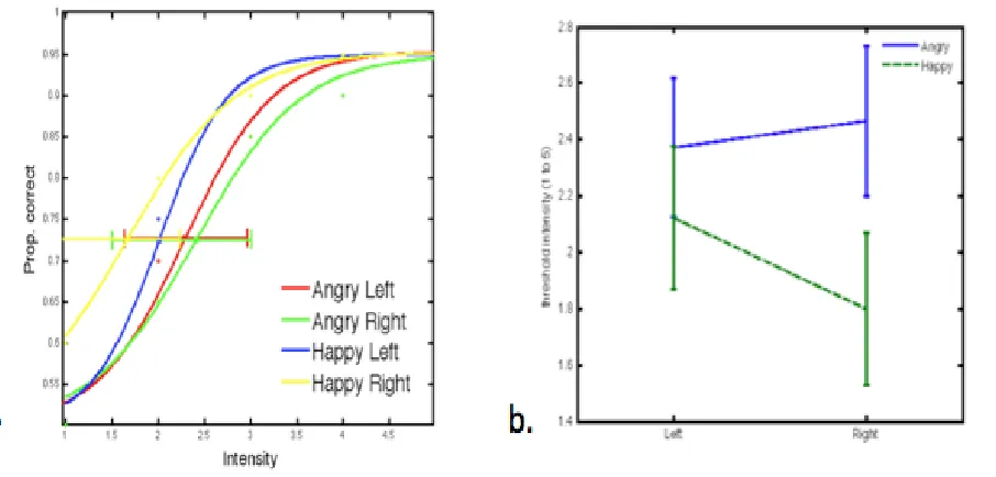
quicker overall; reaction times however differ within each emotion separately, depending on the visual field location of the target face as a function of expression salience. ... 62 Figure 2-9. Line graph displaying mean reaction times (ms) for Angry (blue) and Happy (green) stimuli across the Left and Right visual fields, for expression salience intensity 3, which was the resulting interaction from post-hoc analyses on the 3-way interaction between emotion, VF location and intensity. For expression intensity 3, happy faces are responded to quicker when presented on the right visual field, and angry faces are responded to quicker when presented on the left visual field. ... 62 Figure 2.10. The line graph shows accuracy scores per condition as a function of emotional expression intensity. Each of the colour-coded lines represents one of four conditions of type of emotion and visual field location. Error bars per condition represent Cousineau-corrected, +/-1 within-subjects error. Across conditions, responses become more accurate as the expression intensity is gradually disambiguated from 1 (highly ambiguous) to 5 (exaggerated). Accuracy score significant differences in terms of type of emotion (angry/happy) regardless of visual field location are observed in intensities 2 through to 5; significant differences in terms of visual field location regardless of emotion are only observed for intensity 1. ... 64 Figure 2.11. Interaction plot displaying mean % correct scores for angry (blue line) and happy (green line) stimuli, plotted across the left and right visual fields respectively. It is evident that the overall interaction is driven by happy stimuli only, which displayed higher accuracy when shown on the right visual field as opposed to the left. Angry faces showed a negligible difference in accuracy based on visual field location. ... 65 Figure 2.12. Graph a. displays the mapping of 95% CI against the required intensity (salience) per condition, which is then re-plotted as Intensity by Visual Field with separate error bars representing angry and happy faces on Graph b. ... 66 Figure 3.1. Graphic illustrates the cortical (A) and subcortical (B) retinotectal pathways through
projections from nasal and temporal hemiretinae (figure borrowed from Zackon et al., 1997). ... 77 Figure 3.3. Graphic displays the inverted inputs from the lens to the nasal and temporal hemiretinae; the nasal retina receives input from the temporal hemifield, and conversely the temporal retina receives input from the nasal hemifield (Katz & Crowley, 2002). ... 82 Figure 3.4. Interaction plot displaying mean reaction time (ms) scores for angry (blue line) and happy (green line) emotional face stimuli when plotted across the nasal and temporal hemifields respectively. Happy stimuli when viewed on the temporal hemifield were responded to quicker than when on the nasal hemifield. No such difference was observed for the angry faces. ... 92 Figure 3.5. Line graph representing accuracy scores for each emotion (angry/happy) as a function of expression intensity. Error bars represent Cousineau-corrected, +/-1 within-subjects error. No difference is shown between emotions for intensity 1, and for this expression intensity accuracy scores for both emotions is just above chance. From intensity 2 through to intensity 5, happy faces show consistently higher,
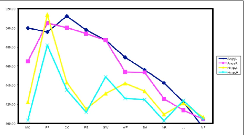
medial geniculate body (grey oval) to the amygdala (red oval) (‘fast route’), or pass through the slower route via the auditory cortex and additional cortical areas (blue route) before reaching the amygdala (‘slow route’). Both routes then result in fear responses from the organism (i.e. elevation of stress hormones, reflex reactions etc.) (Figure from Armony & LeDoux, 2010). ... 109 Figure 4.2. Graphic illustrates the progression of a typical trial. ... 116 Figure 4.3. Line graph depicts left (blue line) and right (green line) visual field faces plotted as a function of expression salience intensity. ... 118 Figure 4.4. Line graph depicts angry (blue line) and happy (green line) faces plotted as a function of visual field presentation (LVF vs RVF). ... 119 Figure 4.5. Line graph depicts reaction times per emotion/sound condition, plotted as a function of
expression salience intensity. Error bars represent Cousineau-corrected +-1 within-subjects error. ... 121 Figure 4.5 Line graph shows mean accuracy scores for angry (blue line) and happy (green line) stimuli respectively in terms of left and right visual field target location. ... 122 Figure 4.6 Line graph displays differences between accuracy scores for angry (blue line) and happy (red line) stimuli respectively, plotted as a function of expression salience intensity. Error bars represent
Cousineau-corrected +-1 within-subjects error... 123 Figure 4.7 Graph displays recognition thresholds for angry (blue) and happy (red) faces respectively, depending on presentation on the Left or Right visual field. ... 124 Figure 4.8 Example of eye masking procedure in preparation for manipulating gaze direction. After
calculating control points around the edge of the eye in the emotional picture of one poser (bottom right square), a corresponding polygon-shaped mask was applied on the same poser’s neutral photograph
(bottom left square). ... 135 Figure 4.9 Example ellipse on a stimulus photograph. The ellipse could be moved between a number of set points to achieve gaze direction manipulation. ... 136 Figure 4.10 Example full set of image intensities and gaze directions from one poser. Each of the five
Table 5.1. Table reports post-hoc t-test statistics for VF comparisons across the five intensities.
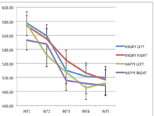
Significant comparisons are denoted by *... 167 Figure 5.2. Line graph plots mean RTs for LVF (blue line) and RVF (green line) target stimuli as a function of expression salience intensity (1-5). ... 168 Figure 5.3. Graphic displays the difference in RT between angry and happy faces (y-axis) and anxiety scores (x-axis). ... 169 Figure 5.4. Graph displays the mean RT distribution for Intensity 1, plotted as a function of centred Q scores ... 170 Figure 5.5. Graph displays the mean RT distribution for Intensity 2, plotted as a function of centred Q scores ... 170 Figure 5.6. Graph displays the mean RT distribution for Intensity 3, plotted as a function of centred Q scores ... 171 Figure 5.9. Graphic displays the significant, moderate positive relationship between response times only to intensity 1 emotional stimuli (y-axis) and anxiety scores (x-axis). The higher the anxiety, the quicker
response times become, but only for highly ambiguous emotional faces. ... 173 Figure 5.10. Graphic displays the significant, moderate negative relationship between angry stimuli (blue line) and response times as a function of anxiety. Significant negative correlations were found for both angry (blue line) and happy (green line) facial expressions, at expression salience intensity 1. ... 174 Figure 5.11. Line graph plots mean accuracy scores for angry (blue line) and happy (green line) stimuli as a function of expression salience intensity. ... 176 Figure 5.12. Line graph plots mean accuracy scores for LVF (blue line) and RVF (green line) target stimuli as a function of expression salience intensity. ... 177 Figure 5.13. Graphic displays the significant, moderate positive relationship between accuracy scores only to intensity 1 emotional stimuli (y-axis) and anxiety scores (x-axis). ... 178 Figure 5.14. Graphic displays the significant, moderate negative relationship between angry stimuli (blue line) and accuracy scores as a function of anxiety. ... 179 Figure 5.15. Graphic displays significant relationships between angry (blue line) and happy (green line) stimuli to detection thresholds (y-axis) as a function of anxiety (x-axis). Correlations are significant for both emotions, with the angry correlation being stronger than that for happy. ... 180 Figure 5.16. Graphic illustrates the difference for thresholds for angry faces relative to happy as a function of increasing anxiety. ... 181
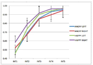
Figure 5.21 Line graph displays mean accuracy scores for angry (blue line) and happy (green line) stimuli when presented on the left and right visual field respectively. ... 190 Figure 5.22 Line graph displays angry (blue line) and happy (red line) mean accuracy scores as a function of expression salience intensity. Error bars represent Cousineau-corrected within-subjects +-1 SE of the mean. ... 191 Figure 5.23 Line graph displays mean accuracy scores for each emotion/visual field location condition as a function of expression salience intensity. Error bars represent Cousineau-corrected within-subjects +-1 SE of the mean. ... 193 Figure 5.24 Scatterplot shows the relationship between the calculated VF difference (LVF-RVF) as a
function of mean-centred questionnaire score (anxiety). ... 194 Figure 5.25 Line graph illustrates the three levels of gaze manipulation (gaze ahead=blue line,
1 CHAPTER 1: GENERAL INTRODUCTION
1.1. THEORETICAL BACKGROUND & OVERVIEW
In 1884, William James submitted a paper to the then-philosophical journal Mind asking why one would run when faced with a wild bear; do we run because we’re afraid of what will happen if we don’t, or are we afraid because we’re running? The
present thesis poses a similar question: to clarify what happens during the stages
from stimulus to feeling, and identify the processes that modulate and facilitate this
timeline of events that come in between. What is it that dictates and guides the way
in which we perceive and respond to the most basic and evolutionarily ingrained of
emotions? The way in which we perceive our environment and function within it is
for the most part dictated by our ability to correctly identify, as well as to efficiently
respond to emotional information.
Efficiency in the way we cognitively process affect is essential to our survival;
accurate detection and classification of environmental emotional stimuli enables us
to detect and respond to potential threat as well as regulating our behaviour and
enabling our social interactions. Emotional perception has been extensively
researched in the cognitive, social and developmental psychological neuroscience
domains since the late 19th Century with no sign of decreasing in momentum as
methodological techniques advance. Emotional perception research has not
however been without theoretical, conceptual, and methodological issues; across
studies, the reporting of conflicting evidence for elements of emotional processing
spanning their definition, classification and categorisation, and how emotions are
processed and responded to from perception to resulting observable behaviour, has
become somewhat of a trademark of emotion research. This chapter will introduce
the underlying theoretical issues informing emotional perception research, present
the evidence so far, and outline the theoretical framework forming the rationale on
1.2 DEFINITIONS OF AFFECT AND LATERALITY
Perhaps one of the most controversial topics in the field of emotion research has
been that of a suggested differential hemispheric contribution, or lateralisation,
specific to the nature of emotional information processed. A large body of evidence
supports the hypothesis of a valence-specific lateralisation (e.g. Reuter-Lorenz,
Kinsbourne, & Moscovitch, 1990; Reuter-Lorenz, Oonk, Barnes, & Hughes, 1995;
Reuter-Lorenz, & Davidson, 1981; Ahern & Schwartz, 1979; Ross, 1977), which has
however been at the forefront of considerable debate as an equally large body of
evidence has conversely suggested that all aspects of emotional processing may be
attributed solely to right hemisphere specialisation (e.g. Borod & Caron, 1980; Borod
et al., 1998; Devinsky, 2000; Dimberg & Petterson, 2000; Ladavas, Umiltà, &
Ricci-Bitti, 1980; Tucker, 1981).
The valence hypothesis suggests that each of the two hemispheres becomes
preferentially engaged depending on the phenomenological nature of emotional
environmental stimuli we are exposed to (Ross, Homan, & Buck, 1994; Schwartz,
Ahern, & Brown, 1979). Emotions that are perceived to be negative or threatening
are suggested to be preferentially processed by the right hemisphere, while the left
hemisphere engages with the processing of positive emotional stimuli (Ross et al.,
1994). The alternative, right hemisphere dominance hypothesis suggests that all
emotional information regardless of valence is unilaterally processed by the right
hemisphere; the underlying rationale in favour of unilateral hemispheric emotional
processing suggests that if a function does not need to be represented bilaterally in
the morphology of the human body (i.e. such as having a left and right arm and leg),
then it is more evolutionarily economical to group all neural connections for said
function close together (Rolls, 1990, 2005).
The theoretical foundations of this debate might be perhaps better understood
through historical attempts at defining, categorising and classifying the concept of
emotions. In the 19th century, William James conceptualised emotions as being
physiological changes occurring in the self following exposure to an arousing stimulus (LeDoux, 2000). James’s definition suggested that the term ‘emotion’
represents a timeline of perceptual and action events, starting from the exposure to
One resulting categorisation of emotions is based on the assumption that they are
individual subjective states concerned with maintaining the life of an organism, and
as such are comprised of a number of attributes relating to instinctual, innate
behaviours (Damasio et al., 2000). Individual emotional states are thence
categorised into primary-basic, secondary-social, and background emotions
according to their phenomenology. Specifically, primary emotions (i.e. fear, anger,
sadness, surprise, disgust, happiness) are shared by numerous animal species and
are grounded in ontologically ancient instinctual behaviours closely connected with
ensuring survival. Secondary (a.k.a. social) emotions (i.e. guilt, pride, empathy,
embarrassment, jealousy) are grounded in experience of social interactions, by
implying the presence of a social audience and do not share the same direct link to
survival as primary emotions. Lastly, background emotions (i.e. wellbeing, malaise, calmness, tension) are connected to organisms’ current individual physiological
states and might act as mediators, modulators, or inhibitors of behaviour (Damasio
et al., 2000; LeDoux, 2000).
The dichotomisation of emotions into positive and negative is predominantly
concerned with primary/basic affect, and links its strong instinct-based attributes to both a basis on an organism’s homeostatic regulation (Damasio et al., 2000), and on
the prompting of cognitive plans for action (Rolls, 1990, 2005). In behaviouristic
terms, positive and negative emotions relate to whether the environmental stimuli
causing them are perceived as possible rewards or possible punishers (Rolls, 2005),
with rewards comprising anything that an organism that will act towards obtaining
(approach), and punishers being anything that an organism will act to avoid
(avoidance).
The grouping of emotions into primary, secondary, background as well as into
positive/negative has also been established on the identification of
neuroanatomically discrete emotional systems, thought to be specific to different
types of basic affect - a type of localised, emotional map comprising subcortical
structures in the limbic system and midline messencephalic structures (Panksepp,
2004, 2005; Panksepp & Zellner, 2004). This map includes a dopamine-facilitated
appetitive motivation seeking system, located in the ventral tegmental area and
terminalis, and the periaqueductal grey of the mesencephalon; a rage system which courses parallel to the fear system’s circuitry from the medial amygdala to the
periaqueductal grey and facilitates aggressive acts and feelings; a system
associated with separation-distress and panic reactions connecting the
periaqueductal grey matter with more rostral brain areas that triggers
separation-distress feelings and mediates panic reactions; regions associated with erotic desire
in basal forebrain and hypothalamic structures connecting them down to the
periaqueductal grey and which are associated mainly with erotic feelings; a care
system which facilitates maternal/paternal nurturing feelings; and lastly a play
system which is associated with youthful rough-and-tumble play and laughter, which
is primarily relevant to positive affect (Panksepp, 2004, 2005).
Specifically relevant to the positive/negative emotion grouping are bottom-up and
top-down perceptual processing networks (Derryberry & Tucker, 1992). For example,
when considering top-down hierarchical cognitive organisation combined with
Panksepp’s more instinctual emotion-mapping system, one observes some overlap.
Specifically, descending neuronal connections allow the cortex to regulate emotional
functions of the limbic system and brainstem, while also controlling finer peripheral
responses (Derryberry & Tucker, 1992). This system includes several circuits of
low-level effectors which process and regulate the endocrine, autonomic and motor
systems, and its projections contribute to specific elements of emotional expressions
(i.e. vocalisations, gestures), as well as to the coordination of eye movements and
postures involved in approach-avoidance behaviours (Derryberry & Tucker, 1992;
Harrison, 2015). The bottom-up organisational system encompasses connections
from the limbic system and brainstem to the cortex, with four ascending and
neurochemically-distinct systems relevant to emotional processing: noradrenergic
projections stemming from the locus coeruleus, serotonergic projections from the
dorsal and medial raphe nuclei, dopaminergic projections from the ventral tegmental
area, and cholinergic projections from the nucleus basalis (de Gelder, van Honk, &
Tamietto, 2011; Derryberry & Tucker, 1992; Harrison, 2015; Pessoa & Adolphs,
2010).
Given the discrete anatomical and functional systems relating to positive and
negative types of primary affect, lateralisation research has attempted to link the
contralateral physiology of the human visual and musculoskeletal systems to the
nature of an emotional stimulus (positive vs. negative). However, thus far
lateralisation of emotional physiology and its expression in behaviour remains
uncertain. The following sections will outline reasons for the continuing uncertainty
by considering the evidence for a functional need for lateralisation, and by
addressing current research.
1.3 REVIEW OF EVIDENCE FOR THE LATERALISATION OF PRIMARY AFFECT
1.3.1 BEHAVIOURAL LATERALISATION LITERATURE
Up to now, a number of studies have examined the lateralised versus
right-hemisphere dominant processing of primary affect, with conflicting evidence reported
in the literature. For example, some behavioural studies report on the overall
right-hemisphere processing for positive and negative, visual and auditory emotional
stimuli (e.g. Borod & Caron, 1980; Borod et al., 1998; Borod, Koff, & White, 1983;
Campbell et al., 1990; Hugdahl, Iversen, & Johnsen, 1993; Ladavas et al., 1980; Ley
& Bryden, 1979; McLaren & Bryson, 1987; Safer, 1981), while other reports suggest
emotion-specific right-biased lateralisation which is less prominent for positive affect
(e.g. Dimond, Farrington, & Johnson, 1976; Ehrlichman & Halpern, 1988; Ley &
Bryden, 1979; Sackeim, Gur, & Saucy, 1978; Sackeim & Gur, 1978). Other work
reports negative emotion-specific laterality effects, with no converse lateralisation for
positive emotions (Best, Womer, & Queen, 1994; Bryden, Free, Gagné, & Groff,
1991; Mandal et al., 1999), while several research reports suggest that positive
emotions are preferentially processed by the left hemisphere, while negative affect is
processed by the right hemisphere, as inferred by visual field asymmetries during
visual presentation of emotional stimuli such as faces and words (Lane et al., 1997;
Moretti, Charlton, & Taylor, 1996; Reuter-Lorenz et al., 1990; Reuter-Lorenz, &
Davidson, 1981; Schwartz et al., 1979; Van Strien & Valstar, 2004; Van Strien & Van
Beek, 2000) (see Table 1 on the following pages for a summary of a selection of
1.3.2 NEUROIMAGING LATERALISATION LITERATURE
More recently, there has been a nearly complete shift of research interest from the
behavioural to the neurological domains. This body of work has produced more
consistent support for the lateralised processing of basic affect, by observing
activation patterns of specific subcortical structures in the basal ganglia and limbic
system, which are also known to activate during states of vigilance and alertness.
Much of what we know in terms of lateralised subcortical activation patterns in the
processing of affect comes from studies on the perception of negative emotional
stimuli. For example, neuroimaging studies investigating subcortical activation during
emotional perception and response have produced a large body of evidence for
fear-specific unilateral activation of fear-specific subcortical structures that are part of the
subcortical vigilance/alarm activation network through the amygdala. For example,
structures such as the superior colliculus (e.g. Cesarei & Codispoti, 2015;
Ellenbogen & Schwartzman, 2009; Vuilleumier, Armony, Driver, & Dolan, 2003),
amygdala (e.g. Adolphs, Russell, & Tranel, 1999; Pessoa & Adolphs, 2010; Pessoa,
2010; van der Zwaag, Da Costa, Zürcher, Adams, & Hadjikhani, 2012), substantia
innominata (e.g. Mesulam, 1998; Viinikainen et al., 2010; Whalen et al., 1998), and
nucleus accumbens (e.g. Carretié et al., 2009; Duncan & Barrett, 2007; Haegelen,
Rouaud, Darnault, & Morandi, 2009; Richter-Levin & Akirav, 2003) have been
identified as structures mediating the speeded processing of self-relevant,
biologically significant, fearful in valence, information. Specifically, this thesis defines
self-relevant, or biologically significant emotional stimuli as any stimuli that the
organism may perceive as having a direct consequence and impact on their
wellbeing. For instance, the nearby sound of a lion’s roar may signify the impending
arrival of an aggressor, thus prompting a person to flee.
Fearful stimuli have been predominantly used in the neuroimaging literature as the
main representation of what constitutes negative affect, with a substantially smaller
amount of research utilising angry or sad stimuli. For example, in fear-conditioning
studies of the subcortical processing of auditory affect, reports from amygdalar lesion
studies have shown both the complete disappearance of fear-specific responses in
fear responses in cases of unilateral partial lesions (Baker & Kim, 2004).The primary
emotion of anger has primarily been examined in relation to its effects on other
higher-order cognitive functions such as working memory (e.g. Jackson, Linden, &
Raymond, 2014; Thomas, Jackson, & Raymond, 2014) and in further relation to
personality or mood disorders like anxiety and depression (e.g. Bradley, Mogg,
Millar, & White, 1995; Eysenck, Derakshan, Santos, & Calvo, 2007; Mogg, Garner, &
Bradley, 2007), and schizophrenia (e.g. Linden et al., 2010; Wolf et al., 2011). It
remains unknown as to whether biologically significant angry stimuli would exhibit
similar lateralised subcortical activation to that reported during processing of fearful
stimuli, as no studies have so far directly compared subcortical engagement
between these two types of negative affect during emotional perception. The thesis
will address the neuroimaging emotional perception literature in detail in chapters 3
and 4, where it is directly relevant to the rationale of the experiments presented
therein.
1.3.3 PRELIMINARY CONCLUSIONS ON LATERALISATION
One possibility for the lack of research interest in examining laterality effects
specific to angry stimuli might be the lack of consensus regarding the definitional
distinction between positive/negative affect and approach/avoidance affect and
resulting behaviour (Wager, Phan, Liberzon, & Taylor, 2003). The two
categorisations have since been used interchangeably in emotion research (i.e. in
early behavioural studies on lateralisation positive affect has been conceptualised as
approach, and negative as avoidance) (Davidson, Jackson, & Kalin, 2000). The
conceptualisation that the positive/negative dichotomy might be equated to the
approach/avoidance distinction however can be somewhat problematic; why is it that
only positive affect should result in approach behaviours? One might argue that negative affect – especially if directly relevant to the organism by signalling the presence of potential threat or danger – might instigate fight behavioural reactions in
the fight or flight dilemma by acting as a potent localiser for the location of threat.
Fight reactions to anger could therefore also be interpreted as approach behaviours,
While studies in the neuroimaging literature on laterality report subliminally
perceived negative affect as being preferentially processed (e.g. Liddell et al., 2005),
behavioural accounts of emotional face perception report biased processing of
positive affect in visual search (e.g.Calvo & Beltrán, 2014; Calvo & Nummenmaa,
2007) and masked emotion tasks(Juth, Lundqvist, Karlsson, & Ohman, 2005;
Leppänen & Hietanen, 2004). An underlying conclusion at this point might be that
differential whole-hemisphere engagement that is thought to depend on either the
positive/negative valence of stimuli, or on the approach/avoidance reactions which
emotional information might inform, might be a somewhat crude generalisation.
Instead, it is specific cortical regions or subcortical structures and areas within them
that have been shown to display distinctly unilateral activation depending on the
nature of the emotional stimulus perceived or being responded to (Wager et al.,
2003).
Given the close, biologically significant relationship between primary affect and
reflexive reactions, it is not surprising that a considerable proportion of lateralisation research focuses on the so-called threat advantage – the suggested preattentive
processing of threatening emotional information (Horstmann, 2007; Horstmann &
Bauland, 2006). Again, mostly fearful stimuli (e.g. fearful facial expressions) tend to
be predominantly used when considering the possibility of processing threatening
environmental information before they pass the awareness threshold. For example,
support for the threat advantage assumption has been provided from studies using
chimeric (i.e. facial expression stimulus which is created by presenting different
stimuli, either all-fearful and all-neutral, or all-fearful and all-happy, to the right and
left visual field simultaneously) and schematic facial expressions of fearful affect
(e.g. Horstmann & Bauland, 2006; Horstmann, 2007; Rafal, Henik, & Smith, 1991),
and negatively-valenced words and scenes (e.g. Fox, 2013; Koster, Crombez,
Damme, & Verschuere, 2004; Yiend, 2010).
1.3.4 MIGHT LATERALITY PATTERNS BE DEPENDENT ON METHODOLOGY?
In behavioural research on lateralisation, a large number of studies have
investigated the potential of preferential engagement of either the left or right
times, saccades latency and direction, physiological measures of arousal, signal detection sensitivity indices/d’prime). A range of experimental paradigms have also
been applied that utilise a variety of primarily visual and spatial attention tasks with
equally varied stimulus types, with subsequent results reported differing in the
directionality of lateralised observable responses. Application of such methodological
variations could have potentially resulted in the resulting variation in patterns of
lateralisation. For example, some studies using valenced facial expressions have
opted to utilising chimeric stimuli (e.g. Jansari, Tranel, & Adolphs, 2000; Lang,
Greenwald, Bradley, & Hamm, 1993), while some opt for photographic facial
expression stimuli (e.g. Bradley et al., 1995; Mogg et al., 2007).
Similarly, behavioural lateralisation studies in the visual domain use a multitude of
stimulus presentation types. For example, facial expression stimuli have been
presented either in an upright (e.g. Horstmann, 2007; Moretti, Charlton, & Taylor,
1996; Reuter-Lorenz, & Davidson, 1981), inverted (e.g. Calvo & Beltrán, 2014; Calvo
& Castillo, 2001), or a combination of both layouts (e.g. Horstmann, 2007; Jansari et
al., 2000). Gaze direction in negative facial expressions of affect also tends to direct
the lateralisation pattern of responses, although in this case laterality appears to
depend on the approach/avoidance dichotomy as opposed to a distinction purely
based on valence. For example, participants responding to happy facial expressions
with gaze directed at them often exhibit motivation to approach behaviours (Adams &
Kleck, 2003a; Davidson, Jackson, & Kalin, 2000). Conversely, a distinctly negative
affect such as anger with eyes directed to the observer might also be expected to
elicit similar motivation to approach behaviours, possibly as a means of intending to
engage with the threat (fight instead of flight) (Adams et al., 2003b). The link
between approach/avoidance behaviours and laterality is founded on earlier reports
of approach/avoidance behaviours suggested as being products of the lateralised
engagement of the right and left hemispheres during visual cognitive activities
concerning personal and extrapersonal space respectively (Heilman, Chatterjee, &
Doty, 1995). In this report, the authors observed right hemisphere activation in visual cognitive activities concerning the space away from the observer’s body (avoidance),
search and forced-choice detection tasks allow valenced stimuli to be displayed up
until participants provide a response (e.g. Moretti et al., 1996; Reuter-Lorenz et al.,
1995; Reuter-Lorenz, & Davidson, 1981), thus resulting in 1 to 3 seconds long
response times. Importantly however, reflex-like responses to valenced stimuli (or
emotion linked to approach behaviours) are more likely to occur following rapid
stimulus display times, as rapid attentional engagement is initiated after
50-100ms-long stimulus display times, with displays of 300ms and 50-100ms-longer resulting in full,
higher-order attentional engagement (Posner, Rafal, Choate, & Vaughan, 1985). The
sheer multitude of emotionally charged environmental stimuli that we are exposed to
on a daily basis necessitates the rapid engagement of attentional vigilance, and
requires a type of filtering mechanism that can efficiently and accurately distinguish
between self-relevant and self-irrelevant information. Facial expressions that convey
emotional nuances are possibly amongst the most attentionally significant stimuli we
are exposed to, not simply due to their automatic recognisability, but also due to the
strong self-preservation relevant signals they might carry. For example, when
observing an emotional face, one is able to identify a friend or a foe, while
automatically initiating plans for appropriate action. When investigating the
perceptual processing properties of primary emotion, facial expressions of affect lend
themselves as being ecologically valid and biologically significant stimuli that could
possibly also apply to other, non human-specific stimuli of affect such as spiders or
snakes. An underlying conclusion, relevant to all different methodologies that have
been used in explorations of lateralised processing of primary affect seems to be that
the lack of consensus in laterality patterns could be due to inconsistency between
methodologies.
1.4 IS THERE A FUNCTIONAL NEED FOR LATERALISATION?
The earliest evidence for lateralised hemispheric contribution comes from clinical
studies on abnormal emotional behavioural manifestations resulting from specific
psychiatric conditions. In a study of epileptic patients who suffered unilateral lesions,
Flor-Henry observed that left-sided lesions resulted in catastrophic emotional
reactions (i.e. tears and dysphoria), while right-sided lesions resulted in emotional
(Flor-Henry, 1983). In another example, in an investigation of pathological laughing
and crying symptoms in patients with nuclear brainstem lesions, Gainotti and
colleagues observed that pathological crying symptomatically occurred in patients
with left lateralised lesions, while pathological laughing occurred in patients with right
lateralised lesions (Gainotti, Antonucci, Marra, & Paolucci, 2001). Examples from the
neuroimaging and neurophysiological literature have also provided support for a
differentially distributed, lateralised hemispheric contribution, which is recruited
accordingly depending on the nature of the emotion perceived. In the non-psychiatric
neuroscience literature, early work in laterality and speech production by Rossi and
Rosadini concluded that the two hemispheres incur opposite influences in the tone of
emotional speech production, as observed in participants having undergone
unilateral hemispheric sedation with sodium amobarbital; the authors noted that the
right hemisphere was recruited during organisation of speech expressions of positive
affect, while the left hemisphere was recruited during negative emotional speech
expression (Rossi & Rosadini, 1967). Further reasoning suggesting a functional
need for lateralisation based on the type of emotional stimulus perceived comes from
emotional attention research. Specifically, attentional networks in the human brain
are fine-tuned to ensuring survival by quickly and correctly identifying relevant
information from our environment and filtering secondary, unnecessary stimuli.
Depending on the biological relevance of a valenced stimulus, we are able to engage
in appropriate action. This reasoning may be derived by linking evidence from the
literature on the asymmetrical attentional processing of the space near or far from
the body, to the suggested lateralised differential hemispheric engagement for
positively and negatively valenced stimuli if one was to assume that positive vs.
negative valence might manifest into approach vs. avoidance behaviours. Left/right
asymmetries have been linked to attention being directed to near/far interpersonal
space; for example, Heilman and colleagues reported that the left hemisphere was
preferentially engaged during visual cognitive activities concerning the space close
to the body, thus drawing attention close to the personal space. Heilman and
colleagues also reported the right hemisphere was preferentially engaged during
visual cognitive activities concerning extrapersonal space, therefore drawing
networks linked to action-readiness. Conversely, if a positively valenced stimulus is
perceived, right-lateralised hemispheric engagement will allow for action to
approach.
1.4.1 MIGHT EMOTIONAL FACIAL EXPRESSIONS ENHANCE THE FUNCTIONAL NEED FOR LATERALITY?
Faces are heavily loaded stimuli regardless of valence; when observing a face,
one is able to identify a multitude of socially-relevant cues which are key to human
interaction such as for example identity, gender and attractiveness (Morris, Ohman, & Dolan, 1999; Vuilleumier, 2005a). In this light, any face – be that emotionally expressive or not – carries some element of biological significance and relevance,
which could in theory mean that all faces irrespective of an emotionally-charged
expression could be fast-tracked through the filters of selective attention. However, in the neuroimaging literature, some basic emotions – particularly those pertaining to threat or danger – have repeatedly been reported as preferentially processed as
attention is biased towards them (e.g. Ledoux, 2000; Morris et al., 1999; Vuilleumier,
2005a, 2005b), with a similar processing preference for threat also displayed in
behavioural studies of emotional processing (Horstmann, 2007; Horstmann &
Bauland, 2006). Contrarily, a number of behavioural accounts of emotional face
perception report biased and preferential processing of positive affect in visual
search and backward masked emotion tasks (e.g.Calvo & Beltrán, 2014; Calvo &
Nummenmaa, 2007; Juth, Lundqvist, Karlsson, & Ohman, 2005; Leppänen &
Hietanen, 2004). Therefore, it may be the case that the two broad categories of basic emotional expressions (positive and negative) – such that do not require more
complex, higher order cognitive disambiguation – impose a stronger bias on
attention than non-emotive faces and may also therefore be sped through attentional
filters. It may also be the case, that when two opposing types of emotional facial
expressions (positive vs. negative) are presented, a form of attentional competition
for their speeded processing takes place, the outcome of which depends on the type
of experimental task employed; for example, in cases of backwards masking (i.e.
Leppänen & Hietanen, 2004) positive affect commands attention, whereas in cases
of simpler emotional stimulus detection (i.e. Horstmann & Bauland, 2006) negative
The sheer multitude of emotionally charged environmental stimuli that we constantly
encounter requires a sophisticated filtering mechanism that distinguishes between
self-relevant and self-irrelevant information. Based on the outcome of this filtering,
the most suitable type of response is selected and initiated, which in turn aids in
managing the high attentional demands involved in deciphering, classifying and
responding to a stimulus (Compton, 2003; Haxby, Hoffman, & Gobbini, 2000).
Similarly, the detection, categorisation, and processing of facial expressions carrying
an emotional load is a process which makes equally strong and biologically-relevant
attentional demands (Palermo & Rhodes, 2007), suggested as being a product of a
complex dynamic network of structures; namely, the amygdala, anterior insula,
brainstem, hypothalamus, and orbital and somatosensory cortices (Dailey, Cottrell,
Padgett, & Adolphs, 2002). The suggestion of increased biological relevance and
hence speeded attentional processing of emotive faces has been confirmed primarily
through studies comparing behavioural responses to basic facial affect to those
resulting from exposure to phobic stimuli. Specifically, some facial expressions are
thought to be processed in a similar way to other highly biologically-relevant stimuli
such as spiders or snakes (Palermo & Rhodes, 2007).
What is it however about faces that makes them as effective as phobic stimuli in
grabbing attention? The answer may lie in specific physiognomic elements of
emotional expressions, which are suggested to be processed independently to
others. For example, facial features which are dynamic and changeable and
therefore can signal subtle changes in emotional expression have been suggested to
be processed differently to other, invariant facial expression features ( Adams &
Kleck, 2003; Haxby et al., 2000; Palermo & Rhodes, 2007). Specifically, once
through the initial encoding stage, dynamic elements of a facial expression such as
eyebrows, mouth movement/shape and eye gaze, are processed independently of
elements which for instance can aid in determining identity (Demaree, Everhart,
Youngstrom, & Harrison, 2005; Haxby et al., 2000). While processing of the dynamic
elements of an expression is facilitated by the superior temporal sulcus (Palermo &
Rhodes, 2007), processing of elements establishing identity are processed through
the lateral fusiform gyrus, via the fusiform face area, and through to anterior temporal
biological significance the attentional networks mediating our behavioural responses
to facial expressions should be the same bottom-up subcortical emotion processing
networks that are not only fine-tuned in correctly detecting and establishing
relevance of the stimuli, but also are asymmetrically distributed in midline
messencephalic structures. The following section will provide an overview of this
asymmetrical attention network distribution, so as to draw parallels with the
hypothesised lateralisation of the resulting behavioural response.
1.4.2 MIGHT DISTRIBUTION ASYMMETRIES OF CORTICAL ATTENTION RESULT IN LATERALISED RESPONSES?
The overall asymmetrical distribution of cortical attentional networks has been
consistently reported in both neuroimaging/neurophysiological and behavioural
literatures. For example, Facoetti and colleagues, found evidence for asymmetrical
attentional control when comparing dyslexics to control participants using a covert
attentional orienting task (Facoetti, Turatto, Lorusso, & Mascetti, 2001). Use of a
covert attentional orientation task allows the attention to shift from one target to the
next without implicating eye movements such as those elicited when participants are
asked to overtly orient their attention to a target stimulus. As a general rule, covert
attention tasks present a cue, followed by a target stimulus. The target may appear
either in a valid location (i.e. location previously cued), or an invalid location (i.e.
uncued location). This type of task hypothesises that response times will be quicker
in valid as opposed to invalid trials. By using such a covert attentional orienting task,
and even though the authors reported the presence of attentional orienting in both
visual fields for both dyslexic and control participants, they also observed significant
differences in response latency in invalid cueing conditions, where dyslexic
participant responses were slower than controls for the left visual field as opposed to
the right (Facoetti et al., 2001). In another study on the control of visuospatial
attention, Spencer and Banich found competing biases from the two hemispheres in the directionality of participants’ attention in a bilateral presentation adaptation of the
flankers task (Spencer & Banich, 2005). In earlier work by Levine and colleagues,
differential hemispheric dominance was observed in a series of perceptual tasks
(1987). Specifically, Levine et al reported that during bilateral stimulus presentation
stimuli, participants showed a left hemisphere advantage for words and a right
hemisphere advantage for faces; pictures of chairs did not produce a lateralised
effect (Levine, Banich, & Kim, 1987).
Hemispheric asymmetries have also often been reported in studies of spatial
attention. Reuter-Lorenz and colleagues had previously reported on the
asymmetrical distribution of spatial attention; they found that the spatial distribution
of attention is biased in the direction contralateral to the more activated hemisphere,
with the rightward bias of the left hemisphere being stronger overall (Reuter-Lorenz
et al., 1990). Conversely, in an fMRI study investigating the distribution of the
network for spatial attention, Gitelman and colleagues used a spatial attention task
that required an equal shift of attention to both left and right visual field, and reported
that the significant majority of participants showed right-lateralised hemispheric
dominance (Gitelman et al., 1999).
There is evidence to suggest that attention networks are asymmetrically
represented across the two hemispheres, in terms of both cortical and subcortical
areas and structures. Although often reported as separate, distinct concepts,
attention and primary emotional perception are intrinsically linked. For example, the
two-step emotional perception model suggested by Haxby and colleagues (Haxby et
al., 2000) highlights that the processing of emotional stimuli is functionally connected
to subcortical attention networks. This model suggests two cognitive stages involved
when perceiving primary affect: encoding/evaluation, and interpretation. During
encoding, the self-relevance of emotional information is evaluated by filtering out
self-irrelevant information; functionally, this is mediated by an attention/vigilance
network well-established in the literature involving structures such as the medial
geniculate nucleus, superior colliculus, pulvinar and amygdala (Vuilleumier & Driver,
2007; Vuilleumier, 2005). In the interpretation stage self-relevant emotional stimuli
having been preferentially processed over other less relevant information are passed
through to selective attention (Adams & Kleck, 2003; Compton, 2003) through a
collaboration of both top-down and bottom-up processes (Compton, 2003; Palermo
& Rhodes, 2002; 2007). One example for the asymmetrical cortical distribution of
attention reports the lateralised engagement of the right and left hemispheres during
the body), and left hemisphere activation during visual cognitive activities concerning
personal space (i.e. near the body). In terms of asymmetrical subcortical activation
depending on the nature of affective stimuli, there are numerous studies in the
neuroimaging literature reporting left lateralisation of the attentional alarm/vigilance
network involving the brainstem, amygdala, pulvinar and superior colliculus and
ending in projections to somatosensory cortex, occurring during perception and
processing of masked negative (typically fearful) stimuli (e.g. de Gelder et al., 2011;
Liddell et al., 2005; Tamietto & de Gelder, 2010; Vuilleumier & Driver, 2007).
A proportion of the literature on hemispheric emotional laterality, or on a
hemispheric preference to specific emotions (e.g. Carver, 2004; Eder, Hommel, & De
Houwer, 2007; Fox, Russo, & Dutton, 2002) has been based on subjective emotional
experience data. Hemispheric asymmetries however are not restricted to the
subjective, physiological experiencing of emotions; they also extend to emotional
perception (Jansari et al., 2000; Reuter-Lorenz, & Davidson, 1981). Using a bilateral
presentation paradigm where a neutral facial expression was paired with an
ambiguous but visible expression of one of the six primary emotions (happiness,
surprise, disgust, fear, or sadness), and by not employing any time constraints on
stimulus display time, Jansari and colleagues were able to detect valence-dependent
laterality effects, and to further report on an overall increase of accuracy during
bilateral stimulus presentation of two emotional facial expressions (Jansari et al.,
2000).
In earlier studies looking at the behavioural response to valenced stimuli, paradigms
by Reuter-Lorenz and Davidson, and Moretti and colleagues used bilateral
presentations of emotional and neutral facial expressions of affect and reported
preferential engagement of the left hemisphere for positive facial expressions and
preferential engagement of the right hemisphere for negative facial expressions
(Moretti, Charlton, & Taylor, 1996; Reuter-Lorenz, & Davidson, 1981). Based on the
above-suggested link between cortical asymmetrical distribution of attention and the
resulting laterality of behavioural responses, particularly when resulting from
valenced stimuli, the following section discusses further links to physiological
1.4.2 MIGHT THE ASYMMETRIC DISTRIBUTION OF CORTICAL EMOTIONAL ATTENTION RELATE TO PHYSIOLOGICAL CONTRALATERALITY?
The asymmetrical distribution of basic attentional emotional processing might be
related to the contralateral anatomy of the human visual, visceral and
muscular-skeletal systems, when considered as a product of visual field dominance and the
subsequent lateralised attentional bias depending on the type of emotion observed.
In the human anatomy, the physiological perception and action networks such as
vision, conjugate lateral saccades, and face muscles involved in the production of
facial expressions and reflexive bodily reactions are contralaterally intertwined. In the
vision literature, evidence suggests that parts of the visual periphery are processed
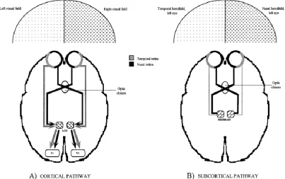
asymmetrically by the human eye, in terms of both resolution, and visual hyperacuity (Fahle, 1987). The human visual system is contralaterally represented in the body’s
morphology (Figure 1), whereby visual input from the left and right visual field passes
through to its contralateral primary visual cortical areas (Felten & Shetty, 2011). This
asymmetry is also evident in lateral eye movements, whereby voluntary saccades
originating from the frontal eye fields cause a strong, rapid deviation of the eyes to
the contralateral side that is often accompanied by movement of the head and trunk
Figure 1. Diagram displaying the contralateral distribution of the human visual system (Figure adapted from Felten & Shetty, 2011).
Projections from the frontal eye fields travel downwards towards the pontine centre
for lateral gaze, from where impulses subsequently ascend through the medial
longitudinal fasciculus to cranial nerve nuclei responsible for ocular movement. The
route of these impulses is nearly fully decussated, with the frontal eye fields of one
hemisphere supplying the contralateral ocular nuclei, resulting in contralateral eye
movements (Merckelbach et al., 1990). The importance of the medial longitudinal
tract involved in the production of conjugate (i.e. combined) lateral eye movements is
evidenced by its presence in all vertebrates, as well as it being the first tract to
myelinate in humans (Willer, 1977); this implies that it is closely linked to reflex
reactions when experiencing environmental emotion-inducing stimuli (Willer, 1977).
Similarly, face musculature is contralaterally represented in the human body, which
might explain evidence for emotion-specific laterality effects in studies of posed facial
expressions reported in the literature (e.g. Yecker et al., 1999). Specifically, when
colleagues reported that negative posed facial expressions were more intensely
represented in the left hemiface, and positive expressions more intensely
represented in the right hemiface (1999).
One observation from the overview of the laterality literature in this section is that
lateralisation might address a specific functional need; lateralised activation specific
to positive and negative emotions mediated respectively by right/left ocular and
musculoskeletal symmetrical cross-laterality needs to occur so that we may be able
to respond to basic emotional stimuli that tap into instinctual reflexes as efficiently as
possible. Although Rolls had previously argued that a lateralised organisation may
not be evolutionarily economical (2002), and although the contralateral nature of
basic emotional perception might not be as elegant as the overall interhemispheric
communication required for more complex, higher-order cognitive functions (and possibly the processing of secondary/social affect), it does serve the specific – albeit somewhat crude – purpose of maintaining survival by ensuring the most efficient
behavioural response is selected and executed.
1.5 SUMMARY AND THESIS OUTLINE
To summarise, emotional lateralisation is evidently a complex automatic
process that has historically proven difficult to disentangle. So far, behavioural
findings towards directionality of lateralisation appear to be closely linked to the type
of methodology, task and stimulus used. Usage of this wide range of methods has
resulted in the lack of consensus as to whether lateralisation occurs for all types of
basic affect, and difficulties establishing whether some basic affect (i.e. negative) is
better suited to engaging and maintaining attention than others. Reports from the
neuroimaging literature consistently point towards the lateralised engagement of
subcortical structures in the processing of specific types of basic affect. Whilst
differential engagement of subcortical structures and activation routes has been well
reported, it still remains to be established how this subcortical lateralisation which is
thought to tap into an attentional activation network of increased vigilance might
translate into observable behaviour.
impact of participant parameters and individual variability (i.e. personality traits such
as anxiety, and psychiatric conditions such as depression or schizophrenia) have
had a significant effect on the directionality of behavioural lateralisation patterns
reported. Up to date however, there has been little interest in establishing the key
factors modulating behavioural lateralisation through using consistent and
comparable methodologies. The current thesis addresses this gap by first developing
a behavioural paradigm to establish the behavioural manifestation of
emotion-specific lateralised processing, then examining whether subcortical lateralisation can
be effectively measured in terms of observable behaviour, and finally by adapting
this paradigm to incorporate both stimulus valence-enhancing factors, as well as participants’ personality traits which have been suggested to modulate this process.