1
DEVELOPMENT OF AN OPTICAL THERMAL HISTORY COATING SENSOR BASED ON THE OXIDATION OF A DIVALENT RARE EARTH ION PHOSPHOR
Alvaro Yanez-Gonzalez1*, [email protected] Enrique Ruiz-Trejo1, [email protected] Berend van Wachem1, [email protected] Stephen Skinner1, [email protected]
Frank Beyrau2, [email protected] Andrew Heyes3, [email protected]
1*Imperial College London, London SW7 2AZ, UK
2Otto-von-Guericke-Universität, Magdeburg 39106, Germany
2 ABSTRACT
The measurement of temperatures in gas turbines, boilers, heat exchangers and other components exposed to hot gases is essential to design energy efficient systems and improve maintenance procedures. When on-line measurements, such as those performed with thermocouples and pyrometers, are not possible or inconvenient, the maximum temperatures of operation can be recorded and measured off-line after operation. Although thermal paints have been used for many years for this purpose, a novel technique based on irreversible changes in the optical properties of thermographic phosphors, can overcome some of the disadvantages of previous methods.
3 INTRODUCTION
Energy efficiency has become increasingly important in recent years. In many engineering components, such as gas turbines, boilers, heat exchangers and fuel cells, efficiency is closely related to the temperature of operation. Accurate measurement of the temperature profiles on surfaces exposed to hot gases is thus essential to increase efficiency, but also to identify components that have been exposed to temperatures outside the design limits so that maintenance procedures can be applied thus reducing system failures.
Several methods are currently available to perform temperature measurements, both in real time (thermocouples, pyrometers) and after operation (thermal paints, thermal crystals). An alternative technique involves the use of thermographic phosphors, which consist of a ceramic host material doped with a rare earth element that emits phosphorescence when excited with a suitable light source. The temporal and spectral characteristics of the phosphorescent emission of these materials can be related to the temperature. These materials have been investigated for on-line temperature measurements with application to surfaces [1-4] and gas flows [5-8]. The off-line version of thermographic phosphors, which is the subject of this work, was recently devised by Feist et al. [9]. In this new technique, irreversible changes in the optical properties of thermographic phosphors are used to record the conditions to which they have been exposed that can be read after operation. Although the sensor cannot be used again similarly to other off-line sensors, the off-line technique presents the advantage that it does not require physical or optical access to the component being measured during operation, and therefore it is simpler to implement. Off-line thermographic sensors record a measure of the thermal exposure, which is a combination of temperature and time so that steady state exposures of known duration are necessary to obtain reliable measurements.
4
possibility of saving up to £3m in engine development programs by using this new technique [11].
Irreversible changes in divalent rare earth ion phosphors have been previously identified, and in particular those due to oxidation of europium in BAM:Eu can be used as an indication of the temperature up to at least 1200 °C [12-14]. BAM:Eu is a highly efficient phosphor commonly used as the blue emitter in fluorescent lamps and plasma display panels (PDPs). Given its availability as a standard product and its high quantum efficiency it has been used as a thermographic phosphor both in the on-line and off-line versions [refs].
In the present paper, BAM:Eu has been deposited as a thermal history sensor coating by the screen printing method onto Inconel 625 substrates, and its temperature sensing capabilities have been investigated in the range from 800 °C to 1100 °C. Ionic diffusion of elements from the substrate alloy into the BAM lattice has been identified in single-layer coatings by means of spectroscopy and material characterisation techniques. Element diffusion, which was responsible for variations in the optical properties of the phosphor coating due to quenching of the europium emission and appearance of new emission lines, was prevented by the use of a suitable diffusion barrier. The successful application of a zirconia-based layer as a diffusion barrier has been demonstrated and the sensitivity achieved with dual-layer coatings was comparable to that of the commercial powder.
METHODS
Coating Manufacture
5
Once the components were completely mixed, the ink was introduced in an ultrasonic bath for 20 minutes to eliminate agglomerates. If lumps were still observed, the ink was then passed through a triple roll mill for 20 minutes. After that, the ink was stored in a refrigerator.
The ink was printed onto the substrate (Inconel 625 10x10x1 mm tiles) through a screen (8x10 325 45° 5m E04 EXP, MCI Precision) with a 20x20 mm square as the printable area. The ink was at room temperature before deposition. After a single layer was printed, the sample was placed inside an air furnace at 100 °C for several hours to dry. After drying, a second layer could be printed on top of the previous one if required.
Once all the required layers were printed, the samples were sintered at 1150 °C for 2 hours in a reducing atmosphere (10 % of H2 and 90 % of N2) to avoid oxidation of the Eu ion. In order to avoid delamination, the heating and cooling rates were kept at 3 °C/min.
Characterisation
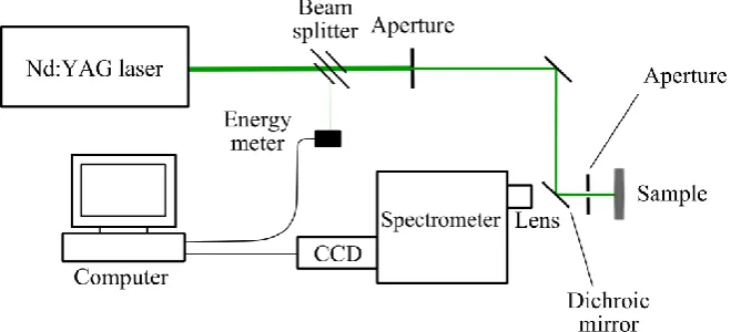
Luminescence. The luminescence properties of the samples were examined at room temperature with the set-up shown in Figure 1. Excitation of the samples was achieved by using the fourth harmonic (266 nm) of a pulsed Nd:YAG laser (Quanta-Ray LAB-150, Spectra Physics), with a repetition rate of 10 Hz and a pulse width of 5-7 ns. The laser energy was maintained constant at approximately 1 mJ/pulse, as measured by an energy meter (PEM-45K, Radiant Dyes), and the laser spot diameter was fixed to 4 mm.
6 Figure 1. Schematic of luminescence detection set-up.
Material characterisation. The coatings manufactured in this work were examined by Scanning Electron Microscopy (SEM) and X-Ray Diffraction (XRD) analysis. The SEM (S-3400N, Hitachi) permits to perform semi-quantitative Energy-Dispersive X-Ray Spectroscopy (EDS), which allows estimating the elemental composition of the sample. The preparation of the coating samples for cross section analysis was crucial in order to obtain a flat surface characteristic of the coating. The samples were mounted in a black phenolic resin (Conducto-Mount, MetPrep) in a hot mounting press (CitoPress-1, Struers). This resin wraps the coating and makes easier the following processes of grinding and polishing the sample surface. Since the resin is conductive, it also facilitates the SEM analysis by reducing the charging effects. The sample surfaces were ground using silicon carbide paper from a grit size of 400 to 2500, and finally polished using a monocrystalline diamond suspension (down to 3 µm).
XRD analysis was performed in a bench machine (D2 Phaser, Bruker) that uses CuKα radiation (1.5418 Å) and permits the scanning 2θ angles from 5 ° to 90 °.
RESULTS AND DISCUSSION Powder preliminary studies
7
the most intense peak of the Eu3+ emission located at 611 nm and the broad band emission peak of Eu2+ at 445 nm. This ratio increases continuously from temperatures of 800 °C up to 1200 °C for the commercial powder used in this study as shown in Figure 3.
At temperatures below 700 °C, the emission from Eu3+ is very weak and cannot be reliable used to define an intensity ratio. In fact, oxidation of BAM:Eu is only reported to start above 500 °C [Hirosawa 2004]. Above 1200 °C, nearly all the Eu2+ emission is vanished and similarly measurements cannot be used to define the intensity ratio. However, it was demonstrated in [ref] that modification of the dopant concentration affects the resistance of the phosphor to oxidation and thus can be used to tune the temperatures at which the sensor is most sensitive to changes in temperature.
In this work, the powder is ball milled as part of the process of coating manufacture in order to reduce the particle size and facilitate the deposition process. The first step in the oxidation of Eu is the adsorption of oxygen on the surface of the BAM:Eu particles [15] and is thus affected by the specific surface area of the powder. A reduction of particle size such as that caused by the milling process increases the specific surface area available and thus increases adsorption. The milled powder is therefore expected to suffer a higher degree of degradation compared to the commercial powder at the same temperature, which is confirmed by the results shown in Figure 3. The intensity ratio of the ball-milled powder after heat treatment at several temperatures in the range 800 – 1200 °C is higher in value than that of the commercial powder, but the sensitivity of the measurand remains nearly constant for both types of powders.
[image:7.595.76.318.531.690.2]8
Figure 3. Intensity ratio of commercial powder and ball milled commercial powder for various heat treatment temperatures and 20 minutes exposure.
Single-layer coating manufacture
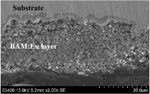
Single-layer coatings were first manufactured by a single pass during the screen printing process. These coatings were analysed by XRD to confirm the presence of the BAM phase and the cross section examined by SEM to study the thickness and microstructure of the coatings. The diffraction patterns of the as-deposited coatings, which are shown in Figure 4, are mainly identified with the BAM phase. This indicates that no structural change in the phosphor occurs that could be identified by this technique. The diffraction peaks of the cubic phase of the substrate are also detected with lower intensity at 43.6 °, 50.8 ° and 74.6 °.
[image:8.595.76.317.84.270.2]9
Figure 4. XRD peaks of the sample and the identified phases of BAM and nickel-based alloy substrate.
Figure 5. SEM cross-section image of the BAM:Eu screen printed coating.
Element diffusion
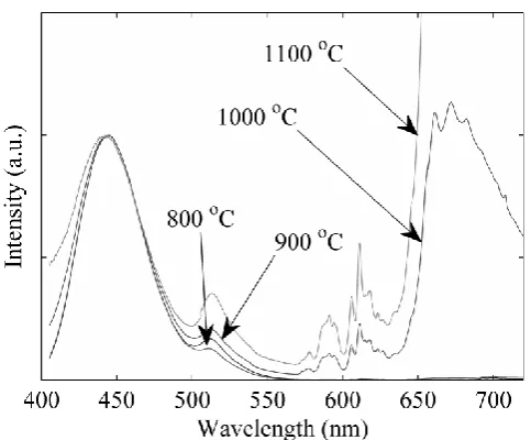
Single-layer screen printed coatings were heat treated for 20 minutes in air at temperatures of 800 °C, 900 °C, 1000 °C and 1100 °C. The emission spectra of these coatings were recorded at room temperature and are shown in Figure 6 normalised to the broad band peak at 445 nm.
Emission spectra of these samples are significantly different compared to the spectra of the commercial powder heat treated in air shown in Figure 2. An emission peak at 513 nm can be now observed whose intensity increases with heat treatment temperature relative to the broad band. A similar peak at this location has been reported previously in emission spectra of
Substrate
[image:9.595.70.310.359.510.2]10
co-doped BAM:Eu,Mn [17, 18], which first suggests the diffusion of transition metal ions from the substrate alloy into the BAM lattice.
In addition to the narrow emission peaks of Eu3+ in the range from 575 nm to 725 nm, a broad bad extending from 640 nm to at least 725 nm can now be observed, which exhibits a much larger intensity than the emission from Eu3+. Eu3+ emission is likely to be quenched by energy transfer to the optical centres responsible for this new broad band emission. Several transition metal ions are known to emit luminescence in the red and near infra-red. Mn4+ emits several broad lines in the range from 620 nm to 700 nm in various host materials [19]. Cr3+ presents emission in several hosts between 680 nm and 720 nm [19-22]. Finally, Fe3+ also exhibits luminescence at wavelengths above 680 nm [19, 23].
[image:10.595.89.331.436.636.2]The possibility of diffusion of transition metal ions in the BAM lattice is supported by the high values of the cation mobility in β-alumina for Ba2+ and Sr2+, this last one with almost the same ionic radius as Eu2+ [16].Furthermore, cation mobility in the conduction layer of BAM:Eu has been previously studied with regards to thermal degradation of the phosphor [16], where diffusion of Eu2+, Eu3+ and Ba2+ ions was measured by X-ray photoelectron spectroscopy (XPS).
Figure 6. Normalised emission spectra of screen printed BAM:Eu single-layer coatings after heat treatment in air at 800 °C, 900 °C, 1000 °C and 1100 °C.
11
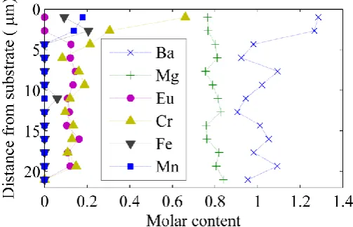
detected elements was then normalised to the content of aluminium (Al10), which was considered to remain constant in the BAM lattice and the results are shown in Figure 7. This assumption is reasonable since Mn is known to substitute for Mg in the spinel block of the BAM lattice [18]. Cr3+ and Fe3+ have similar ionic radii and thus are expected to occupy a similar site within the crystal lattice [24].
[image:11.595.76.328.433.595.2]In Figure 7, the content of Ba oscillates around 1 mol at all locations in accordance with the chemical formula of BAM. The content of Mg is however lower than expected from the stoichiometry of BAM, and it oscillates around 0.8 mol. This could easily be explained by substitution of Mg by transition metal ions in the spinel block as mentioned before. All three elements Cr, Fe and Mn are detected by EDS. Presence of Fe and Mn is only significant at locations close to the substrate, with levels close to 0.15 mol. At distances longer than 2.5 μm these two elements are not detected by this technique. Cr ion, however, is detected at all locations across the coating. Near the substrate, there is a steep gradient of concentration from 0.7 mol to approximately 0.13 mol at distances longer than 5 μm, which is indicative of diffusion. Finally, the Eu content is nearly constant with a value of 0.13 mol at all positions, which is an expected value for this dopant in commercial BAM:Eu [25].
Figure 7. Molar content of various elements calculated by EDS at different locations across the thickness of the coating after heat treatment of the sample in air at 1100 °C for 20 minutes.
12
the broad band in the wavelength range 640 – 725 nm could be the result of contributions from Mn4+, Cr3+ and/or Fe3+.
In order to discern the effect of each of these ions, samples were prepared by mixing 1 mg of the commercial powder with 2 wt % powder of one of each of the transition metal ions Mn, Cr and Fe. Each of the three mixtures was thoroughly ground to ensure a homogeneous mixture and then heat treated for 20 minutes in air at 1000 °C. The emission spectra of these samples were then measured at room temperature and they are shown in Figure 8 in the wavelength range from 550 nm to 725 nm. The emission spectra of the commercial powder, ball-milled powder and screen printed coatings (note that dual-layer coatings will be introduced in the following section) heat treated under the same conditions of 20 minutes exposure at 1000 °C are shown for comparison.
Analysis of spectra in Figure 8 confirms that the presence of Cr and Fe ions does not affect the emission spectrum, as these two mixtures exhibit the same emission peaks as the commercial powder alone. On the other hand, the mixture with Mn exhibits a similar emission to that of the commercial powder up to 640 nm, but an additional broad band is visible at longer wavelengths. This broad band extends over the same wavelengths as that observed in the single-layer coatings, although its intensity is much lower compared to the coating samples. This result indicates that Mn2+ present in fresh samples might oxidise to Mn4+ during the heat treatment thus giving rise to the broad band emission between 640 – 725 nm and quenching the Eu3+ emission.
13
Figure 8. Normalised emission spectra of Eu3+ of various mixtures of commercial powder and transition metal ions, the ball-milled powder and single and dual-layer coatings.
Dual-layer coating manufacture
A dual-layer coating was envisaged that incorporates an intermediate layer between the substrate and the phosphor coating. This layer should act as a diffusion barrier, be chemically compatible with the phosphor and the substrate and be easily deposited by screen printing. Potential candidates might be for example alumina and zirconia, being the latter selected due to the availability of a suitable ink for deposition.
14
Figure 9. SEM cross-section image of the BAM:Eu screen printed coating with an interlayer of SSZ.
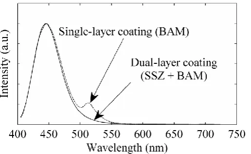
The emission spectrum of the coating after the manufacture process was compared to that of the single-layer coating and it is shown in Figure 10. The broad band emission corresponding to Eu2+ is present in both coatings, but the peak at 513 nm, which was ascribed to the presence of Mn2+ in the single-layer coating, is missing in the dual-layer coating. This confirms the possibility to use a SSZ interlayer to stop diffusion of transition metal ions from the substrate.
The emission spectrum of the dual-layer coating after heat treatment in air at 1000 °C, which was shown previously in Figure 8, suggests that some diffusion is still present as the broad band ascribed to emission from Mn4+ is clearly visible. However, the relative intensity of this broad band is drastically reduced compared to that of the single-layer coating, which indicates a much lower degree of diffusion.
[image:14.595.75.333.569.727.2]15 Comparison of intensity ratio
The diffusion of transition metal ions from the substrate causes changes in the emission spectra of coating samples. These changes affect the shape of the spectra and the relative intensities of the peaks. Therefore, temperature measurements based on the intensity ratio as defined in previous sections may be affected by this diffusion.
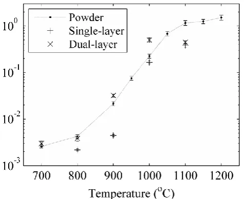
The intensity ratio was measured for samples whose emission spectra are plotted in Figure 8. Therefore, the intensity ratios at 1000 °C shown in Figure 11 correspond to those samples. Analysis of ρ for powder samples mixed with transition metal ions suggests that presence of Cr does not affect the value of ρ. Since the emission spectrum of this sample also showed a similar shape to that of the commercial powder, it can be concluded that Cr does not play a significant role in the luminescence process. The intensity ratio of the sample mixed with Fe has a lower value than that of the commercial powder. Therefore, although Fe does not affect the emission spectrum, it quenches the Eu3+ emission thus leading to a lower value of ρ. The sample mixed with Mn presents a value of ρ almost an order of magnitude lower than that of the commercial powder, which was expected by the high level of quenching observed in the emission spectrum of this sample.
16
Figure 11. Intensity ratio of various mixtures of commercial powder and transition metal ions, the ball-milled powder and single and dual-layer coatings.
17
Figure 12. Intensity ratio of single and dual/layer coatings compared to the commercial powder calibration curve.
CONCLUSIONS
Thermographic phosphor BAM:Eu has been applied as a coating sensor and the possibility to measure surface temperatures successfully demonstrated for the first time.
Single and dual-layer coatings of thermographic phosphor BAM:Eu have been deposited on Inconel 625 substrates by the screen printing method with a thickness that varied from 15 – 20 μm. This thickness proved to be sufficient to perform luminescence measurements even after heat treatment, where the luminescence is largely reduced due to oxidation of Eu2+. The microstructure of the coating consisted of a stack of particles with numerous voids, which indicates that sintering of the particles cannot be achieved at the relatively low temperatures of the annealing.
[image:17.595.85.323.83.277.2]18
spectra. Other minor changes in the emission spectra were ascribed to the ball-milling of the powder during coating manufacture.
The sensing capabilities of the sensor were also investigated by using the intensity ratio between the emission lines of Eu3+ (611 nm) and Eu2+ (445 nm). Analysis of powder samples heat treated at 1000 °C for 20 minutes revealed that diffusion of Mn caused a drastic reduction of the intensity ratio compared to the commercial powder. Although diffusion of Fe did not affect the emission spectra, it affected the emission efficiency and also induced a reduction in the intensity ratio. The presence of Cr showed no effect on the optical properties of the phosphor.
The emission spectra and thus the intensity ratio of single-layer coatings were greatly affected by diffusion of elements from the substrate. A dual-layer coating in which the phosphor layer was separated from the substrate by a suitable diffusion barrier was manufactured. An interlayer made of zirconia proved successful in preventing diffusion. The intensity ratio of dual-layer coatings showed a similar sensitivity to that of the powder. Therefore, the use of a diffusion barrier is essential in the performance of BAM:Eu as a sensor coating.
While the present study focuses on the intensity ratio technique, further work should look into the lifetime decay method, which has been successfully demonstrated for BAM:Eu in previous publications [ref]. Additionally, the presence of transition metal ions diffusing from the substrate into the phosphor coating shown in this work is, to the knowledge of the authors, the first experimental evidence of the diffusion mechanism described in the patent by Feist et al. [9]. The possibility to use this mechanism to perform successful temperature measurements needs to be further investigated.
NOMENCLATURE
BAM:Eu Europium-doped barium magnesium aluminate (BaMgAl10O17:Eu) CCD Charge-coupled device
EDS Energy dispersive X-ray spectroscopy
Nd:YAG Neodymium-doped yttrium aluminium garnet PDP Plasma Display Panel
REACH Registration, Evaluation, Authorisation and Restriction of Chemicals SEM Scanning electron microscopy
19
XPS X-ray photoelectron spectroscopy XRD X-ray diffraction
ρ Intensity ratio
ACKNOWLEDGEMENTS
The authors would like to thank the financial support from The Energy Futures Lab.
REFERENCES
1. Tobin, K., et al., High-temperature phosphor thermometry of rotating turbine blades.
AIAA Journal, 1990. 28(8): p. 1485-1490.
2. Allison, S.W. and G.T. Gillies, Remote thermometry with thermographic phosphors: Instrumentation and applications. Review of Scientific Instruments, 1997. 68(7): p. 2615-2650.
3. Khalid, A.H. and K. Kontis, Thermographic phosphors for high temperature measurements: Principles, current state of the art and recent applications. Sensors, 2008. 8(9): p. 5673-5744.
4. Feist, J., et al., Application of an Industrial Sensor Coating System on a Rolls-Royce Jet Engine for Temperature Detection. Journal of Engineering for Gas Turbines and Power, 2013. 135(1): p. 012101-012101-9.
5. Omrane, A., et al., Simultaneous 2D flow velocity and gas temperature measurements using thermographic phosphors. Applied Physics B, 2008. 92(1): p. 99-102.
6. Fond, B., et al., Simultaneous temperature, mixture fraction and velocity imaging in turbulent flows using thermographic phosphor tracer particles. Optics Express, 2012. 20(20): p. 22118-22133.
7. Abram, C., et al., High-speed planar thermometry and velocimetry using thermographic phosphor particles. Applied Physics B-Lasers and Optics, 2013. 111(2): p. 155-160.
8. Fond, B., C. Abram, and F. Beyrau, On the characterisation of tracer particles for thermographic particle image velocimetry. Applied Physics B, 2015: p. 1-7.
9. Feist, J., J. Nicholls, and A. Heyes, Determining thermal history of components, 2007. 10. REACH, Registration, Evaluation, Authorisation and Restriction of Chemicals
(REACH), Regulation (EC) No 1907/2006 of the European Parliament and of the Council, in OJ of the European Union L 396/1 18.12.2006.
11. Feist, J.P., et al. Off-Line Temperature Profiling Utilising Phosphorescent Thermal History Paints and Coatings. in ASME Turbo Expo 2014: Turbine Technical Conference and Exposition. 2014. American Society of Mechanical Engineers.
12. Rabhiou, A., A. Kempf, and A. Heyes, Oxidation of divalent rare earth phosphors for thermal history sensing. Sensors and Actuators B-Chemical, 2013. 177: p. 124-130. 13. Yañez Gonzalez, A., et al., Reusable Thermal History Sensing via Oxidation of a
Divalent Rare Earth Ion Based Phosphor Synthesized by the Sol-Gel Process. Heat Transfer Engineering, 2015. 36(14-15): p. 1275-1281.
14. Yañez Gonzalez, A., et al., A detailed characterization of BaMgAl10O17:Eu phosphor
20
15. Bizarri, G. and B. Moine, On BaMgAl10O17:Eu2+ phosphor degradation mechanism:
thermal treatment effects. Journal of Luminescence, 2005. 113(3-4): p. 199-213. 16. Lacanilao, A., et al., Structural analysis of thermal degradation and regeneration in
blue phosphor BaMgAl10O17:Eu2+ based upon cation diffusion. Solid State Ionics,
2013. 253: p. 32-38.
17. Smets, B. and J. Verlijsdonk, The luminescence properties of Eu2+-and Mn2+-doped
barium hexaaluminates. Materials Research Bulletin, 1986. 21(11): p. 1305-1310. 18. Yang, P., G.-Q. Yao, and J.-H. Lin, Energy transfer and photoluminescence of
BaMgAl10O17 co-doped with Eu2+ and Mn2+. Optical Materials, 2004. 26(3): p.
327-331.
19. Shionoya, S. and W.M. Yen, Phosphor handbook. 1999: CRC Press.
20. Qiao, B., et al., Study on ZnGa2O4:Cr3+ a.c. powder electroluminescent device.
Materials Letters, 2007. 61(2): p. 401-404.
21. Chao, C.-C., Charge-transfer luminescence of Cr3+ in magnesium oxide. Journal of
Physics and Chemistry of Solids, 1971. 32(11): p. 2517-2528.
22. Wojtowicz, A., Luminescence of Cr3+ in kyanite. Journal of Luminescence, 1991.
50(4): p. 221-230.
23. Pott, G. and B. McNicol, Zero‐Phonon Transition and Fine Structure in the Phosphorescence of Fe3+ Ions in Ordered and Disordered LiAl
5O8. The Journal of
Chemical Physics, 1972. 56(11): p. 5246-5254.
24. Shannon, R., Revised effective ionic radii and systematic studies of interatomic distances in halides and chalcogenides. Acta Crystallographica Section A: Crystal Physics, Diffraction, Theoretical and General Crystallography, 1976. 32(5): p. 751-767.
25. Kim, K.-B., et al., Structural and optical properties of BaMgAl10O17:Eu2+ phosphor.