City, University of London Institutional Repository
Citation
:
Kyriacou, P. A. and Shafqat, K. (2012). Heart Rate Variability (HRV) andcardiovascular dynamic changes during local anesthesia. In: Kamath, M.V., Watanabe, M.A. and Upton, A.R.M. (Eds.), Heart Rate Variability (HRV) Signal Analysis: Clinical Applications. (pp. 221-240). CRC Press. ISBN 1439849803
This is the draft version of the paper.
This version of the publication may differ from the final published
version.
Permanent repository link:
http://openaccess.city.ac.uk/3538/Link to published version
:
Copyright and reuse:
City Research Online aims to make research
outputs of City, University of London available to a wider audience.
Copyright and Moral Rights remain with the author(s) and/or copyright
holders. URLs from City Research Online may be freely distributed and
linked to.
City Research Online: http://openaccess.city.ac.uk/ [email protected]
Heart Rate Variability (HRV) and cardiovascular dynamic changes
during local anesthesia
P A Kyriacou and K Shafqat
School of Engineering and Mathematical Sciences, City University London, London UK
P A Kyriacou, Ph.D
School of Engineering and Mathematical Sciences City University London
Northampton Square EC1V 0HB
London, UK
Tel:
+44 (0)20 7040 8131
Email: [email protected]
Key words
Contents
Heart Rate Variability (HRV) and cardiovascular dynamic changes during local anesthesia ... 1
Abstract: ... 3
1
Cardiovascular and HRV changes during local anesthesia in animal studies ... 4
2
Effects of local anesthesia on the HRV in dentistry... 6
3
Comparison of cardiovascular and hemodynamic effects of local vs general anesthesia .... 10
4
HRV and cardiovascular effects of local anesthesia ... 13
5
Discussion and Conclusions: ... 23
Abstract:
The analysis of Heart Rate Variability (HRV), the beat to beat fluctuation of the heart rate, is a
non-invasive technique with a main aim in gaining information about the autonomic neural regulation of the heart. Assessment of heart rate variability has been shown to aid clinical diagnosis and intervention strategies. However, due to the complex nature of different mechanisms that affect the HRV and the
large number of signal processing techniques that have been used for HRV analysis there are quite a few conflicting reports on HRV that perhaps impede its use as a reliable clinical tool. This chapter will provide an overview of some of the work carried out on animals and humans in an effort to investigate the effect of local anesthesia (LA) on HRV and cardiovascular dynamics. Also, another part of this chapter
will deal with various studies where the use of local anesthesia has been compared with general anesthesia (GA) for different surgical procedures. The last section of the chapter focuses and reports on the work performed to investigate the effect of different local anesthetic techniques and anesthetic
1
Cardiovascular and HRV changes during local anesthesia in animal studies
The effect of different anesthetic drugs on cardiovascular hemodynamics have been studied since the 1960s (37,67-69). However, most of these studies focus their analysis on the effect of local anesthetic drugs on heart rate, blood pressure and other cardiovascular and hemodynamic parameters, without
any in depth investigation on Heart Rate Variability (HRV) and/or Blood Pressure Variability (BPV). This could represent a major drawback, as small transient changes occurring due to the application of local anesthetic drugs could be missed by examining the above mentioned global variables. Nevertheless these studies could be useful in providing some indication of hemodynamic and cardiovascular effects
[image:5.612.70.534.419.705.2]due to local anesthesia (LA). Some of the studies carried out on animals to analyze the effect of LA are summarized in Table 1.
Table 1: Animal studies involving different local anesthetic drugs
Authors Animal model
Local anesthesia (LA) Summary
Fogarty et al (20) Rabbits Pentazocine Transient increase and subsequent decrease in arterial pressure. Abnormalities of the QRST complex.
Basil et al. (5) Cuinea-pigs, cats and dogs
Hydrochloride (M&B 17,803A)/ practolol and propranolol
Propranolol more potent than M&B 17,803A and practolol
Munson et al. (46) Rhesus monkeys
Etidocaine, bupivacaine, and lidocaine
Central nervous toxicity of Lidocaine 4 times less than etidocaine and bupivacaine.
Kotelko at al. (36) Sheep Bupivacaine and lidocaine Arrhythmias more common in the animals that received bupivacaine.
Hotvedt at al. (30) Dogs Bupivacaine Bupivacaine can enhance susceptibility to reentrant arrhythmias.
Gerard et al. (23) Mongrel dogs
Bupivacaine and/or diazepam
major neural blockade.
Edouard et al. (18) Dogs Lidocaine after an intravenous bolus of bupivacaine or normal saline with concurrent 40 minute infusions of equihypotensive doses of verapamil.
Regional anesthesia should be applied with caution in patients treated with calcium entry blockers.
DeKock at al. (35) Rats Bupivacaine
5 intravenous digoxin or saline
The threshold doses of bupivacaine toxic effects and its serum concentrations were lower in the digoxin group.
Pitkanen et al. (52) Rabbits Ropivacaine, bupivacaine and lidocaine
Bupivacaine is cardio depressant and arrhythmogenic.
Freysz at al. (21) Pigs Bupivacaine Bupivacaine should be used with caution in the condition of ischemia.
Oliveira at al. (50) Rats Prilocaine chloridate (alone and mixed with felypressin and Epinephrine (E))
E must be used with Prilocaine
Stewart et al. (64) used HRV analysis to study the role of the Autonomic Nervous System (ANS) in mediating eye temperature responses during painful procedures (sham handling or surgical castration) using thirty-four month old bull calves. The maximum eye temperature, Heart Rate (HR), and HRV were
recorded continuously from 25 minutes before to 20 minutes after castration. The results showed that LA reduced the response to painful procedure but did not completely eliminate this response. It was concluded that HR, HRV, and infrared thermography measurements when used together could provide a noninvasive means to assess ANS responses as indicators of acute pain.
Due to the highly vascular nature of the application area local anesthesia during oral surgery could significantly affect the cardiovascular dynamics. For this reason, a significant amount of work is presented in the literature that deals with the use of local anesthesia in dentistry. Some of this work will
2
Effects of local anesthesia on the HRV in dentistry
The effects of anesthetic drugs given during dental procedures can vary significantly depending on the
[image:7.612.72.545.207.709.2]condition of the patient. Table 2 summarizes some key studies which describe cardiovascular and/or hemodynamic changes that could occur during dental anesthesia.
Table 2: Effects of anesthetic drugs in dentistry
Authors Study Summary
Hass et al. (26) LA during dental treatment in patient with and without cardiac disease.
Cardiac patients suffer with significantly higher ST segment depression during tooth extraction.
Cintron et al. (12) Cardiovascular effects and safety of dental anesthesia in patients with recent
myocardial infarction.
Limited dental anesthesia and dental interventions were tolerated by patients with recent myocardial infarction.
Rengo et al. (54) Effect of LA (mepivacaine hydrochloride 2% plus adrenalin 1:200.000) on cardiopathic patients.
HR, Systolic Blood Pressure (SBP) and Diastolic Blood Pressure (DBP) significantly increased during tooth extraction.
Davenport et al. (14) Hemodynamic effects of 2% lidocaine with and without E 1:100,000 on patients with cardiovascular disease.
The cardiac effects of local anesthetics containing E are small and they can be safely used in patients with stable cardiovascular disease.
Goldstein et al. (24) The effects of sedation with intravenous diazepam and of inclusion of E with the local anesthetic during molar extraction.
Increase in HR (25%), SBP (13%) and CO (34%). Diazepam sedation abolished the
Norepinephrine (NE) response without significantly affecting the HR or SBP responses. Cardiac output and mean plasma E were increased fivefold with the inclusion of E
Chernow at al. (11) Hemodynamic effects of LA following inferior alveolar nerve block with E-and NE-containing lidocaine hydrochloride.
Lidocaine alone caused no change in MAP or HR. Lidocaine with E caused a transient increase in HR and no change in MAP.
Köhler et al. (34) Cardiovascular risk due to the presence of E in LA 2.0 2% lidocaine with and without 20 or 80 E.
Lidocaine caused no changes. Lidocaine with 20 E caused increase in plasma E concentration and HR and decrease MAP. Similar changes occur earlier with 80 of E.
Hempenstall et al. (2 8)
Comparison of LA and GA in dental surgery.
The reports that were discussed so far focused on the hemodynamic changes that occur during dental LA on the basis of blood pressure and heart rate changes. The changes occurring in parasympathetic
activity due to LA were studied by Kawano et al. (32) in 52 patients undergoing dental treatment. Coefficient of variation of the R-R interval (CVR-R=SD/MEANx100%) was used as an index of parasympathetic activity. Comparisons between a control group and another group receiving atropine sulfate were made. The results showed consistently low CVR-R values, higher SBP, DBP, HR and longer
recovery time in the sulfate group compared to the control group. Matsumura et al. (56) also studied the changes caused by LA (2% lidocaine containing 1:80,000 epinephrine) on HR, Blood Pressure (BP) and HRV during dental surgery. The study included 40 patients (mean age: 42.7+/‐3.0 years), who
underwent tooth extraction. A Holter monitor was used to record the ECG signal. The power in Low Frequency (LF=0.041 to 0.140 Hz), High Frequency (HF=0.140 to 0.50 Hz), and Total Power (TP=0.000 to 4.000 Hz) was calculated, and the ratio of power between LF and HF region (LF:HF) and normalized
power in the HF region ( / 00) were used as indices of sympathetic and parasympathetic activities, respectively. The results showed that after the administration of the local anesthetic, both blood pressure and pulse rate increased. Patients that were 40 years of age or older experienced an increase in BP while the LF:HF ratio decreased. In contrast, in patients that were <40 years of age, the
%HF decreased and the LF:HF increased indicating that regulation of the ANS during dental surgery differs between younger and older patients.
In another study Carrera et al. (10) compared three anesthetics drugs in combination with different
vasoconstrictors in the surgical removal of lower third molars. The study consisted of three groups (n=15) split according to the anesthetic solution and associated vasoconstrictor administered (4% articaine + epinephrine 1:200,000; 3% mepivacaine without vasoconstrictor; and 3% prilocaine + felypressin 1:1,850,000). Heart rate, SBP and DBP, and oxygen saturation were recorded
were found to be more stable with articaine + epinephrine 1:200,000, although the three studied
solutions caused no significant hemodynamic changes with respect to the basal values.
The changes in the cardiovascular dynamics during dental surgery in 18 hypertensive patients were compared with age and sex matched control group (normotensive patients) by Miura et al. (43). Physiological parameters of HR, BP and HRV were monitored before and during the dental surgery. From
the HRV analysis, the LF, HF, and TP spectral powers were calculated, and the ratio of power (LF:HF) and %HF were used as indices of sympathetic and parasympathetic activities. The increase in BP during tooth extraction did not different significantly between the two groups. Administration of anesthesia significantly decreased the %HF in normotensive patients (before vs. after anesthesia; 22.3 +/‐ 2.4 vs.
3.8 +/‐ 2.7%, ). In contrast, the LF:HF significantly decreased during LA and tooth extraction in hypertensive patients. These results indicate that pressor response induced by tooth extraction did not
differ between the two groups.
Ishida et al. (31) studied the effects of LA and periodontal surgery on autonomic nervous activities using power spectral analysis of HRV on ten patients undergoing periodontal surgery. Heart rate and BP were also measured during LA and surgery. The results showed that the LF:HF ratio increased significantly before and during LA, about three minutes after LA, and before surgery. Peak of the plasma epinephrine
concentration occurred almost simultaneously with the increase in LF:HF ratio after the administration of LA containing epinephrine. Mental stress was the contributing factor in the increase in LF:HF ratio before anesthesia while, the later increase was due to the physical stress and epinephrine presence in
anesthesia. However, there were no significant changes in the power of the HRV bands during periodontal surgery.
In 2001 Nakamura et al. (47) also studied the changes in HRV, BP and blood variables in eleven normotensive patients (age, 22.5+/‐0.7 years) during dental surgery. Baseline readings of these variables
2% Lidocaine with 1:80,000 adrenaline. The results showed that during dental surgery there was a significant increase in SBP (+ 0.8+/‐3.5 mm g) but it was not correlated with baseline SB or with
24 hour averaged BP, LF:HF ratio or HF power. Hence, it was concluded that ambulatory measurements of blood pressure and HRV over 24 hours cannot predict the responses of BP during dental surgery. Blood pressure, HR and temperature variability during periodontal surgery was also analyzed by Gedik et al. (22) in 127 healthy patients (43 males, 84 females) aged 26+/‐ 2 years. Patients were
divided into four groups (gingivectomy, periodontal flap surgery, frenectomy and curettage) and were anesthetized using Ultracain DS containing 0.06 adrenaline. A significant decrease was observed in
all parameters (blood pressure: systolic .3+/‐20. , diastolic 67.7+/‐ 3. , pulse rate: 87.8+/‐ 4.9,
temperature: 36.3+/‐0.3) for all patients. owever, the changes significantly decreased after the operations (BP: systolic 05.9+/‐ 9.7, diastolic 62.6+/‐ .3, pulse rate: 84.0 +/‐ 3. , temperature: 36.2+/‐0.3). emale patients, without age differentiation, showed statistically significant decreases in all
parameters ( ).
The effect of epinephrine in local dental anesthesia in patients with coronary artery disease was analyzed in sixty-two patients aged (58.7+/‐8.8) by Neves et al. (48). Anesthesia was applied with 2%
lidocaine with epinephrine (E group) (n=30) and without epinephrine (NE group) (n=32). The results showed that there were no significant differences in the two groups with respect of BP, HR and number of arrhythmic episodes. Based on these results it was concluded that epinephrine could be employed safely during dental anesthesia for patients with coronary artery disease.
Hemodynamic changes during the surgical removal of lower third molars have also been studied more recently by Alemany et al. (2) in eighty normotensive individuals (40 females and 40 males) patients. Local anesthesia was applied using 4% articaine adrenalin (1:100.000). The parameters of SBP, DBP, HR,
and oxygen saturation (SpO2) were measured. Also, patient anxiety was determined using Corah's
by means of a visual analog scale. The results showed that female patients showed higher levels of anxiety. Heart rate and BP changes during molar extraction were within the normal limits. The SpO2
values showed no significant changes. These results showed that most of the cardiovascular changes could be associated with the anxiety and stress induced by surgery. A comparison between 2 ml of 2% lidocaine with clonidine (15 ) or epinephrine (12.5 ), used for the extraction of upper third
molar was also carried out by Brkovic et al. (7) in 40 patients. The results showed that 10 minutes after
surgery the HR and SBP reduced significantly in the lidocaine+clonidine group while HR increased significantly in the lidocaine+epinephrine group. Changes in all other hemodynamic parameters were similar in both groups. It was therefore concluded that lidocaine+clonidine combination could safely
replace lidocaine+epinephrine for intraoral infiltration anesthesia.
3
Comparison of cardiovascular and hemodynamic effects of local vs general
anesthesia
Due to its perceived advantages such as faster recovery time, fewer requirements of post-operative analgesia, cost effectiveness and more stable cardiovascular and hemodynamics conditions LA has been preferred over GA in many surgical procedures. A large amount of research has been carried out to
compare the use of local and general anesthesia for different surgical procedures. This section will focus in some of the studies carried out to compare the hemodynamic and cardiovascular changes caused by local and general anesthesia.
In 1990 Takolander et al. (65) studied 75 patients to compare the hemodynamics and cardiovascular
changes caused by LA and GA during coronary surgery. Arterial plasma catecholamines, BP and HR were determined before, during and after carotid endarterectomy. Patients were divided into three groups. The LA-group (n=28) received LA given as a cervical block with skin infiltration containing 200
containing 200 adrenaline, while, the GAo-group (n=15) received GA without skin infiltration. The results showed that plasma nonadrenaline (P-NA) levels were significantly higher in the LA-Group ( during anesthesia and surgery while it decreased in the GAo-group ( ) and remained
unaltered in the GAs-group. Incidences of hypotensive blood pressure reaction (SBP < 100 ; LA vs. GAo, ) were higher in the GAs-group (8 patients) as compared to the LA-group (2 patients)
and GAo (7 patients). Ten patients in the LA-group also showed a hypertensive blood pressure reaction.
These results indicate that both types of anesthesia have their disadvantages in patients with an increased risk for cardiovascular morbidity/mortality.
Børdahl et al. (6) also compared local and general anesthesia for laparoscopic sterilization by randomly
allocating 125 women to receive either LA or GA. Midazolam was used as premedication. For LA, lidocaine with adrenaline was infiltrated infraumbilically and bupivacaine was applied to each tube. In the GA group, alfentanil and propofol was used for intubation and atracurium was used as muscle
relaxant. The results showed that LA performed better than GA in terms of operating time, preoperative discomfort, equipment costs, postoperative abdominal pain, analgesics requirement and patient’s recovery time.
Comparison between LA and GA for cataract surgery in 169 patients was performed by
Campbell et al. (9). Oxygen saturation, BP and HR parameters were monitored during anesthesia and in immediate recovery period. The results showed that oxygen desaturation occurred at least once in 19% of the patients in the GA group compared to none in the LA group. In the GA group 61% of the patients
also experienced more than 30% decreased in systolic pressure. Based on these results the authors have concluded that there was no significant difference between the performances of LA and GA in cataract surgery.
The hemodynamic effect preoperative stressor events during rhinoplasty was also studied by
24 hours starting on the day before the operation and continuing throughout the procedure. All patients received 10 of 2% lidocaine with 1:80,000 adrenaline 15 minutes after intubation. Frequency domain
HRV parameters, HR and non-invasive BP were measured during the study. The results showed that mild to moderate tachycardia occurred in the majority of the patients before induction of anesthesia. Similar change was also detected after the infiltration of lidocaine/adrenaline and during lateral osteotomies. However, preoperative stressors (with the exclusion of GA induction, intubation, and extubation) did not
cause any significant changes in the BP. Tachycardia before induction was caused by the increase in the sympathetic activity due to the patient’s anxiety. hese results show that these patients would benefit from routine use of premedications and that a lidocaine/adrenaline combination is a safe addition to GA
in rhinoplasty patients.
In another study patient's stress response during asleep-awake craniotomy has been studied by Conte et al. (13) by quantifying the sympathovagal balance using HRV analysis. Twenty-one patients
aged (22 to 53 years) undergoing tumor resection with language testing were recruited for the study. Heart rate and SBP were collected at five time points: T1: preanesthesia; T2: dura mater opening; T3: cortical mapping; T4: subcortical mapping; T5: dura mater suturing. The patients were anesthetized
with propofol/remifentanil infusion and ventilated via laryngeal mask during T2, but were awakened for language testing at T3 and T4, and resedated with remifentanil during T5. At each of these five points the HRV frequency domain parameters of TP, power in Very Low Frequency (VLF), LF, HF band and LF:HF ratio were estimated. The results showed that compared to T1, significant increase in HR and BP
were observed from T3 through T5 ( ). The LF:HF ratio progressively increased, reaching significant level ( ) during T4. However, the ratio values returned to the level of T1 during the T5
individualize the protocol and the duration of the awaken phase according to the patient’s autonomic
response.
4
HRV and cardiovascular effects of local anesthesia
Local anesthesia has been extensively used in numerous surgical procedures and depending on the site and technique of application the amount of anesthetic dose could differ significantly. This could result in
varying cardiovascular and hemodynamic effects during the application of anesthesia. In the literature there are many studies in which different anesthetic drugs and/or anesthetic techniques have been compared. These studies, which will be the main focus of this section, have analyzed the effect of
different techniques and/or drugs on patient’s data such as R, B , eart Rate Variability (HRV), etc.
Barman et al. (4) undertook a study to determine intraoperatively the hemodynamic effects of LA of the carotid sinus nerve during carotid dissection in preparation for endarterectomy. In a control group (n=10) saline solution was infiltrated into the carotid bifurcation. In another group (n=10), 5 of
2% lidocaine hydrochloride was infiltrated. Heart rate and BP values were recorded at baseline and
interoperatively every 2 minutes during a 10-minute period. Interoperatively the lidocaine group showed higher increase in systolic pressure ( and mean pressure ( ). From these
results it was concluded that local anesthetic injection of the carotid sinus nerve before carotid
dissection and endarterectomy is unnecessary when nerve-sparing dissection is performed.
In another study Keyl et al. (33) investigated the stability in cardiac autonomic tone after GA or LA in 28 patients undergoing cataract surgery. Group 1 (n=14) received GA (premedication: clorazepate;
anesthetic induction: propofol, alfentanil, atracurium; anesthetic maintenance: isoflurane, alfentanil; airway management: laryngeal mask airway) and Group 2 (n=14) received LA (retrobulbar block with bupivacaine/mepivacaine). HRV frequency domain analysis was carried out intraoperatively and up to
reduced intraoperatively and increased slowly during the postoperative period. Postoperatively, the HR and the LF:HF ratio values were significantly increased in the LA group compared to the GA group. From
these findings it was concluded that during ophthalmic surgery, in terms of preoperative cardiac autonomic tone, GA has no disadvantage compare to LA. The hemodynamic and electrocardiographic responses to LA (midazolam) was studied by Middlehurst et al. (41) in 75 patients with heart disease. After splitting the patients into two groups, anesthesia was applied using lignocaine 2%, adrenaline
1:50,000, and vasopressin 0.25 IU, either alone or with midazolam. During the study the parameters relating to HR, BP, and ECG values were recorded, and the rate-pressure product and pressure-rate quotient were calculated as indicators for myocardial ischemia. Significant changes were observed in HR,
SBP and mean blood pressure due to sedation and anesthesia. For the anesthetic group the maximum value for the rate-pressure product and the minimum value for the pressure-rate quotient were 12168 (95% CI=1368) and 1.39 (95% CI = 0.04) respectively while, for the sedated group these values
were 9882 (95% CI=1226), and 1.13 (95% CI=0.06) respectively. Based on these results the authors concluded that the treatment could not be associated with significant ischemic risk.
The use of Total Intravenous Anesthesia (TIVA) was also compared with LA during cataract surgery by
Schwall et al. (57). Patients were randomly assigned to peribulbar local block (n=10) or TIVA (n=10). Propofol and alfentanil were used for TIVA. Parameters related to HR, BP, plasma concentrations of catecholamines, cortisol, and glucose were assessed at seven pre-, intra-, and post-operative time points. The results showed that LA cause no significant change in plasma concentrations of E, NE and
cortisol whereas, in the case of TIVA, plasma E, NE and cortisol decreased approximately by 66%, 51% and 61% respectively. Blood pressure and HR did not change significantly during LA while, SBP decreased by 30%, and heart rate by 12 bpm during TIVA. The results presented in this study showed that LA
In another study Dogru et al. (17) investigated the effect of high/low doses of epinephrine (E) during axillary brachial plexus block in 60 ASA I and II patients, which were divided randomly in three groups.
Patients in group 1 received 5 of saline containing 25 E and then 35 of 1.5% lidocaine; patients in group 2 received 5 of saline alone and then 200 of epinephrine mixed with 35 of 1.5% lidocaine; patients in group 3 received 5 of saline alone and then 35 of 1.5% lidocaine.
Starting from one minute after axillary injection the hemodynamic data were measured in an interval of
1 minute for a duration of 10 minutes. The results of the study indicated that complete anesthesia was achieved in 85% of the patients in groups 1 and 3 and 90% in group 2. Group 2 had a significantly longer duration of motor block compared to groups 1 and 3 while, analgesia duration was significantly longer in
groups 1 and 2 than in group 3. During the course of a 10 minute recording, HR, systolic arterial pressure and diastolic arterial pressure were higher in group 2 as compared to groups 1 and 3 (p 0.05). From these results it could be seen that lower dose of E provided more stable hemodynamic and similar
blockade therefore, its use would be beneficial for patients with higher risk of tachycardia and/or hypertension.
Aydin et al. (3) in another study analyzed the effect of single-dose fentanyl on the cardiorespiratory
system in 70 ASA I, II and III elderly patients (>60 years) undergoing cataract surgery with phacoemulsification method. One group (n=35) of patients received fentanyl in 0.7 bolus doses in a 2 balanced salt solution, while the other group (n=35) received 2 balanced salt solutions
without any analgesic drug. Hemodynamic parameters of SBP, DBP, MAP, HR, peripheral oxygen
saturation (SpO2), respiratory rate (RR), end-tidal carbon dioxide (ETCO2), inspired CO2 concentration
and sedation scores were measured preoperatively and at 5, 10, 15, 20, and 30 minutes intraoperatively. The results showed that in the fentanyl group, no significant differences were observed in SPB, DBP, MAP, RR, or peripheral SpO2. Whereas in the control group, RR was higher than the baseline
Intraoperatively both the groups had higher end-tidal CO2 and inspired CO2 levels. Compared to the
control group, the fentanyl group showed higher intraoperative ETCO2 levels. From these results it was
concluded that Fentanyl (0.7 ) could improve the comfort levels of elderly patients without any cardiorespiratory side effects undergoing cataract surgery with topical anesthesia.
Shafqat et al. have also studied the effect of LA on HRV parameters during axillary brachial plexus block.
Fourteen ASA I and II patients (7 males and 7 females) mean age 50.6 ± 20.7 years and mean weight 67 ± 15.3 Kg undergoing elective general surgery under LA were recruited to the study. Patients with known cardiovascular and respiratory problems and those suffering from diabetes were excluded from the study. In all cases the transarterial approach was used for the brachial plexus block. A
combination of 30 ml of 1% Lignocaine and 20 ml of 0.5% Bupivacaine with 1:200000 part adrenaline was used as anesthetic agent. Anesthesia was applied without the use of neural simulator. Midazolam
was used if extra anesthesia was required during the surgery.
ECG monitoring started about 30 minutes before the start of the block and continued for approximately
another 30 minutes after the surgery in the recovery ward. The lead II ECG signals were digitized at 1 kHz sampling frequency to reduce the error is HRV parameters estimation (1,40). The heart timing
[image:17.612.73.531.455.623.2]signal (38) was used for the HRV signal representation and also for the correction of missing and/or ectopic beats. The performance of the heart timing representation and the beat correction algorithm
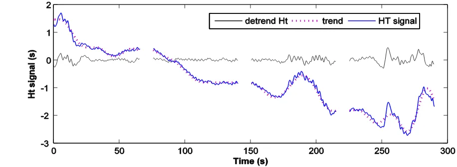
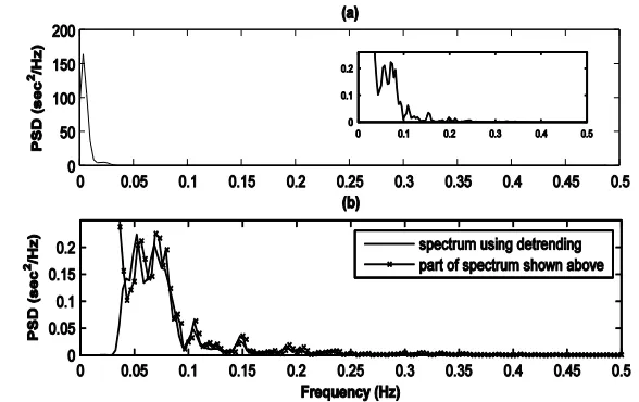
has been validated in previous studies (39,58). The VLF component of the signal was removed by detrending the signal using a wavelet packet analysis which have been validated previously (60). The effect of detrending the HRV signal is shown in Figure 1 and Figure 2 respectively. Figure 1 shows the
time domain representation while Figure 2 shows the spectrum, of the same signal used in Figure 1, before and after detrending. The respiration signal was estimated from the ECG signal using the ECG Derived Respiration (EDR) technique (44-45).
The authors proposed a new method for calculating the variable boundaries associated with the LF and the HF region of the HRV signal. The HF band boundaries were defined using the cross-spectrum between the HRV signal and the estimated respiration signal. From the cross-spectrum the Centre
Frequency (CF) and the Standard Deviation Spectral Extension (SDSE) were estimated using Eq.
1
andEq.
2
respectively. [image:18.612.154.448.57.242.2]̅
∫ (
∫ (
1
(
∫ (
̅ )
(
∫ (
)
2
Where in Eq.
1
and Eq.2
( represents the frequency domain representation and the integral limits and will represent the upper and lower boundaries of the region (LF, HF).Using the estimate of the CF and the SDSE, the range of the HF band was defined as
S S . The CF and the SDSE related to the LF region of the signal was also calculated, but in this case the estimate wascarried out using the frequency domain representation of the HRV signal. In the case where the lower boundary of the HF component was below 0.15 Hz then this lower boundary was used in the estimation of the LF component ICF and the boundaries, otherwise the estimation was done in the frequency range
of 0.04-0.15 Hz. The CF and the boundaries were smoothed using a median filter with a length of ten seconds to avoid sharp fluctuations in these parameters. Two examples of LF and the HF boundary
[image:19.612.119.511.57.196.2]estimation along with the corresponding HRV and the estimated respiration signals are shown in Figure
4
and Figure3
respectively.In both cases (Figure 4 and Figure 3) the estimation was carried out on five minute segments of data. In the case of Figure 4 (c) the cross-spectrum shows a single well defined peak at a frequency of 0.3 Hz. This indicates that the power related to the respiration component (HF) is confined to a narrow band
that lies within the fixed limits defined for the HF component. Figure 4 (d) (HRV signal spectrum) confirms the information represented by the cross-spectrum and shows a well defined respiration related component around 0.3 Hz. In this case the respiration component is easily distinguishable from the LF component whose CF is around 0.1 Hz. Figure 4 (d) also shows that the range S S of each
band (LF and HF) estimated for power estimation in the variable boundary method and represented by horizontal magenta lines quite adequately covers major parts of the signal in these two regions. The situation is quite different in the second example of the boundary estimation presented in Figure 3; in
this case the respiration signal (see Figure 3 (b)) shows more complex dynamics as compared to the respiration signal of the first example (see Figure 4 (b)). Because of this reason, the cross-spectrum between the HRV signal and the estimated respiration signal shown in Figure 3 (c) is spread over a larger
[image:20.612.117.514.58.207.2]frequency range slightly below the fixed lower boundary of the HF region and shows more than one component. By looking at the cross-spectrum shown in part (c) it can be seen that if fixed boundaries
were used to calculate the power then some of the power which might be due to the respiration component would be wrongly assigned to the LF region of the signal. However, the variable boundary method will be able to take into account the part of the spectrum below the fixed lower boundary of the HF region as in this case the range of the HF component is defined by using the cross-spectrum between
the HRV and the respiration signal. By looking at the magenta horizontal line (in the high frequency region) in Figure 4 (d) it can be seen that the boundary of the HF region as defined by the variable boundary method indeed extends below 0.15 Hz, which is considered to be the lower limit of the HF
region in the fixed boundary method. The results from both examples (see Figure 4 (d) andFigure 3 (d)) showed that the major parts of the signal power are covered by the LF and the HF region as defined by the variable boundary method. In order to see the effect of variable boundaries, on the estimation of
the HRV parameters, these quantities were also estimated using the fixed (traditional) range of the LF (0.04-0.15 Hz) and the HF (0.15-0.4 Hz) bands.
The frequency domain analysis of these signals were carried out using non-parametric (Welch’s
periodogram) method (59), parametric (Autoregressive modeling) method (58), Continuous Wavelet Transform (CWT) (61), Smoothed-Pseudo Wigner-Ville Distribution (SPWVD) (62) and Empirical mode decomposition (EMD) (63). The results obtained in these studies showed that after the application of the local anesthetic drug the LF:HF ratio increased initially and then decreased reaching a minimum value.
Compared to the variations observed in other parts of the data during this decrease and sometime after reaching the minimum, the variations in the ratio values were significantly low until it recovered from the minimum phase after the block. The decrease in the ratio values were observed in each case within an hour of the application of the anesthetic block. Apart from the LF:HF ratio value, significant changes
normalized LF power showing a decreased and normalized HF power showing an increase after the application of the anesthetic block. The statistical analysis also showed that similar results were
obtained using the fixed (traditional) boundary method and variable boundary method. The parameter values during the surgery were not included in the statistical analysis as in this case it might not be possible to separately identify the changes in the HRV parameter due to the local anesthetic drug and
the changes occurring due to the surgical procedure.
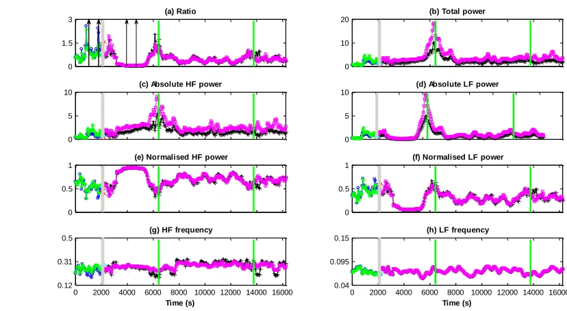
The parameters estimated with the CWT data analysis obtained from one of the patients included in the study are presented in Figure 5. The Figure shows that in most regions similar changes are observed in the values estimated by both the fixed and the variable boundary method. However, there are
incidences (e.g. around 6000 seconds) where different values of the parameters are obtained from the two methods. In all these cases the CF estimated from the two methods shows more difference between them as compared to the other regions of the data. The use of the cross-spectrum between
HRV signal and estimated respiration signal would allow the variable boundary method to consider the effect of the respiration signal below 0.15 Hz which is not possible with the fixed boundary method. Due to this difference, when the respiration frequency will be close or lower than 0.15 Hz the parameter
During the analysis of the data from locally anaesthetized patients two distinguishable changes were observed in the LF:HF ratio values after the application of LA. Firstly, the presence of Adrenaline in the
LA mixture was considered to be the major factor behind a transient short lived increase in the LF:HF ratio which was observed in almost all the patients included in this study. Secondly, the anesthetic mixture would cause a sympathetic impairment and/or vagal enhancement resulting in a decrease in the
LF:HF ratio values.
Due to their superior capability of detecting transient changes occurring in the signals, the Time-Frequency methods (CWT, SPWVD and EMD) managed to detect changes in the LF:HF ratio values in
[image:23.612.82.496.77.304.2]more patients as compared to the parametric and non-parametric methods. The CWT and the EMD were the most successful methods and detectedLF:HF ratio values changes in thirteen out of fourteen
Figure 5 Results obtained from the CWT analysis of a patient undergoing local anesthetic procedure. In each plot the grey vertical block represents the time of block (anesthesia) application and the green vertical lines represent start and end of the surgery. The vertical arrow pairs in part (a) show the data segment before and after the application of block which was used in statistical analysis. Each plot shows the parameter values estimated using both the fixed and the variable boundary method. Lines in green and magenta color represent the parameter values before and after the block application estimated using fixed boundary method respectively. In the case of variable boundary method the same information is presented with blue and black color lines respectively. The units on y-axis for the subplots (b, c and d) showing absolute power values are and for the subplots (g and h) showing frequency values is Hz
0 1.5 3 (a) Ratio 0 10 20
(b) Total power
0 5 10
(c) Absolute HF power
0 5 10
(d) Absolute LF power
0 0.5 1
(e) Normalised HF power
0 0.5 1
(f) Normalised LF power
0 2000 4000 6000 8000 10000 12000 14000 16000 0.12
0.31 0.5
(g) HF frequency
Time (s)
0 2000 4000 6000 8000 10000 12000 14000 16000 0.04
0.095 0.15
(h) LF frequency
patients included in the study. The performance of the SPWVD method suffered due to the presence of interference terms, present due to the bilinear nature of the distribution, which caused an error in the
power estimation. The better performance of the CWT method over the SPWVD method in detecting the transient changes occurring in the signal has also been reported by other researchers (19,49).
The analysis of the data from locally anesthetized patients showed that during brachial plexus block
using a mixture of Lignocaine and Bupivacaine there is a noticeable and almost consistent change in the sympathovagal balance (LF:HF ratio decreases) which can be detected through appropriate and structured analysis of HRV.
5
Discussion and Conclusions
Due to the high success rate and the low number of serious (life threatening) incidents that are usually associated with the application of LA there are not many studies that analyzed the hemodynamic and
cardiovascular data, including HRV analysis, extensively during various procedures involving LA.
The current recommendations regarding maximum doses of local anesthetics presented in textbooks, or by pharmaceutical industries, are not evidence based (i.e., determined by randomized and controlled studies). Usually, recommendations are in the form of a total amount of the drug which does not take in to account the patient’s body mass index. Also, other factors such as the physical state of the patients
and site of the application have great effect of the dosage of the anesthetic drug. Due to all these uncertainties the maximum dosage of different anesthetic drugs varies from country to country as
Table 3: Officially Recommended Highest Doses of Local Anesthetics in Finland, Germany, Japan,
Sweden, and the United States (55).
Finland
Germany
Japan
Sweden
US
ChloroprocaineWith epinephrine
--- --- --- --- 800
--- --- 1000 --- 1000
Procaine
With epinephrine
--- 500 600 (epidural) --- 500
--- 600 --- --- ---
Articaine
With epinephrine
7 4 --- --- ---
7 4 --- --- ---
Bupivacaine
With epinephrine
175 (200 *)
(400 /24 h)
150 100 (epidural) 150 175
175 150 --- 150 225
Levobupivacaine
With epinephrine
150
(400 /24 h)
150 --- 150 150
--- --- --- --- ---
Lidocaine
With epinephrine
200 200 200 200 300
500 500 --- 500 500
Mepivacaine
With epinephrine
--- 300 400 (epidural) 350 400
--- 500 --- 350 550
Prilocaine
With epinephrine
400 --- --- 400 ---
600 --- --- 600 ---
Ropivacaine
With epinephrine
225 (300 *)
(800 /24 h)
No mention 200 (epidural)
300 (infiltr)
225 225
(300 *)
225 No mention --- 225 225
(300 *)
[image:25.612.73.534.123.692.2]The technique of HRV variability has been applied in many research studies in the field of anesthesia both general and local, however so far HRV has not reached the point to be accepted as a clinical
diagnostic tool, as many of the results obtained from such studies are either inconclusive or not in agreement. However, with the continuous advancement of computational and signal processing techniques there is hope that HRV will be able to provide more robust information which will aid in the better anesthetic monitoring of patients. Also, in order to get a better understanding of the complex
cardiovascular changes occurring due to local anesthesia the information obtained from the linear and non-linear analysis of HRV signals should be combined with the information obtained from other physiological signals such as, Electroencephalogram (EEG), BVP etc. Combining information from such
physiological signals would allow for better prediction of future changes in the patient’s cardiovascular state. This would also help in tailoring the delivery and management of anesthesia according to individual patient requirements.
Another issue which impedes the more conclusive results in HRV studies during anesthesia is the lack of control in the studies involving patients. As the patient’s safety during anesthesia is of paramount importance it is quite difficult to analyze the effect of anesthesia in varying levels of anesthetic dosages.
For this more attention should be given on animal studies where studied parameters could be controlled in a more systematic manner.
Also, as commented above, the current availability of extensive computational recourses has caused a rapid increase in the number of signal processing techniques available for the analysis of physiological
data. This makes the comparison and validation of results from different studies quite difficult. It is essential that advance signal processing methods should be applied with care as minor changes could cause a great deal of ambiguity and potentially render the results meaningless. For instance,
patients undergoing epidural anesthesia. The problem with this study in that the authors have used wavelet analysis with irregularly sample signal (tachogram) and have associated the coefficients from
specific wavelet decomposition levels to the two (HF and LF) bands of the HRV signal. Estimating HRV parameters in this way could result in error as due to irregular sampling of the data there could be
significant overlap of scales (frequencies) between levels (42).
Finally, researchers should make every effort to make their data sets available in open-excess database via common internet protocols. This will allow easier comparison of the results obtained from different analysis techniques and facilitate rapid development and better understanding of the research area.