BJP
Themed Section: Therapeutics for Dementia and Alzheimer's Disease:
New Directions for Precision Medicine
R E V I E W A R T I C L E
The potential of memory enhancement through modulation of
perineuronal nets
James A. Duncan
1|
Richard Foster
1,2|
Jessica C.F. Kwok
3,41
School of Chemistry, University of Leeds, Leeds, UK
2
Astbury Centre for Structural Molecular Biology, University of Leeds, Leeds, UK 3
School of Biomedical Sciences, University of Leeds, Leeds, UK
4
Institute of Experimental Medicine, Czech Academy of Science, Prague, Czechia
Correspondence
Jessica C. F. Kwok, School of Biomedical Sciences, University of Leeds, Leeds LS2 9JT, UK.
Email: [email protected]
Funding information
The Leverhulme Trust; Alzheimer Research UK —Yorkshire Network; Medical Research Coun-cil (MRC)
With an increasingly aging global population, the incidence of neurological diseases
such as dementia is set to increase to unmanageable levels, yet there are currently
only symptomatic therapies available for treatment. The mechanisms underlying the
development of some forms of dementia, such as Alzheimer's disease (AD), are not
yet completely elucidated with several competing hypotheses existing. During the
closure of the critical period in the brain, significant compositional changes occur to
the neural extracellular matrix (ECM). Specifically, condensed mesh
‐
like structures
called perineuronal nets (PNNs) form around subsets of neurons and have a profound
effect on axonal growth and limit neuronal plasticity. These PNNs act as a
morpho-logical checkpoint and can influence memory and cognition. Manipulating these
important ECM structures may provide the key to reactivating plasticity and restoring
memory, both of which are severely impaired in AD and other associated neurological
diseases. This review explores the current understanding of how PNNs are
manipu-lated and examines potential new methods for PNN modulation.
LINKED ARTICLES:
This article is part of a themed section on Therapeutics for
Dementia and Alzheimer's Disease: New Directions for Precision Medicine. To view
the other articles in this section visit http://onlinelibrary.wiley.com/doi/10.1111/
bph.v176.18/issuetoc
1
|I N T R O D U C T I O N
Alzheimer's disease (AD) is the most common form of dementia and is clinically characterised by a progressive loss of memory and functional cognition as well as other non‐cognitive disturbances such as anxiety and delusions (Yiannopoulou & Papageorgiou, 2013). The leading risk factor for AD is age, one in 23 people over the age of 65 suffer from this
form of dementia (Prince et al., 2014). As our understanding of the dis-ease state has expanded, four main hypotheses of the cause of AD have been developed: the cholinergic hypothesis, the amyloid hypothesis, the Tau hypothesis, and the genetic hypothesis. Since its initial discovery over a century ago, a definitive cure for AD is still to be found, despite numerous attempts at realising one. Regrettably, the few treatments cur-rently available are not considered to be disease‐modifying in nature but
-This is an open access article under the terms of the Creative Commons Attribution License, which permits use, distribution and reproduction in any medium, provided the original work is properly cited.
© 2019 The Authors. British Journal of Pharmacology published by John Wiley & Sons Ltd on behalf of British Pharmacological Society.
Abbreviations:ACAN, aggrecan; AD, Alzheimer's disease; BCAN, brevican; CS, chondroitin sulfate; CSPGs, chondroitin sulfate proteoglycans; ECM, extracellular matrix; GAG, glycosaminoglycan; GalNAc,N‐acetylgalactosamine; GlcA, glucuronic acid; HA, hyaluronan; Hapln, hyaluronan and proteoglycan link protein; NCAN, neurocan; PNNs, perineuronal nets; Tn‐R, tenascin‐R
Correction added on 24th June 2019 after first online publication: ORCID IDs were added for James A. Duncan and Richard Foster.
rather, symptomatic and have been the first line of treatment for the last 30 years (Ballard et al., 2011; Cummings, Morstorf, & Zhong, 2014).
In the last two decades, attention has shifted towards treating the more recently appreciated disease pathologies, notably targeting and preventing the build‐up ofamyloidβ(Aβ)proteins (Selkoe & Hardy, 2016) and the microtubule associated phosphoprotein,Tau(Iqbal, Liu, Gong, & Grundke‐Iqbal, 2010), both of which are increasingly becoming explicitly linked to one another (Bloom, 2014). Several anti‐Aβ monoclo-nal antibody therapies (Doody et al., 2014; Vandenberghe et al., 2016) and a selection ofβ‐secretase (BACE1)inhibitors (Vassar, 2014) have progressed as far as phase III clinical trials but have failed to translate into commercial medicines. These recent high‐profile failures have led some to believe that new hypotheses of AD are required, as the current understanding has provided no progress to a medicine (Kepp, 2017). This has led researchers to inspect features of the brain other than the neural cells, such as the extracellular matrix (ECM), for novel inspiration. Synaptic plasticity plays a key role in memory throughout the critical period of development, during intense periods of learning and as we age. Recent experiments have shown that neural plasticity can be restored to a juvenile‐like state through modulation of a neuronal ECM component known as perineuronal nets (PNNs), resulting in the restoration of cognition in a mouse model (Rowlands et al., 2018; Sorg et al., 2016; Yang et al., 2017). In this review, we discuss the emerging role that PNNs play in controlling plasticity and memory, presenting the unique structural and functional features of these complex ECM components and the evolving methodologies used to modulate them.
2
|K E Y C O M P O N E N T S A N D S T R U C T U R E
O F P N N S
Neural cells produce specialised and distinctive ECM molecules that fill the diffuse space between neurons and glial cells (Gundelfinger,
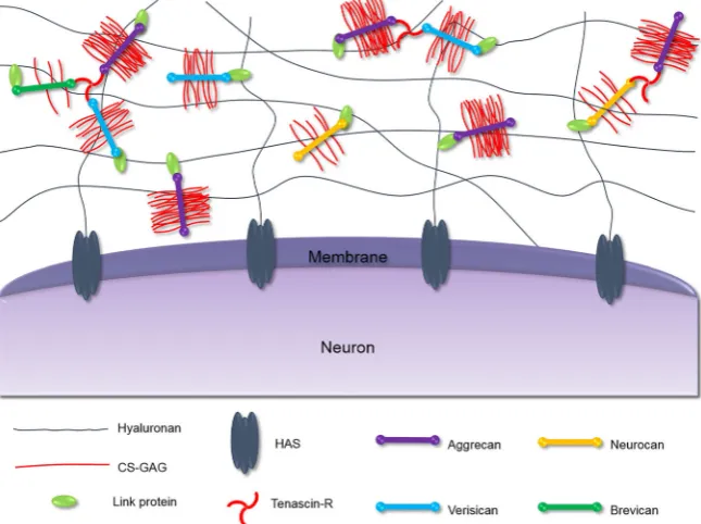
Frischknecht, Choquet, & Heine, 2010). These molecules can con-dense and ensheath specific neurons forming PNNs. These nets were first observed by Camillo Golgi in 1889, but only recently has there been an interest in their structure and function (Spreafico, De Biasi, & Vitellaro‐Zuccarello, 1999). The revelation that PNNs play a signifi-cant role in regulating plasticity and memory has intensified research efforts into elucidating their molecular composition and connectivity in recent years. PNNs are composed of several components, the struc-ture of which is shown in Figure 1 (Kwok, Dick, Wang, & Fawcett, 2011).Hyaluronan(HA), the most abundant and most crucial compo-nent of PNNs, forms a backbone mesh‐like structure which allows for the binding of other important components such as chondroitin sul-fate proteoglycans (CSPGs) and ultimately dictates the overall struc-ture of the ECM (McRae & Porter, 2012). HA consists of alternating
N‐acetylglucosamine and glucuronic acid (GlcA) units forming long non‐sulfated polysaccharide chains which vary in length, ranging from 25 to 1,000 kDa (Viapiano & Matthews, 2006). HA is synthesised by the transmembrane enzyme hyaluronan synthase (HAS) which anchors the PNNs to the neuronal surface (Kwok, Carulli, & Fawcett, 2010). Lecticans are the major CSPG family in the brain ECM and are able to bind an array of matrix molecules; as a result, these CSPGs are con-sidered the organisers of the ECM (Yamaguchi, 2000). The lectican family include the non‐CNS‐specific lecticans: aggrecan (ACAN) and versican (Glumoff, Savontaus, Vehanen, & Vuorio, 1994; Popp, Ander-sen, Maurel, & Margolis, 2003), and the CNS‐specific lecticans: neurocan (NCAN) and brevican (BCAN; Watanabe et al., 1995; Yamada, Watanabe, Shimonaka, & Yamaguchi, 1994).
[image:2.595.51.374.486.727.2]Lecticans are different from other CSPGs in the ECM as they are composed of globular domains enabling them to interact with HA and tenascin‐R (Tn‐R) simultaneously. All lecticans have an N‐terminal globular (G1) domain containing an immunoglobulin‐like loop repeat capable of binding HA chains and certain link proteins (Haplns) and up to two link modules at the C‐terminus (Yamaguchi, 2000). The
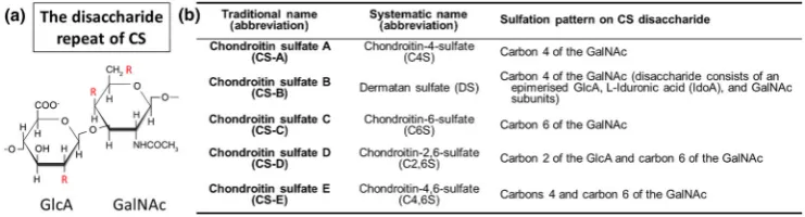
central protein core has covalently attached chondroitin sulfate gly-cosaminoglycan (CS‐GAG) chains which extend in a brush‐like manner perpendicular to the core protein. The CS‐GAG chains are composed of alternatingN‐acetylgalactosamine (GalNAc) and GlcA (Figure 2a). These extensions can be sulfated at various positions, typically carbon 4 and/or carbon 6 of the GalNAc subunit and/or carbon 2 of the GlcA subunit, which gives rise to multiple versions of chondroitin sulfate (CS) shown in Figure 2b (Mikami & Kitagawa, 2013; Silbert & Sugumaran, 2002). The CS chains vary in number, length, and pattern of sulfation, and this has a significant effect on their functions (Bandtlow & Zimmermann, 2000). The C‐terminal globular (G3) domain allows for the binding of tenascins—typically, this is Tn‐R in the PNNs. As a trimeric modular glycoprotein, Tn‐R serves to strengthen the overall macromolecular structure of PNNs through binding multiple lecticans (Lundell et al., 2004; Figure 1). Visualisation of the PNNs is commonly done using the lectin Wisteria floribunda
agglutinin (WFA) staining; however, it is still unclear which component of the nets WFA binds to (Härtig, Brauer, & Brückner, 1992).
3
|T H E R O L E O F P N N S I N P L A S T I C I T Y A N D
M E M O R Y
3.1
|Plasticity
Plasticity is the ability of neurons to reorganise and reassemble synap-tic connectivity in response to experiences and external stimuli and is governed by a wide range of interrelated factors. It was previously thought that neurons and glial cells which constitute much of the brain volume were the exclusive directors of this adaptability. However, ECM molecules which form the vital links between these cells are increasingly becoming associated with neuroplasticity. As the critical period closes for plasticity, PNNs rapidly form predominantly around parvalbumin‐positive (PV+) GABAergic interneurons. This creates a lattice structure which blocks the formation of new synapses (Deepa et al., 2006; Kosaka & Heizmann, 1989; Tsien, 2013).
These lattice structures are dynamic, being turned over throughout the lifetime of the neuron, regulating communication, and acting as the gateway to the neuron. Several recent studies have provided insight into the multifaceted role PNNs play in controlling plasticity. The PNNs act not only as a physical barrier between the neurons and the rest of the ECM but also as a mediator in the binding and
movement of critical binding proteins and membrane bound neuronal proteins, respectively (Dick et al., 2013; Frischknecht et al., 2009). The PNNs can be considered to act as a cationic buffer for the neurons, as they possess an unusually high negative charge density (Morawski et al., 2015). This provides protection from oxidative stress caused by cations such as Fe3+ (Härtig et al., 1999; Suttkus et al., 2014). Additionally, the repulsive axon guidance moleculesemaphorin 3A (Sema3A) specifically binds to the glycosaminoglycan (GAG) chondroitin‐4,6‐sulfate (C4,6S), which is enriched in the PNNs. When bound to PNNs, Sema3A enhances the inhibition of PNNs to neuronal growth and is involved in restricting plasticity (Boggio et al., 2019; Dick et al., 2013). Moreover, PNNs limit the lateral movement of
AMPA‐type glutamate receptorson the cell surface. Removing PNNs allows these receptors to diffuse laterally, leading to an increased paired‐pulse ratio, a readout of short‐term synaptic plasticity and is recorded using whole‐cell patch clamp. This suggests that suppressing membrane protein mobility is another way in which PNNs inhibit synaptic plasticity (Frischknecht et al., 2009).
[image:3.595.111.482.48.148.2]The important role PNNs play in regulating plasticity has been shown using several animal models with focus on the visual cortex. The first evidence of this was seen in dark‐rearing experiments using cats and mice. The visual cortex critical period was extended through enhanced plasticity as a result of attenuating the expression of CSPGs and stalling PNN formation (Lander, Kind, Maleski, & Hockfield, 1997; Pizzorusso et al., 2002). Additionally, PNN formation is hindered by reducing overall neuronal activity and thus providing a potential continuation of the critical period. Using organotypic mouse brain slices and non‐specific suppression of neuronal activity by blocking voltage‐ gated sodium channels, Reimers, Hartlage‐Rübsamen, Brückner, and Roßner (2007) were able to postpone the development of PNNs and maintain synaptic plasticity. Moreover, through a knockout (KO) mouse model, the hapln1 gene which encodes the vital PNN component hyaluronan and proteoglycan link protein 1 (Hapln1), the development of PNNs can be attenuated, leading to the adult mice visual and somatosensory systems plasticity being greatly enhanced to levels that are comparable to juvenile animals (Carulli et al., 2010). Finally, adult rats suffering from amblyopia, a visual acuity disorder developed during the critical period for vision, were provided with a stimuli‐enriched environment resulting in the reduction of the density of the PNNs as well as the restoration of visual acuity and ocular dominance (Sale et al., 2007). These few examples provide compelling evidence for the close relationship thought to exist between PNNs and brain plasticity.
3.2
|Memory
Synaptic plasticity has a long history of being linked to the encoding, storage, and retrieval of information in the form of memory (Hebb, 1949; Jones, 1994; Martin, Grimwood, & Morris, 2000). As a result, PNNs have been implicated in controlling various forms of memory. Recently, it has been reported that digestion of the PNNs wrapped around neurons in the secondary visual cortex (V2L) interrupts the recall of long‐term fear memory in rats. In contrast, more recent fear memory was undisturbed by the same change to the ECM (Thompson et al., 2017). Several studies prior to this work also showed similar remote fear memory recall impairment when PNNs were disrupted in various regions of the brain (Gogolla, Caroni, Lüthi, & Herry, 2009; Hylin, Orsi, Moore, & Dash, 2013). This suggests that PNNs stabilise existing synaptic connec-tions and block the formation of new synapses between these neurons. PNNs in the perirhinal cortex have also been shown to affect a different form of memory known as object recognition (OR) memory in mice. Two mouse models with Tau pathology showing significant impairment in OR memory were injected with chondroitinase ABC (ChABC) at the site of the perirhinal cortex, in order to enzymi-cally digest the PNNs present. One week after treatment, the Tau mice demonstrated similar levels of OR memory and synaptic transmission to control animals, suggesting that ChABC may be effective in restor-ing memory loss in neurodegenerative disorders such as AD (Yang et al., 2015). A different study sought to genetically attenuate PNNs and investigate the effects this had on long‐term OR memory. Using the same adulthapln1KO mouse model used by Carulli and colleagues to investigate the effects of PNN removal on plasticity, OR memory was greatly enhanced in the absence of PNNs (Romberg et al., 2013). Furthermore, both perirhinal basal synaptic transmission and
long‐term depression were measured, following on from the notion that these are the core physiological mechanisms underpinning long‐ term OR memory. These parameters were enhanced by the removal of PNNs (Romberg et al., 2013).
This evidence inevitably guides research towards novel methods of altering the PNNs to enhance plasticity for memory‐related deficiencies. As previously mentioned, the most telling and disruptive symptom of AD is a loss of memory. Modulation of the PNNs may provide improvements to impaired neuronal connectivity seen in AD patients' brains, altogether bypassing the pathologies of AD.
4
|M O D U L A T I O N O F T H E P N N S
4.1
|Removing PNNs
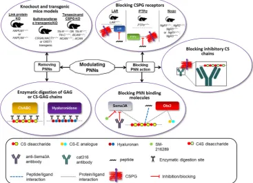
[image:4.595.114.482.445.710.2]Several molecular structures including HA backbone, link proteins such as Hapln1 and Tn‐R, and the major CSPGs are essential for main-taining the structure and function of the PNNs (Kwok et al., 2010; Suttkus et al., 2014). As these components are exposed to the diffuse ECM, many extracellular macromolecules can recognise and bind to specific molecular sequences (Figure 3). An example of this is the bac-terial enzyme ChABC which can indiscriminately recognise and digest CS‐GAG chains present on the CSPGs into disaccharides and partially digest HA (Saito & Yamagata, 1968). Without the CS and HA chains, the structural integrity of the CSPG is compromised resulting in a complete collapse of the PNN structure into diffuse ECM. ChABC has been used extensively in a range of experiments to investigate the effects of PNN removal on various parameters and indications including plasticity, memory, and spinal cord injury recovery (Bradbury
et al., 2002; Kwok, Afshari, García‐Alías, & Fawcett, 2008; Pizzorusso et al., 2002; Romberg et al., 2013).
Levels of the brain‐specific lectican BCAN were significantly ele-vated in AD patients, contributing to a loss of synaptic plasticity observed prior to neuronal cell death (Howell, Bailey, Cozart, Gannon, & Gottschall, 2015). Aβ protein can directly interact with BCAN in vitro, as well as disrupting the proteolytic cleavage mechanisms involved in BCAN processing, potentially accentuating the inhibition of synaptic plasticity (Ajmo et al., 2010; Howell et al., 2015). Injection of ChABC into the hippocampus of 15‐month‐old double transgenic APPswe/PS1dE9 mice which have greatly increased Aβprotein produc-tion and severe synaptic deficits with age, removed the CS chains on lecticans and the effects on Aβplaques, was monitored. Interestingly, the application of ChABC resulted in a significant reduction in Aβ bur-den and an increase in synaptic bur-density (Howell et al., 2015). These results introduce the possibility of targeting perisynaptic lecticans as a starting point for an AD therapy. The APPswe/PS1dE9 mice model has also been used to demonstrate the up‐regulation of several ECM proteins including Hapln1, NCAN, BCAN, and Tn‐R that coincides with an early increase in synaptic Aβlevels, as well as LTP and contextual memory impairment. Treatment of the ECM with ChABC was able to reverse these adverse effects, suggesting that increasing ECM levels contributes to early memory deficits in AD (Végh et al., 2014).
The relative sulfation patterns on CSPGs notably influence the formation of PNNs and the effect they have on axon growth. During development and maturation, the ratio of chondroitin‐4‐sulfate (C4S) to chondroitin‐6‐sulfate (C6S) in the PNNs gradually increases (Miyata, Komatsu, Yoshimura, Taya, & Kitagawa, 2012). This is due to both the depletion of C6S over time and the increase in C4S during progression into adulthood (Miyata et al., 2012). The change in C4S/C6S ratio is partly the result of the change in activity of both chondroitin 6‐sulfotransferase‐1 (C6ST1) and chondroitin 4‐sulfotransferase‐1 (C4ST1), Golgi‐resident enzymes which are responsible for the sulfation of unsulfated chondroitin to the 6‐ sulfated and 4‐sulfated forms of chondroitin, respectively (Mikami & Kitagawa, 2013; Silbert & Sugumaran, 2002). The changes in sulfation observed during development continue into adulthood and aging where C6S sulfation drops further (Foscarin, Raha‐Chowdhury, Fawcett, & Kwok, 2017). C6S has been shown to be permissive to axonal growth and regeneration (Kitagawa, Tsutsumi, Tone, & Sugahara, 1997; Lin, Rosahl, Whiting, Fawcett, & Kwok, 2011). In con-trast, C4S is thought to be the most inhibitory form of CS to axonal growth and guidance (Deepa et al., 2006; Wang et al., 2008). The shift in C4S/C6S ratio is crucial for successful PNN development and restriction of axon growth during the closure of the critical period. If C6S is up‐regulated by overexpression of C6ST1, then PNN formation in the visual cortex is severely impaired and the mice are more plastic (Miyata et al., 2012). On the contrary, reducing C6S level inc6st1KO mice shows a reduction in axonal regeneration after a CNS lesion (Lin et al., 2011). An increase in C6S also surprisingly leads to an increase in the proteolysis of ACAN by a disintegrin and metalloproteinase domain with thrombospondin motif (ADAMTS) protease, further disrupting PNN formation (Miyata & Kitagawa, 2016).
Besides using ChABC to digest PNNs, some studies have used hyal-uronidase to specifically target HA chains and disrupt the entire PNN (Frischknecht et al., 2009). Happel and colleagues injected hyaluronidase into the auditory cortex of adult Mongolian gerbils and investigated the effects this had on cognitive flexibility in reversal learning. They found that removal of the PNN through this method improved the activity‐ dependent reorganisation of existing synaptic networks during reversal learning and an overall increase in synaptic plasticity (Happel et al., 2014). In some cases, ChABC and hyaluronidase have been used in com-bination to eradicate all traces of the CS and HA in the ECM (Hylin et al., 2013). Enzymic degradation using ChABC and hyaluronidase can be considered a crude tool for modulating the ECM. In the context of treating AD patients, using enzymes such as ChABC is not considered practical for targeting the large volume of an adult brain (Fawcett, 2015). More discrete methods, such as specifically targeting and altering the molecular composition of PNNs, must be used if fine modifications to neuronal plasticity are required (van't Spijker & Kwok, 2017).
Several animal KO and transgenic mouse models have been devel-oped to prevent or reduce the formation of PNNs around neurons. As mentioned previously, removing Hapln1 through gene deletion of
hapln1has consistently resulted in the attenuation of PNNs (Carulli et al., 2010; Czipri et al., 2003; Romberg et al., 2013). Other link pro-teins that are found in the PNNs such as the brain link protein 2 (Bral2) have also been removed in KO mouse models to inhibit the development of PNNs (Bekku et al., 2012). A useful alternative approach to PNN degradation was shown through the overexpression of C6S to disrupt the accrual of ACAN viac6st1 transgenic mice (Miyata & Kitagawa, 2016; Miyata et al., 2012). KO mice lacking the chondroitin sulfateN‐acetylgalactosaminyltransferase‐1 (CSGalNAcT‐ 1) enzyme have been used as an alternative way to interrupt CSPG production. Interestingly, these mice still formed structurally identifi-able, albeit abnormal, PNNs (Yoshioka et al., 2017). KOs of the gene encoding the trimeric Tn‐R have been conducted, resulting in the dis-ruption (but not complete removal) of the PNNs. This was thought to be due to the absence of two types of CSPGs (phosphacan and NCAN) in the nets (Haunsoø et al., 2000; Suttkus et al., 2014).
There have been examples of multiple KO mice—notably, work by Geissler et al. (2013) generated quadruple KO mice preventing the expression of tenascin‐C, Tn‐R, BCAN, and NCAN and causing severely shrunken PNNs to form. Recently, a novel animal model was developed in which the levels of ACAN were reduced in vivo through targeted
ACANgene deletion. This attenuated the PNNs and was shown to result in reinstating of the juvenile ocular dominance plasticity, as well as providing enhancements in OR memory (Rowlands et al., 2018). Despite the numerous successes seen with transgenic animals in removing PNNs and enhancing plasticity, this method of modulating PNNs has both practical and moral hurdles preventing it from being a viable treatment for neurodegenerative disorders in patients at present.
4.2
|Blocking PNN action
of the PNNs. For example, as discussed previously, Sema3A has been shown to bind specifically to certain CS moieties on CSPGs. This includes chondroitin‐4,6‐sulfate(C4,6S) but not chondroitin‐2,6‐ sulfate (C2,6S), despite both forms being disulfated chondroitins, suggesting that Sema3A binding has sulfation pattern specificity rather than overall sulfation quantity specificity (Corredor et al., 2016; Dick et al., 2013). As a chemorepulsive axon guidance molecule, the pres-ence of Sema3A in the PNNs potentiates PNN inhibition on neurite outgrowth and new synaptic connections (Dick et al., 2013). Low MW compounds can be used to block Sema3A and controllably coun-teract its inhibitory nature in PNNs on axonal growth. For instance, a selective Sema3A inhibitor, SM‐216289, was identified from a fungal strain fermentation broth and shown to regenerate or preserve injured axons both in vitro and in vivo (Kaneko et al., 2006). Several Sema3A C‐terminus‐derived basic peptides have also been reported to inter-rupt the Sema3A–CS‐GAG interactions (Corredor et al., 2016). A recent paper by Boggio et al. (2019) used adeno‐associated virus to overexpress the soluble fragment of neuropilin 1, the receptor of Sema3A, resulting in an enhanced ocular dominance plasticity in the visual cortex. Additionally, anti‐Sema3A monoclonal antibodies have been developed (Yamashita et al., 2015); while not being designed to specifically target Sema3A in the PNNs, these antibodies could be probed for their relevance in blocking Sema3A binding to PNNs both in vitro and in vivo.
Another PNN binding protein is orthodenticle homeobox 2 (Otx2). This transcription factor also binds to C4,6S in the PNNs of the PV+ interneurons, is then internalised, and modulates gene expression for the maturation of PV+ neurons. It plays an important role in the commencement and termination of the critical period for plasticity (Bernard & Prochiantz, 2016). Moreover, it is thought that Otx2 can facilitate its own uptake in a positive feedback loop by binding to increasingly thriving PNNs (Beurdeley et al., 2012). The transfer of Otx2 into the PV+ interneurons can be blocked by various peptide and CS mimetics and thus reduce PNNs surrounding the cells. A sequence of amino acids which traverses the N‐terminal domain and homeodomain of Otx2 was described as being a putative GAG binding motif (Beurdeley et al., 2012). This motif contains an RK doublet pep-tide sequence which has proved to be crucial for CS‐GAG binding. An RK peptide was created, and this peptide was used to outcompete binding of an Otx2 to PNNs in cortical cells and thus prevent the internalisation of Otx2 in vitro and in vivo (Beurdeley et al., 2012). Additionally, the RK doublet interacted specifically with C2,6S and C4,6S, both of which contain carbon 6 sulfation (Beurdeley et al., 2012). This again highlights the importance of sulfation patterns on CSPGs for coordination of the ECM and specifically the PNNs. An alternative approach to blocking Otx2–CS‐GAG interactions has been explored using CS analogues (Despras et al., 2013). The preparation of hexasaccharide C4,6S analogues from lactose was first described and then followed up by in vitro and in vivo studies to assess whether they successfully mimicked natural C4,6S. Using a gel shift assay, C4,6S analogues were shown to bind to Otx2, presumably at the RK doublet peptide sequence (Despras et al., 2013). Additionally, infusion of a par-ticular hexasaccharide C4,6S analogue reduced the Otx2 internalised
by PV+ interneurons as well as slightly inhibiting WFA staining, sug-gesting disruption of the PNNs around these cells (Despras et al., 2013). Lastly, inducing point mutations in the RK doublet motif of the Otx2 gene in knock‐in mice, the localisation and accumulation of Otx2 in the PV+ were disrupted (Lee et al., 2017). Furthermore, this led to a delay in PNN expression and the extension of the critical period for plasticity (Lee et al., 2017).
Recently, antibodies that bind to and block the brush‐like CS chains on PGs in PNNs have been explored in vivo (Figure 3). The Cat316 antibody can specifically recognise C4S, blocking the usual inhibitory effect on axonal growth associated with C4S on CSPGs (Yang et al., 2017). It was also noted that the binding of Cat316 to the PNNs moderately prevented the binding of Sema3A to the PNNs, accentuating the constructive effect on axon growth (Yang et al., 2017). Despite restoring memory function in mice with Tauopathies, this approach did not significantly alter disease progression. Addition-ally, there remains the necessity to inject Cat316 directly into the brain. In order to address this, the authors suggest that a better blocking agent should be developed that would be able to cross the blood brain barrier, if this approach is to be used in a therapeutic set-ting in the future for neurodegenerative diseases such as AD (Yang et al., 2017).
There are several known membrane‐bound cell surface CSPG receptors that contribute to the inhibitory nature of CSPGs and control of neural plasticity (Miao, Ye, & Zhang, 2014). These include the recep-tor protein tyrosine phosphataseσ(PTPσor RTP Type S)and the leu-kocyte common antigen‐related phosphatase (LAR or RTP Type F; l; Fisher et al., 2011; Shen et al., 2009), as well as the Nogo receptors, NgR1 and NgR3 (Dickendesher et al., 2012), which have all been reported to have high binding affinity for CSPGs. Axonal growth inhi-bition from CSPGs was shown to be reduced when a double knockout of thePTPσgene was carried out in neuronal cell culture (Shen et al., 2009). Similarly, dorsal root ganglion (DRG) neurons derived from
LARKO mice do not suffer a restriction in neurite outgrowth in the presence of CSPG substrate (Fisher et al., 2011) indicating the impor-tance of the LAR–CSPG interaction in plasticity. To further confirm these results, two sequence‐targeting peptides, extracellular LAR pep-tide and intracellular LAR peppep-tide, were used to treat CSPG substrate cultured DRG neurons resulting in an increase in neurite length as expected (Fisher et al., 2011). More recently, a membrane‐permeable peptide mimetic was developed and utilised, binding to PTPσ and preventing the interaction with CSPGs (Lang et al., 2015). The peptide mimetic known as intracellular sigma peptide represented the highly conserved wedge domain on PTPσand treatment of adult sensory neurons with intracellular sigma peptide allowed axons to sprout and cross through a CSPG gradient (Lang et al., 2015). To assess the signif-icance of the Nogo receptors on axon regeneration, several Nogo receptor KO mice were generated and studied (Dickendesher et al., 2012). Following an optic nerve crush injury, the regeneration of reti-nal ganglion cell axons was assessed in the various mutants. Triple mutantNgR1−/−; NgR2−/−; NgR3−/−mice and double mutantNgR1−/−;
NgR3−/− mice showed improved axon regeneration when compared
5
|F U T U R E D I R E C T I O N S
The extensive and thorough research already carried out and presented here has clearly demonstrated the promise in targeting PNNs for the treatment of several neurological conditions. Much of the current methodology for modulating PNNs involves the use of macromolecules such as digestive enzymes or antibodies due to the relative ease of use and production in the early stages of therapy development. Additionally, genetic modifications such as KO models are frequently used to abolish or attenuate PNNs. These methods are very useful to establish the proof of concept that modulating PNNs can have a significant effect on plasticity and memory; however, they are not easy to translate into clinically relevant therapies. Commonly, low MW compounds are developed to try and mimic the effects that macromolecules and/or KO models have on phenotype (Samanen, 2013). There are considerable benefits to using low MW compounds over other forms of therapy such as biologics and genetic modifications. One of the main advantages, especially in the early stages of development, is the ease of optimisation. Making subtle adjustments to biologics to improve the effectiveness is seldom successful due to the size and complexity of these species, whereas modifying low MW compounds can easily be achieved through chem-ical synthesis; guidance by methods such as structure‐based drug design can aid in improving efficacy of the drug (Samanen, 2013). Additionally, low MW compounds are usually orally administered which is commonly preferred over the parenteral administration required for biologics due to their poorly defined and adverse physio-chemical properties (Samanen, 2013).
One of the most obvious targets for the development of low MW compounds would be the transmembrane enzyme HAS. Inhibiting HAS would prevent the synthesis of the HA potentially resulting in the breakdown of PNNs and disrupting the ECM overall. HA synthesis has been successfully targeted before on numerous occasions, albeit in alternative indications to AD (Nagy et al., 2015). The commercially available drug 4‐methylumbelliferone has frequently been used for this purpose, due to its ability to deplete one of the substrates (uridine diphosphate GlcA) required for HA synthesis (Nagy et al., 2015). The fact that HA is found throughout all ECMs and not just in neural ECMs and around the PNNs, coupled with the promiscuity of 4‐methylumbelliferone, means that targeted treatment to specifically disrupt HA synthesis in the PNNs may be a challenge (Garg & Hales, 2004; Nagy et al., 2015).
A promising target for intervention with low MW compounds on PNNs and axonal growth may be the CSPGs. More specifically, preventing the biosynthesis of these structures by targeting the sulfotransferase enzymes that provide the CS chains has been shown to be an effective method to renew axonal growth. In recent studies, the Golgi‐residentN‐acetylgalactosamine 4‐sulfate 6‐sulfotransferase (GALNAC4S‐6ST) was shown to be modestly inhibited by a low MW compound that had been optimised using a high throughput screening and medicinal chemistry (Cheung et al., 2017). The most potent compound decreased the overall levels of C4,6S and overall sulfation in vitro as well as reversing the inhibition of
axonal growth caused by CSPGs in DRG neurons (Cheung et al., 2017). Despite being selective towards membrane‐bound GAG sulfotransferases compared to cytosolic sulfotransferases, the optimised compound was indiscriminately inhibitory towards several membrane bound GAG sulfotransferases including the closely related C4ST1 (Cheung et al., 2017). This observation suggests that the rever-sal of inhibited axonal growth seen in DRG neurons could be due to pan‐sulfotransferase inhibition. Clearly, this highlights the potential hurdles involved in achieving drug–protein interaction specificity, which may pose a challenge for the further development of low MW sulfotransferase inhibitors.
There are numerous other components in the PNNs that can potentially be targeted by low MW compounds, including the CNS‐ exclusive tenascin, TN‐R, and the HA–CSPG bridging link proteins. Additionally, it can be envisaged that interruption of the binding of the known PNN binding molecules Sema3A and Otx2 may provide success. Further research into the druggability of these proteins is required before an inhibitor can be realised. Other therapeutic avenues may be pursued. However, given the benefits of developing a low MW modulator, as previously mentioned, coupled with the pit-falls frequently encountered with these other modalities, discovery of a low MW drug seems to provide the most promise going forward.
6
|C O N C L U S I O N S
Increasingly, the emerging evidence suggests that PNNs have a vital role to play in controlling plasticity, regulating axonal growth and regeneration and memory storage during development and throughout adulthood. This has obvious implications in several neurological dis-eases, including the many forms of dementia, in which the underlying mechanisms involved in disease progression are not fully understood. The molecular make‐up and function of the PNNs are increasingly being appreciated, when assessing the scientific literature currently available. This understanding has been used to target and disrupt the PNNs using numerous methods to increase neuronal plasticity. This includes enzymic degradation of the nets, genetic therapy to prevent PNN formation, and blocking of PNN action using low MW compounds and/or biomolecules. The abundance of potential protein targets in the PNNs should inspire the development of novel therapeutic agents with a focus on utilising the ease of discovery and optimisation of low MW compounds to inhibit PNNs, in order to reactivate plasticity and restore cognition in neurological disorders such as AD.
6.1
|Nomenclature of targets and ligands
A C K N O W L E D G E M E N T S
This work was supported by the Medical Research Council (MRC), Alzheimer Research UK—Yorkshire Network, and The Leverhulme Trust.
C O N F L I C T O F I N T E R E S T
The authors declare no conflicts of interest.
O R C I D
James A. Duncan https://orcid.org/0000-0002-0044-4977
Richard Foster https://orcid.org/0000-0002-2361-3884
Jessica C.F. Kwok https://orcid.org/0000-0002-9798-9083
R E F E R E N C E S
Ajmo, J. M., Bailey, L. A., Howell, M. D., Cortez, L. K., Pennypacker, K. R., Mehta, H. N.,…Gottschall, P. E. (2010). Abnormal post‐translational and extracellular processing of brevican in plaque‐bearing mice over‐ expressing APPsw.Journal of Neurochemistry,113, 784–795. https:// doi.org/10.1111/j.1471‐4159.2010.06647.x
Alexander, S. P. H., Fabbro, D., Kelly, E., Marrion, N. V., Peters, J. A., Faccenda, E.,… CGTP Collaborators (2017). The Concise Guide to PHARMACOLOGY 2017/18: Enzymes.British Journal of Pharmacol-ogy,174, S272–S359. https://doi.org/10.1111/bph.13877
Alexander, S. P. H., Kelly, E., Marrion, N. V., Peters, J. A., Faccenda, E., Harding, S. D.,…CGTP Collaborators (2017). The Concise Guide to PHARMACOLOGY 2017/18: Other proteins. British Journal of Pharmacology,174, S1–S16. https://doi.org/10.1111/bph.13882 Alexander, S. P. H., Peters, J. A., Kelly, E., Marrion, N. V., Faccenda, E.,
Harding, S. D.,…CGTP Collaborators (2017). The Concise Guide to PHARMACOLOGY 2017/18: Ligand‐gated ion channels.British Journal of Pharmacology,174, S130–S159. https://doi.org/10.1111/bph.13879 Ballard, C., Gauthier, S., Corbett, A., Brayne, C., Aarsland, D., & Jones, E. (2011). Alzheimer's disease.Lancet,377, 1019–1031. https://doi.org/ 10.1016/S0140‐6736(10)61349‐9
Bandtlow, C. E., & Zimmermann, D. R. (2000). Proteoglycans in the developing brain: New conceptual insights for old proteins.Physiological Reviews,80, 1267–1290. https://doi.org/10.1152/physrev.2000.80.4.1267 Bekku, Y., Saito, M., Moser, M., Fuchigami, M., Maehara, A., Nakayama, M.,
…Oohashi, T. (2012). Bral2 is indispensable for the proper localization of brevican and the structural integrity of the perineuronal net in the brainstem and cerebellum.The Journal of Comparative Neurology,520, 1721–1736. https://doi.org/10.1002/cne.23009
Bernard, C., & Prochiantz, A. (2016). Otx2‐PNN interaction to regulate cor-tical plasticity.Neural Plasticity, 2016, 1–7. https://doi.org/10.1155/ 2016/7931693
Beurdeley, M., Spatazza, J., Lee, H. H. C., Sugiyama, S., Bernard, C., Di Nardo, A. A.,…Prochiantz, A. (2012). Otx2 binding to perineuronal nets persistently regulates plasticity in the mature visual cortex.The Journal of Neuroscience, 32, 9429–9437. https://doi.org/10.1523/ JNEUROSCI.0394‐12.2012
Bloom, G. S. (2014). Amyloid‐βand tau: The trigger and bullet in Alzheimer disease pathogenesis.JAMA Neurology,71, 505–508. https://doi.org/ 10.1001/jamaneurol.2013.5847
Boggio, E. M., Ehlert, E. M., Lupori, L., Moloney, E. B., De Winter, F., Vander Kooi, C. W.,…Pizzorusso, T. (2019). Inhibition of semaphorin3A pro-motes ocular dominance plasticity in the adult rat visual cortex.
Molecular Neurobiology. https://doi.org/10.1007/s12035-019-1499-0 Bradbury, E. J., Moon, L. D. F., Popat, R. J., King, V. R., Bennett, G. S., Patel,
P. N., … McMahon, S. B. (2002). Chondroitinase ABC promotes
functional recovery after spinal cord injury. Nature, 416, 636–640. https://doi.org/10.1038/416636a
Carulli, D., Pizzorusso, T., Kwok, J. C. F., Putignano, E., Poli, A., Forostyak, S.,…Fawcett, J. W. (2010). Animals lacking link protein have attenu-ated perineuronal nets and persistent plasticity. Brain, 133, 2331–2347. https://doi.org/10.1093/brain/awq145
Cheung, S. T., Miller, M. S., Pacoma, R., Roland, J., Liu, J., Schumacher, A. M., & Hsieh‐Wilson, L. C. (2017). Discovery of a small‐molecule modu-lator of glycosaminoglycan sulfation. ACS Chemical Biology, 12, 3126–3133. https://doi.org/10.1021/acschembio.7b00885
Corredor, M., Bonet, R., Moure, A., Domingo, C., Bujons, J., Alfonso, I.,… Messeguer, À. (2016). Cationic peptides and Peptidomimetics bind glycosaminoglycans as potential Sema3A pathway inhibitors. Biophys-ical Journal, 110, 1291–1303. https://doi.org/10.1016/j.bpj.2016. 01.033
Cummings, J. L., Morstorf, T., & Zhong, K. (2014). Alzheimer's disease drug‐ development pipeline: Few candidates, frequent failures.Alzheimer's Research & Therapy,6, 37. https://doi.org/10.1186/alzrt269
Czipri, M., Otto, J. M., Cs‐Szabó, G., Kamath, R. V., Vermes, C., Firneisz, G.,
…Glant, T. T. (2003). Genetic rescue of chondrodysplasia and the peri-natal lethal effect of cartilage link protein deficiency.The Journal of Biological Chemistry,278, 39214–39223. https://doi.org/10.1074/jbc. M303329200
Deepa, S. S., Carulli, D., Galtrey, C., Rhodes, K., Fukuda, J., Mikami, T.,… Fawcett, J. W. (2006). Composition of perineuronal net extracellular matrix in rat brain: A different disaccharide composition for the net‐ associated proteoglycans. The Journal of Biological Chemistry, 281, 17789–17800. https://doi.org/10.1074/jbc.M600544200
Despras, G., Bernard, C., Perrot, A., Cattiaux, L., Prochiantz, A., Lortat‐ Jacob, H., & Mallet, J. M. (2013). Toward libraries of biotinylated chon-droitin sulfate analogues: From synthesis to in vivo studies.Chemistry,
19, 531–540. https://doi.org/10.1002/chem.201202173
Dick, G., Tan, C. L., Alves, J. N., Ehlert, E. M., Miller, G. M., Hsieh‐Wilson, L. C.,…Kwok, J. C. (2013). Semaphorin 3A binds to the perineuronal nets via chondroitin sulfate type E motifs in rodent brains.The Journal of Biological Chemistry,288, 27384–27395. https://doi.org/10.1074/jbc. M111.310029
Dickendesher, T. L., Baldwin, K. T., Mironova, Y. A., Koriyama, Y., Raiker, S. J., Askew, K. L.,…Giger, R. J. (2012). NgR1 and NgR3 are receptors for chondroitin sulfate proteoglycans.Nature Neuroscience,15, 703–712. https://doi.org/10.1038/nn.3070
Doody, R. S., Thomas, R. G., Farlow, M., Iwatsubo, T., Vellas, B., Joffe, S.,… Solanezumab Study Group (2014). Phase 3 trials of solanezumab for mild‐to‐moderate Alzheimer's disease.The New England Journal of Med-icine,370, 311–321. https://doi.org/10.1056/NEJMoa1312889 Fawcett, J. W. (2015). The extracellular matrix in plasticity and
regenera-tion after CNS injury and neurodegenerative disease. Progress in Brain Research, 218, 213–226. https://doi.org/10.1016/bs.pbr.2015. 02.001
Fisher, D., Xing, B., Dill, J., Li, H., Hoang, H. H., Zhao, Z.,…Li, S. (2011). Leukocyte common antigen‐related phosphatase is a functional recep-tor for chondroitin sulfate proteoglycan axon growth inhibirecep-tors.The Journal of Neuroscience, 31, 14051–14066. https://doi.org/10.1523/ JNEUROSCI.1737‐11.2011
Foscarin, S., Raha‐Chowdhury, R., Fawcett, J. W., & Kwok, J. C. F. (2017). Brain ageing changes proteoglycan sulfation, rendering perineuronal nets more inhibitory.Aging (Albany NY), 9, 1607–1622. https://doi. org/10.18632/aging.101256
from Erschienen in Nature Neuroscience, vol 12, pg 897‐804, 2009).E‐ Neuroforum,15, 94–95.
Garg, H. G., & Hales, C. A. (2004). Chemistry and biology of hyaluronan. Amsterdam: Elsevier.
Geissler, M., Gottschling, C., Aguado, A., Rauch, U., Wetzel, C. H., Hatt, H., & Faissner, A. (2013). Primary hippocampal neurons, which lack four crucial extracellular matrix molecules, display abnormalities of synaptic structure and function and severe deficits in perineuronal net forma-tion. The Journal of Neuroscience, 33, 7742–7755. https://doi.org/ 10.1523/JNEUROSCI.3275‐12.2013
Glumoff, V., Savontaus, M., Vehanen, J., & Vuorio, E. (1994). Analysis of aggrecan and tenascin gene expression in mouse skeletal tissues by northern and in situ hybridization using species specific cDNA probes.
Biochimica et Biophysica Acta, 1219, 613–622. https://doi.org/ 10.1016/0167‐4781(94)90220‐8
Gogolla, N., Caroni, P., Lüthi, A., & Herry, C. (2009). Perineuronal nets pro-tect fear memories from erasure. Science (New York, N.Y.), 325, 1258–1261.
Gundelfinger, E. D., Frischknecht, R., Choquet, D., & Heine, M. (2010). Converting juvenile into adult plasticity: A role for the brain's extracel-lular matrix. The European Journal of Neuroscience, 31, 2156–2165. https://doi.org/10.1111/j.1460‐9568.2010.07253.x
Happel, M. F. K., Niekisch, H., Castiblanco Rivera, L. L., Ohl, F. W., Deliano, M., & Frischknecht, R. (2014). Enhanced cognitive flexibility in reversal learning induced by removal of the extracellular matrix in auditory cor-tex.Proceedings of the National Academy of Sciences,111, 2800–2805. https://doi.org/10.1073/pnas.1310272111
Harding, S. D., Sharman, J. L., Faccenda, E., Southan, C., Pawson, A. J., Ireland, S., … NC‐IUPHAR (2018). The IUPHAR/BPS Guide to PHARMACOLOGY in 2018: Updates and expansion to encompass the new guide to IMMUNOPHARMACOLOGY. Nucl Acids Res, 46, D1091–D1106. https://doi.org/10.1093/nar/gkx1121
Härtig, W., Brauer, K., & Brückner, G. K. (1992). Wisteria floribunda
agglutinin‐labelled nets surround parvalbumin‐containing neurons.
Neuroreport, 3(10), 869–872. https://doi.org/10.1097/00001756‐ 199210000‐00012
Härtig, W., Derouiche, A., Welt, K., Brauer, K., Grosche, J., Mäder, M.,… Brückner, G. (1999). Cortical neurons immunoreactive for the potas-sium channel Kv3.1b subunit are predominantly surrounded by perineuronal nets presumed as a buffering system for cations.Brain Research, 842, 15–29. https://doi.org/10.1016/S0006‐8993(99) 01784‐9
Haunsoø, A., Ibrahim, M., Bartsch, U., Letiembre, M., Celio, M. R., & Menoud, P. A. (2000). Morphology of perineuronal nets in tenascin‐R and parvalbumin single and double knockout mice. Brain Research,
864, 142–145. https://doi.org/10.1016/S0006‐8993(00)02173‐9 Hebb, D. O. (1949).The organization of behavior: A neuropsychological
the-ory. New York: John Wiley & Sons, Inc. London, Chapman & Hall, Limited.
Howell, M. D., Bailey, L. A., Cozart, M. A., Gannon, B. M., & Gottschall, P. E. (2015). Hippocampal administration of chondroitinase ABC increases plaque‐adjacent synaptic marker and diminishes amyloid burden in aged APPswe/PS1dE9 mice. Acta Neuropathologica Communications,
3, 54. https://doi.org/10.1186/s40478‐015‐0233‐z
Hylin, M. J., Orsi, S. A., Moore, A. N., & Dash, P. K. (2013). Disruption of the perineuronal net in the hippocampus or medial prefrontal cortex impairs fear conditioning. Learning & Memory, 20, 267–273. https:// doi.org/10.1101/lm.030197.112
Iqbal, K., Liu, F., Gong, C.‐X., & Grundke‐Iqbal, I. (2010). Tau in Alzheimer disease and related tauopathies. Current Alzheimer Research, 7, 656–664. https://doi.org/10.2174/156720510793611592
Jones, E. G. (1994). Santiago Ramon y Cajal and the croonian lecture.
Trends in Neurosciences,17, 190–192. https://doi.org/10.1016/0166‐ 2236(94)90100‐7
Kaneko, S., Iwanami, A., Nakamura, M., Kishino, A., Kikuchi, K., Shibata, S.,
…Okano, H. (2006). A selective Sema3A inhibitor enhances regenera-tive responses and functional recovery of the injured spinal cord.
Nature Medicine,12, 1380–1389. https://doi.org/10.1038/nm1505 Kepp, K. P. (2017). Ten challenges of the amyloid hypothesis of Alzheimer's
disease.J. Alzheimer's Dis.,55, 447–457. https://doi.org/10.3233/JAD‐ 160550
Kitagawa, H., Tsutsumi, K., Tone, Y., & Sugahara, K. (1997). Developmental regulation of the sulfation profile of chondroitin sulfate chains in the chicken embryo brain. The Journal of Biological Chemistry, 272, 31377–31381. https://doi.org/10.1074/jbc.272.50.31377
Kosaka, T., & Heizmann, C. W. (1989). Selective staining of a population of parvalbumin‐containing GABAergic neurons in the rat cerebral cortex by lectins with specific affinity for terminal N‐acetylgalactosamine.
Brain Research, 483, 158–163. https://doi.org/10.1016/0006‐8993 (89)90048‐6
Kwok, J. C. F., Afshari, F., García‐Alías, G., & Fawcett, J. W. (2008). Proteo-glycans in the central nervous system: Plasticity, regeneration and their stimulation with chondroitinase ABC.Restorative Neurology and Neuro-science,26, 131–145.
Kwok, J. C. F., Carulli, D., & Fawcett, J. W. (2010). In vitro modeling of perineuronal nets: Hyaluronan synthase and link protein are necessary for their formation and integrity. Journal of Neurochemistry, 114, 1447–1459. https://doi.org/10.1111/j.1471‐4159.2010.06878.x Kwok, J. C. F., Dick, G., Wang, D., & Fawcett, J. W. (2011). Extracellular
matrix and perineuronal nets in CNS repair.Developmental Neurobiol-ogy,71, 1073–1089. https://doi.org/10.1002/dneu.20974
Lander, C., Kind, P., Maleski, M., & Hockfield, S. (1997). A family of activity‐ dependent neuronal cell‐surface chondroitin sulfate proteoglycans in cat visual cortex. The Journal of Neuroscience, 17, 1928–1939. https://doi.org/10.1523/JNEUROSCI.17‐06‐01928.1997
Lang, B. T., Cregg, J. M., Depaul, M. A., Tran, A. P., Xu, K., Dyck, S. M.,… Silver, J. (2015). Modulation of the proteoglycan receptor PTPσ pro-motes recovery after spinal cord injury. Nature, 518, 404–408. https://doi.org/10.1038/nature13974
Lee, H. H. C., Bernard, C., Ye, Z., Acampora, D., Simeone, A., Prochiantz, A.,
…Hensch, T. K. (2017). Genetic Otx2 mis‐localization delays critical period plasticity across brain regions. Molecular Psychiatry, 22, 680–688. https://doi.org/10.1038/mp.2017.1
Lin, R., Rosahl, T. W., Whiting, P. J., Fawcett, J. W., & Kwok, J. C. F. (2011). 6‐Sulphated chondroitins have a positive influence on axonal regener-ation.PLoS ONE,6, e21499.
Lundell, A., Olin, A. I., Mörgelin, M., Al‐Karadaghi, S., Aspberg, A., & Logan, D. T. (2004). Structural basis for interactions between tenascins and lectican C‐type lectin domains: Evidence for a crosslinking role for tenascins. Structure, 12, 1495–1506. https://doi.org/10.1016/j. str.2004.05.021
Martin, S. J., Grimwood, P. D., & Morris, R. G. M. (2000). Synaptic plasticity and memory: An evaluation of the hypothesis. Annual Review of Neuroscience, 23, 649–711. https://doi.org/10.1146/annurev.neuro. 23.1.649
McRae, P. A., & Porter, B. E. (2012). The perineuronal net component of the extracellular matrix in plasticity and epilepsy. Neurochemistry International, 61, 963–972. https://doi.org/10.1016/j.neuint.2012. 08.007
Mikami, T., & Kitagawa, H. (2013). Biosynthesis and function of chondroi-tin sulfate. Biochim. Biophys. Acta ‐ Gen. Subj., 1830, 4719–4733. https://doi.org/10.1016/j.bbagen.2013.06.006
Miyata, S., & Kitagawa, H. (2016). Chondroitin 6‐sulfation regulates perineuronal net formation by controlling the stability of aggrecan.
Neural Plasticity,2016, 7–9.
Miyata, S., Komatsu, Y., Yoshimura, Y., Taya, C., & Kitagawa, H. (2012). Per-sistent cortical plasticity by upregulation of chondroitin 6‐sulfation.
Nature Neuroscience,15, 414–422. https://doi.org/10.1038/nn.3023 Morawski, M., Reinert, T., Meyer‐Klaucke, W., Wagner, F. E., Tröger, W.,
Reinert, A.,…Arendt, T. (2015). Ion exchanger in the brain: Quantita-tive analysis of perineuronally fixed anionic binding sites suggests diffusion barriers with ion sorting properties. Scientific Reports, 5. https://doi.org/10.1038/srep16471
Nagy, N., Kuipers, H. F., Frymoyer, A. R., Ishak, H. D., Bollyky, J. B., Wight, T. N., & Bollyky, P. L. (2015). 4‐Methylumbelliferone treatment and hyaluronan inhibition as a therapeutic strategy in inflammation, auto-immunity, and cancer. Frontiers in Immunology, 6. https://doi.org/ 10.3389/fimmu.2015.00123
Pizzorusso, T., Medini, P., Berardi, N., Chierzi, S., Fawcett, J. W., & Maffei, L. (2002). Reactivation of ocular dominance plasticity in the adult visual cortex.Science (80‐.),298, 1248–1251.
Popp, S., Andersen, J. S., Maurel, P., & Margolis, R. U. (2003). Localization of aggrecan and versican in the developing rat central nervous system.
Developmental Dynamics, 227, 143–149. https://doi.org/10.1002/ dvdy.10282
Prince, M., Knapp, M., Guerchet, M., McCrone, P., Prina, M., Comas‐ Her-rera, M.,…Salimkumar, D. (2014).Dementia UK: Update(2nd ed.). Reimers, S., Hartlage‐Rübsamen, M., Brückner, G., & Roßner, S. (2007).
Formation of perineuronal nets in organotypic mouse brain slice cul-tures is independent of neuronal glutamatergic activity.The European Journal of Neuroscience, 25, 2640–2648. https://doi.org/10.1111/ j.1460‐9568.2007.05514.x
Romberg, C., Yang, S., Melani, R., Andrews, M. R., Horner, A. E., Spillantini, M. G.,…Saksida, L. M. (2013). Depletion of perineuronal nets enhances recognition memory and long‐term depression in the perirhinal cortex.
The Journal of Neuroscience,33, 7057–7065. https://doi.org/10.1523/ JNEUROSCI.6267‐11.2013
Rowlands, D., Lensjø, K. K., Dinh, T., Yang, S., Andrews, M. R., Hafting, T.,… Dick, G. (2018). Aggrecan directs extracellular matrix‐mediated neuro-nal plasticity.Journal of Neuroscience,38, 10102 LP–10113.
Saito, H., & Yamagata, T. (1968). Enzymatic methods for the determination of small quantities of isomeric chondroitin sulfates enzymatic quanti-ties methods for the determination of small of isomeric chondroitin sulfates.The Journal of Biological Chemistry,243, 1536–1542. Sale, A., Maya Vetencourt, J. F., Medini, P., Cenni, M. C., Baroncelli, L., De
Pasquale, R., & Maffei, L. (2007). Environmental enrichment in adult-hood promotes amblyopia recovery through a reduction of intracortical inhibition.Nature Neuroscience,10, 679–681. https://doi. org/10.1038/nn1899
Samanen, J. (2013). Similarities and differences in the discovery and use of biopharmaceuticals and small‐molecule chemotherapeutics. In Introduc-tion to biological and small molecule drug research and development: Theory and case studies(pp. 161–203). Amsterdam: Elsevier.
Selkoe, D. J., & Hardy, J. (2016). The amyloid hypothesis of Alzheimer's dis-ease at 25 years.EMBO Molecular Medicine,8, 595–608. https://doi. org/10.15252/emmm.201606210
Shen, Y., Tenney, A. P., Busch, S. A., Horn, K. P., Cuascut, F. X., Liu, K.,… Flanagan, J. G. (2009). PTPσis a receptor for chondroitin sulfate pro-teoglycan, an inhibitor of neural regeneration. Science (80‐.), 326, 592–596.
Silbert, J. E., & Sugumaran, G. (2002). Biosynthesis of chondroitin/ dermatan sulfate.IUBMB Life,54, 177–186. https://doi.org/10.1080/ 15216540214923
Sorg, B. A., Berretta, S., Blacktop, J. M., Fawcett, J. W., Kitagawa, H., Kwok, J. C. F., & Miquel, M. (2016). Casting a wide net: Role of perineuronal nets in neural plasticity. The Journal of Neuroscience, 36, 11459–11468. https://doi.org/10.1523/JNEUROSCI.2351‐16.2016 Spreafico, R., De Biasi, S., & Vitellaro‐Zuccarello, L. (1999). The
perineuronal net: A weapon for a challenge.Journal of the History of the Neurosciences, 8, 179–185. https://doi.org/10.1076/jhin.8.2.179. 1834
Suttkus, A., Rohn, S., Weigel, S., Glöckner, P., Arendt, T., & Morawski, M. (2014). Aggrecan, link protein and tenascin‐R are essential components of the perineuronal net to protect neurons against iron‐induced oxida-tive stress.Cell Death & Disease,5, e1119.
van't Spijker, H. M., & Kwok, J. C. F. (2017). A sweet talk: The molecular systems of perineuronal nets in controlling neuronal communication.
Frontiers in Integrative Neuroscience,11, 1–10.
Thompson, E. H., Lensjø, K. K., Wigestrand, M. B., Malthe‐Sørenssen, A., Hafting, T., & Fyhn, M. (2017). Removal of perineuronal nets disrupts recall of a remote fear memory.Proceedings of the National Academy of Sciences. 201713530. https://doi.org/10.1073/pnas.1713530115 Tsien, R. Y. (2013). Very long‐term memories may be stored in the
pattern of holes in the perineuronal net.Proceedings of the National Academy of Sciences, 110, 12456–12461. https://doi.org/10.1073/ pnas.1310158110
Vandenberghe, R., Rinne, J. O., Boada, M., Katayama, S., Scheltens, P., Vellas, B.,…Bapineuzumab 3000 and 3001 Clinical Study Investigators (2016). Bapineuzumab for mild to moderate Alzheimer's disease in two global, randomized, phase 3 trials.Alzheimer's Research & Therapy,8, 18. https://doi.org/10.1186/s13195‐016‐0189‐7
Vassar, R. (2014). BACE1 inhibitor drugs in clinical trials for Alzheimer's disease.Alzheimer's Res. Ther.,6, 89. https://doi.org/10.1186/s13195‐ 014‐0089‐7
Végh, M. J., Heldring, C. M., Kamphuis, W., Hijazi, S., Timmerman, A. J., Li, K. W.,…van Kesteren, R. E. (2014). Reducing hippocampal extracellular matrix reverses early memory deficits in a mouse model of Alzheimer's disease.Acta Neuropathologica Communications,2, 76.
Viapiano, M. S., & Matthews, R. T. (2006). From barriers to bridges: Chondroitin sulfate proteoglycans in neuropathology.Trends in Molecu-lar Medicine, 12, 488–496. https://doi.org/10.1016/j.molmed.2006. 08.007
Wang, H., Katagiri, Y., McCann, T. E., Unsworth, E., Goldsmith, P., Yu, Z.‐X.,
… Geller, H. M. (2008). Chondroitin‐4‐sulfation negatively regulates axonal guidance and growth.Journal of Cell Science,121, 3083–3091. https://doi.org/10.1242/jcs.032649
Watanabe, E., Aono, S., Matsui, F., Yamada, Y., Naruse, I., & Oohira, A. (1995). Distribution of a brain‐specific proteoglycan, neurocan, and the corresponding mRNA during the formation of barrels in the rat somatosensory cortex. The European Journal of Neuroscience, 7, 547–554. https://doi.org/10.1111/j.1460‐9568.1995.tb00659.x Yamada, H., Watanabe, K., Shimonaka, M., & Yamaguchi, Y. (1994).
Molecular‐cloning of brevican, a novel brain proteoglycan of the aggrecan versican family. The Journal of Biological Chemistry, 269, 10119–10126.
Yamaguchi, Y. (2000). Lecticans: Organizers of the brain extracellular matrix.Cellular and Molecular Life Sciences,57, 276–289. https://doi. org/10.1007/PL00000690
lipopolysaccharide‐treated mice. International Immunology, 27, 459–466. https://doi.org/10.1093/intimm/dxv014
Yang, S., Cacquevel, M., Saksida, L. M., Bussey, T. J., Schneider, B. L., Aebischer, P.,…Spillantini, M. G. (2015). Perineuronal net digestion with chondroitinase restores memory in mice with tau pathology.
Experimental Neurology, 265, 48–58. https://doi.org/10.1016/j. expneurol.2014.11.013
Yang, S., Hilton, S., Alves, J. N., Saksida, L. M., Bussey, T., Matthews, R. T.,
…Fawcett, J. W. (2017). Antibody recognizing 4‐sulfated chondroitin sulfate proteoglycans restores memory in tauopathy‐induced neurode-generation. Neurobiology of Aging, 59, 197–209. https://doi.org/ 10.1016/j.neurobiolaging.2017.08.002
Yiannopoulou, K. G., & Papageorgiou, S. G. (2013). Current and future treatments for Alzheimer's disease. Therapeutic Advances in
Neurological Disorders, 6, 19–33. https://doi.org/10.1177/17562856 12461679
Yoshioka, N., Miyata, S., Tamada, A., Watanabe, Y., Kawasaki, A., Kitagawa, H., … Igarashi, M. (2017). Abnormalities in perineuronal nets and behavior in mice lacking CSGalNAcT1, a key enzyme in chondroitin sul-fate synthesis.Molecular Brain,10, 1–10.
How to cite this article: Duncan JA, Foster R, Kwok JCF. The potential of memory enhancement through modulation of perineuronal nets. Br J Pharmacol. 2019;176:3611–3621.