(b) I do not wish my thesis to be made available to readers
Q
without my written consent for . . . months.(2) I agree that my thesis, or a copy, may be sent to another institution under conditions determined by the Librarian.
(b) I do not wish my thesis, or a copy, to be sent to another institution without my written consent for ... months.
(3)
Q
I agree that my thesis may be copied for Library use (b) I do not wish my thesis to be copied for Library use for... months.
s·
d/It_
<t:
~---~~ ~---~~
Date /
J
/2./27~
The copyright of this thesis belongs to the author. Readers must sign their name in the space below to show that they recognise this. They are asked to add their permanent address.
Corticosterone responses to captivity and sampling
stress in the mallard (Anas platyrhynchos) and
'
grey duck (Anas superciliosa)
A thesis presented in partial fulfiln1ent of the requiren1ents for the
degree of
Masters of Science
in Physiology
at Massey University
Mark Forn1an
i i
Abstract
1. The aim of this study ·was to investigate the influence of capture and captivity stress on plasma corticosterone levels and breeding success in
mallard and grey ducks. Measurements of plasma corticosterone levels were
used to identify factors that cause stress and to identify individual birds with
low stress responses and a greater likelihood of a successful breeding in
captivity than their peers. The effect on corticosterone levels of the stress
associated with the collection of blood samples was investigated and
different sampling regimes for measuring corticosterone responses to stress
were examined. The use of exogenous glucose administration and the
placing of ducks into darkened boxes to lm,ver corticosterone levels was also
studied.
2. Corticosterone levels in v:ild mallards after capture were higher than
levels in ducks held captive for 3 or 5 months. Corticosterone levels
decreased in captive ducks in relation to the time spent in captivity and
amount of contact spent with people.
3. Corticosterone levels repeatedly measured over 8 months varied
between individual captive grey duck. The only 2 female ducks to rear
ducklings, and their mates, all had lmver corticosterone levels before the
breeding season than the remaining 4 female and 3 male ducks.
4. The variation in corticosterone levels and responses between
individual grey duck and the negative relationship between corticosterone
levels and body weight may have been due, in part, to the existence of a
dominance hierarchy amongst the grey ducks.
5. Corticosterone levels in winter may indicate potential breeding success
and, can be used to identify stressful factors in the captive environment.
6. The observation of an increase in corticosterone levels with the
sampling and handling of ducks depends on the magnitude of levels in the
first sample obtained and on the frequency with which samples are obtained
thereafter.
7. High corticosterone levels will decrease if a duck is placed in a
iii
glucose.
8. It is concluded that the measurement of corticosterone levels can be
used to indicate factors that may affect the breeding performance of birds.
Methods for minimising the stress associated with the capture and captivity
of wild birds can also be identified from the measurement of corticosterone
iv
Acknowledgements
I would like to sincerely thank my supervisor Dr. J.F. Cockrem for his
assistance and advice throughout this work. I would also like to thank Prof.
D. J. Mellor for his support, encouragement and many helpful discussions
during this study.
Thanks are also due to the follmving:
Don Thomas and Mark Powell for assistance in the trapping of wild
ducks.
Debbie Antony, Natalie Petrie and Duncan Stewart for helping with
the sampling of the ducks in this study.
Departments of Soil Science and Veterinary Pathology and Public
Health for the use of scintillation counters.
Dr. J.F. Cockrem and Margaret Scott for help ·with computer problems.
Dr. J.F. Cockrem, Prof D.J. Mellor and Assoc. Prof. A.S. Davies for
proof-reading and comments.
Special thanks and appreciation go to my parents and sister, flatmates
and many friends who have given me much moral support and
encouragement during this time; especially Natalie Petrie, Shauna Silvester,
Suzanne Hodgekinson, Andre,\' Dinnis, Joseph Bateson, Richard Kingston
and members of the Massey University Athletic and Harrier Club.
Finally, I would like to thank all the staff and post-graduate students
associated with the Department of Physiology and Anatomy for their
V
Contents
PAGE
1 Title
ii Abstract
iv Acknowledgements
v Contents
Chapter 1: General introduction
1 1.1 Stimuli, stressors, stress and distress
3 1.2 Physiological measurements of stress
5 1.3 Plasma corticosterone levels and the
hypothalamo-pituitary-adrenal axis
Chapter 2: Influence of capture, captivity and sampling stress on
plasma corticosterone levels in the mallard (Anas
pla tyrhynchos)
8 Abstract
9 2.1 Introduction
13 2.2 Materials and methods
13
14
14
15
16
16
17
2.2.1 Sample collection
2.2.2 Comparisons of plasma corticosterone levels in
different groups of mallards
2.2.2.1 Plasma corticosterone levels in wild and
captive mallards
2.2.2.2 Plasma corticosterone responses of wild and
captive mallards to repeated sampling at 20
min intervals
2.2.2.3 Effects on plasma corticosterone levels of the
time taken to obtain a blood sample, the order
of sampling and various frequencies of
repeated sampling
2.2.3 Measurement of plasma corticosterone
19
19
20
21
21
21
23
23
27
28
28
29
2.3
2.4
vi
Results
2.3.1 Plasma corticosterone levels in wild and captive
mallards
2.3.2 Plasma corticosterone responses of wild and captive
mallards to repeated sampling at 20 min intervals
2.3.3 Effects on plasma corticosterone levels of the time
taken to obtain a blood sample, the order of sampling
and various frequencies of repeated sampling
2.3.3.1 Effects on plasma corticosterone levels of the
time taken to obtain a blood sample and of
the order of sampling
2.3.3.2 Plasma corticosterone responses of mallards
to various frequencies of repeated sampling
Discussion
2.4.1 Plasma corticosterone levels in wild and captive
mallards
2.4.2 Plasm.a corticosterone responses of wild and captive
mallards to repeated sampling at 20 min intervals
2.4.3 Effects on plasma corticosterone levels of the time
taken to obtain a blood sample, the order of sampling
and various frequencies of repeated sampling
2.4.3.1 Effects on plasma corticosterone levels of the
time taken to obtain a blood sample and of
the order of sampling
2.4.3.2 Plasma corticosterone responses of mallards
to various frequencies of repeated sampling
Chapter 3: Plasma corticosterone levels and response to
sampling stress in relation to breeding success in the
grey duck (Anas superciliosa)
31 Abstract
vii
36 3.2 Materials and methods
36 36 37 37 37 38 38 39 39
3.2.1 Sample collection
3.2.2 Variation between ducks in initial plasma
corticosterone levels
3.2.3 Variation between ducks m plasma corticosterone
responses to sampling stress
3.2.4 Measurement of social status, territorial area and
body ,,veight and condition
3.2.4.1 Social status
3.2.4.2
3.2.4.3
Terri tori al area
Body weight and condition
3.2.5 Measurement of plasma corticosterone
3.2.6 Statistical analyses
41 3.3 Results
41 41 41 42 42 42 43
3.3.1 Individual variation in initial plasma corticosterone
levels and responses to sampling stress
3.3.1.1 Individual variation in plasma corticosterone
levels
3.3.1.2 Individual variation in the plasma
corticosterone response to sampling stress
3.3.2 Possible causes of the variation in plasma
corticosterone levels and responses to sampling
stress
3.3.2.1
3.3.2.2
3.3.2.3
Effects on plasma corticosterone levels of the
time taken to obtain a blood sample and of
the order of sampling
Relationship between plasma corticosterone
levels and the social status of individual male
grey duck
Relationship between plasma corticosterone
levels and the territorial area of individual
43 44 44 44 3.3.2.4 viii
Relationship between plasma corticosterone
levels and the body weight and condition of
grey duck
3.3.3 Monthly variation in plasma corticosterone levels
and responses to sampling stress
3.3.3.1
3.3.3.2
Monthly variation in plasma corticosterone
levels
Monthly variation in the plasma
corticosterone response to sampling stress
46 3.4 Discussion
46 46 47 48 48 49 49 50
3.4.1 Individual variation in plasma corticosterone levels
and responses to sampling stress
3.4.1.1 Individual variation in plasma corticosterone
levels
3.4.1.2 Individual variation in the corticosterone
response to sampling stress
3.4.2 Possible causes of the variation in plasma
corticosterone levels and responses to sampling
stress
3.4.2.1
3.4.2.2
3.4.2.3
3.4.2.4
Effects on plasma corticosterone levels of the
time taken to obtain a blood sample and of
the order of sampling
Relationship bet"\veen plasma corticosterone
levels and the social status of individual male
grey duck
Relationship between plasma corticosterone
levels and the territorial area of individual
grey duck
Relationship between plasma corticosterone
levels and the body weight and condition of
51
51
52
ix
3.4.3 Monthly variation in plasma corticosterone levels
and responses to sampling stress
3.4.3.1 Monthly variation in plasma corticosterone
levels
3.4.3.2 Monthly variation in the plasma
corticosterone response to sampling stress
Chapter 4: The plasma corticosterone and glucose responses to
sampling stress and exogenous glucose
administration in the mallard (Anas
platyrhvnchos)
53 Abstract
54 4.1 Introduction
57 4.2 Materials and methods
57
58
58
58
59
4.2.1 Sample collection and exogenous glucose
administration
4.2.2 Experiments
4.2.2.1 Plasma corticosterone and glucose responses
to sampling and handling stress
4.2.2.2 Exogenous glucose administration and the
plasma corticosterone and glucose responses
to sampling stress
4.2.3 Measurement of plasma glucose
59 4.2.4 Measurement of plasma corticosterone
59 4.2.5 Statistical analyses
61 4.3 Results
61
61
61
4.3.1 Plasma corticosterone and glucose responses to
sampling and handling stress
4.3.1.1 Effect on plasma corticosterone levels in the
first blood sample of the time taken to obtain
a blood sample and of the order of sampling
4.3.1.2 Plasma corticosterone and glucoses responses
62
63
63
65
X
4.3.1.3 Plasma corticosterone and glucose responses
to sampling and additional handling stress
4.3.2 Exogenous glucose administration and the plasma
corticosterone and glucose responses to sampling
stress
4.3.2.1
4.3.2.2
Oral glucose administration and the plasma
corticosterone and glucose responses to
sampling stress
Intravenous glucose administration and the
plasma corticosterone and glucose responses
to sampling stress
67 4.4 Discussion
67 4.4.1 Plasma corticosterone and glucose responses to
sampling and handling stress
67
68
70
70
70
72
4.4.1.1 Effects on plasma corticosterone levels in the
first blood sample of the time taken to obtain
4.4.1.2
4.4.1.3
a blood sample and of the order of sampling
Plasma corticosterone and glucose responses
to sampling stress
Plasma corticosterone and glucose responses
to sampling and additional handling stress
4.4.2 Exogenous glucose administration and the plasma
corticosterone and glucose responses to sampling
stress
4.4.2.1
4.4.2.2
Oral glucose administration and the plasma
corticosterone and glucose responses to
sampling stress
Intravenous glucose administration and the
plasma corticosterone and glucose responses
to sampling stress
Chapter 5: General discussion
xi
75 5.2 Selection of individuals with a greater likelihood of
successful breeding in captivity
76 5.3 Adjustment to captivity and breeding success
77 5.4 Methods for studying the influence of stress on plasma
corticosterone levels
78 References
xii Appendix A: Monthly variation in body weight and condition in
Chapter 1:
1
.L
General introductionCaptive breeding of some native New Zealand birds occurs for
conservation purposes. The removal of birds from predation should help to
increase breeding success while guarantee a readily obtainable food supply.
However despite the placement of birds in a captive environment which, as
based on habitat studies, mimic natural habitats the birds may not breed at
all or even die. In such situations the birds are unable to adapt to captivity
and are said to be under stress.
In this study the stress associated with capture and captivity was
measured in ·wild mallard ducks, ·with plasma levels of the adrenal gland
hormone corticosterone used as a measure of stress. Ways to reduce capture
and captivity stress were examined. The variation between individual
mallards in their corticosterone responses to capture and captivity was noted
and further investigated an1ongst a group of native New Zealand Grey duck
over 8 months and discussed in conjunction with their breeding
performance during this time.
Grey ducks were used in this study as they provided a species of
waterfowl known to be reluctant breeders in captivity (Marchant and
Higgins, 1990) and hence more likely to be influenced by any stress
associated ,vith captivity than their Mallard counterparts. Mallards were
used for studies of, and ,-vays to relieve, stress associated with capture and
captivity as they provided a species of v,aterfowl abundant around the area
of study and hence readily obtainable by trapping.
Ll
Stimuli, stressors, stress and distressBefore discussing stress associated with the capture and captivity of
wild birds it is important to define the context in which the terms stress,
distress and stimuli that are stressful (stressors) are used. Firstly, stimuli that
elicit physiological responses that may include a rise in corticosterone levels
are discussed. Next the variation in the range of physiological responses that
2
discussed. Finally the context in which stress is used in this study is
explained.
Numerous stimuli, both artificial and natural, such as temperature
extremes, starvation, disease organisms, electric shock, handling, repeated
sampling, transportation, chasing and loud noises have been found to
increase corticosterone levels in birds (reviewed by Mench, in press). Such
stimuli have also been found to induce increases in the catecholamines,
adrenaline and noradrenaline, released from the adrenal gland (reviewed by
Harvey et. al., 1984). Other responses exhibited by birds towards the above stimuli may be stimulus specific such as neurally mediated skin
vasodilation in hot te1Ttperatures and withdrawal behaviour from the
source of an electric shock (Siegel, 1971). Corticosterone, catecholamine and stimulus-specific responses are said to occur \Vhen the bird has interpreted
the stimuli as threatening, or perceiving to threaten, its homeostasis or
well-being.
In order to encompass the corticosterone, catecholamine and
stimulus-specific responses of an animal exposed to stimuli affecting (or perceived to
affect) its homoeostasis Selye (1936) used the term General Adaptation Syndrome (G.A.S.). The G.A.S. consists of three distinct sequential stages or
reactions; the alarm reaction, the stage of resistance and finally the stage of
exhaustion. Siegel (1980), cited Selye (1963) in further stating that the stimuli inducing such responses as outlined in the G.A.S. are stressors and stress is
the general term used to describe the animal's defensive response to the
stressor.
The alarm reaction involves responses such as catecholamine
mediated vasodilation and stimulus specific behaviour such as ruffling of
feathers to increase cooling in response to hot temperatures. The stage of
resistance involves the release of corticosteroids while the exhaustion stage
occurs when stimuli such as disease organisms cannot be either avoided or
adapted to by corticosterone, catecholamine or stimulus-specific responses.
The problem with defining stress in terms of the G.A.S. is the presence
3
could be thought of as an alarm reaction and hence interpreted as indicative
of stress. As stress is generally perceived as having a negative influence
upon an animal's well-being the use of stress in describing an animal's
defensive response to such environmental stimuli is confusing (Harvey et.
al., 1984; Freeman, 1985). However, Williams (1984) stated that the use of the
term stress is generally positive and adaptive in that it describes reactions
combating or accommodating stinrnli.
Although still describing alarm reactions as indicative of stress, Henry
(1993) cited Seyle (1974), in using the term "distress" to describe a response to
a stressor that involves corticosterone release while stress without distress
occurs in predominantly alarm reactions to stressors. Other authors further
defined distress in describing corticosteroid responses to stressors only
where the magnitude and duration of the response is above that exhibited
by the animal in response to innocuous stimuli such a midday rise in
ambient temperature (Ewbank, 1973; Harvey et. al., 1984; Freeman, 1985).
Hence distress is used to describe situations \vhich are unpleasant, incur
some cost or damage to the animal and possibly causes suffering (Ewbank,
1992).
In this study, the use of the \\Tord stress is limited to describing the
presence of stimuli believed to have induced a rise in corticosterone levels
in the birds studied. For example, "capture stress" is used to describe the
stimuli associated with the capture of birds which elevated their
corticosterone levels. The individual stimuli which may have contributed
to the overall capture stress, such as the presence of humans or being
contained within a trap, are labelled stressors. The use of the term distress is
avoided in this study as it is not known whether any of the observed
corticosterone responses of the mallards or grey ducks are any different from
responses that could result from innocuous stimuli.
1.2 Physiological measurements of stress
The use of corticosterone levels as one physiological parameter in
4
demonstrating elevation of corticosteroid levels in animals when they are
exposed to stimuli labelled stressful e.g. restraint (Harvey et. al., 1984). Corticosterone levels can be used to measure and compare the severity of
stress in birds (Brmvn, 1961) as the magnitude and duration of any
corticosterone response depends on the stressor applied (Harvey et. al., 1984). However, the use of parameters such as plasma corticosterone levels in
quantifying the disturbance caused by certain stimuli means all stimuli
which increase corticosterone levels, including egg laying in chickens, are
termed stressors (Freeman, 1985). Ewbank (1992) avoids describing a
relatively innocuous increase in corticosterone levels as being indicative of
stress by comparing any increase in levels ·with previous corticosterone
responses induced by stinrnli that caused some damage or disadvantage to
the animal, such as debeaking of chickens, that is presumably noxious.
Duncan (1981) suggests using more than one physiological parameter
·when deciding whether a stimulus is stressful to a bird. Situations could
exist where the absence of a corticosterone response could be interpreted to
indicate the absence of any disturbance created by certain stimuli, but the
catecholamine response could be as marked as responses to other stimuli
said to be stressful.
Despite the limitations of using only plasma corticosterone levels to
detect stress, measurements of corticosterone levels could help to detect
stimuli in the environment of birds which are stressful and that could be
removed to improve the bird's welfare. For example, comparisons of the
corticosterone responses of birds placed in solitary as opposed to communal
confinement for transportation may reveal ,vhich type of confinement is
the least stressful (Freeman, 1971). The maintenance of low levels of
environmental stress is especially important in captive breeding
programmes where greatest success will be achieved from captive
Ll
Plasma corticosterone levels and thehvpothalamo-pituitary-adrenal axis
5
In understanding why stressful stimuli inducing an increase in plasma
corticosterone levels should be avoided among captive birds if possible, an
appreciation of how corticosterone acts to resist or adjust to such stimuli
within the bird is useful. Increased circulating levels of corticosterone can
induce the mobilization of energy stores (gluconeogenesis), suppression of
reproduction, immunosuppression and suppress inflammation (Siegel 1971,
1980; Harvey et. al., 1984; Axelrod and Reisine 1984). The suppression of
reproduction is of greatest concern in captive breeding programmes of wild
birds.
For stressful stimuli to induce an increase in plasma corticosterone
levels, a series of hormonal events must occur within the bird before the
presence of such stimuli in the birds environment leads to elevated
corticosterone levels. The hypothalamo-pituitary-adrenal (HPA) axis
modulates the corticosterone response to stress in birds. The release of
corticotrophic releasing factor (CRF) and arginine vasotocin (A VT) from the
hypothalamus triggers the release of adrenocorticotrophic hormone (ACTH)
from the pituitary gland which in turn releases corticosterone from the
adrenal gland (Harvey and Hall, 1990).
CRF and AVT are released from paraventricular nuclei (PVN) neurons
m the hypothalamus when they are stimulated by the catecholamine
neurotransmitters adrenaline and noradrenaline and other fs-adrenergic
agonists (Rees ct. al., 1985a). Serotonin and gamma-amino-butyric acid
(GABA) are another two neurotransmitters that are released from neurons
synapsing on PVN neurons and they may stimulate CRF and AVT release
(Harvey and Hall, 1990).
Certain stimuli are implicated in increasing the turnover of the
neurotransmitters that induce CRF and AVT release. For example,
adrenaline and noradrenaline content and turnover in hypothalamic
neurons is increased when birds are starved while serotonin turnover is
6
1990). Hence it is the presence of stressful stimuli that triggers hypothalamic
neural activity, HPA activation and finally corticosterone release.
CRF is secreted into the hypothalamic portal vasculature from CRF
containing neurons while A VT containing neurons terminate on the
posterior pituitary. CRF and A VT act synergistically to evoke the release of
ACTH at the pituitary with CRF also inducing the transcription of
pro-opiomelanocortin (POMC) mRNA, the precursor for ACTH synthesis
(Harbuz and Lightman, 1992).
Although the release of ACTH from the pituitary is primarily
controlled by levels of CRF and AVT, its release may be inhibited by
hypothalamic soITtatostatin (SRIF) and by corticosterone negative feedback
(Harvey and Hall, 1990). Corticosterone feedback regulation of ACTH is
directed towards inhibiting a surge in ACTH release that occurs in response
to stressors (Harbuz and Lightman, 1992). CRF levels are also controlled by a
corticosterone feedback inhibition which regulates basal CRF levels rather
than inhibiting any surge in CRF as seen in response to stressors (Harbuz
and Lightman, 1992).
Corticosterone secretion is primarily stimulated by ACTH, though
ACTH-like factors originating from the pineal or hypothalamic sites possibly
also have a stimulatory effect (Holmes and Phillips, 1976; Harvey et. al.,
1984). ACTH causes the proliferation of adrenocortical tissue and induces
concentration-dependent increases in the release and biosynthesis of
corticosterone. Corticosterone release and biosynthesis may also occur
autonomously from adrenal gland cells (Holmes and Phillips, 1976).
Other hormones that can stimulate corticosterone release include
pituitary growth hormone (GH) and prolactin (PRL) from the pituitary
gland (Carsia et. al., 1984, 1985; Cheung et. al., 1988), while parathyroid hormone (PTH) from the parathyroid gland can induce corticosterone
biosynthesis in the same manner as ACTH (Rosenberg et. al., 1988).
Triiodothyronine (T
3) may reduce corticosterone release as hypothyroidism
(low T
3) potentiates corticosterone synthesis (Carsia et. al., 1985) while
prolonging the half-life of corticosterone in plasma (Kovacs and Peczely,
7
adrenomedullary cells in the adrenal may potentiate ACTH induced
corticosterone secretion (Rees et. al., 1985).
The effect of gonadal steroids on plasma corticosterone levels is
uncertain. Assenmacher et. al. (1975) state that testosterone increases plasma levels of both corticosterone and corticosterone binding proteins while
Carsia et. al., (1987) found that testosterone inhibits corticosterone biosynthesis. These variable effects of testosterone on corticosterone
biosynthesis and release could be due to seasonal variation in the
responsiveness of the adrenocortical tissue to gonadal steroids (Silverin,
1979).
Other factors that may cause increased plasma levels of corticosterone
include alterations in adrenal blood flow (Harvey ct. al., 1984) and changes
in the characteristics of corticosterone binding proteins (Gould and Siegel,
1978; Wingfield et. al., 1984). Increased plasma levels of corticosterone decrease the sensitivity of adrenocortical cells to ACTH, demonstrating a
negative feedback relationship of corticosterone on its mvn biosynthesis and
release (Carsia et. al., 1983).
In conclusion, ·while an increase in plasma corticosterone levels can be
due to the presence of stress and subsequent activation of the HP A axis,
other factors such as the plasma levels of gonadal or thyroid hormones may
Chapter 2:
Influence of capture, captivity and sampling stress
on plasma corticosterone levels in the mallard
8
Abstract
1. The aim of this study ,vas to measure corticosterone responses to capture and captivity in mallards and to examine variations in responses
with duration and situation of captivity. The use of different sampling
regimes in measuring corticosterone responses was also examined.
2. Corticosterone levels in wild mallards after capture were higher
than levels in ducks held captive for 3 or 5 months, with levels decreasing
among captive ducks in relation to the time spent in captivity.
Corticosterone levels ,vere lo·west in ducks either reared indoors or held in
an aviary for one month.
3. High corticosterone levels ,vill decrease if the duck is placed in
either a darkened comnrnnal or individual box.
4. If blood samples are collected at 20 min intervals a corticosterone
response to san-1pling will apparently only occur if initial corticosterone
levels are basal.
5. Sampling at 5 min intervals increases corticosterone levels
whereas sampling at 20, 30 or 60 min intervals does not.
6. In conclusion, placement of ,vild ducks into darkened boxes may
reduce stress associated '"'ith capture. The duration and possibly
environment of captivity may affect corticosterone levels. An allowance of a
acclimatisation period to captivity should be made before anticipation of
breeding, while the captive e1wironment may determine the duration of
9
2.1 Introduction
The captive breeding of birds for conservation purposes is based on the
belief that placing birds in captivity may enhance breeding success. The
environment in which they are held should contain a guaranteed supply of
food, ample nesting sites and be free of predators. Initially the birds have to
undergo the stress of capture and transportation before being released into
their captive environment. N'ovel and potentially stressful situations which
may be present in a captive environment include confinement in an aviary,
presence of humans and the use of feeders and pelleted feed. Some birds
adapt to captivity and breed, some do not and some may even die.
By using physiological and/ or behavioural parameters it is possible to
quantify the degree of stress involved in capture and captivity. Ways to
minimise or neutralise any stress involved in capture and captivity could be
discovered thereby accelerating the acclimatisation of the bird to captivity
and improving reproductive performance.
One physiological pararneter used to quantify the stress associated with
capture is plasma corticosterone levels. Increased corticosterone levels after
capture have been found in the starling (Sturnus vulgaris, Dawson and Howe, 1983t \Nhite-throated sparrow (Zonotrichia albicollis, Schawbl et. al.,
1988), Garden ,,varbler (Sylvia borin, Sch,vabl et. al., 1991) and Harris Hawk
(Parabutco unicinct11s, Mays ct. al., 1991). The magnitude of any such increase in corticosterone levels however, may vary ·with reproductive state,
as demonstrated in the female White-cro,vned sparrow (Zonotrichia leucophrys, Wingfield ct. al., 1982).
Holding birds in communal boxes for transportation after capture
maintains raised corticosterone levels resulting from capture (Wingfield et.
al., 1982). Birds placed in individual boxes may also demonstrate raised
corticosterone levels. Chickens (Gallus domesticus ) placed in a box for several hours either as a group (Beuving and Yonder, 1978) or individually
(Craig and Craig, 1985) responded vvi th increased corticosterone levels.
Birds placed in captive environments can also maintain elevated
White-1 0
crowned sparrows held in outdoor aviaries were just as high as levels
measured after capture from the wild (Wingfield et. al., 1982).
Measurements of corticosterone levels in birds could indicate ways to
accelerate acclimatisation to captivity. The identification of factors that
increase corticosterone levels could allow aviculturalists to then remove
these factors from the bird's environment. In chickens factors that have
been shO\vn to affect corticosterone levels include cage design (Compton et. al., 1981, Gibson et. al., 1986), group size (Jones and Harvey, 1987) and heat stress (Edens and Siegel, 1975). Similarly, factors that affect corticosterone
levels in birds other than chickens include heat stress in turkeys
(El-Halawani et. al., 1973), cold stress in the pigeon (Columbia livia, Jeronen et. al., 1976) and duck (Landsberg and \,Veiss, 1975) and intrusion by foreign birds on Japanese Quail (Satterlee et. al., 1983).
To investigate the degree of stress imposed by capture and captivity as
measured by corticosterone levels it is important to knmv the basal
corticosterone levels in birds prior to capture. There is a lag of 60 sec in the
pekin duck (Harvey ct. al., 1980) and starling (Dawson and Hmve, 1983) between the capture or removal of the bird from its normal environment
and an increase in corticosterone levels. Corticosterone levels in samples
collected in less than 60 sec ·will therefore reflect true basal levels without
sampling artefacts.
Characterisation of the corticosterone response to different factors such
as cage design requires the collection of several blood samples, usually
within one hour. Repeated handling and blood sampling of the birds may
cause a corticosterone response in its ov;n right (Wingfield et. al., 1992) thereby disguising effects on corticosterone levels of the factor being studied.
This potential problem can be overcome if samples are collected far enough
apart to allow any sampling induced increase in corticosterone levels to
abate.
When investigating corticosterone responses one must recognise that
variation between individual birds may occur due to differences in age
1 1
obtained has also been shm\'n to affect corticosterone responses in mice
(Guila, 1991) and may also do so for birds.
The magnitude of a corticosterone response may depend upon the
time of day at which samples have been collected. Daily rhythms in
corticosterone levels have been found in ducks (Wilson et. al., 1982t turkeys
(El-Halawani ct. al., 1973 and Proudman, 1991), pigeons (Joseph and Meier,
1973) and quail (Boissin and Assenmacher, 1970; Kovacs and Peczely, 1983).
In hens, a daily rhythm in corticosterone levels occurs with a peak that
coincides with egg laying (Beuving and Yonder, 1977; Wilson and
Cunningham, 1981). However, Freeman and Flack (1980) found no daily
rhythm in corticosterone levels in 5 different strains of non-laying chicks.
There is apparently only one study in v:hich corticosterone levels were
measured in v,'ild birds from capture through to captivity (Wingfield et. al.,
1982). Ho,vever suggestions of ways to reduce corticosterone responses to
capture and accelerate adjustment to captivity were not reported in this
study nor ,,vas any individual variation that may have occurred.
The corticosterone responses to various factors have been investigated
as previously stated, but any variation in these responses between birds
from different environments has not. Discovering such environmental
variability could be important as information could be gained that may help
in decisions about ·which environments most suit particular birds.
Furthermore, individual variability in the corticosterone responses to a
novel stressor could indicate which birds 1'\'0tlld or would not habituate well
to other novel stressors such as captivity.
The study described here therefore had three main objectives. (1)
Corticosterone levels were measured in mallards after capture and after
various periods in several captive environments in order to allow ways of
lowering corticosterone levels after capture and during captivity to be
evaluated. (2) Repeated sampling ,vas used to investigate if the
corticosterone responses of ducks to the novel stressor of sampling vary
between groups of ducks from different environments. Individual
variability in this corticosterone response ,vas considered, together with
1 2
breed in captivity. (3) Corticosterone responses to sampling were also
investigated to discover the best sampling regime to use for observing and
1 3
2.2 Materials and methods
Blood samples were collected from mallards (Table 2.1) obtained from
the wild (Groups A to D) or held captive in the Avian Physiology Unit,
Massey University for various lengths of time after capture from the wild
(Groups E to J). Blood samples were also collected from a group of ducks
reared in captivity at the Avian Physiology Unit (Group K).
All results were obtained over a 6 month period from January in mid
summer to June in mid winter, with sampling starting at 9.30am and
finishing by 11.00am to minimise any diurnal rhythm effect. All ducks
housed at the Avian Physiology Unit had access to food (duck pellets, J.F.
Cockrem, Massey University) and water ad libitum.
Sample collection
Blood samples (0.5 - 2.0 ml) from the brachia! vein were taken into
heparinised syringes, centrifuged and the plasma frozen for later analysis of
corticosterone levels.
For ducks in groups A to H and K initial blood samples were obtained
in less than 90 sec from the duck being handled. If corticosterone levels do
not increase in response to handling within 90 sec then levels in these
samples reflect levels in the duck prior to handling and can be designated to
occur at 0 min (Table 2.1). The collection of further samples allowed the
measurement of any change in corticosterone levels in response to
sampling. If no sample had been obtained by 90 sec sampling was abandoned
to ensure that the duration of sampling experienced by the duck was the
same at each sampling time.
Blood samples from group I and J ducks were collected 5 and 30 min
after initial handling respectively (Table 2.1). A maximum of four samples
was collected from each duck during an experiment, so the omission of a
sample at 0 min allowed later sampling to coincide with samples collected
from some other groups. Again, sampling was abandoned if no sample had
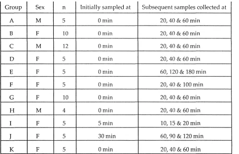
Table 2.1: Experimental groups
Group Sex n Initially sampled at Subsequent samples collected at
A M 5 0 min 20, 40 & 60 min
B F 10 0min 20, 40 & 60 min
C M 12 0min 20, 40 & 60 min
D F 5 0 min 20, 40 & 60 min
E F 5 0min 60, 120 & 180 min
F F 5 0min 20, 40 & 100 min
G F 10 0 min 20, 40 & 60 min
H M 4 0 min 20, 40 & 60 min
I F 5 Smin 10, 15 & 20 min
J
F 5 30min 60, 90 & 120 min2.2.2 Comparisons of plasma corticosterone levels in different
groups of mallards
i 4
Ducks vvere obtained from wild or captive situations, so a comparison
of corticosterone levels in mallards in response to capture and after various
periods of captivity could be made using initial samples from group A to H
ducks. Samples collected· at 20 min intervals were used to compare
corticosterone responses to sampling stress between groups of ducks
obtained from wild or captive situations. Along with samples collected at 20
min intervals, samples collected at various other frequencies were used to
describe corticosterone responses of ducks to sampling stress.
2.2.2.1 Plasma corticosterone levels in wild and captive
mallards
Vvild ducks 1\'ere caught in traps placed on the edge of ponds 2 km
from the Avian Physiology Unit. The traps consisted of two compartments
(2.0 x 1.0 x 1.0111 and 2.0 x 2.0 x 1.0m; length x vvidth x height) with funnel
entrances from the outside into one compartment and between
compartn1ents. The traps were baited ,vith wheat after several days of
pre-baiting and checked the following morning. After the trap had been
darkened with a large cover ducks were removed and sampled individually.
After initial blood sample collection ducks were placed into individual
darkened boxes (0.40 x 0.25 x 0.30m; length x width x height) and further
samples collected every 20 min for 1 h. Ducks sampled in this way are
included in groups A and B (Table 2.1).
Some ducks (groups C and D) were taken from the trap and placed into
a darkened communal box (0.94m x 0.56m x 0.34m; length x width x height),
transported to the Avian Physiology Unit (a journey which lasted 15 to 30
min), left for 1 h and then sampled. In comparison with groups A and B
ducks, group C and D ducks allow effects of communal boxing on
1 5
were collected every 20 min for 1 hr with the ducks being placed in
individual boxes betv:een sampling.
Captive ducks were held at the Avian Physiology Unit in either
outdoor pens or aviaries. On experimental days ducks in outdoor pens were
herded into an aviary and confined ·within a darkened space approximately
1 m in diameter. Ducks \Vere individually removed from the group and
taken to another room \Vere they \Vere restrained by a handler and the
initial blood sample taken: Ducks san1pled in this way are included in
groups E, F and G v,1ith the time spent in captivity prior to sampling being of
1, 6 or 26 weeks duration respectively. After the initial blood sampling,
ducks v,:ere placed in individual darkened boxes and further samples
collected at various frequencies.
Ducks housed in aviaries were also confined within a darkened space
approximately 1 m in diameter within their aviary before being individually removed and taken to another room for sampling. Again, samples were
collected every 20 min for 1 h with these ducks being included in group H.
An additional group of ducks ,vere reared indoors from the day of
hatching as part of a separate experiment performed by Dr. Cockrem and
were sampled for con1parison (group K). The ducks reared indoors were
sampled in the same ,vay as those in the aviary with samples also being
collected at 20 IT1in intervals for 1 h.
Ambient temperatures at a site 1 km from the Avian Physiology Unit
were obtained from the Grasslands Division, Department of Scientific and
Industrial Research, Palmerston North to determine whether temperature
differences may have been responsible for the variation in corticosterone
levels bet\-veen groups E to H ducks who were sampled on different days.
2.2.2.2 Plasma corticosterone responses of wild and captive
mallards to repeated sampling at 20 min intervals
As shown in Table 2.1, groups A to D, G, H and K were sampled every
20 min for 1 h. This standard sampling regime allmved comparisons of the
captive ducks .
2.2.2.3
1 6
Effects on plasma corticosterone levels of the time
taken to obtain a blood sample, the order of sampling
and various frequencies of repeated sampling
Only captive female mallards held in an outdoor pen were used for
this experiment (groups E to G, I and J). Ducks ,,vere sampled as described earlier for ducks from the outdoor pen. After the first sample (or, in groups I
and
J,
handling but not sampling) ducks ,vere placed in individual darkenedboxes until and after the collection of subsequent samples as outlined in
Table 2.1.
Measurement of plasn1a corticosterone
Corticosterone levels were n1easured by radioimmunoassay in
extracted plasma. Briefly, 100µ1 plasma samples v,rere extracted into 1 ml
dichlorornethane. After mixing and centrifugation a 500µ1 aliquot of the
dichlorornethane ·were removed and placed in another test tube and dried
under a stream of air at 37()c. Dried extracts were reconstituted in 200µ1 of
phosphate-buffered saline ,vi th gel a tin and left overnight at 4°c. Aliquots of
buffer (20 µ1) ·were removed, n1ade up to 100µ1 and frozen at -20°c until
assayed. The extraction efficiency measured using a spike of tritiated
corticosterone ·was 93.8 ± 2.7%.
The antiserum (batch No. B3-163, Endocrine Sciences, Tarzana, CA)
cross-reacted with other steroids at < 1.0%: progesterone (0.6%),
desoxycorticosterone (0.2%), testosterone (0.1 %), estradiol (0.03%) and
aldosterone (0.02%). Reconstituted extracts were incubated with 100µ1 of
antiserum and 100µ1 of tritiated corticosterone (approximately 5,000 cpm;
Amersham) overnight at 4°C. Separation of bound and free hormone was
achieved by the addition of 0.5 ml of dextran coated charcoal. Tubes were
1 7
promptly centrifuged at 3000 rpm in a Heraeus Christ 5000S refrigerated
centrifuge. A 500µ1 aliquot of the supernatant (containing bound labelled
corticosterone) was removed and placed in a disposable scintillation vial.
Scintillant mixture (0.005g dimethylPOPOP, 0.05g POP, 2 1 toluene) was
added (3 ml), and the tubes shaken for 1 h in an orbital shaker. After 1 h
vials were counted in a Beckmann LS7500 scintillation counter for 10 min.
Displacement curves for increasing amounts of duck plasma and of the
corticosterone standard ,,vere compared following logit-log transformation
and found to be parallel. Corticosterone (0.62 to 10.00 ng/ml) was added to
duck plasma and ,vas quantitatively recovered (102.2 ± 5.6%, n=4). The limit
of sensitivity of the assay ,vas 0.40 ng/ml. The intra- and inter-assay
coefficients of variation were 6.sc1c, and 9.3% respectively.
2.2.4 Statistical analyses
Corticosterone levels in the first sample obtained from each duck were
compared between groups using one-way ANOV A with Bonferroni
adjusted probability levels used for subsequent post-hoc tests. Where the
Bartletts test indicated heterogeneity of variance comparisons of
corticosterone levels between each group was performed using
Kruskal-Wallis nonparametric ANOV A.
The effect of tirne, or sampling stress, on corticosterone levels within
each group of ducks was also investigated by one-v;ay repeated measures
ANOVA with Bonferroni adjusted probability levels used for· subsequent
post-hoc tests. Where the Bartletts test indicated heterogeneity of variance
the effect of sampling stress on corticosterone levels within each group was
investigated by using Friedn-1an nonparametric ANOVA.
Relationships between corticosterone levels and either ambient
temperature or the time taken to obtain a blood sample were examined
using regression analysis. Effects of sampling order on corticosterone levels
were examined using the Kruskal-Wallis nonparametric ANOVA
1 8
Log transformed data ,vere used for statistical analysis. Summary data
1 9
2.3 Results
2.3.1 Plasma corticosterone levels in ,vild and captive mallards
There was large variation in corticosterone levels between groups of
mallards obtained from various environments (Fig 2.01). Corticosterone
levels in both male (group A) and female ducks (group B) were significantly
higher when they v,'ere sampled directly from the trap than if they were
obtained from a darkened comnnmal box 1 h after transportation from the
trap (p<0.05, group C and D).
For the ducks kept in captivity, corticosterone levels were significantly
higher in ducks held in an outdoor pen for 1 (group E) or 6 weeks (group F)
than levels in ducks held for 5 months (group G, p<0.01). The ducks held in
an outdoor pen for either 1 or 6 weeks also had significantly higher
corticosterone levels than ducks held in aviaries (group H, p<0.05) or reared
in an indoor room (group K, p<0.01). The ducks held for 5 months in an
outdoor pen (group G) had similar corticosterone levels to ducks held in
aviaries (group H, p=0.719), but significantly higher levels than in ducks
reared in an indoor roon1 (group K, p<0.05). The corticosterone levels in
ducks reared in an indoor room \Vere not different from levels in ducks
held in aviaries (group H, p=0.395).
Some experimented groups had a larger range of corticosterone values
than other groups, as shown in Figure 2.02. All the male ducks from the trap
(group A) had higher corticosterone levels than male ducks sampled from a
communal box (group C). For the female ducks, only 3 of 7 ducks sampled
from the trap (group B) had corticosterone levels higher than the females
from a communal box (group D).
The effect of time in captivity on corticosterone levels can also be seen
by comparing individual corticosterone values. Of the ducks held for 1
(group E) or 6 '"'eeks (group F) in an outdoor pen only 1 of the 10 ducks had
corticosterone level as low as the levels in any of the ducks that had spent 5
months (group G) in cap ti vi ty. All of the 9 ducks held for 5 months in an
,--...
E
...
0) C:
._,..,
(1) C:
0
~
(1)
,4--1 (./)
0
u
,4--1
~
0
u
80-(7)
60-
T
a
b
C
d
e
f
g
h
a &
b -
Male (a) and female (b) ducks from trap (groups A and B respectively)c
&d -
Male (c) and female (d) ducks trapped, transported and left for 1 h in a communal box (groups C and Drespectively)
e tog -
Female ducks after 1 wk (e), 6 wks (f) and 5 months (g) in outdoor pen (groups E, F and G respectively)h - Male ducks after 2 months in an outdoor pen followed by 1 month in an aviary (group H)
i - Female ducks 3 months old, reared from hatch in an indoor room (group K)
[image:36.588.70.505.89.594.2]125
-1111111
E
100-,
0) C ...__., Q) C 0 l... Q) -4-1 (/) 0u
-4-1 l... 0u
Ill 75-11111111111111Ill Ill
Ill
50 -
Ill25
-I 1111111 1111111 I
1111111
I 11111111111111111111111
I
1111111 I I - 11111111111111111111111 I
- 111111111111111111111 I 1111111
I 1111111111 I
Ill Ill Ill
Ill I
Ill I Ill
. , . 1111
111111111 111
Ill 1 111 111•1 I 111111111 Ill Ill
I I Ill I
Ill 111111 1111111 Ill 0-'--,--'--,c--_._--r-___.__,c--_._-.-___.__c--_._---.-_.__-.-~=·~·~·=
a
b
C
d
e
f
g
h
a & b - Male ( a) and female (b) ducks from trap (groups A and B respectively)
c
&d -
Male ( c) and female ( d) ducks trapped, transported and left for 1 h in a communal box (groups C and Drespectively)
e tog -
Female ducks after 1 wk (e), 6 wks (f) and 5 months (g) in outdoor pen (groups E, F and G respectively)h -
Male ducks after 2 months in an outdoor pen followed by 1 month in an aviary (group H) [image:37.567.68.514.179.699.2]i -
Female ducks 3 months old, reared from hatch in an indoor room (group K)20
held in aviaries (group H), but only 1 of the 9 ducks had levels as low as
levels in ducks reared in an indoor room (group K).
The variation in corticosterone levels between groups of ducks held for
varying lengths of time in captivity was not related to the ambient
temperature on the day they '\Vere sampled (p=0.617, Fig 2.03).
Plasma corticosterone responses of '\vild and captive
mallards to repeated sampling at 20 min intervals
Mean corticosterone levels in both male (group A) and female ducks
(group B) obtained from the trap were significantly lower at 20 min than
levels at 0 min, with this decrease sustained until 60 min (p<0.05, Fig 2.04).
Corticosterone levels in all 5 n1ale (group A) and 7 female ducks (group B)
were lower at 20 rn.in than levels at 0 min with this decrease sustained in all
5 male ducks and in all 5 of the female ducks from which complete sets of
blood samples were obtained (Fig 2.05).
No change in mean corticosterone levels was seen in response to
repeated blood san1pling in either male (group C, p=0.438) or female ducks
(group D, p=0.714) obtained from a communal box (Fig 2.04). Mean
corticosterone levels at 20, 40 and 60 min in both male and female ducks
obtained from the communal box ,vere not significantly different from
levels measured at 20, 40 and 60 min in ducks obtained directly from the
trap. Corticosterone levels generally remained relatively constant during
repeated sampling of individual male ducks obtained from the communal
box (group C, Fig 2.05). In the female ducks from the communal box (group
D) corticosterone levels in 2 of the 5 ducks remained below 12 ng/ml
whereas the other 3 female ducks had high levels (Fig 2.05).
Mean corticosterone levels in female ducks from the outdoor pen
(group G) decreased in relation to levels measured at 0 min (p<0.05, Fig
2.04). A decrease in corticosterone levels was seen in 7 of these 9 ducks (Fig
2.05). No change in mean corticosterone levels (p=0.272) was seen for the
male ducks from an aviary (group H, Fig 2.04) with levels remaining
125
-,,--..._
E
'
100
-0)
C
____..
IllQ.)
C
0
~
Q.)
...,
(/)0
u
...,
~
0
u
75-Ill 75-Ill
Ill
50-
Ill Ill11111 Ill 11111111 Ill
11111
••
11111111111 1111111111 11111111 11111
I
II Ill 111125-
Ill Ill 1111·=
1111
11111 Ill 11111 Ill Ill Ill 11111 II Ill
1111
..
11111 •1111
1111 1111 1111
1111 II 1111 11111
0
-2.5
5.0
7.5
10.0
12.5
15.0
Temperature (°C)
[image:39.568.84.502.96.553.2]80
a
8060 60
40 40
20 (9) (8) (10)
(8)
0 0
80
b
80f
(7)
60
T
60,,,..._
l
E
40 (9) (7) 40T T
...
l. .L (4) (4)
0) T ..,.
C 20 20 .1
..._.,
....(])
0
C 0
0 80 c8o
g
~
(]) .µ
60 60
(/) 0
u
(9).µ 40 T
~ (12)
0 T
u
20 l 20 (4)T
0 0
0 20 40 60
80
d
60
40
(5) (5)
T T
20 l
l
0
0 20 40 60
Time (min)
a & b - Male (a) and female (b) ducks from trap (groups A and B respectively)
C & d - Male (c) and female (d) ducks after transportation and 1
h in communal box (groups C and D respectively)
e - Female ducks after 5 months in an outdoor pen (group G) f - Male ducks after 2 months in an outdoor pen followed by
1 month in an aviary (group H)
g - Female ducks 3 months old, reared from hatch in an indoor room (group K)
[image:40.567.132.511.18.757.2],--....
E
'
0)C
.,__,.
(l) C 0 "-(l) +J U) 0u
+J "-0u
80 80
e
60 60
40 40
II
20 20
0 0
120 80
f
100
60 80
60 40
40
20
20
t
=
~~ Fl ::S0 0
80
C
80g
60 40 60
/
40 20 0 20&::=:
0 80d
0 20 40 6060
40
20
0
0 20 40 60
Time (min)
a & b - Male (a) and female (b) ducks from trap (groups A and B respectively)
c & d - Male (c) and female (d) ducks after transportation and 1 h in communal box (groups C and D respectively)
e - Female ducks after 5 months in an outdoor pen (group G) f - Male ducks after 2 months in an outdoor pen followed by
1 month in an aviary (group H)
g - Female ducks 3 months old, reared from hatch in an indoor room (group K)
21
No significant change in mean corticosterone levels (p=0.168) occurred
with sampling in female ducks obtained from an indoor room (group K, Fig
2.04). Hmvever, in 4 of the 5 ducks corticosterone levels at either 20 or 40
min had reached 12 ng/ml or more and were higher than levels measured
at O min. Corticosterone levels in one group K duck never rose above 4 ng/ ml (Fig 2.05).
2.3.3 Effects on plasma corticosterone levels of the time taken to
obtain a blood sample, the order of sampling and various
frequencies of repeated sarnpling
2.3.3.1 Effects on plasma corticosterone levels of the time
taken to obtain a blood sample and of the order of
sampling
Corticosterone levels in female ducks from groups E, F and G did not
exhibit any significant trend ·with time '"·hen samples were obtained within
90 sec of the duck being handled (p=0.567, Fig 2.06). The removal of a duck
from its group did not have any significant effect on the corticosterone
levels in the re1T1aining ducks of that group, because there ivas no
relationship bet,veen the order in which a duck was picked up for sampling
and its corticosterone level (p=0.132, Fig 2.07).
2.3.3.2 Plasma corticosterone responses of mallards to various
frequencies of repeated sampling
Mean corticosterone levels in female ducks sampled every 20 min for 1
h (group G) decreased in relation to levels measured at 0 min (p<0.05, Fig
2.08). A decrease in corticosterone levels was seen in 7 of these 9 ducks (Fig
2.09).
Sampling every 20 min for 40 min and again at 100 min (group F), did
not significantly change mean corticosterone levels from levels at 0 min
,,,--....
120
E
...
•
0)
100
C
...__,
•80
(l) • •
C • •
0
60
•
L •
(l) •
.j-1
40
• • • • • • • • • • •(/)
··= •
0 a • • • •• a • a
u
20
• • • • • • •.j-1 a • • • a • ••
L • a a a •
0
0
• ••
• • • •u
40
50
60
70
80
90
Time taken to obtain
a blood sample (sec)
~
50
E
( 11 )
...
40
T
0)
C
..._,,
(J.)
30
C
0
!...
(J.)
20
.µ
(/')
0
u
10
.µ!...
0
u
0
1
2
3
4
5
Order of sampling
Q) C:
0
~
Q)
.µ
(J)
0
u
.µ~
0
u
80
60
40
20
0 I I
111 Sampled every 20 min (group G)
□ Sampled at 0, 20, 40 & 100 min (group F) • Sampled every 60 min (group E)
o Sampled at 30, 60, 90 & 1 20 min, handled only at 0 min (group J)
x Sampled at 5, 10, 1 5 & 20 min, handled only at 0 min (group I)
I I I I
0
10 20
30 40
60 90 100 120 180
Time (min)
E
'
0)C
..._,,,
Q) C 0 ~ Q) .l,,J (/) 0u
.l,,J ~ 0u
80 60 40 100 80 60 40 20 0 80 60 40 20 0a
80d
60
40
20
0
0 20 40 60 0 30 60 90 120
b
0 20 40 60 80 1 00
C
0 60 120 180
100 80 60 40 20 0 0
Time (min)
a Sampled every 20 min (group G)
5
b
Sampled at 0, 20, 40 & 100 min (group F) c Sampled every 60 min (group E)10 15 20
d
Sampled at 30, 60, 90 & 120 min, handled only at 0 min (group J)e Sampled at 5, 10, 1 5 & 20 min, handled only at 0 min (group I)