R E S E A R C H
Open Access
2-Cys peroxiredoxin is required in
successful blood-feeding, reproduction, and
antioxidant response in the hard tick
Haemaphysalis longicornis
Kodai Kusakisako
1,2, Remil Linggatong Galay
3, Rika Umemiya-Shirafuji
4, Emmanuel Pacia Hernandez
1,2,
Hiroki Maeda
1,2, Melbourne Rio Talactac
1,2, Naotoshi Tsuji
5, Masami Mochizuki
1, Kozo Fujisaki
6and Tetsuya Tanaka
1,2*Abstract
Background:Ticks are obligate hematophagous arthropods that feed on vertebrate blood that contains iron. Ticks also concentrate host blood with iron; this concentration of the blood leads to high levels of iron in ticks. The host-derived iron reacts with oxygen in the tick body and this may generate high levels of reactive oxygen species, including hydrogen peroxide (H2O2). High levels of H2O2cause oxidative stress in organisms and therefore,
antioxidant responses are necessary to regulate H2O2. Here, we focused on peroxiredoxin (Prx), an H2O2-scavenging
enzyme in the hard tickHaemaphysalis longicornis.
Methods:The mRNA and protein expression profiles of 2-Cys peroxiredoxin (HlPrx2) inH. longicorniswere investigated in whole ticks and internal organs, and developmental stages, using real-time PCR and Western blot analysis during blood-feeding. The localization of HlPrx2 proteins in tick tissues was also observed by
immunostaining. Moreover, knockdown experiments ofHlPrx2were performed using RNA interference to evaluate its function in ticks.
Results:Real-time PCR showed thatHlPrx2gene expression in whole ticks and internal organs was significantly upregulated by feeding. However, protein expression, except in the midgut, was constant throughout blood-feeding. Knockdown of theHlPrx2gene caused significant differences in the engorged body weight, egg weight and hatching rate for larvae as compared to the control group. Finally, detection of H2O2after knockdown ofHlPrxs
in ticks showed that the concentration of H2O2significantly increased before and after blood-feeding.
Conclusion:Therefore, HlPrx2 can be considered important for successful blood-feeding and reproduction through the regulation of H2O2concentrations in ticks before and after blood-feeding. This study contributes to the search
for a candidate target for tick control and further understanding of the tick’s oxidative stress coping mechanism during blood-feeding.
Keywords:Peroxiredoxin,Haemaphysalis longicornis, RNA interference, Hydrogen peroxide
(Continued on next page)
* Correspondence:tetsuya@ms.kagoshima-u.ac.jp
1Laboratory of Infectious Diseases, Joint Faculty of Veterinary Medicine,
Kagoshima University, Korimoto, Kagoshima 890-0065, Japan
2Department of Pathological and Preventive Veterinary Science, United
Graduate School of Veterinary Science, Yamaguchi University, Yoshida, Yamaguchi 753-8515, Japan
Full list of author information is available at the end of the article
(Continued from previous page)
Abbreviations:H2O2, Hydrogen peroxide; Prx, Peroxiredoxin; HlPrx2,Haemaphysalis longicornis2-Cys peroxiredoxin;
ROS, Reactive oxygen species; SDS-PAGE, Sodium dodecyl sulfate-polyacrylamide gel electrophoresis; HlPrx,Haemaphysalis longicornis1-Cys peroxiredoxin; PBS, Phosphate buffered saline; IFAT, Indirect
immunofluorescent antibody test; RNAi, RNA interference;dsHlPrx2, The double-stranded RNA ofHlPrx2;dsHlPrx, The double-stranded RNA ofHlPrx;dsLuc, The double-stranded RNA of the fireflyluciferase;dsDouble, The double-stranded RNA mixture ofdsHlPrxanddsHlPrx2at 1μg concentration each; SA, The acinar cells of the salivary glands; SGG, The granular cells of the salivary glands; SD, The salivary duct
Background
In high concentrations, hydrogen peroxide (H2O2) is
known to be a harmful chemical compound to aerobic organisms due to its ability to seriously damage mem-brane lipids, nucleic acids, and proteins [1]. Almost all aerobic organisms have developed defense systems to scavenge H2O2. Catalases, peroxidases, and
peroxiredox-ins (Prxs) are scavengers of H2O2 [2]. Prxs are
ubiqui-tous antioxidant enzymes investigated in various organisms [3]. Particularly, high levels of Prxs are pro-duced in mammalian cells, including erythrocytes [4]. Erythrocytes are exposed to more oxidative stress than any other cell type, due to the abundance of heme iron and oxygen, which can generate H2O2 [5]. These
indi-cate that Prxs may have important roles in peroxide de-toxification in cells.
Prxs can be divided into two groups according to the presence of one or two highly conserved cysteines in or-ganisms, 1-Cys or 2-Cys Prx [3]. 2-Cys Prxs are identified by two conserved cysteines, peroxidatic and resolving cys-teines [6]. On the other hand, the 1-Cys Prxs have a con-served peroxidatic cysteine and do not contain a resolving cysteine [7]. Enzymes of the Prx family exhibit antioxidant activity that catalyzes the reduction of H2O2 into water
(H2O), with thioredoxin as an immediate hydrogen donor
or donor thiol, respectively [7].
In some endoparasites, such as Plasmodium and Fas-ciolaparasites, Prxs have been characterized as antigens or secreted proteins, suggesting that endoparasite Prxs may participate in interactions between the parasites and their hosts [8, 9]. Therefore, to evaluate the efficacy of antigens for these endoparasites, basic biological and bio-histological analyses such as mRNA and protein ex-pression profiles, and the localization of proteins in these parasites have been studied.
Ticks need blood meals to develop from one stage to the next and for reproduction. Blood-feeding and the di-gestion of blood provide nutrition and energy for molt-ing, development, and the vitellogenesis of ticks [10]. Ticks feed on vertebrate blood that contains iron, such as heme, ferrous iron, and other pro-oxidants. Ticks also concentrate host blood with iron; this concentration of the blood leads to high levels of iron in ticks.
Host-derived iron may react with oxygen in the tick body, and then high levels of reactive oxygen species (ROS), in-cluding H2O2, may be generated [11]. Haemaphysalis
longicornis 1-Cys Prx (HlPrx) has been reported previ-ously; however, there is still little knowledge about the biological functions of Prxs in ticks [12].
In the present study, we analyzed mRNA and protein expression profiles and the localization of proteins in tick tissues of H. longicornis 2-Cys Prx, HlPrx2, identi-fied previously [13]. Moreover, HlPrx and/or HlPrx2
gene silencing was performed to clarify their functions in ticks using RNA interference. Finally, we demon-strated that the double knockdown ofHlPrxand HlPrx2
led to increased oxidative stress in ticks.
Methods Ticks and animals
The parthenogenetic Okayama strain of H. longicornis
has been maintained by blood-feeding on the ears of Jap-anese white rabbits (KBT Oriental Co. Ltd, Saga, Japan) in the Laboratory of Infectious Diseases, Joint Faculty of Veterinary Medicine, Kagoshima University [14]. Rabbits were cared for in accordance with the guidelines ap-proved by the Animal Care and Use Committee of Kago-shima University (Approval no. VM13007) and maintained under regulated conditions throughout the experiments.
Total RNA extraction and cDNA synthesis
To extract total RNA, whole ticks were homogenized using an Automill (Tokken, Chiba, Japan), while dissected organs were disrupted using a pellet pestle motor (Sigma-Aldrich, St. Louis, MO, USA). The extracted RNA was purified using TRI Reagent (Sigma-Aldrich), and then treated with an RQ1 RNase-Free DNase (Promega, Madison, WI, USA). cDNA synthesis was performed with ReverTra Ace-α- (Toyobo, Osaka, Japan) fol-lowing the manufacturer’s protocol using 1 μg of total RNA.
Expression analysis ofHlPrx2mRNA
SYBR qPCR Mix (Toyobo) with a 7300 real-time PCR system (Applied Biosystems, Foster City, CA, USA). Gene-specific primers were designed to target HlPrx2
and the internal control genes, as shown in Table 1. Standard curves were made from four-fold serial dilu-tions of the cDNA of adult ticks fed for three days. The PCR cycle profile was as follows: initial denaturation at 95 °C for 10 min, 40 cycles of a denaturation step at 95 °C for 15 s, and an annealing/extension step at 60 °C for 60 s. The data was analyzed with 7300 system SDS software (Applied Biosystems). At the first step of real-time PCR,
actin, tubulin, P0, and L23 genes were evaluated for standardization andL23was selected as the tick reference in the current study.
Production of an antiserum against recombinant HlPrx2 To prepare mouse anti-HlPrx2 sera, 100 μg of recom-binant HlPrx2 (rHlPrx2; [13]) was completely mixed with Freund’s complete adjuvant (Sigma-Aldrich) and in-traperitoneally injected into ddY female mice (four weeks old, Kyudo, Saga, Japan). After two weeks, these mice were injected with 100μg of rHlPrx2 with Freund’s
incomplete adjuvant (Sigma-Aldrich) twice at a two-week interval to boost the generation of antibodies against rHlPrx2. Their blood was collected two weeks after the third immunization to obtain specific antisera to rHlPrx2.
Protein extraction and Western blot analysis
[image:3.595.57.545.374.724.2]Homogenized ticks were suspended in phosphate buff-ered saline (PBS) and ultrasonicated three times, two mi-nutes each (Vibra-CellTM; Sonics and Materials, Newtown, CT, USA) on ice and finally centrifuged at 500× g. The supernatant was resolved in a 12 % SDS-PAGE gel under reducing conditions. After sodium do-decyl sulfate-polyacrylamide gel electrophoresis (SDS-PAGE), the proteins were transferred onto a polyvinyli-dene difluoride membrane (Immobilon -P; Millipore, Danvers, MA, USA). The membrane was blocked over-night with 3 % skim milk in PBS (pH 7.4) (blocking solu-tion); it was incubated with a 1:500 dilution of anti-rHlPrx2 mouse sera in blocking solution at 37 °C for 1 h. For loading control, tubulin was detected using anti-serum against recombinant H. longicornis tubulin [15].
Table 1Gene-specific primers used in this study
Primer Sequence (5'–3')
HlPrx2 RT-F TATGCCTAAGCTGGCGAAGC
HlPrx2 RT-R CAGGCGAGGTGAGAGAAGTG
HlPrx RT-F ATGAGGTCCTCCGTGCTACT
HlPrx RT-R TGCCACACCGTCATAAGCAT
HlPrx2 real time-F GTGTGCCCTGCTAACTGGAA
HlPrx2 real time-R ATGAGACACACGGGGCTTTG
HlPrx2 T7-F TAATACGACTCACTATAGGGATCAAGCTGTCCGATTACAAGAAC
HlPrx2 T7-R TAATACGACTCACTATAGGTTCCAGTTAGCAGGGCACACT
HlPrx2 RNAi-F GATCAAGCTGTCCGATTACAAGAAC
HlPrx2 RNAi-R TTCCAGTTAGCAGGGCACACT
HlPrx T7-F TAATACGACTCACTATAGGCACCACGGTTGGATCAAGGA
HlPrx T7-R TAATACGACTCACTATAGGTTTGCAGAGCCACCACTCAA
HlPrx RNAi-F CACCACGGTTGGATCAAGGA
HlPrx RNAi-R TTTGCAGAGCCACCACTCAA
Actin RT-F CCAACAGGGAGAAGATGACG
Actin RT-R ACAGGTCCTTACGGATGTCC
Actin real time-F ATCCTGCGTCTCGACTTGG
Actin real time-R GCCGTGGTGGTGAAAGAGTAG
Tubulin real time-F TTCAGGGGCCGTATGAGTAT
Tubulin real time-R TGTTGCAGACATCTTGAGGC
P0 real time-F CTCCATTGTCAACGGTCTCA
P0 real time-R TCAGCCTCCTTGAAGGTGAT
L23 real time-F CACACTCGTGTTCATCGTCC
L23 real time-R ATGAGTGTGTTCACGTTGGC
After washing five times in PBS containing 0.05 % Tween 20 (PBS-T), the membrane was incubated with a 1:50,000 dilution of horseradish peroxidase (HRP)-con-jugated sheep anti-mouse IgG (Dako, Glostrup, Denmark) in blocking solution at 37 °C for 1 h. After washing five times in PBS-T, bands were detected using AmershamTMECLTMPrime Western Blotting Detection Reagent (GE Healthcare, Buckinghamshire, UK) and viewed using FluorChem FC2 software (Alpha Inno-tech, San Leandro, CA, USA). To accurately determine differences in the protein expression, band densitometry analysis was performed using Alpha View Software (Alpha Innotech). The band densitometry analysis re-sults shown in this study represent the mean of three tri-als of Western blot analysis.
Immunostaining
To confirm the localization of HlPrx2 in tick tissues, in-direct immunofluorescent antibody tests (IFAT) were performed. Engorged ticks were dissected under a stereomicroscope (SZX10, Olympus, Tokyo, Japan) for collecting tick internal organs. Dissected organs were fixed in a 4 % paraformaldehyde phosphate buffer solu-tion (pH 7.4) that included 0.1 % glutaraldehyde at 4 °C overnight. After washing with a sucrose series, organs were embedded in a Tissue-Tek O.C.T Compound (Sakura Finetek, Torrance, CA, USA). Frozen sections from each internal organ were cut to a thickness of 10 μm using Kawamoto's film method (Leica Microsys-tems, Tokyo, Japan) and a cryostat (Leica CM 1850, Leica Microsystems, Wetzlar, Germany). The films were blocked with 5 % skim milk in PBS (pH 7.4) (blocking solution) at 37 °C for 1 h, and then incubated with 1:50 dilution in a blocking solution of anti-HlPrx2 mouse serum at 37 °C for 1 h. For the negative control, normal mouse serum was used. After washing three times in PBS, the slides were incubated at 37 °C for 1 h with Alexa Fluor 594 goat anti-mouse IgG (Invitrogen, Carlsbad, CA, USA) with 1:1,000 dilution in the blocking solution. After removing the antibody by washing three times with PBS, the films were placed on a glass slide and mounted with DAPI (VECTASHIELD ; Vector La-boratories, Burlingame, CA, USA), and then covered with a cover glass. The images were recorded using a confocal laser scanning microscope (LSM700, Carl Zeiss, Jena, Germany). Hemocytes were prepared using a glass slide instead of film as described previously by Galay et al. [16]. Briefly, hemolymph collected from ticks by am-putating the legs was smeared directly on glass slides and air-dried. After drying, hemocyte smears were fixed with 4 % paraformaldehyde in PBS that included 0.1 % glutaraldehyde. Thereafter, the same method used for the internal organs’IFAT was performed.
RNA interference (RNAi)
Two separate PCR reactions of approximately 469 bp with a single T7 promoter were generated using the fol-lowing primer sets: a T7-attached gene-specific forward primer (HlPrx2 T7-F) and gene-specific reverse primer (HlPrx2 RNAi-R) and a T7-attached gene-specific re-verse primer (HlPrx2 T7-R) and gene-specific forward primer (HlPrx2 RNAi-F) (Table 1). After gel purification of PCR products using a GENECLEAN II KIT (MP Biomedicals, Irvine, CA, USA), double-stranded RNA of
H. longicornis 2-Cys peroxiredoxin (dsHlPrx2) was syn-thesized using the T7 RiboMAX™Express RNAi System (Promega) with two separate single-promoter templates in accordance with the manufacturer’s protocol. Double-stranded RNA of H. longicornis 1-Cys peroxiredoxin (dsHlPrx) was also synthesized usingHlPrxgene-specific primers (Table 1). The firefly luciferase (Luc) gene [17] was used for control (dsLuc group). One microgram of
dsHlPrx, dsHlPrx2, or dsDouble (dsHlPrx and dsHlPrx2
were mixed at 1 μg concentration each) was injected into 30 unfed adult female ticks in each experimental group and dsLuc group through the fourth coxae into the hemocoel. Injected ticks were observed for one day and subsequently transferred to rabbits with each group infesting separate ears. Three to four days after attach-ment, three ticks were manually detached to confirm gene silencing using RT-PCR. The remaining ticks were allowed to feed until engorgement, and the total number of engorged ticks, the engorged body weight, the ovipos-ition, and the hatching rate were assessed.
Detection of hydrogen peroxide (H2O2) in adult female
ticks during blood-feeding
The H2O2 concentration in ticks was measured using
the ferrous oxidation of xylenol orange assay [18]. Briefly, homogenized unfed and partially fed ticks were suspended in 200μl of Milli-Q H2O, while homogenized
engorged ticks were suspended in 900 μl of Milli-Q H2O. The samples were centrifuged at 500× g, and the
supernatant was collected. The supernatant from the engorged ticks was further diluted 10 times in Milli-Q H2O. Ninety microliters of the supernatant from unfed
and partially fed ticks or the diluted supernatant from engorged ticks was used for a sample solution as described later. The assay reagent consisted of 125 μM xylenol orange, 250 μM ammonium iron (II) sulfate, 100 mM sorbitol, and 25 mM sulfuric acid. One hun-dred microliters of the sample solutions was added to a 1-ml assay reagent. The mixture was vortexed immedi-ately, left at room temperature for 30 min, and measured at 560 nm using a spectrophotometer (Ultrospec 2100 pro; GE Healthcare, Pittsburgh, PA, USA). Finally, the ratio of the H2O2concentration (μM) to the corresponding
Statistical analysis
All experiments were conducted in two or three separate trials. Data except for hatching rate were statistically an-alyzed using Welch’s t-test. Hatching rate analysis was done using the chi-square test. P< 0.05 and P< 0.01 were considered to be statistically significantvscontrol.
Results
Transcription profiles ofHlPrx2
The mRNA levels of HlPrx2 in whole female ticks and internal organs during blood-feeding and in different de-velopmental stages (egg, larval, nymphal and adult
stages) were investigated using real-time PCR. HlPrx2
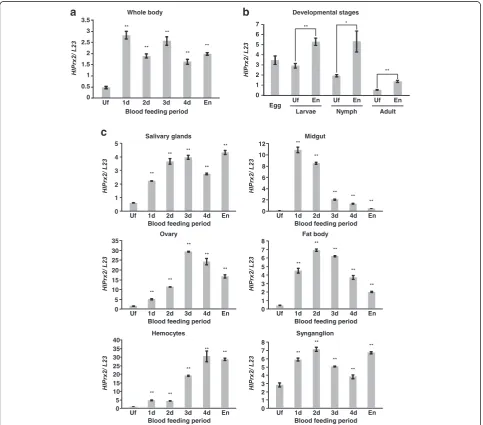
mRNA was upregulated in whole female ticks, develop-mental stages, and all internal organs (salivary glands, midgut, ovaries, fat bodies, synganglia and hemocytes) during blood-feeding (Fig. 1). In the whole body, mRNA was upregulated at day 1 and, in spite of higher expres-sion levels as compared to those of unfed stage (Welch’s
t-test: t(2)= 23.43, P= 0.002), gradually decreased
there-after (Fig. 1a). Upregulation of the mRNA level was also observed in the developmental stages from unfed to en-gorgement, and the immature stages, including the egg, showed higher expression levels as compared to the
Fig. 1aTranscription profiles ofHlPrx2in whole ticks during blood-feeding analyzed by real-time PCR (Uf, unfed females; 1d-4d, adults partially fed for 1–4 days).bTranscription profiles ofHlPrx2in unfed and engorged tick developmental stages.cTranscription profiles ofHlPrx2in the internal organs: salivary glands, midgut, ovary, fat body, hemocytes, synganglion).L23was used as the internal control. Data are presented as the mean ± standard deviation (SD).*P< 0.05;**P< 0.01, significant differencesvs dsLucby Welch’st-test.Abbreviations: Uf, unfed ticks; En,
[image:5.595.57.541.249.674.2]adult stage (Welch’s t-test: Larvae, t(2)= 9.77, P= 0.002;
Nymph, t(2)= 5.65, P= 0.030; Adult, t(2)= 12.39, P=
0.006) (Fig. 1b). In the midgut, mRNA drastically in-creased at day 1 (Welch’s t-test: t(2)= 36.31, P= 0.001)
and decreased thereafter (Fig. 1c, Midgut). In the ovary, the expression level gradually increased until day 2, then drastically increased at day 3, and decreased thereafter (Fig. 1c, Ovary) (Welch’s t-test: day 2, t(2)= 81.42, P<
0.001; day 3, t(2)= 174.42, P< 0.001). In the hemocytes,
the expression level increased at day 1 and remained al-most the same at day 2, drastically increased from day 3 to day 4 (Welch’st-test: day 1,t(2)= 34.28,P= 0.001; day
2,t(2)= 40.81,P= 0.001; day 3,t(2)= 86.14, P< 0.001; day
4, t(2)= 16.25, P= 0.004), and then slightly decreased at
the engorged state (Fig. 1c, Hemocytes). The expression levels of HlPrx2 gene in ovaries and hemocytes were higher than those of other internal organs. In other tis-sues, such as the salivary glands, fat bodies and syngan-glia, mRNA was upregulated from unfed to day 1 and remained at a high level until engorgement (Fig. 1c). These results indicate that the mRNA of HlPrx2 gene was upregulated in ticks by blood-feeding. The high levels of mRNA expression in the ovaries and hemocytes suggest that HlPrx2 gene may be related to the reproduction and immune response of ticks.
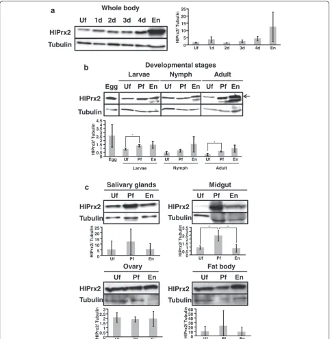
Protein expression profiles of HlPrx2
The protein expression levels of HlPrx2 in whole female ticks and internal organs during blood-feeding and in different developmental stages were investigated by Western blot analysis using HlPrx2-specific antisera. The predicted molecular mass of HlPrx2 protein is ap-proximately 22 kDa, and the theoretical isoelectric point (pI) is 6.8; the signal peptide and glycosylation sites were not found with sequence analysis [13]. However, the cal-culated molecular mass in Western blot analysis was ap-proximately 26 kDa. The mobility of native HlPrx2 protein in Western blot analysis decreased because the pI = 6.8 is slightly low. HlPrx2 expression was generally upregulated during blood-feeding in the whole body, the developmental stages, and the midgut (Fig. 2). In the whole body and the developmental stages, the HlPrx2 expression level was upregulated from unfed to engorge-ment (Fig. 2a, b). Notably, in the developengorge-mental stages, protein expression levels seemed to be almost the same, although immature stages, including the egg, showed higher mRNA expression levels as compared to those of the adult stage (Figs. 1b and 2b). In Fig. 2b, other bands under HlPrx2 band at the engorged state of all stages can be seen. These bands are considered to be non-specific bands derived from the blood of the host rabbit (see Additional file 1: Figure S1). These non-specific bands in the rabbit blood cross-reacted with HlPrx2 antisera; thus, these are considered to be a candidate
cross-reacting protein related to 2-Cys peroxiredoxin (see Additional file 1: Figure S1 and Additional file 2). Moreover, in the knockdown ticks, the band of HlPrx2 protein was decreased as compared to the control group (see Additional file 1: Figure S2). Therefore, the anti-HlPrx2 mouse serum used in this study was considered as specifically working.
In the midgut, although the protein expression level was very low in the unfed stage, it significantly increased from unfed to partially fed states (Welch’s t-test: t(2)=
3.66, P= 0.049) and significantly decreased in the engorged state (Welch’s t-test: t(2)= 3.34, P= 0.034)
(Fig. 2c, Midgut). In the salivary glands, ovaries, and fat bodies, the protein expression levels of HlPrx2 were constant during blood-feeding (Fig. 2c). These results in-dicate that the protein expression of HlPrx2 is strongly upregulated in the whole body, especially in the midgut, by blood-feeding; however, the expression levels of HlPrx2 protein in the other tissues, such as the salivary glands, ovaries and fat bodies, were constant during blood-feeding. The drastic increase of HlPrx2 protein ex-pression in the midgut during blood-feeding suggests that HlPrx2 protein could be related to the antioxidant re-sponse in this tissue because ticks’midgut may be exposed to high concentrations of ROS during blood-feeding.
Localization of HlPrx2 in the salivary glands, midgut, ovaries, and hemocytes from engorged adult female ticks Western blot analysis showed the high expression of HlPrx2 protein in the whole body and internal organs. To determine localization in the cells of internal organs, IFAT was performed using some internal organs of engorged female ticks. In the salivary glands, positive fluorescence was detected in the cell membrane of the acinar cells (SA) and granular cells (SGG) and in the basal lamina of the salivary duct (SD) (Fig. 3, Salivary glands). In the midgut, positive fluorescence was de-tected in the basal lamina of the digestive cells (Fig. 3, Midgut). In the ovary, positive fluorescence was detected in the cell membrane of the oocytes and basal lamina of the oviduct (Fig. 3, Ovary), whereas in the hemocytes, positive fluorescence was detected in the cell membrane (Fig. 3, Hemocytes). These results demonstrate that the HlPrx2 protein was associated to the tissue membranes.
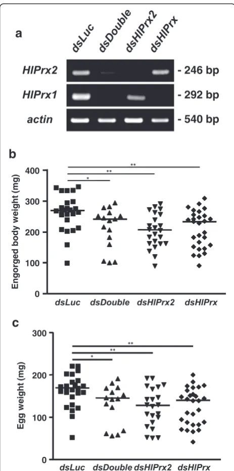
Effects ofHlPrxand/orHlPrx2gene silencing on the blood-feeding and reproduction of female ticks
To clarify the functions of the HlPrx and HlPrx2
genes, gene silencing using the RNAi method was conducted. Gene silencing was confirmed by semi-quantitative RT-PCR and Western blot analysis (Fig. 4a). The knockdown of HlPrx and/or HlPrx2
The ticks’ engorged body weight and egg weight sig-nificantly decreased (Fig. 4b and c). Notably, double knockdowns, wherein both HlPrx and HlPrx2 were si-lenced, showed almost the same results as HlPrx si-lencing. HlPrx2 silencing resulted in a greater
decrease in engorged body weight and egg weight when compared to those of dsDouble and dsHlPrx si-lencing. However, the hatching rates of dsHlPrx and
dsHlPrx2 groups were similar (Table 2). These results suggest that the knockdown of HlPrx and/or HlPrx2
[image:7.595.57.540.87.584.2]genes significantly decreased engorged body weight, egg weight and hatching rate as compared to the
dsLuc group.
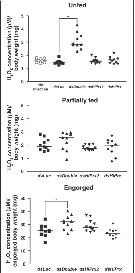
Double knockdown ofHlPrxgenes increased the concentration of H2O2before and after blood-feeding
To elucidate the observed effects of HlPrx and/or
HlPrx2 gene silencing during blood-feeding, H2O2
con-centrations were measured in female ticks. Gene silen-cing was also confirmed by semi-quantitative RT-PCR (data not shown). In the unfed and engorged states, the
dsDouble group showed significantly higher concentra-tions of H2O2as compared to thedsLuc group (Welch’s
t-test: Unfed,t(10)= 7.77,P< 0.001; Engorged,t(17)= 2.72,
P= 0.014) (Fig. 5, Unfed and Engorged).HlPrxorHlPrx2
gene-silenced groups only showed slightly higher con-centrations of H2O2 as compared to the dsLuc-injected
group in the unfed state (Fig. 5, Unfed). On the other hand, in the engorged state, the HlPrx2 gene-silenced group showed a slightly higher concentration of H2O2,
whereas theHlPrxgene-silenced group showed a slightly lower concentration of H2O2as compared to thedsLuc
-injected group (Fig. 5, Engorged). These results demon-strate that the knockdown of HlPrx and HlPrx2 genes leads to a high concentration of H2O2 in ticks before
and after blood-feeding.
Discussion
To protect against the toxicity of H2O2, aerobic
organ-isms have evolved antioxidant enzymes, such as cata-lases, peroxidases, and Prxs [2]. Moreover, ticks lack heme synthesis and catabolism pathways because they are unable to prepareσ-aminolevulinic acid, a heme pre-cursor, even at genomic levels [3, 19]. Therefore, they rely on heme from their host and store it in hemosomes of the midgut without digestion [4]. These facts suggest that ticks might face difficulties in producing proteins that contain heme, such as catalase and peroxidase, which are both H2O2-scavenging enzymes [20].
More-over, ticks must acquire nutrients from the host blood meal and metabolize these nutrients via catabolism and anabolism [21].Plasmodiumparasites also take in nour-ishment from host blood and are likely to utilize mem-bers of the Prx family as the principal enzymes for reducing peroxides, including H2O2, because they lack
catalase and peroxidase [22]. Therefore, Prxs might be similarly essential to the regulation of the H2O2
concen-tration for ticks.
In this study, we found thatHlPrx2mRNA expression was upregulated by blood-feeding (Fig. 1). On the other hand, HlPrx2 protein expression was almost stable dur-ing blood-feeddur-ing, except in the midgut (Fig. 2). In the whole body, although mRNA expression was upregu-lated by blood-feeding when compared to the unfed state (Fig. 1a, b), protein expression seemed to be con-stant in all states of blood-feeding except for the engorged state, where it showed an increased expression level (Fig. 2a, b). Fasciola parasites secrete Prxs into
Fig. 4aKnockdown confirmation ofHlPrxand/orHlPrx2genes in partially fed adult ticks. Each tick total RNA was extracted from 3 ticks pooled. The left column indicates the detection primer set; actinwas used as a control. The right column indicates the size of the PCR products.bColumn graph for engorged body weight in the knockdown experiment.cColumn graph for egg weight after finishing oviposition by engorged adult ticks in the knockdown experiment. Horizontal lines indicate the median values.Abbreviations: dsLuc, double-strandedLuciferase-injected group;dsHlPrx2, double-strandedHlPrx2-injected group;dsHlPrx, double-strandedHlPrx-injected group;dsDouble, both double-strandedHlPrx- andHlPrx2-injected group. *P< 0.05;**P< 0.01, significant differencesvs dsLucby Welch
[image:9.595.57.289.85.552.2]their hosts to regulate their environment for survival in the host body [9]. Our results suggest that ticks may also secrete HlPrx2 protein into hosts as Fasciola parasites do, and the inconsistency of protein expression in com-parison with mRNA expression may be due to the re-lease of HlPrx2 proteins. Protein expression in the whole body increased according to the state of engorge-ment (Fig. 2c). This drastic change seems to be related to body size, because tick body weight notably increases from day 4 to engorgement, and the increase in body weight is about 100-fold compared to unfed ticks [23]. It may be also in response to the very large amounts of blood ingested during the rapid engorgement stage, which may expose ticks to higher levels of ROS. Al-though other developmental stages (larval and nymphal stages) also showed similar tendencies in HlPrx2 protein expression (Fig. 2b),HlPrx2 mRNA expression in larval and nymphal stages was higher than in the adult stage (Fig. 1b). This result suggests that HlPrx2 protein might have an important role in the molting and survival of immature stages during blood-feeding and after engorgement.
In the internal organs, especially the midgut, HlPrx2 mRNA and protein expression was consistent (Figs. 1c and 2c). The mRNA and protein expression levels were negligible in the unfed midgut (Figs. 1c and 2c). Blood-feeding acts as a trigger, inducing the upregulation of HlPrx2 mRNA and protein expres-sion. In IFAT examination of the midgut, HlPrx2-specific fluorescence was detected in the basal lamina (Fig. 3). There have been some reports that the mul-timer of 2-Cys Prxs are associated with membranes, such as red blood cells [24, 25]. Our results, along with those of previous reports, suggest that HlPrx2 protects digestive cells against membrane oxidation and suppresses unnecessary diffusion of H2O2 from
midgut lumen and digestive cells. On the other hand,
[image:10.595.59.539.101.176.2]the midgut, ovaries, and fat bodies are known to pro-duce vitellogenin, a phospholipoglycoprotein and a member of the lipid transfer protein superfamily that is the precursor of major yolk proteins in all ovipar-ous organisms [26, 27]. During blood-feeding, the ex-pression patterns of tick vitellogenin are upregulated from day 3 to engorgement; the highest expression of mRNA and protein is observed upon engorgement [27]. Vitellogenin also has a positive effect on oxida-tive stress resistance in bees and is a preferred target of oxidative carbonylation in comparison with hemolymph proteins in adult bees [28]. In addition, in the ovaries and fat bodies, HlPrx2 mRNA expres-sion was upregulated from around day 3, and protein expression was present stably (Figs. 1c and 2c). This indicates HlPrx2 protein could protect vitellogenin and the organs synthesizing vitellogenin, such as the midgut, the fat bodies, and the ovaries, from the oxi-dative stress that occurs during blood-feeding. In the salivary glands, HlPrx2 mRNA expression was upreg-ulated during blood-feeding (Fig. 1c), while protein expression was upregulated from unfed to partially fed states (Fig. 2c). Moreover, in the case of HlPrx, the other known peroxiredoxin of H. longicornis, mRNA is upregulated in the salivary glands, and HlPrx protein is also highly expressed in the salivary glands [12]. An anti-HlPrx antibody was detected in the host serum after several repeated tick infestations [12], suggesting that the HlPrx was released from ticks into the host eliciting to produce anti-HlPrx on im-mune response. In Fasciola parasites, infective parasites excyst from a dormant state following ingestion and pene-trate the intestinal wall before migrating to the liver; in this nutrient- and oxygen-rich environment, the parasites undergo rapid growth and development, and energy is supplied by aerobic respiration [29]. This developmental situation of Fasciola parasites is similar to the
Table 2Effects ofHlPrxand/orHlPrx2gene silencing in ticks
Knockdown groups Infest No. Drop No. Engorged body weight (mg)
Egg weight (mg) Ratio of egg weight/engorged body weight Hatching rate (%)
dsLuc 30 25 263.7 ± 58.9 162.1 ± 38.9 0.61 ± 0.03 100
dsDouble 30 15 218.8 ± 66.2a 130.3 ± 46.7d 0.59 ± 0.08 87
dsHlPrx2 30 22 204.4 ± 56.3b 116.7 ± 45.3e 0.55 ± 0.09g 77i
dsHlPrx 30 28 210.0 ± 59.8c 124.8 ± 43.4f 0.58 ± 0.06h 78j
a
Significant difference as compared with thedsLucgroup by Welch’st-test:t(27)= 3.16,P= 0.020 b
Significant difference as compared with thedsLucgroup by Welch’st-test:t(45)= 3.53,P= 0.001 c
Significant difference as compared with thedsLucgroup by Welch’st-test:t(50)= 3.24,P= 0.002 d
Significant difference as compared with thedsLucgroup by Welch’st-test:t(26)= 2.22,P= 0.036 e
Significant difference as compared with thedsLucgroup by Welch’st-test:t(42)= 3.67,P= 0.001 f
Significant difference as compared with thedsLucgroup by Welch’st-test:t(51)= 3.30,P= 0.002 g
Significant difference as compared with thedsLucgroup by Welch’st-test:t(25)= 2.85,P= 0.009 h
Significant difference as compared with thedsLucgroup by Welch’st-test:t(41)= 2.46,P= 0.018 i
Significant difference as compared with thedsLucgroup by Chi-square test:χ2
= 6.36,df= 1,P= 0.012
j
development of ticks during blood-feeding. In addition,
Fasciolaparasites secrete Prxs into their host to regulate their environment for survival in the host body [9]. These findings strongly suggest that tick Prxs may be also
secreted into the host’s body in a way similar to that of
Fasciolaparasites.
In hemocytes, HlPrx2 mRNA expression was upregu-lated during blood-feeding, and a specific fluorescence was also detected in cell membranes of the hemocytes (Figs. 1c, 3). In Ixodes ricinus, two Prx homologous genes (Accession nos. AY333958 and AY333959) were strongly induced in the hemolymph after Borrelia burg-dorferi infection [30]. Furthermore,Borrelia exploits the salivary Salp25D, a protein homologous to Prx in Ixodes scapularis, for protection against reactive oxygen inter-mediates generated by the mammalian neutrophils at the vector-host interface [31]. These results indicate that HlPrx2 might be related to immune response, e.g. diges-tion of foreign bodies such asBorreliaandBabesia para-sites in hemocytes. In the mosquitoAnopheles stephensi, 2-Cys Prx (AsPrx-4783) expression induced in the mid-gut was two to seven times higher in malaria parasite-infected insects than in unparasite-infected mosquitoes [32]. Two
Prx genes of I. ricinuswere also induced in the midgut by B. burgdorferi infection [30]. HlPrx2 in the midgut may also be involved in immune response; however, fur-ther investigation is necessary.
We also performed knockdown experiments of HlPrx
and/or HlPrx2 genes and measured the H2O2after the
knockdown of these genes (Table 2 and Figs. 4, 5). The H2O2 concentration of no injection group in the unfed
state was about 3μM (data not shown). In comparison with insects, the H2O2 concentration in normal state
silkworms was also reported at about 3 μM [33]. These observation may suggest that at a normal state, tick and silkworm H2O2 concentrations might have the same
range. Therefore, this detection method of H2O2
con-centration was considered as functionally acceptable. In the unfed and engorged states, the dsDouble group showed significantly higher concentrations of H2O2 as
compared to the dsLuc group. These results suggest a synergistic regulation of H2O2by HlPrx and HlPrx2. In
addition, phenotype evaluation after the knockdown of
HlPrxand/orHlPrx2 demonstrated significant decreases in the engorged body weight, egg weight and hatching rate, particularly after HlPrx2 knockdown. The antioxi-dant activities evaluated by a metal-catalyzed oxidation system seemed to be almost the same comparing 1-Cys Prx and 2-Cys Prx from the bumblebee Bombus ignites
[34]. The donors of 1-Cys Prxs and 2-Cys Prxs are thiol and thioredoxin, respectively [7]. Thioredoxin is a major disulfide reductase system which can provide electrons to a large range of enzymes and is found to be critical for DNA synthesis and defense against oxidative stress [35]. Taken together, the 1-Cys and 2-Cys Prxs seemed to have almost the same antioxidant activity but their donors are different. These data indicate that 2-Cys Prx is more related to cell metabolism through the
[image:11.595.56.293.90.568.2]antioxidant activity because of its utilization of thiore-doxin as donor, thus, HlPrx2 knockdown in the ticks led to the significant decrease in engorged body weight, egg weight, and hatching rate in spite of no significant effect to H2O2 concentrations in the knockdowned ticks.
Therefore, these findings suggest that HlPrxs play an im-portant role in successful blood-feeding and reproduction, with HlPrx2 being apparently more significant. Addition-ally, the observed effects in the dsDouble group were milder than those of thedsHlPrx2group. H2O2can
acti-vate signaling pathways to stimulate cell proliferation, dif-ferentiation and migration in multicellular organisms [36]. These results suggest that thedsDoublegroup, but not the
dsHlPrx2group, was exposed to a high concentration of H2O2, leading to higher engorged body weight, egg weight
and hatching rate as compared to thedsHlPrx2group. In endoparasites, Prx has been shown to be the most important detoxifying enzyme for their survival [8, 9] making it a candidate for use in vaccine development and a therapeutic target in treating endoparasitic infec-tious diseases [30, 32]. In ticks, there have been a few re-ports on Prxs. However, anti-HlPrx antibody was detected in the host serum after several repeated tick in-festations [12], suggesting that HlPrx was released from ticks into the host and the amount of released HlPrx protein was quite small since several infestations of ticks were done to detect the anti-HlPrx antibody. In addition, ticks ingest and concentrate large amounts of the host-derived blood [23], it can be suggested that the anti-HlPrx antibody would be concentrated in tick's body. In the present study, anti-HlPrx2 antibody cross-reacted with some rabbit Prx from normal rabbit blood (see Additional file 1: Figure S1), giving some concerns whether HlPrx2 can be a good vaccine candidate. How-ever, the knockdown ofHlPrx and/orHlPrx2 genes sig-nificantly affected tick blood-feeding, reproduction and antioxidant activity (Table 2 and Figs. 4, 5). Therefore, tick Prx can be a potential target for tick control and provide further understanding of the oxidative stress coping mechanisms in ticks during blood-feeding.
Conclusion
In summary, we investigated mRNA and protein expres-sion profiles of HlPrx2 and the localization of this protein in tick tissues. Real-time PCR showed that
HlPrx2gene expression in whole bodies and internal or-gans was significantly upregulated during blood-feeding. However, protein expression was constant throughout blood-feeding. Moreover, a knockdown experiment of
HlPrx2 was performed using RNAi to evaluate its func-tion in ticks. The knockdown of theHlPrx2gene caused significant differences in body weight, egg weight and hatching rate in engorged ticks as compared to those of the control group. Finally, the detection of H2O2 after
the double knockdown of HlPrxs in ticks showed that H2O2 concentration increased before and after
blood-feeding. Therefore, HlPrx2 can be considered important for successful blood-feeding and reproduction through the regulation of H2O2 concentrations in ticks during
blood-feeding. This study contributes to the search for a candidate target for tick control and furthers under-standing of the tick’s oxidative stress coping mechanism during blood-feeding.
Additional files
Additional file 1:Figure S1.Comparison of normal rabbit blood and engorged-state samples in developmental stages using Western blot analysis. The top arrow indicates native HlPrx2 protein, the middle arrow indicates non-specific band 1 and the bottom arrow indicates non-specific band 2 (M, marker).Figure S2.Confirmation of antibody’s specificity inHlPrxand/orHlPrx2 genes-silencing partially fed adult ticks. Each tick’s total protein was extracted from 3 ticks pooled. The left column indicates the specific anti-serum. For loading control, tubulin was detected. (PPTX 11304 kb)
Additional file 2:Candidates for non-specific bands from Japanese white rabbit blood in Western blot analysis. (DOCX 13 kb)
Acknowledgements
We are grateful to Dr. T. Masatani of the Transboundary Animal Diseases Research Center, Joint Faculty of Veterinary Medicine, Kagoshima University, for his helpful comments and suggestions on this work.
Funding
This work was supported by the Japan Society for the Promotion of Science (JSPS) KAKENHI Grant Numbers 25292173, 26660229, 16H05028, and 16 J08221, and Cooperative Research Grant (27-joint-11) of the National Research Center for Protozoan Diseases, Obihiro University of Agriculture and Veterinary Medicine. K. Kusakisako is supported by a Grant-in-Aid for JSPS fellows.
Availability of data and materials
The datasets supporting the conclusions of this article are included within the article and its additional files. The sequences ofHlPrx,HlPrx2,actin, tubulin,P0, andL23are deposited in the GenBank database under the accession number AB038382 (HlPrx), LC049075 (HlPrx2), AY254898 (actin), AB642157 (tubulin), EU048401 (P0), and DQ849041 (L23).
Authors’contributions
KK, MM and TT designed the experiments. KK, EPH, HM, and MRT performed the experiments. KK, RLG, RUS, NT, MM, KF and TT analyzed the data. KK wrote the manuscript. All the authors checked and approved the final version of the manuscript to be published.
Competing interests
The authors declare that they have no competing interests.
Consent for publication
Not applicable.
Ethics approval and consent to participate
Not applicable.
Author details 1
Laboratory of Infectious Diseases, Joint Faculty of Veterinary Medicine, Kagoshima University, Korimoto, Kagoshima 890-0065, Japan.2Department of
Pathological and Preventive Veterinary Science, United Graduate School of Veterinary Science, Yamaguchi University, Yoshida, Yamaguchi 753-8515, Japan.3Department of Veterinary Paraclinical Sciences, College of Veterinary Medicine, University of the Philippines Los Baños, Los Baños, Laguna 4031, Philippines.4National Research Center for Protozoan Diseases, Obihiro
Hokkaido 080-8555, Japan.5Department of Parasitology, Kitasato University
School of Medicine, Minami, Sagamihara, Kanagawa 252-0374, Japan.
6National Agricultural and Food Research Organization, Tsukuba, Ibaraki
305-0856, Japan.
Received: 27 June 2016 Accepted: 11 August 2016
References
1. Robinson MW, Hutchinson AT, Dalton JP, Donnelly S. Peroxiredoxin: a central player in immune modulation. Parasite Immunol. 2010;32:305–13. 2. Rhee SG. H2O2, a necessary evil for cell signaling. Science. 2006;312:1882–3. 3. Hall A, Nelson K, Poole LB, Karplus PA. Structure-based insights into the
catalytic power and conformational dexterity of peroxiredoxins. Antioxid Redox Signal. 2011;15:795–815.
4. Chae HZ, Kim HJ, Kang SW, Rhee SG. Characterization of three isoforms of mammalian peroxiredoxin that reduce peroxides in the presence of thioredoxin. Diabetes Res Clin Pract. 1999;45:101–12.
5. Lee TH, Kim SU, Yu SL, Kim SH, Park DS, Moon HB, et al. Peroxiredoxin II is essential for sustaining life span of erythrocytes in mice. Blood. 2003;101:5033–8.
6. Hofmann B, Hecht HJ, Flohé L. Peroxiredoxins. Biol Chem. 2002;383:347–64. 7. Choi HJ, Kang SW, Yang CH, Rhee SG, Ryu SE. Crystal structure of a novel
human peroxidase enzyme at 2.0 A resolution. Nat Struct Biol. 1998;5:400–6. 8. Kawazu S, Komaki-Yasuda K, Oku H, Kano S. Peroxiredoxins in malaria
parasites: parasitologic aspects. Parasitol Int. 2008;57:1–7. 9. Dalton JP, Robinson MW, Mulcahy G, O'Neill SM, Donnelly S.
Immunomodulatory molecules ofFasciola hepatica: candidates for both vaccine and immunotherapeutic development. Vet Parasitol. 2013;195:272–85. 10. Grandjean O. Blood digestion inOrnithodorus moubataMurraysensu stricto
Walton females (Ixodoidea: Argasidae) II. Modifications of midgut cells related to the digestive cycle and to the triggering action of mating. Ann Parasitol Hum Comp. 1983;58:493–514.
11. Citelli M, Lara FA, da Silva VIJ, Oliveira PL. Oxidative stress impairs heme detoxification in the midgut of the cattle tick,Rhipicephalus(Boophilus) microplus. Mol Biochem Parasitol. 2007;151:81–8.
12. Tsuji N, Kamio T, Isobe T, Fujisaki K. Molecular characterization of a peroxiredoxin from the hard tickHaemaphysalis longicornis. Insect Mol Biol. 2001;10:121–9.
13. Kusakisako K, Masatani T, Miyata T, Galay RL, Maeda H, Talactac MR, et al. Functional analysis of recombinant 2-Cys peroxiredoxin from the hard tick Haemaphysalis longicornis. Insect Mol Biol. 2016;25:16–23.
14. Fujisaki K. Development of acquired resistance precipitating antibody in rabbits experimentally infested with females ofHaemaphysalis longicornis (Ixodoidea: Ixodidae). Natl Inst Anim Health Q (Tokyo). 1978;18:27–38. 15. Umemiya-Shirafuji R, Tanaka T, Boldbaatar D, Tanaka T, Fujisaki K. Akt is an
essential player in regulating cell/organ growth at the adult stage in the hard tickHaemaphysalis longicornis. Insect Biochem Mol Biol. 2012;42:164–73. 16. Galay RL, Takechi R, Umemiya-Shirafuji R, Talactac MR, Maeda H,
Kusakisako K, et al. Impaired cellular immune response to injected bacteria after knockdown of ferritin genes in the hard tick Haemaphysalis longicornis. Parasitol Int. 2016;65:251–7.
17. Oba Y, Yoshida M, Shintani T, Furuhashi M, Inouye S. Firefly luciferase genes from the subfamilies Psilocladinae and Ototretinae (Lampyridae,
Coleoptera). Comp Biochem Physiol B Biochem Mol Biol. 2012;161:110–6. 18. Low FM, Hampton MB, Peskin AV, Winterbourn CC. Peroxiredoxin 2
functions as a noncatalytic scavenger of low-level hydrogen peroxide in the erythrocyte. Blood. 2007;109:2611–7.
19. Perner J, Sobotka R, Sima R, Konvickova J, Sojka D, Oliveira PL, et al. Acquisition of exogenous haem is essential for tick reproduction. Elife. 2016;5:e12318.
20. Wood ZA, Schröder E, Robin Harris J, Poole LB. Structure, mechanism and regulation of peroxiredoxins. Trends Biochem Sci. 2003;28:32–40. 21. Tsuji N, Miyoshi T, Battsetseg B, Matsuo T, Xuan X, Fujisaki K. A cysteine
protease is critical forBabesiaspp. transmission inHaemaphysalisticks. PLoS Pathog. 2008;4:e1000062.
22. Mitozo PA, de Souza LF, Loch-Neckel G, Flesch S, Maris AF, Figueiredo CP, et al. A study of the relative importance of the peroxiredoxin-, catalase-, and glutathione-dependent systems in neural peroxide metabolism. Free Radic Biol Med. 2011;51:69–77.
23. Kitaoka S. Physiological and ecological studies on some ticks. VII. Parthenogenetic and bisexual races ofHaemaphysalis bispinosain Japan and experimental crossing between them. Natl Inst Anim Health Q (Tokyo). 1961;1:142–9.
24. Schröder E, Littlechild JA, Lebedev AA, Errington N, Vagin AA, Isupov MN. Crystal structure of decameric 2-Cys peroxiredoxin from human erythrocytes at 1.7 A resolution. Structure. 2000;8:605–15.
25. Hall A, Karplus PA, Poole LB. Typical 2-Cys peroxiredoxins–structures, mechanisms and functions. FEBS J. 2009;276:2469–77.
26. Avarre JC, Lubzens E, Babin PJ. Apolipocrustacein, formerly vitellogenin, is the major egg yolk precursor protein in decapod crustaceans and is homologous to insect apolipophorin II/I and vertebrate apolipoprotein B. BMC Evol Biol. 2007;7:3.
27. Boldbaatar D, Umemiya-Shirafuji R, Liao M, Tanaka T, Xuan X, Fujisaki K. Multiple vitellogenins from theHaemaphysalis longicornistick are crucial for ovarian development. J Insect Physiol. 2010;56:1587–98.
28. Seehuus SC, Norberg K, Gimsa U, Krekling T, Amdam GV. Reproductive protein protects functionally sterile honey bee workers from oxidative stress. Proc Natl Acad Sci USA. 2006;103:962–7.
29. Sekiya M, Mulcahy G, Irwin JA, Stack CM, Donnelly SM, Xu W, et al. Biochemical characterisation of the recombinant peroxiredoxin (FhePrx) of the liver fluke. Fasciola hepatica FEBS Lett. 2006;580:5016–22.
30. Rudenko N, Golovchenko M, Edwards MJ, Grubhoffer L. Differential expression ofIxodes ricinustick genes induced by blood feeding orBorrelia burgdorferiinfection. J Med Entomol. 2005;42:36–41.
31. Narasimhan S, Sukumaran B, Bozdogan U, Thomas V, Liang X, DePonte K, et al. A tick antioxidant facilitates the Lyme disease agent's successful migration from the mammalian host to the arthropod vector. Cell Host Microbe. 2007;2:7–18.
32. Peterson TM, Luckhart S. A mosquito 2-Cys peroxiredoxin protects against nitrosative and oxidative stresses associated with malaria parasite infection. Free Radic Biol Med. 2006;40:1067–82.
33. Zhang L, Lu Z. Expression, purification and characterization of an atypical 2-Cys peroxiredoxin from the silkworm. Bombyx mori Insect Mol Biol. 2015;24:203–12.
34. Hu Z, Lee KS, Choo YM, Yoon HJ, Lee SM, Lee JH, et al. Molecular cloning and characterization of 1-Cys and 2-Cys peroxiredoxins from the bumblebeeBombus ignitus. Comp Biochem Physiol B Biochem Mol Biol. 2010;155:272–80.
35. Lu J, Holmgren A. The thioredoxin antioxidant system. Free Radic Biol Med. 2014;66:75–87.
36. Veal EA, Day AM, Morgan BA. Hydrogen peroxide sensing and signaling. Mol Cell. 2007;26:1–14.
• We accept pre-submission inquiries
• Our selector tool helps you to find the most relevant journal • We provide round the clock customer support
• Convenient online submission • Thorough peer review
• Inclusion in PubMed and all major indexing services • Maximum visibility for your research
Submit your manuscript at www.biomedcentral.com/submit