R E S E A R C H
Open Access
Feasibility and safety of low-flow
extracorporeal carbon dioxide removal to
facilitate ultra-protective ventilation in
patients with moderate acute respiratory
distress syndrome
Vito Fanelli
1*, Marco V. Ranieri
2, Jordi Mancebo
3, Onnen Moerer
4, Michael Quintel
4, Scott Morley
5,
Indalecio Moran
3, Francisco Parrilla
3, Andrea Costamagna
1, Marco Gaudiosi
1and Alain Combes
6Abstract
Background:Mechanical ventilation with a tidal volume (VT) of 6 mL/kg/predicted body weight (PBW), to maintain
plateau pressure (Pplat) lower than 30 cmH2O, does not completely avoid the risk of ventilator induced lung injury
(VILI). The aim of this study was to evaluate safety and feasibility of a ventilation strategy consisting of very low VT
combined with extracorporeal carbon dioxide removal (ECCO2R).
Methods:In fifteen patients with moderate ARDS, VTwas reduced from baseline to 4 mL/kg PBW while PEEP was
increased to target a plateau pressure–(Pplat) between 23 and 25 cmH2O. Low-flow ECCO2R was initiated when
respiratory acidosis developed (pH < 7.25, PaCO2> 60 mmHg). Ventilation parameters (VT, respiratory rate, PEEP),
respiratory compliance (CRS), driving pressure (DeltaP = VT/CRS), arterial blood gases, and ECCO2R system operational
characteristics were collected during the period of ultra-protective ventilation. Patients were weaned from ECCO2R
when PaO2/FiO2was higher than 200 and could tolerate conventional ventilation settings. Complications, mortality
at day 28, need for prone positioning and extracorporeal membrane oxygenation, and data on weaning from both MV and ECCO2R were also collected.
Results:During the 2 h run in phase, VTreduction from baseline (6.2 mL/kg PBW) to approximately 4 mL/kg PBW
caused respiratory acidosis (pH < 7.25) in all fifteen patients. At steady state, ECCO2R with an average blood flow of
435 mL/min and sweep gas flow of 10 L/min was effective at correcting pH and PaCO2to within 10 % of baseline
values. PEEP values tended to increase at VTof 4 mL/kg from 12.2 to 14.5 cmH2O, but this change was not
statistically significant. Driving pressure was significantly reduced during the first two days compared to baseline (from 13.9 to 11.6 cmH2O; p < 0.05) and there were no significant differences in the values of respiratory system
compliance. Rescue therapies for life threatening hypoxemia such as prone position and ECMO were necessary in four and two patients, respectively. Only two study-related adverse events were observed (intravascular hemolysis and femoral catheter kinking).
Conclusions:The low-flow ECCO2R system safely facilitates a low volume, low pressure ultra-protective mechanical
ventilation strategy in patients with moderate ARDS.
(Continued on next page)
* Correspondence:vito.fanelli@unito.it
1Department of Anesthesia and Critical Care - AOU Città della Salute e della
Scienza di Torino, University of Turin, Corso Dogliotti 14, 10126 Torino, Italy Full list of author information is available at the end of the article
(Continued from previous page)
Keywords:Acute respiratory distress syndrome, Protective mechanical ventilation, Extracorporeal carbon dioxide removal, Extracorporeal membrane oxygenation, Positive end-expiratory pressure, Driving pressure, Ventilator-induced lung injury
Background
Over-distention of the normally aerated lung and/or open-ing and closopen-ing of collapsed alveoli may worsen pulmon-ary damage in patients with acute respiratory distress syndrome (ARDS). Current guidelines for ARDS recom-mend a protective ventilation strategy based on limitation of tidal volume (VT) to 6 mL/kg predicted body weight
and plateau pressure (Pplat) to 30 cmH2O, an approach
that has been shown in a randomized clinical trial to re-duce mortality by 9 % [1]. However, recent studies have shown that ARDS patients who are ventilated according to the ARDS Network (ARDSnet) protective ventilatory strategy may still be exposed to forces that can induce lung injury [2–5], thus challenging current recommenda-tions on how to minimize the risk of ventilator-induced lung injury (VILI) [3]. Moreover, Hager and coworkers [6] showed that mortality decreases as Pplatis reduced.
How-ever, as this relationship appears to be linear [6], several authors have postulated that an ultra-protective ventila-tion strategy based on further reducventila-tion in VTfrom 6–4
mL/kg and Pplat from 30–25 cmH2O may improve
out-comes [3]. Such tidal volumes reduce alveolar ventila-tion resulting in respiratory acidosis, which can be mitigated through the application of extracorporeal carbon dioxide removal (ECCO2R) [7–9].
The feasibility and safety of ultra-protective ventilation strategies facilitated by ECCO2R has been tested in
sev-eral studies using a pump-less arteriovenous device op-erating at a blood flow rate of 1.0–1.5 L/min [10–12]. Information on feasibility and safety of ultra-protective ventilation strategies facilitated by low-flow venous-venous ECCO2R are limited to a single-center study [8].
The aim of the current study was to assess in a multi-center trial the feasibility and safety of an ultra-protective ventilation strategy facilitated by low-flow veno-venous ECCO2R in patients with moderate ARDS.
We used an ECCO2R system (Hemolung Respiratory
Assist System, ALung Technologies), which is specific-ally designed to provide clinicspecific-ally significant CO2
re-moval at low blood flow rates (350–550 mL/min).
Methods
Patients were enrolled in four European intensive care units of academic hospitals. Local ethics committees approved the study protocol. Informed consent was obtained from the patients. In the case of incompetent patients, consent was obtained in accordance with local ethics committee procedures [13].
Patients
The study included fifteen adult patients with moderate ARDS according to the Berlin definition (PaO2/FiO2(P/
F) 100–200 mmHg, with positive end-expiratory pres-sure (PEEP) >5 cmH2O) [14], who were mechanically
ventilated with an expected duration of ventilation lon-ger than 24 h. Exclusion criteria were age <18 years, pregnancy, decompensated heart insufficiency or acute coronary syndrome, severe chronic obstructive pulmon-ary disease (COPD), respiratory acidosis with arterial PCO2 (PaCO2) >80 mmHg, acute brain injury, severe
liver insufficiency (Child-Pugh score >7) or fulminant hep-atic failure, heparin-induced thrombocytopenia, contra-indication for systemic anticoagulation, patient moribund, decision to limit therapeutic interventions, catheter access to femoral vein or jugular vein impossible, pneumothorax, or platelet count <50 × 103/mL.
ECCO2R System
Low-flow ECCO2R was provided with the Hemolung
Respiratory Assist System (RAS) (ALung Technologies, Inc, Pittsburgh, PA, USA) [15]. Briefly, venous blood is cir-culated through a 15.5-Fr dual lumen venous catheter (jugular or femoral) by a magnetically driven centrifugal pump at a flow rate of 350–550 mL/min. The pump is in-tegrated within a cylindrical bundle of hollow fiber mem-branes, creating a flow pattern, which improves CO2
transfer efficiency relative to passive oxygenators. Sweep gas (air or 100 % O2) is drawn through the hollow fibers
under negative pressure by a vacuum pump, creating a gradient for CO2 diffusion. Maintaining the sweep gas
under negative pressure mitigates the risk of air embolism across the membrane, and also allows for automatic re-moval of plasmatic water condensation from the fiber lu-mens in order to preserve gas exchange efficiency. Level of blood flow, pump speed (RPM) and extracorporeal CO2removal rate (vCO2) are displayed on a controller.
Study protocol
Patients were sedated, paralyzed and ventilated in accordance with the EXPRESS trial protocol [16]: VT of
6 mL/kg (ideal body weight); PEEP set to achieve Pplatof
28–30 cmH2O; respiratory rate (RR) set to 20–35 to
activated at a blood flow rate of 350–550 mL/min and a sweep gas of 0 L/min such that no CO2removal was
ini-tially performed.
Following a 2-h run-in time, VT was gradually reduced
from 6 to a minimum value of 4 mL/kg by 0.5 mL/kg every 30 minutes and PEEP was increased to target a Pplatbetween
23 and 25 cmH2O . If arterial pH was <7.25 with PaCO2>60
mmHg, despite an increase in RR up to 35/min, sweep gas through the ECCO2R device was switched on with 100 %
oxygen at 10 L/min to obtain an arterial pH≥7.25 with a PaCO2≤60 mmHg and RR ≤35/min. If PaCO2 was >75
mmHg and/or pH <7.20, despite a respiratory rate of 35/ min and optimized ECCO2R, sodium bicarbonate could be
infused. If undesirable hypercapnia/acidosis persisted, VT
was increased at the discretion of the treating physician. Re-fractory hypoxemia and/or hypercapnia could be managed at the discretion of the attending physician using veno-venous extracorporeal membrane oxygenation (ECMO), prone positioning, or nitric oxide (NO) inhalation. If PaCO2was constantly <35 mmHg and/or pH was >7.50
under the aforementioned ECCO2R settings, the
respira-tory rate was decreased to 18–22/min and sweep gas flow was decreased to 2–5 L/min.
The ECCO2R-facilitated ultra-protective ventilation
strategy was continued for at least 24 h. The potential for weaning from ultra-protective ventilation and ECCO2R was assessed daily if PaO2/FiO2(P/F) was >200
by setting mechanical ventilation according to conven-tional ARDSnet settings (VT= 6 mL/kg, PEEP 5–10
cmH2O, RR 20–30/min, inspired O2fraction (FiO2) = 40
%) and switching off sweep gas through the ECCO2R
de-vice. Under these conditions, if the patient remained stable for at least 12 h with Pplat<25 cmH2O and PaCO2
<50 mmHg (allowing for RR up to 30–35/min), ECCO2R
was discontinued and the venous catheter removed. ECCO2R parameters (blood flow, sweep gas flow, and
CO2removal rate), ventilator settings (VT, PEEP, RR, Pplat,
mean airway pressure, minute ventilation, inspiratory-to-expiratory ratio, inspired fraction of oxygen), hemodynamics (mean arterial pressure, heart rate, dose of vasopressor) and arterial blood gas values (pH, PaO2,
PaCO2,HCO3–, lactate), heparin dose and activated partial
thromboplastin time ratio (aPTTr) were collected at base-line, after run-in time, 30 minutes after every VTreduction
and at least twice a day (08:00 am ± 2 h and 08:00 pm ± 2 h) in the subsequent days on ECCO2R. Blood chemistry
data were collected daily. Respiratory system compliance [17] and driving pressure [18] were calculated according to the standard formula.
Serious adverse events (SAE) were prospectively defined as: (a) any event that is fatal or immediately life threaten-ing, permanently disablthreaten-ing, severely incapacitating or re-quires prolonged hospitalization; or (b) any event that may jeopardize the patient and requires medical or
surgical intervention to prevent one of the outcomes listed above; and (c) which the attending physician perceives might be directly related to enrollment in the clinical trial. Adverse events were considered to be study-related if the event followed a reasonable sequence from a study pro-cedure and could readily have been produced by the study procedure. Adverse events were considered non study-related if they were study-related primarily to the underlying dis-ease or to ARDS and its sequelae. Other adverse events not fulfilling the above definition were recorded in the pa-tients’case report forms (CRFs). Following discontinuation of ECCO2R, subjects were monitored for adverse events
until hospital discharge or day 8 after enrollment, which-ever occurred first.
Data are expressed as mean ± SD. Statistical analysis was performed by one-way analysis of variance for repeated mea-sures, followed by Bonferroni post-hoc test for comparison between different time points (Stata Corp, College Station, TX, USA).P<0.05 was considered statistically significant.
Results
Fifteen patients with moderate ARDS were included in the period April to November 2014. Baseline characteris-tics of patients enrolled in the study are shown in Table 1.
Ventilation settings during the VT reduction phase are
[image:3.595.304.538.429.731.2]shown in Table 2. At baseline, all patients had PaO2/
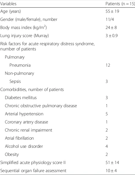
Table 1Baseline characteristics of patients
Variables Patients (n = 15)
Age (years) 55 ± 19
Gender (male/female), number 11/4 Body mass index (kg/m2) 24 ± 8
Lung injury score (Murray) 3 ± 0.9 Risk factors for acute respiratory distress syndrome,
number of patients Pulmonary
Pneumonia 12
Non-pulmonary
Sepsis 3
Comorbidities, number of patients
Diabetes mellitus 3
Chronic obstructive pulmonary disease 1
Arterial hypertension 5
Coronary artery disease 1
Chronic renal impairment 2
Atrial fibrillation 2
Alcohol use disorder 4
Obesity 2
FiO2≤200, they were ventilated with a conventional
pro-tective ventilation strategy according to the EXPRESS trial protocol, and were paralyzed for a median time of 1 day kk. The initial stepwise reduction in VT, without
ECCO2R, resulted in significant respiratory acidosis (pH
<7.25) in all 15 patients at a mean VT of 3.96 ± 0.1 mL/
kg. After initiation of ECCO2R, a VTof 4.29 ± 0.5 mL/kg
was achieved and respiratory acidosis was significantly corrected, with pH and PaCO2returning to within 10 %
of baseline values obtained at VT= 6 mL/kg. The median
number of days on ECCO2R was 3 (2–4). The reduction
in VTwas associated with a significant reduction in Pplat
from 27.7 ± 1.6 to 23.9 ± 1 cmH2O (p <0.05) at day 1
and this difference remained significant throughout the
study period (Table 3). PEEP tended to increase from 12 ± 3 to 14 ± 2 cmH2O at day 1, however, this difference
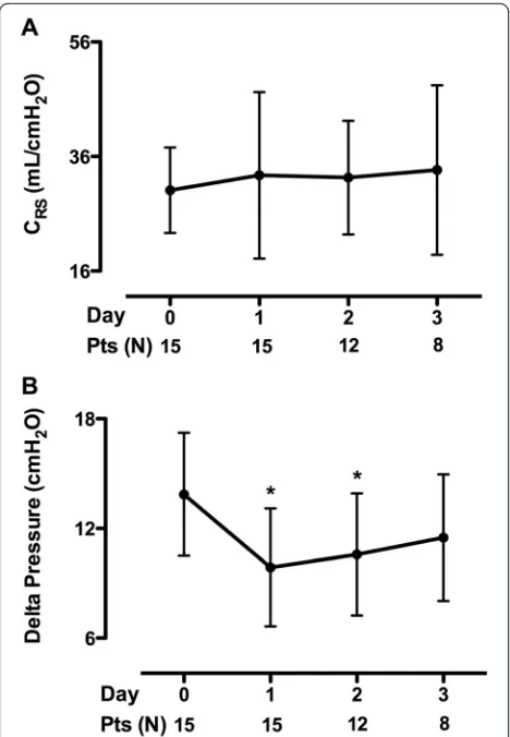
was not statistically significant over time (Table 3). Driv-ing pressure (Pplat – PEEP) was significantly reduced
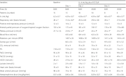
during the first 2 days compared to baseline (p <0.05); there were no significant differences in the values of re-spiratory system compliance (Fig. 1). At day 1, the ECCO2R device provided CO2 removal of 81 ± 9 mL/
[image:4.595.56.541.100.182.2]min at a blood flow rate of 435 ± 60 mL/min and sweep gas flow rate of 10 ± 0.3 L/min. The efficiency of the ECCO2R system was stable throughout the study period (Table 3). Infusion of bicarbonate and renal replacement therapies for acute kidney injury were never used in this cohort of patients.
Table 2Time course of ventilation variables during the run-in phase
Variables Baseline VT5 mL/kg VT4.5 mL/kg VT4 mL/kg
VT(mL/kg) 6.2 ± 0.7 5.02 ± 0.1* 4.48 ± 0.1* 3.96 ± 0.1*
Respiratory rate (beats/minute) 28 ± 7 29 ± 4 30 ± 4* 30 ± 5*
Positive end-expiratory pressure (cmH2O) 12 ± 3 13.8 ± 3 13.6 ± 4 13.0 ± 4.0
Plateau pressure (cmH2O) 27.7 ± 1.6 25.2 ± 1.6* 23.6 ± 1.3* 22.7 ± 1.8*
Patients who reached the pH threshold for ECCO2R, n 0 2 15
*P<0.05 vs baseline.VTtidal volume,ECCO2Rextracorporeal carbon dioxide removal
Table 3Time course of ventilation variables, blood gases, ECCO2R operational characteristics and hemodynamics at VT4 mL/kg plus ECCO2R
Variables Baseline VT4 mL/kg plus ECCO2R
Day 1 Day 2 Day 3
Time (h) 8.00 8.00 8.00 20.00 8.00 20.00
Patients (number) 15 15 10 10 8 6
VT(mL/kg) 6.2 ± 0.7 4.29 ± 0.5* 4.58 ± 0.7* 4.59 ± 0.8* 4.8 ± 0.7* 4.8 ± 0.7*
Respiratory rate (beats/minute) 28 ± 7 31.6 ± 4.6* 29.6 ± 6.8 29.6 ± 6.8 28 ± 7 27.4 ± 8.6 Positive end-expiratory pressure (cmH2O) 12 ± 3 14 ± 2 13 ± 3 12 ± 4 13 ± 5 13 ± 3
Arterial partial pressure of oxygen/inspired oxygen fraction 159 ± 34 175 ± 45 185 ± 91 190 57 176 ± 59 176 ± 80 Plateau pressure (cmH2O) 27.7 ± 1.6 23.9 ± 1* 24 ± 4* 24 ± 3* 24 ± 3* 23 ± 3*
Blood flow (ml/min) 435 ± 60 424 ± 63 423 ± 35 424 ± 29 436 ± 39
Rotations per minute (RPM) 1407 ± 26 1408 ± 30 1409 ± 32 1411 ± 36 1414 ± 41
Sweep gas (L/min) 8.6 ± 3.5 9.2 ± 2.9 9 ± 3 9.9 ± 0.3 8.7 ± 3.2
CO2removal (ml/min) 81 ± 9 70 ± 29 70 ± 31 81 ± 22 71 ± 11
Ph 7.36 ± 0.1 7.33 ± 0.1 7.39 ± 0.1 7.36 ± 0.1 7.33 ± 0.1 7.4 ± 0.1
PaO2 (mmHg) 95 ± 29 90 ± 22 91 ± 26 84 ± 10 81 ± 15 99 ± 29
PaCO2 (mmHg) 51 ± 15 53 ± 15 51 ± 18 52 ± 17 55 ± 20 49 ± 11
HCO3 (mmol/L) 28 ± 5 27.6 ± 6.1 28.7 ± 6.2 28. ± 6.9 28.1 ± 7.4 28.3 ± 6.03 Lactate (mmol/L) 2 ± 1 2.9 ± 4.8 1.9 ± 1.7 1.8 ± 1.8 1.9 ± 2.0 1.3 ± 0.4 Heart rate (beats/minute) 76 ± 9 90 ± 17 86 ± 15 95 ± 21 94 ± 19 94 ± 17 Mean arterial pressure (mmHg) 98 ± 20 74 ± 14 80 ± 10 76 ± 12 76 ± 19 85 ± 14 Norepinephrine dose (mcg/kg/min) 0.51 ± 0.6 0.45 ± 0.4 0.34 ± 0.3 0.29 ± 0.27 0.57 ± 0.4 0.5 ± 0.6
[image:4.595.57.540.428.725.2]At day one, the heparin dosage was 341 ± 138 IU/kg/day with an aPTT ratio of 1.77 ± 0.7, and remained stable over time. The average baseline value of hemoglobin (Hb) was 10.4 ± 2 gr/dL, and the median Hb threshold for transfu-sion was 6.9 gr/dL (6.9–7.0). On day 1, four patients re-ceived 2.25 ± 0.5 red blood cell (RBC) units and 1.25 ± 2.5 pools of platelets. On day 2, only two patients received 1.5 ± 0.7 units of RBCs. On day 3, only one patient re-ceived two units of RBCs.
Two study-related adverse events were observed. In one patient, intravascular hemolysis (plasma-free hemoglobin 401.4 mg/dL) was observed resulting in a discontinu-ation of ECCO2R after 2 days. Kinking of the ECCO2R
catheter caused a reduction in circuit blood flow in an-other patient. Individual data for patients who needed rescue therapies for worsening hypoxemia are given in Table 4. The overall mortality at day 28 was 47 %. Among the eight survivors, six were successfully weaned from both ECCO2R and mechanical ventilation;
while two patients were still dependent on ventilator support at 28 days.
Discussion
The main finding of the current study is that the low-flow ECCO2R system effectively prevents respiratory
acidosis consequent to the reduction of tidal volume to 4 mL/kg in patients with moderate ARDS. Severe hypox-emia occurred in about one third of the patients and was managed by prone positioning in conjunction with ECCO2R; conversion to VV-ECMO was required in two
patients. Side effects related to ECCO2R (intravascular
hemolysis and pump malfunction by femoral catheter kinking) were observed in two patients.
The landmark study by the ARDSnet demonstrated a 9 % mortality reduction in patients with ARDS by limiting plateau pressure to <30 cmH2O [1]. However,
[image:5.595.57.291.87.425.2]recent studies have shown that patients with ARDS may still be at risk of VILI despite values of Pplat≤30 cmH2O
[2, 4, 5]. Terragni [2] and Bellani [4] showed that in some patients, tidal hyperinflation may still occur des-pite limiting VTto 6 mL/kg and Pplatto 30 cmH2O, and
that this is associated with biological signs of VILI, such as higher levels of inflammatory mediators and increased metabolic activity of the lungs. Grasso and coworkers showed that hyperinflation might be attenuated by redu-cing the levels of PEEP based on the shape of the airway pressure time curve and that this is associated with lower levels of pulmonary inflammatory cytokine release [5]. Moreover, Hager and coworkers found that ARDS patients may benefit from VT reduction even if they
already have Pplat<30 cmH2O [6].
ECCO2R has been proposed to partially clear CO2and
consequently reduce the need of minute ventilation as delivered by conventional mechanical ventilation [9]. The first evidence that ECCO2R might be a safe
adjunct-ive therapy to conventional mechanical ventilation for ARDS patients dates back to the early 1980s [19, 20]. At that time, a modified veno-venous ECMO circuit with a blood flow not >1 L/min allowed for dramatic reduc-tions of minute ventilation and ventilator-applied pressure [19]. More recently, Terragni and colleagues
Fig. 1Time course of respiratory system compliance (CRS) (a) and
driving pressure (b). *P<0.05 vs day 0.Ptspatients
Table 4Oxygenation and outcomes of patients who required rescue therapies
Patients Time of PaO2/FiO2
worsening
PaO2/FiO2before
rescue therapy
Outcome (at 28 days) (day) Prone position ECMO
# 1 5 108 Dead
# 5 6 78 Alive
# 8 2 75 Dead
# 11 2 115 Alive
# 12 1 158 Dead
# 13 1 162 Alive
PaO2/FiO2arterial partial pressure of oxygen/inspired oxygen fraction,
[image:5.595.305.539.111.230.2]demonstrated that a modified renal replacement therapy circuit with an oxygenator in series with the hemofilter could facilitate an ultra-protective ventilation strategy, which may mitigate VILI [8]. Our results extend these findings by performing a prospective multicenter study in 15 ARDS patients across four European ICUs.
Monitoring of respiratory mechanics may help clini-cians in assessing the risk of VILI [17]. In the presence of normal chest wall elastance, values of Pplat around
30 cmH2O may increase the risk of alveolar
hyperin-flation, which is the main determinant of VILI [2]; analysis of a large dataset including ARDS patients previously enrolled in clinical trials, shows that driving pressure (i.e., the ratio of tidal volume to respiratory sys-tem compliance) is an independent risk factor for hospital mortality, because regardless of the changes in VT and
PEEP, only changes in driving pressure affected the out-come [18]. Our data show that an ultra-protective ventila-tion strategy facilitated by low-flow veno-venous ECCO2R
resulted in values of respiratory mechanics associated with enhanced protection from VILI as: (1) values of Pplat
around 25 cmH2O, as achieved in this study, have
been associated with lower serum levels of the pro-inflammatory cytokine interleukin-6 and a smaller percentage of lung hyperinflation, as demonstrated by computed tomography [2, 7, 8]; and (2) our ultra-protective ventilation strategy resulted in a significant reduction in driving pressure. These findings allow us to speculate that in our patients, global interaction between moderate-to-high levels of PEEP and very low tidal volume, facilitated by ECCO2R, might be
beneficial to minimize the risk of VILI.
In recent years, advances in technology have generated renewed interest in all extracorporeal support tech-niques. While high flow veno-venous ECMO has been increasingly used to treat life-threatening hypoxemia [21, 22], ECCO2R systems are used to provide
partial-to-full CO2 removal with minimal effects on oxygenation
[9]. Appropriate strategies to manage worsening hypox-emia during ECCO2R treatment is a compelling issue. In
fact, depending on the severity of lung injury, patients with ARDS may experience worsening of arterial oxy-genation that might be life threatening [23]. Moreover, the use of very low tidal volumes may expose patients to the risk of de-recruitment in the event that insufficient PEEP is applied. Consequently, prone positioning while the patient is still on ECCO2R, or shift to high-flow
veno-venous ECMO, might be necessary. In this regard, prone positioning has been demonstrated to be effective not only in improving oxygenation but also in decreas-ing early and late mortality [24]. In the current study, in-dication for rescue therapies for profound hypoxemia was not established by protocol. Within the first week of enrollment 27 % of patients in our cohort required
prone positioning when their PaO2/FiO2 dropped to a median value of 137. Notably, patients underwent prone position without any interruption of ECCO2R and had
improvement in arterial oxygenation. Only two patients required escalation from ECCO2R to ECMO because of
life-threatening hypoxemia. Mortality at 28 days was 47 %, which was expected in a cohort of patients with moderate and severe ARDS.
Previous-generation ECCO2R systems have carried a
high rate of mechanical complications such as pump malfunction, membrane clotting, and catheter displace-ment [8]. In the current study, target blood flow rates were not reached in one case only due to a kinked cath-eter; otherwise, the treatment was consistent over time. Compared with arterio-venous systems in which limb ischemia, compartment syndrome, and intracranial hemorrhage have been described [7], only one case of intravascular hemolysis requiring transfusion was re-ported in our series.
Some limitations of this study should be addressed. First, inferences from this study are limited by its small sample size. Second, although we speculate that the strategy tested in this multicenter study was lung-protective, we did not measure pro-inflammatory media-tors associated with VILI and we did not evaluate lung volume/densities distribution with computed tomog-raphy of the chest. The precise impact of worsening oxy-genation during ECCO2R treatment is also not clear.
Consequently, this approach will be systematically tested in an upcoming randomized clinical trial, such as that under the auspices of European Society of Intensive Care Medicine, which will test the feasibility, safety and efficacy of several ECCO2R systems to facilitate
ultra-protective ventilation - VT of 4 mL/kg predicted
body weight (PBW) and Pplat <25 cmH2O - in patients
with moderate and severe ARDS (SUPERNOVA: a strat-egy of ultraprotective lung ventilation with extracorpor-eal CO2 removal for new-onset moderate to severe ARDS) [25].
Conclusions
In conclusion, low-flow ECCO2R is feasible, safe and
ef-ficient. It facilitates an ultra-protective mechanical venti-lation strategy for reducing tidal volume to 4 mL/kg to maintain Pplat <25 cmH2O. This approach allows
Key messages
Low-flow extracorporeal CO2removal safely and
effectively facilitates an ultra-protective ventilation strategy in patients with moderate ARDS
The current study provides a clinical and physiological rationale to study whether ultra-protective mechanical ventilation improves clinical outcomes of patients with moderate and severe ARDS in randomized control trials
Competing interests
V. Fanelli: speaker honoraria for educational programs from ALung Technologies. J. Mancebo: steering committee member for ALung Technologies Registry. O. Moerer: none. M. Quintel: none. S. Morley: employee of ALung Technologies receiving salary and stock compensation. I. Moran: none. F. Parrilla: none. A. Costamagna: none. M. Ranieri: Medical advisory board for ALung Technologies. A. Combes: Medical advisory board and speaker honoraria from ALung Technologies.
Authors’contributions
Study conception and design: AC and VMR. Acquisition of data: VF, VMR, JM, OM, MQ, IM, FP, AC MG, and AC. Analysis and interpretation of data: VF, VMR, JM, OM, and AC. Drafting the manuscript: VF. Critical revision of the manuscript for important intellectual content: VF, VMR, JM, OM, AC, and SM. Supervision: VMR and AC. All authors read and approved the final manuscript.
Acknowledgements
Financial support for the study was provided by ALung Technologies, Inc.
Author details
1Department of Anesthesia and Critical Care - AOU Città della Salute e della
Scienza di Torino, University of Turin, Corso Dogliotti 14, 10126 Torino, Italy.
2Dipartimento di Anestesia e Rianimazione, Ospedale Policlinico Umberto I,
Sapienza Università di Roma, Rome, Italy.3Servei de Medicina Intensiva,
Hospital de Sant Pau, Barcelona, Spain.4Department of Anesthesiology,
Emergency and Intensive Care Medicine, University Medical Center Göttingen, Göttingen, Germany.5ALung Technologies, Pittsburgh, USA. 6Service de Réanimation Médicale, iCAN, Institute of Cardiometabolism and
Nutrition, Hôpital de la Pitié-Salpêtrière, Assistance Publique-Hôpitaux de Paris, Paris, France.
Received: 28 September 2015 Accepted: 31 January 2016
References
1. ARDSnet. Ventilation with lower tidal volumes as compared with traditional tidal volumes for acute lung injury and the acute respiratory distress syndrome. The Acute Respiratory Distress Syndrome Network. N Engl J Med. 2000;342(18):1301–8.
2. Terragni PP, Rosboch G, Tealdi A, Corno E, Menaldo E, Davini O, Gandini G, Herrmann P, Mascia L, Quintel M, et al. Tidal hyperinflation during low tidal volume ventilation in acute respiratory distress syndrome. Am J Respir Crit Care Med. 2007;175(2):160–6.
3. Slutsky AS, Ranieri VM. Ventilator-induced lung injury. N Engl J Med. 2013;369(22):2126–36.
4. Bellani G, Guerra L, Musch G, Zanella A, Patroniti N, Mauri T, Messa C, Pesenti A. Lung regional metabolic activity and gas volume changes induced by tidal ventilation in patients with acute lung injury. Am J Respir Crit Care Med. 2011;183(9):1193–9.
5. Grasso S, Stripoli T, De Michele M, Bruno F, Moschetta M, Angelelli G, Munno I, Ruggiero V, Anaclerio R, Cafarelli A, et al. ARDSnet ventilatory protocol and alveolar hyperinflation: role of positive end-expiratory pressure. Am J Respir Crit Care Med. 2007;176(8):761–7.
6. Hager DN, Krishnan JA, Hayden DL, Brower RG. Tidal volume reduction in patients with acute lung injury when plateau pressures are not high. Am J Respir Crit Care Med. 2005;172(10):1241–5.
7. Bein T, Weber-Carstens S, Goldmann A, Muller T, Staudinger T, Brederlau J, et al. Lower tidal volume strategy (approximately 3 ml/kg) combined with extracorporeal CO2 removal versus‘conventional’protective ventilation (6 ml/kg) in severe ARDS: the prospective randomized Xtravent-study. Intensive Care Med. 2013;39(5):847–56.
8. Terragni PP, Del Sorbo L, Mascia L, Urbino R, Martin EL, Birocco A, et al. Tidal volume lower than 6 ml/kg enhances lung protection: role of extracorporeal carbon dioxide removal. Anesthesiology. 2009;111(4):826–35.
9. Fanelli V, Costamagna A, Ranieri VM. Extracorporeal support for severe acute respiratory failure. Semin Respir Crit Care Med. 2014;35(4):19–527. 10. Bein T, Weber F, Philipp A, Prasser C, Pfeifer M, Schmid F-X, et al. A new
pumpless extracorporeal interventional lung assist in critical hypoxemia/ hypercapnia. Crit Care Med. 2006;34(5):1372–7.
11. Elliot SC, Paramasivam K, Oram J, Bodenham AR, Howell SJ, Mallick A. Pumpless extracorporeal carbon dioxide removal for life-threatening asthma. Crit Care Med. 2007;35(3):945–8.
12. Flörchinger B, Philipp A, Klose A, Hilker M, Kobuch R, Rupprecht L, et al. Pumpless Extracorporeal Lung Assist: A 10-Year Institutional Experience. Ann Thorac Surg. 2008;86(2):410–7.
13. Ranieri VM, Thompson BT, Barie PS, Dhainaut JF, Douglas IS, Finfer S, et al. Drotrecogin alfa (activated) in adults with septic shock. N Engl J Med. 2012;366(22):2055–64.
14. Ranieri VM, Rubenfeld GD, Thompson BT, Ferguson ND, Caldwell E, Fan E, et al. Acute respiratory distress syndrome: the Berlin Definition. JAMA. 2012;307(23):2526–33.
15. Burki NK, Mani RK, Herth FJ, Schmidt W, Teschler H, Bonin F, et al. A novel extracorporeal CO(2) removal system: results of a pilot study of hypercapnic respiratory failure in patients with COPD. Chest. 2013;143(3):678–86. 16. Mercat A, Richard JC, Vielle B, Jaber S, Osman D, Diehl JL, et al. Positive
end-expiratory pressure setting in adults with acute lung injury and acute respiratory distress syndrome: a randomized controlled trial. JAMA. 2008;299(6):646–55.
17. Grasso S, Fanelli V, Cafarelli A, Anaclerio R, Amabile M, Ancona G, et al. Effects of high versus low positive end-expiratory pressures in acute respiratory distress syndrome. Am J Respir Crit Care Med. 2005;171(9):1002–8. 18. Amato MB, Meade MO, Slutsky AS, Brochard L, Costa EL, Schoenfeld DA, et
al. Driving pressure and survival in the acute respiratory distress syndrome. N Engl J Med. 2015;372(8):747–55.
19. Gattinoni L, Pesenti A, Mascheroni D, Marcolin R, Fumagalli R, Rossi F, et al. Low-frequency positive-pressure ventilation with extracorporeal CO2 removal in severe acute respiratory failure. JAMA. 1986;256(7):881–6. 20. Gattinoni L, Agostoni A, Pesenti A, Pelizzola A, Rossi GP, Langer M, et al.
Treatment of acute respiratory failure with low-frequency positive-pressure ventilation and extracorporeal removal of CO2. Lancet. 1980;2(8189):292–4. 21. Peek GJ, Mugford M, Tiruvoipati R, Wilson A, Allen E, Thalanany MM, et al.
Efficacy and economic assessment of conventional ventilatory support versus extracorporeal membrane oxygenation for severe adult respiratory failure (CESAR): a multicentre randomised controlled trial. Lancet. 2009;374(9698):1351–63.
22. Noah MA, Peek GJ, Finney SJ, Griffiths MJ, Harrison DA, Grieve R, et al. Referral to an extracorporeal membrane oxygenation center and mortality among patients with severe 2009 influenza A(H1N1). JAMA. 2011;306(15):1659–68.
23. Briel M, Meade M, Mercat A, Brower RG, Talmor D, Walter SD, et al. Higher vs lower positive end-expiratory pressure in patients with acute lung injury and acute respiratory distress syndrome: systematic review and meta-analysis. JAMA. 2010;303(9):865–73.
24. Guerin C, Reignier J, Richard JC, Beuret P, Gacouin A, Boulain T, et al. Prone positioning in severe acute respiratory distress syndrome. N Engl J Med. 2013;368(23):2159–68.